H2A.Z in Early Xenopus laevis
Development
Karl David Brown
December 2007
A thesis submitted for the degree of Master of Philosophy
of the Australian National University
College of Medicine and Health Sciences Graduate Program
John Curtin School of Medical Research
Australian National University
Statement
The research described in this thesis was solely and entirely conducted by the author
unless acknowledgement is made in the text. It has not presented for any other degree.
Karl David Brown
Acknowledgements
I would like to thank my supervisory panel for their support. Dr Patricia Ridgway was
my primary supervisor whose guidance throughout this project was invaluable, likewise
for Dr David Tremethick, leader of the Chromatin and Transcriptional Regulation
group. Eldon Ball taught me the nuances of in situ hybridisation, and Sudha Rao
provided valuable guidance relating to RNA-based techniques.
In addition to my panel, the wider scientific community at the ANU should be thanked.
Among this community I would like to specifically thank: Danny Rangasamy and
Torsten Juerlich for explaining the arcana of real-time PCR; Dave Hayward for showing
me the minutiae of the molecular aspects of in situ hybridisation; and members of the
Chromatin and Transcriptional regulation, and Gene Expression and Epigenomics
Groups for their input and companionship over the period. Electrophoresis of
sequencing reaction products and automated sequence data collection was performed by
the staff of the Australian Cancer Research Foundation (ACRF) Biomolecular Resource
Facility (BRF). The ANU and JCSMR provided generous scholarships and funding that
made this work possible.
On a more personal note I would like to thank Jun Fan and Stephen Olms for taking a
stray into their home.
I would like to thank my families for their support; my mother, father, my brother
Nathan, Rose and Richard Bowman, Kylie, Damon, Sten, and Carolyn Jakobsen.
Finally, my patient partner Elizabeth deserves special gratitude.
This document is dedicated to the memory of Dennis Woodhouse. He will remain an
Abstract
The genome of eukaryotes is packaged into the small volume of the nucleus in an
organised manner. This structure of DNA and associated proteins is called chromatin.
The basic unit of chromatin is the nucleosome; an octomer of core histone proteins and
associated DNA. Other proteins such as linker histones can also associate with the DNA
or the core histones. The modular structure of chromatin allows for structural variation
with functional consequences including activation or repression of transcription.
Alterations can include post-translational modifications to histones, remodelling by
multi-protein complexes, DNA methylation, and non-allelic variants of the canonical
histones. Changes to chromatin structure have an important impact on all DNA
processing events.
This thesis investigated the histone variant H2A.Z, a variant of the canonical core
histone H2A. H2A.Z is highly conserved and essential in a number of species
suggesting it has a critical function. Preliminary work using the Xenopus laevis
developmental model system had revealed that disruption of H2A.Z function resulted in
defective embryo morphology consistent with disrupted gastrulation and mesoderm
development (Ridgway et al., 2004a). This led to the following hypothesis: H2A.Z is
important to gastrulation and mesodermal development in X laevis because it plays a
developmental role.
Temporal and spatial expression patterns of H2A.Z mRNA demonstrated in this study
are consistent with a role in mesoderm development. Peak H2A.Z mRNA expression
levels occur during gastrulation. H2A.Z mRNA is enriched in the marginal zone of the
late blastula, involuting tissue in the gastrula and in notochord (a mesodermal tissue) in
dorsal cells of the early blastula before zygotic transcription, indicating that H2A.Z may
play a role in determining polarity of the dorsal ventral axis.
Two important processes for development were examined in this thesis: cell fate and
cell movement. Determination of mRNA levels and localisations for a selection of
mesodermal marker genes indicates that cell fate programs progress normally in
embryos where H2A.Z function is disrupted. However, the localisation of mesoderm
derived cells is perturbed suggesting cell movement is perturbed. Taken together these
studies suggest the H2A.Z histone variant has a specific role in regulating cell mobility
Publication arising in part from this
research
Ridgway, P., Brown, K.D., Rangasamy, D., Svensson, U. and Tremethick, D.J. (2004).
Unique residues on the H2A.Z containing nucleosome surface are important for
Table of contents
THE ROLE OF THE HISTONE VARIANT H2A.Z IN EARLY XENOPUS LAEVIS
DEVELOPMENT...I
STATEMENT...II
ACKNOWLEDGEMENTS...III
ABSTRACT...IV
PUBLICATION ARISING IN PART FROM THIS RESEARCH...VI
TABLE OF CONTENTS...VII
LIST OF ABBREVIATIONS...XI
1 INTRODUCTION... 1
1.1 Chromatin structure... 1
1.1.1 Nucleosome assembly and structure...1
1.1.2 Higher order chromatin structures ...3
1.1.3 Alterations to chromatin structure ...7
1.1.3.1 Chromatin remodelling machines...8
1.1.3.2 Post translational modification ...11
1.1.3.3 DNA methylation ...15
1.1.3.4 Histone variants...15
1.2 The histone variant H2A.Z... 16
1.2.1 Gene and transcript ...16
1.2.2 Protein ...17
1.2.3 Incorporation of H2A.Z into chromatin ...19
1.2.4 H2A.Z and transcription ...21
1.3 Chromatin and transcriptional regulation during metazoan development ... 24
1.4 H2A.Z is essential for metazoan development ... 26
1.4.1.1 H2A.Z is targeted to a selection of foregut genes during C. elegans development...26
1.4.1.2 Essential regions of H2Av during fly development ...27
1.4.1.3 H2A.Z localisation in early mouse development ...28
1.4.2 H2A.Z is essential for correct mesoderm development in X. laevis...29
1.4.2.2 A histidine motif on the H2A.Z-containing nucleosome surface is important for vertebrate
development...30
1.5 Scope of thesis... 33
2 MATERIALS AND METHODS... 35
2.1 Production of plasmids ... 35
2.1.1 Plasmids constructed for production of mRNA for microinjection...36
2.1.2 Plasmids constructed for production of RNA probes for in situ hybridisation. ...36
2.1.3 Polymerase chain reaction ...36
2.1.4 Molecular cloning ...38
2.1.4.1 Restriction Digests ...38
2.1.4.2 DNA Ligation...38
2.1.4.3 Gel extraction for plasmid purification...38
2.1.4.4 Agarose gel electrophoresis ...38
2.1.4.5 Agarose gel extraction...39
2.1.4.6 DNA Sequencing ...39
2.1.4.7 Preparation of electrocompetent E. coli bacterial cells ...39
2.1.4.8 Transformation of electrocompetent E. coli bacterial cells...40
2.1.4.9 Plasmid amplification ...40
2.2 In vitro transcription... 40
2.2.1 Determination of nucleic acid concentration...41
2.3 X. laevis as a model animal ... 42
2.3.1 In vitro fertilization and culture of X. laevis embryos...42
2.3.2 mRNA microinjection...43
2.4 Whole mount in situ hybridisation... 43
2.5 Gene expression analysis... 44
2.5.1 Total RNA extraction from whole embryos...44
2.5.2 Reverse transcription ...45
2.5.3 Densitometry of PCR products...45
2.5.3.1 Calibration of RT-PCR ...45
2.5.4 RT real-time PCR...46
2.5.4.1 Real-time PCR conditions and primers ...46
2.5.4.2 Analysis of real-time PCR data...47
2.6 Regulatory Considerations ... 48
3 TEMPORAL AND SPATIAL EXPRESSION OF H2A.Z MRNA DURING EARLY X. LAEVIS DEVELOPMENT 49 3.1 Introduction... 49
3.1.1 Overview of X. laevis development to gastrulation ...50
3.1.1.1 Early cleavage divisions...50
3.1.1.2 Mid-blastula transition ...50
3.1.1.3 Gastrulation and mesoderm formation ...51
3.1.2 Expression and localisation of H2A.Z mRNA in early X. laevis development ...51
3.1.3 Experimental approach ...52
3.2 Results ... 53
3.2.1 H2A.Z mRNA levels in the early embryo ...53
3.2.2 Determining H2A.Z mRNA levels by RT real-time PCR ...57
3.2.2.1 Real-time PCR optimisation ...57
3.2.2.2 Endogenous H2A.Z mRNA levels peak at gastrulation...59
3.2.3 H2A.Z mRNA localisation during development ...60
3.2.3.1 H2A.Z mRNA is enriched in a subset of blastomeres at stage 5...60
3.3 H2A.Z mRNA is enriched in the marginal zone and some mesodermal tissues... 61
3.4 Discussion ... 63
4 H2A.Z AND CELL FATE IN MESODERMAL LINEAGES... 72
4.1 Introduction... 72
4.1.1 A possible role for H2A.Z in the blastula...73
4.1.2 A possible role for H2A.Z in the gastrula ...74
4.1.3 Marker genes for mesoderm ...75
4.1.4 Experimental approach ...77
4.2 Results ... 78
4.2.1 Effect of perturbing H2A.Z function on the expression levels of mesodermal marker genes ...78
4.2.1.1 Optimisation of microinjection ...78
4.2.1.2 mRNA levels of mesodermal marker genes are unaffected when H2A.Z function is perturbed 80 4.2.2 Impaired H2A.Z function and the localisation of mesodermal mRNA ...92
4.3 Discussion ... 94
5.1 Introduction... 98
5.1.1 Cell movement during early development ...99
5.1.2 H2A.Z expression has a role in convergent extension? ...102
5.2 Results ... 105
5.2.1 Is H2A.Z affecting morphology by disrupting cell movements?...105
5.2.1.1 Localisation of notochord tissue is altered when H2A.Z function is perturbed. ...105
5.2.1.2 Perturbing H2A.Z function does not alter the expression levels of Dishevelled....108
5.3 Discussion ... 111
6 GENERAL DISCUSSION... 114
6.1 H2A.Z incorporation in chromatin structure and gene expression ... 114
6.2 H2A.Z and cell movement specification from the early blastula... 117
6.2.1 H2A.Z is co-localized to PCP pathway components in mesoderm ...118
6.3 H2A.Z and developmental processes... 119
6.3.1 Gastrulation, neurulation, and neural tube defects ...120
6.3.2 Wider Implications...122
6.4 Future work... 123
6.4.1 Localization of Dsh...123
6.4.2 Gene expression/rescue experiments...124
6.4.3 Functional studies ...124
6.5 Conclusion... 125
List of abbreviations
A. thaliana: Arabidopsis thaliana
Ab: antibody
Abs: Absorbance
Ac: acetylation
ACF: ATP-utilising chromatin-assembly and –remodelling factor
ACRF: Australian Cancer Research Foundation
ACT: Australian Capital Territory
AEEC: Animal Experimentation Ethic Committee
ANU: The Australian National University
Arp: Actin related protein
AQIS: Australian Quarantine Inspection Service
AS: anti-sense
ATP: adenosine triphosphate
BAF: BRG associated factor
BAP: Brahma associated protein
BB/BA: Benzyl-benzoate / benzyl alcohol
Bdf1: Bromo-domain factor one
bp: base pairs
BRF: Biomolecular Resource Facility
BRG: Brahma related gene
Brm: Brahma
C. elegans: Caenorhabditis elegans
cDNA: complementary DNA
CENP-A: centromere protein A
CHD: chromodomain helicase DNA-binding protein
CHRAC: chromatin-accessibility complex
Chz1: chaperone for H2A.Z-H2B dimers
CRE: Core enhancer region
Ct: cycles at threshold
CTP: cytosine triphosphate
C-terminal: carboxy-terminal of an amino acid chain
DEPC: diethyl pyrocarbonate
DIG: digoxigenin
DMSO: dimethyl sulfoxide
DNA: deoxynucleic acid
dNTP: deoxyribonucleotide triphosphate
D. melanogaster: Drosophila melanogaster
DOM: Domino
ds: double stranded
DSB: double strand break (in DNA)
Dsh:named for the null mutants effect on fly hair orientation (Wallingford et al., 2002;
Wallingford et al., 2000).
E. coli: Escherichia coli
EDTA: ethylene diamine tetraacetic acid
EGFP: enhanced green fluorescent protein
EGTA: ethylene glycol tetraacetic acid
Fz: Frizzled
GAPDH: Glyceraldehyde 3-phosphate dehydrogenase
Gsc: Gooscoid
gDNA: genomic DNA
GTP: guanine triphosphate
hBRM: human Brahma
H. sapiens: Homo sapiens
H1: histone 1
H2A: histone 2A
H2A-Bbd: histone 2A Barr-body deficient
H2A.Zdn defect: The defect arising from the injection of mRNA encoding the dominant
negatives H2A.ZNQ and H2A.ZCS into Xenopus laevis embryos at the two cell
stage.
H2B: histone 2B
H3: histone 3
H4: histone 4
HAT: histone acetyltransferase
HDAC: histone deacetylase
HEPES: 4-(2-hydroxyethyl)-1-piperazineethanesulfonic acid
HP1: heterochromatin protein 1
HPRI: human placental RNase inhibitor
INCENP: inner centromere protein
INO80: complex named for the gene product ino80 first identified from a mutation
causing inositol auxotrophy in yeast
K: lysine
kb: kilobase pairs
LB: Luria broth
LBA: Luria broth with ampicillin.
MBT: mid-blastula transition.
MBSH: modified Barth’s saline with HEPES
me: methylation
MEMFA: MOPS, EDTA, MgSO4, formaldehyde
Mi-2: human dermatomyositis-specific antigen recognised by patient Mitchell
autoimmune antibodies 2
MMR: Marc’s Modified Ringer’s Solution
MOR: Moira
mRNA: messenger ribonucleic acid
myo-2: Myosin-2
N-terminal: amino-terminal
NHMRC: National Health and Medical Research Council
Nhp: non-histone protein
NQdn: H2A.ZNQ dominant negative mRNA injected embryos
nt: nucleotides
NTD: neural tube defect.
NTP: nucleotide triphosphate
NuRD: nucleosome-remodelling histone-deacetylation
NuRF: nucleosome remodelling factor
OD: optical density
PBAF: polybromo BRG associated factor
PBAP: polybromo Brahma associated protein
PBS: phosphate buffered saline
PBT: Phosphate buffered saline with 0.1% v/v Tween 20
PCR: polymerase chain reaction.
PEH: paired ends of helices
RbAp: retino-blastoma associated protein
RIPA: radio-immuno-precipitation assay (buffer)
RNA: Ribonucleic acid
RNAi: RNA interference
RSC: remodel structure of chromatin
RT: reverse transcription
Rvb: RuvB-like protein
S: serine
S. cerevisiae: Saccharomyces cerevisiae
SD: standard deviation
SEM: standard error of the mean
siRNA: short interfering RNA
Snf: sucrose non-fermenting
Spp.: species
ss: single stranded
Stbm: Stabismus
SUMO: small ubiquitin-like modifier
SWI/SNF: switching defective/sucrose non-fermenting
T: threonine
TAE: Tris-acetate-EDTA
TE: Tris-EDTA
Tip60: Tat interacting protein 60
Tm: melting temperature
TSA: trichostatin A
UTP: uracil triphosphate
UTR: untranslated region
UV: ultraviolet
Wnt: these genes are named for two members of the family, wingless of D.
melanogaster (Nusslein-Volhard and Wieschaus, 1980) and integrated from
vertebrates (Nusse and Varmus, 1982).
X. laevis: Xenopus laevis
Xbcan: Xenopus brevican
Xbra: Xenopus brachyury
XMyoD: Xenopus myogenic determinant
1 Introduction
1.1 Chromatin structure
The eukaryotic cell stores a large amount of DNA in the small volume of the nucleus;
the human genome is over a metre in length. This is achieved by packaging DNA into a
structure called chromatin (Horn and Peterson, 2002; Vermaak and Wolffe, 1998).
Variations in chromatin structure can influence almost all DNA-related cellular
processes including transcription (Li et al., 2007), recombination, repair, and replication
(Ehrenhofer-Murray, 2004). This thesis will investigate one alteration to chromatin,
incorporation of variant histone H2A.Z, in the context of development.
1.1.1 Nucleosome assembly and structure
The basic unit of chromatin is the nucleosome. The principle components of the
nucleosome are DNA and histones (Figure 1). Histones are small basic proteins that
have a structured central region and unstructured tails (Luger et al., 1997). A histone’s
central region interacts with the other histones, DNA of the nucleosome (Felsenfeld and
Groudine, 2003) and other non-histone proteins (Wolffe and Hayes, 1999). Early
dialysis studies indicated the nucleosome consisted of a (H3/H4)2 histone tetramer and
two H2A/H2B histone heterodimers associated with a length of DNA (Eickbush and
Moudrianakis, 1978; Ruiz-Carrillo et al., 1979). Nucleosome assembly in vitro at
physiological ionic strength begins with formation of (H3/H4)2 tetramers on the DNA
followed by H2A/H2B heterodimers (Ruiz-Carrillo et al., 1979). However, in vivo
nucleosome assembly is not spontaneous or unregulated. Histone chaperone proteins
assembly complexes (Akey and Luger, 2003). The histones of the octomer are known as
the core histones and are the principle protein components of chromatin (Figure 1)
(Arents and Moudrianakis, 1993; Luger et al., 1997; Wolffe and Hayes, 1999). The four
canonical core histones all have the histone fold consisting of two tandem
helix-strand-helix motifs that enables hetero-dimerisation and combine to form DNA interaction
sites (Arents et al., 1991; Arents and Moudrianakis, 1995; Baxevanis et al., 1995). The
histone fold domains provide each histone dimer in the octomer with three DNA
binding sites: two β bridge motifs and a single PEH (paired ends of helices) motif
(Arents et al., 1991; Luger et al., 1997). The β bridges begin with two positively
charged amino acids (Arents et al., 1991; Luger et al., 1997). Fourteen α helices per
nucleosome have amino terminals in the path of DNA around the octomer that form
hydrogen bonds with the DNA (Luger et al., 1997). In addition to these interactions
within the path of DNA around the octomer, the histones’ protruding tails are positively
charged, thus tail-DNA charge interactions can alter the accessibility of negatively
charged DNA (Arents et al., 1991; Arents and Moudrianakis, 1993; Luger et al., 1997).
X-ray crystallography of the nucleosome demonstrated that almost two turns of DNA
(approximately 147bp) wraps around the nucleosome core in the completed nucleosome
(Figure 1) (Arents et al., 1991; Luger et al., 1997). The complete nucleosome is a
flattened ‘disc’ (Figure 1) with the DNA wrapped around the outer edge (Arents et al.,
Figure 1 Nucleosome core particle. A) Octomer of histone proteins (yellow, H2A; red, H2B; blue, H3; green, H4) with 147bp of DNA (phosphodiester backbones in dark green and brown), view from above the DNA superhelix. B) As ‘A’ except the view is perpendicular to the DNA superhelix. (Adapted from (Luger et al., 1997)).
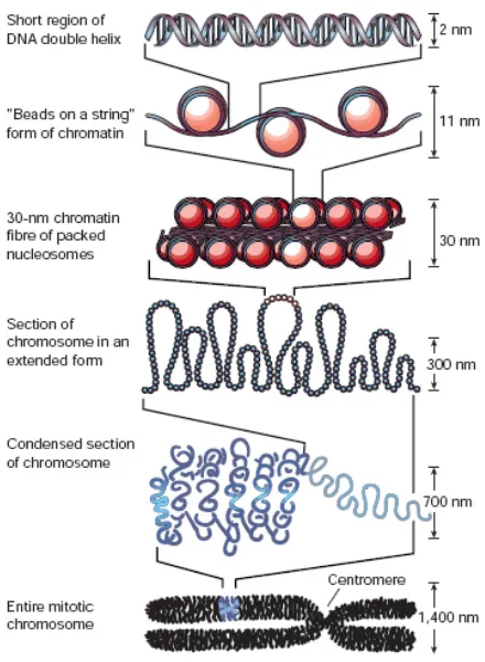
1.1.2 Higher order chromatin structures
The structure of chromatin is hierarchical (Figure 2). At the lowest level nucleosomes
on DNA can be envisaged as ‘beads on a string’, though the DNA wraps around the
nucleosome cores rather than passing through them (Arents et al., 1991; Luger et al.,
1997). This structure is known as the 10nm fibre corresponding to the diameter of
nucleosomes (Horn and Peterson, 2002; Wolffe and Hayes, 1999). Between each
nucleosome a linker histone such as H1 can associate with the DNA (Doenecke et al.,
1997; Horn and Peterson, 2002). Other nuclear proteins can also associate with either
the DNA or the histones (refer to 1.1.3). For example transcription factors, proteins that
bind to specific DNA sequences to regulate transcription, can function by altering
chromatin structure such as promoting loops in 10nm fibres (Green, 2005; Hahn, 2004;
Figure 2 Chromatin folding. The first level of compaction is the association of DNA with nucleosomes to create the 10nm ‘beads on a string structure’. This can condense into a 30nm diameter fibre. Further compaction of chromosomes is observed in vivo, 60-300nm diameter fibres are observed at interphase, these can be further compacted into fibres of around 700nm diameter and finally fully condensed mitotic chromosomes occur during mitosis and meiosis (Adapted from (Felsenfeld and Groudine, 2003)).
Interactions between nucleosomes and between nucleosomes and linker histones can
compact the 10nm ‘beads on a string’ into higher order structures (Belmont and Bruce,
1994) (Figure 3). In vitro compaction of 10nm fibres first produces 30nm fibres, and
30nm fibres are also observed in fixed cells (Belmont and Bruce, 1994; Gordon et al.,
2005). Interactions between H4 amino-terminal (N-terminal) tails and an H2A acidic
patch domain on adjacent nucleosomes are required for 30nm fibre formation
(Robinson and Rhodes, 2006). Currently, two models compete for the structure of the
30nm fibre (Figure 3). The solenoid model places nucleosomes into a simple helix
‘zigzag’ model the 10nm fibre is twisted (-71.3o rotation of adjacent nucleosomes) so
nucleosomes alternate between two halves of a two start helix (Bednar et al., 1998;
Dorigo et al., 2004; Schalch et al., 2005) (Figure 3). The zigzag model is supported by
cross-linking studies on arrays of 12 nucleosomes (Dorigo et al., 2004) and a low
resolution (9Å) crystal structure for an array of four nucleosomes condensed into a short
30nm fibre sufficient to determine the positions and orientations of nucleosomes
[image:21.612.251.413.267.407.2](Schalch et al., 2005).
Figure 3 Models of the 30nm Chromatin Fibre: (Left) In the solenoid model proposed by Rhodes and colleagues, the fibre is an interdigitated one-start helix (Robinson and Rhodes, 2006). Alternative helical gyres are coloured blue and magenta. The linker DNA has not been modelled. (Right) In the zigzag model suggested by Richmond and colleagues, the fibre is a two-start helix with the linker DNA criss-crossing between the adjacent rows of nucleosomes(Dorigo et al., 2004). Alternative gyres are coloured blue and orange. Views have the fibre axis running vertically. Image courtesy of D. Rhodes, figure from (Tremethick, 2007).
At higher ionic strengths in vitro the 30nm fibres oligomerise to form thicker
chromonema fibres (Gordon et al., 2005). Fibres 60-400nm in diameter are observed in
fixed cells at interphase (Belmont and Bruce, 1994). During mitosis condensed
chromatids (anaphase and telophase) have 400-600nm diameters (Gordon et al., 2005;
Woodcock and Dimitrov, 2001) (Figure 2), however the exact structures and formation
in vivo of fibres are unknown (Woodcock and Dimitrov, 2001).
In vivo chromatin domains can be classified by the degree of compaction as either
euchromatin or heterochromatin (Grewal and Jia, 2007). Euchromatin is largely
heterochromatin continues to be condensed throughout interphase and provides
regulated activation of some genes as well as repression (Grewal and Jia, 2007).
Heterochromatin is further divided into two classes, constitutive and facultative
heterochromatin (Sexton et al., 2007). Constitutive heterochromatin occurs at regions
along the DNA that are in this condensed form in all cells of the organism. Constitutive
heterochromatin forms on regions containing a high density of repetitive DNA
sequences such as the satellite sequences found within the constitutive heterochromatin
of the centromere. Facultative heterochromatin loci are differentially condensed during
development in different cell types. Heterochromatin has traditionally been associated
with transcriptional repression while euchromatin was associated with activation of
transcription. This notion was supported by work in several systems including
positional variegation and artificially inserting endogenously active genes within the
telomeres in D. melanogaster, and mating-type loci in S. cerevisiae (Laurenson and
Rine, 1992). However, for some genes heterochromatin formation is required for
activation because heterochromatin has structural diversity allowing some genes within
heterochromatin to be more accessible than others (Hahn, 2004).
Higher order chromatin structures beyond the degree of compaction of the 10nm fibres
include well-known large scale chromatin structures such as the condensed
chromosomes of mitosis. That individual chromosomes occupy specific domains within
the nucleus has been known for some time (Heard and Bickmore, 2007). More recently
it was discovered that chromatin from different chromosomes protrudes into the
inter-chromosome domain and within this domain gene-poor chromatin regions are more
centrally located (Heard and Bickmore, 2007; Misteli, 2007). Evidence has emerged for
high order structural organisation such as large scale looping of chromatin into
chromosomes (Akhtar and Gasser, 2007; Sexton et al., 2007). Chromatin dynamically
associates with the nuclear envelope with transcriptionally active chromatin
predominantly attached to nuclear pores (Akhtar and Gasser, 2007). To summarise,
chromatin structure is hierarchical, dynamic, and varies non-randomly across the
genome and the cells of the organism (Misteli, 2007).
1.1.3 Alterations to chromatin structure
That the nucleosome’s octomer core has subunits and contains sites for
post-translational modification allows variations in chromatin structure influencing
accessibility or affinity of chromatin components to each other or other proteins, such as
the transcriptional machinery, chromatin remodellers, transcription factors, and
enzymes responsible for histone post-translational modification, or
methylation/demethylation of the DNA, thereby altering transcription (Fan et al., 2002;
Kuo et al., 1998; Li et al., 1998; Nagy et al., 1997; Varga-Weisz et al., 1995;
Whitehouse et al., 1999). Chromatin higher-order folding is dependent on interactions
within and between nucleosomes, nucleosomes and linker histones, and the spacing of
nucleosomes along the DNA. At the nucleosome level these interactions can be
modified through chromatin remodelling machines, post-translational histone
modifications, and incorporation of histone variants. For example, in the condensed
structure of the centromere, H3 is replaced by centromere protein A (CENP-A)
(Sullivan and Karpen, 2001; Sullivan, 2001). Short interfering RNAs (siRNAs) also
have roles in targeting heterochromatin initiation sites, though at this point the
mechanism remains unknown (Grewal and Jia, 2007). In reality several alterations to
chromatin contribute to any functional state. For example in X. laevis histone
acetylation by p300 and CREB binding protein (CBP) at promoters for some
recruitment of the TATA-binding protein (a transcription factor), and eventual
recruitment of remodelling machinery for transcription (Hecht et al., 2000).
Recently, genes encoding proteins that remodel chromatin or post-translationally
modify histones were found to have a large number of connections (hub genes) within
genetic networks for developmental signalling genes in C. elegans (Lehner et al., 2006).
These hub genes were experimentally confirmed to be key modulators of numerous
biological processes, including developmental signalling, indicating the central role of
chromatin (Lehner et al., 2006).
1.1.3.1 Chromatin remodelling machines
Chromatin remodelling machines are multi-protein complexes containing an ATPase
subunit, ATP hydrolysis is necessary to mobilise otherwise stable nucleosomes (Bao
and Shen, 2007; Bouazoune and Brehm, 2006; Mohrmann and Verrijzer, 2005).
Chromatin remodelling outcomes include: separation of end-DNA from the
nucleosome, sliding of histone octomers relative to the DNA, removal of histones as
single proteins, dimers or octomers, and substitution of histone variants (Akey and
Luger, 2003; Belotserkovskaya et al., 2003; Jin et al., 2005; Li et al., 2007).
Nucleosomes can act as an obstacle to transcription, replication, and repair, or in some
cases to improve transcription by positioning factors and promoting access to DNA
(Ehrenhofer-Murray, 2004; Jin et al., 2005; Kireeva et al., 2002; Li et al., 2007). Yeast
studies have shown DNA surrounding transcription start sites is globally depleted of
nucleosomes; an outcome of remodelling machine action (Owen-Hughes and
Engeholm, 2007). The actions of remodelling machines alter the accessibility of DNA
to transcription factors, sites on histones for post translational modification, or other
protein-protein interactions (Hirschhorn et al., 1992; Horn and Peterson, 2002). Other
binding, complex assembly or stability, post translational modification, targeting of
specific chromatin regions, or protein binding (Mohrmann and Verrijzer, 2005).
Variation in remodelling machine subunit composition enables different complexes to
remodel chromatin in ways appropriate to a range of biological functions at specific
regions of the genome (Akey and Luger, 2003; Belotserkovskaya et al., 2003; Li et al.,
2007; Saha et al., 2006).
There are five families of chromatin remodelers; SWI/SNF, ISWI, NuRD, INO80 and
SWR1 (Driscoll et al., 2007; Felsenfeld and Groudine, 2003; Mueller et al., 1985;
Nathan et al., 2006; Peterson and Laniel, 2004; Xu et al., 2005; Zhang et al., 2003)
(Table 1, complex names are expanded in the List of Abbreviations). RNA and DNA
polymerases are chromatin remodelling machines directly involved in transcription
(Kireeva et al., 2002; Wolffe and Hayes, 1999). The remodelling complexes and their
individual subunits are conserved. For example two SWI/SNF family members, S.
cerevisiae SWI/SNF and H. sapiens PBAF, share multiple homologous subunits
including SWI2/SNF2 and BRG1, SWI3 and BAF55, and SNF5 subunits (Table 1).
Conservation of chromatin remodelling machinery indicates the importance of
chromatin remodelling for an organism’s survival. Interestingly, it has been shown that
remodelling machines show stages and tissues specific enrichment during X. laevis
development (Linder et al., 2004) and CHD4 has been demonstrated to have a
developmental role in X. laevis (Linder et al., 2007). Other proteins associated with
chromatin, such as those responsible for post translational modifications to histones, are
Remodelling complex family
Example remodelling complexes (species)
Known biological functions Known subunits (equivalents) References
SWI/SNF (S. cerevisiae)
PolII activation, elongation, double strand break (DSB) repair, gene activation and repression
SWI2/SNF2, SWP61/ARP7, SWP59/ARP9 SWI1/ADR6, SWI3, SWP73,
SNF5, SWP82, SWP29/TFG3/TAF30/ANC1, SNF6, SNF11. (Mohrmann and Verrijzer, 2005), (Mohrmann and Verrijzer, 2005)
SWI/SNF
PBAF (H. sapiens)
Development, tumour suppressor, cell cycle
BRG1 (SWI2/SNF2, STH1), Polybromo/BAF180 (RSC1, RSC2, RSC4), BAF170 and BAF55 (SWI3), BAF57 (BAP111), BAF60A OR BAF60B (SWP73), BAF53 (BAP55), Actin, HSNF5/INI1 (SNF5)
(Mohrmann and Verrijzer, 2005; Pepin et al., 2007; Saha et al., 2006)
CHRAC (H. sapiens) Nucleosome assembly and
spacing SNF2H (ISWI, SNF2L, ISW2), WCRF180, CHRAC15, CHRAC17, (Mohrmann and Verrijzer, 2005; Pepin et al., 2007; Saha et al., 2006)
ISWI
NuRF (H. sapiens)
Transcriptional activation SNF2L (ISWI, ISW2) , BPTF, RBAP46, RBAP48 (Bao and Shen, 2007; Saha et al., 2006)
INO80 (S. cerevisiae)
DNA double strand break repair (recruited by phosphorylated H2A.X), PolII activation, gene specific regulation.
INO80, RVB1, RVB2, ARP4, ARP5, ARP8, Actin, NHP10, ANC1/TAF14, IES1 to 6,
(Bao and Shen, 2007; Saha et al., 2006)
INO80
INO80
(A. thaliana)
Homologous recombination, gene transcription
INO80, RVB1, RVB2 (Bao and Shen, 2007; Saha et
al., 2006; Wu et al., 2005) SWR1
(S. cerevisiae)
DNA double strand break repair, Htz1 deposition,
SWR1 (DOM-A, SRCAP, P400), ARP4, ARP6, SWC3, SWC4/EAF2/GOD1, SWC5/AOR1, SWC6/VPS71, SWC7, SWR2/VP372, YAF9, BDF1, Actin, RVB1, RVB2
(Bao and Shen, 2007; Bouazoune and Brehm, 2006; Saha et al., 2006)
SWR1
Tip60
(D. melanogaster)
DNA double strand break repair,
H2A.Z deposition and acetylation DOM-A (SWR1, SRCAP, P400),TIP60, PON, REP, MRG15, H2AV, TRA1, GAS41, ING3, E(PC), H2B (Denslow and Wade, 2007; Saha et al., 2006) NuRD
(H. sapiens)
Transcriptional repression and silencing, histone deacetylation, development
Mi-2, MBD2 OR 3, HDAC1, HDAC2, RBA P46, RBAP48, P66Α OR P66Β,
MTA1, 2, OR 3 (MTA), (Bouazoune and Brehm, 2006; Denslow and Wade, 2007)
NuRD/Mi-2/CHD
NuRD (D.
melanogaster)
Transcriptional regulation, development
Mi-2, P55, RPD3 (HDAC1, HDAC2), MBD2 OR 3, MTA, P66/68 (Saha et al., 2006)
1.1.3.2 Post translational modification
Post translational histone modifications include phosphorylation (ph), ubiquitylation,
ADP-ribosylation, methylation (me), SUMOylation, and acetylation (ac) of histone
amino acid residues (Berger, 2007; Kouzarides, 2007). Proline isomerisation (Nelson et
al., 2006) and deamination (arginine to citrulline conversion) can also occur (Cuthbert
et al., 2004). Lysines (K) can be mono-, di- or tri-methylated (Me, Me2, Me3)
(Kouzarides, 2007). Arginines (R) can be mono- or di-methylated (Kouzarides, 2007).
Both canonical and variant histones can be modified (Kouzarides, 2007). Over 60
different residues on histones are known to harbour modifications (Kouzarides, 2007).
Modifications thus far described are mostly in tail regions (Xu et al., 2005), however
some residues within core domains can be modified, including H3 lysine 56 acetylation
(H4K56ac) in S. cerevisiae and methylation of the equivalent residue (H4K59me) in
mammals (Zhang et al., 2003). The number, sites, and types of post translational
modification taken together provide a system with huge combinatorial power. These
modifications are non-randomly distributed, most are reversible (no known enzyme can
reverse arginine deamination or arginine methylation (Berger, 2007; Kouzarides,
2007)), and can alter chromatin’s structural and functional properties providing a
sophisticated regulatory system for the genome. Post translational modification function
can be context dependent. Function can sometimes be determined directly by structure
or indirectly by reading by ‘adaptors’, that are often components of remodelling
machinery.
Post translation modification is required for in vivo chromatin assembly. Nucleosome
assembly requires (H3/H4)2 tetramers with particular post translational modifications
(Sobel et al., 1995). Histone post translational modification can affect higher order
to the 30nm fibres formation and higher order folding of these fibres (Shogren-Knaak et
al., 2006). All core histone tail domains can shield the charge of DNA though they
repress or potentiate oligomerisation of 30nm fibres into larger structures largely
through other undefined interactions (Gordon et al., 2005; Wolffe and Hayes, 1999).
Post-translational modification can modify transcription directly, indirectly or both.
Directly, by altering electrostatic charge or interactions between nucleosomes or
histones and DNA, thus changing the accessibility of targets for DNA binding proteins
(Becker, 2006; Peterson and Laniel, 2004). For example, histone hyperacetylation
results in 10nm fibres decompaction providing access for transcription factors, one
mechanism of this decompaction, alteration of the charge of the H4 N-terminal tail (free
amino group terminal of an amino acid chain) by H4K16ac (acetylation neutralises the
basic lysine) weakens interactions with the acidic patch of H2A on adjacent
nucleosomes (Shogren-Knaak et al., 2006). An example of alteration to histone–DNA
electrostatic interactions may be H3K56ac, since acetylation neutralises this residue’s
charge and fills most of the DNA-histone gap (Bernstein and Hake, 2006;
Ehrenhofer-Murray, 2004; Shogren-Knaak et al., 2006). Post-translational modifications can act
indirectly by altering/creating binding sites for chromatin binding proteins or complexes
(Berger, 2007). For example H3K9me at promoter regions recruits HP1, promoting
repressive heterochromatin formation (Ayyanathan et al., 2003) and H4K16ac prevents
the action of the ACF remodelling complex (Shogren-Knaak et al., 2006).
Traditionally, particular post translational modifications at specific sites were correlated
with either transcription or repression. More recently it has become apparent that some
post translational modifications can recruit transcriptional activators and repressors. For
example H3K9 methylation in mammals is restricted to promoter regions of inactive
2005), the mark is also enriched in pericentric heterochromatin where it is thought to
promote silencing (Schotta et al., 2004). Variable function is due to the chromatin
context, including variable protein-protein interaction sites, and cell type specific
availability of associating proteins (Li et al., 2007; Peterson and Laniel, 2004). Context
for post translational modifications can also be established by post translational
modifications affecting each other, this can occur in several ways. Firstly, multiple
modifications cannot occur on the same site so they are mutually exclusive, for example
SUMOylation is repressive to transcription because it prevents acetylation or
ubiquitylation of the modified residue (Nathan et al., 2006). Secondly, interference by
adjacent modifications, for example binding of HP1 to H3K9me is blocked by H3S10ph
(S: serine) (Fischle et al., 2005). Thirdly, alteration of an enzyme’s recognition epitope
reduces the enzyme’s catalytic activity, for example isomerisation of H3P38 affects
H3K36me by Set2 (Nelson et al., 2006). Also additional modifications enhance
recognition of binding site by enzymes, for example the HAT GCN5 may recognise H3
better if H3S10 is phosphorylated (Lo et al., 2000). Finally, combinations of
modifications on different tails can act together, for example H3K9me and H4K20me3
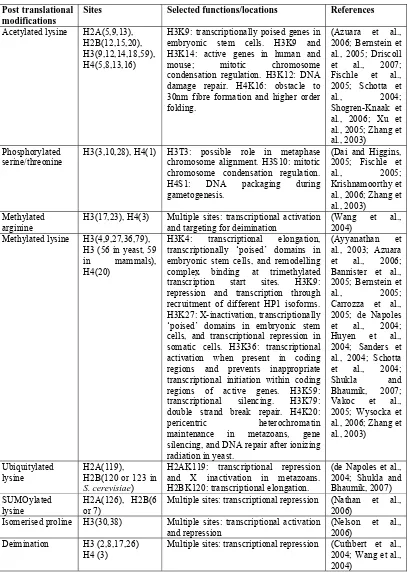
Post translational modifications
Sites Selected functions/locations References
Acetylated lysine H2A(5,9,13), H2B(12,15,20), H3(9,12,14,18,59), H4(5,8,13,16)
H3K9: transcriptionally poised genes in embryonic stem cells. H3K9 and H3K14: active genes in human and mouse; mitotic chromosome condensation regulation. H3K12: DNA damage repair. H4K16: obstacle to 30nm fibre formation and higher order folding.
(Azuara et al., 2006; Bernstein et al., 2005; Driscoll et al., 2007; Fischle et al., 2005; Schotta et al., 2004; Shogren-Knaak et al., 2006; Xu et al., 2005; Zhang et al., 2003)
Phosphorylated serine/threonine
H3(3,10,28), H4(1) H3T3: possible role in metaphase chromosome alignment. H3S10: mitotic chromosome condensation regulation. H4S1: DNA packaging during gametogenesis.
(Dai and Higgins, 2005; Fischle et al., 2005; Krishnamoorthy et
al., 2006; Zhang et al., 2003)
Methylated arginine
H3(17,23), H4(3) Multiple sites: transcriptional activation and targeting for deimination
(Wang et al., 2004)
Methylated lysine H3(4,9,27,36,79), H3 (56 in yeast, 59 in mammals), H4(20)
H3K4: transcriptional elongation, transcriptionally ‘poised’ domains in embryonic stem cells, and remodelling complex binding at trimethylated transcription start sites. H3K9: repression and transcription through recruitment of different HP1 isoforms. H3K27: X-inactivation, transcriptionally ‘poised’ domains in embryonic stem cells, and transcriptional repression in somatic cells. H3K36: transcriptional activation when present in coding regions and prevents inappropriate transcriptional initiation within coding regions of active genes. H3K59: transcriptional silencing. H3K79: double strand break repair. H4K20:
pericentric heterochromatin maintenance in metazoans, gene
silencing, and DNA repair after ionizing radiation in yeast.
(Ayyanathan et al., 2003; Azuara et al., 2006; Bannister et al., 2005; Bernstein et
al., 2005; Carrozza et al., 2005; de Napoles et al., 2004; Huyen et al., 2004; Sanders et al., 2004; Schotta et al., 2004; Shukla and Bhaumik, 2007; Vakoc et al., 2005; Wysocka et al., 2006; Zhang et al., 2003)
Ubiquitylated
lysine H2A(119), H2B(120 or 123 in
S. cerevisiae)
H2AK119: transcriptional repression and X inactivation in metazoans. H2BK120: transcriptional elongation.
(de Napoles et al., 2004; Shukla and Bhaumik, 2007) SUMOylated
lysine H2A(126), H2B(6 or 7) Multiple sites: transcriptional repression (Nathan et al., 2006) Isomerised proline H3(30,38) Multiple sites: transcriptional activation
and repression (Nelson et al., 2006) Deimination H3 (2,8,17,26)
[image:30.612.131.538.102.681.2]H4 (3) Multiple sites: transcriptional repression (Cuthbert et al., 2004; Wang et al., 2004)
1.1.3.3 DNA methylation
As well as histone post translational modification, the chromatin’s DNA component can
be chemically altered by methylation of cytosine (Doerfler, 1981; Wyatt, 1951),
typically at CG dinucleotides or CNG trinucleotides (Doerfler, 1981; Grandjean et al.,
2007), though some cytosine methylation is targeted to other sites (Grandjean et al.,
2007). DNA methylation is generally associated with transcriptional repression
(Doerfler, 1981; Naveh-Many and Cedar, 1981) and commonly occurs in repeats
(Bestor, 2000; Doerfler, 1981; Roizes, 1976), transposons (Bestor, 2000; Walsh et al.,
1998), and within genes (Bestor, 2000; Doerfler, 1981; Miller et al., 1978). DNA
methylation is excluded from most promoter regions at CpG islands (Bestor, 2000;
McClelland and Ivarie, 1982) further linking DNA methylation with transcriptional
regulation.
1.1.3.4 Histone variants
The canonical histones make up the bulk of the histone content in all mitotic eukaryotic
cells and are defined by several features: they have multiple gene copies in clustered
cassettes in the genome, the genes contain introns, the non-polyadenylated transcripts
are produced exclusively in S-phase, and the proteins are incorporated behind the
replication fork during this period (Bernstein and Hake, 2006; Jin et al., 2005). Histone
variants all have the histone fold though they need not have all the features of canonical
histones listed above (Jin et al., 2005). Variants have different tails and can alter the
octomer core structure, therefore variant incorporation can alter chromatin function by
changing post translational modification sites, protein-protein interactions with
remodellers or transcription factors, or by directly altering chromatin structure (eg.
10nm fibre compaction) and thereby DNA accessibility (Fan et al., 2002; Jin et al.,
2005). These changes to function can be global, regional, or loci specific. Some
examples: the variant H1o globally represses transcription in early
development (Costanzi et al., 2000), CENP-A (an H3 variant) is required for
centromere formation (Suto et al., 2000), while the variant H1b interacts with Msx1 to
form a complex that binds the Core Enhancer Region (CER), an important regulatory
DNA sequence for MyoD (Lee et al., 2004), to induce a repressive chromatin state
(Bernstein and Hake, 2006).
Variant histones can replace H2A, H2B, H3, or linker histones but no H4 variants have
been uncovered. Most known variants are of the H2A histone family (Costanzi et al.,
2000). A selection from the numerous H2A variants known is given in Table 3. The
most abundant H2A variants are H2A.Z and H2A.X (Costanzi et al., 2000). Studies in
multiple organisms have found H2A.Z makes up approximately 10% of the H2A
content (Jin et al., 2005; Kobor et al., 2004; Mizuguchi et al., 2004; Redon et al., 2002).
The variant H2A.Z is the focus of this project.
H2A variant Function/expression. Reference
MacroH2A1.2 Concentrated on the inactive X of female mammals. (Costanzi et al., 2000) H2A.Bbd Almost completely excluded from the inactive X
chromosome in mammals.
(Chadwick and Willard, 2001)
H2AX Phosphorylated at double strand breaks in mammals. Present in X. laevis oocytes.
(Paull et al., 2000; Rogakou et al., 1998)
H2AvD D. melanogaster H2A.Z and has functional activities of
both H2A.Z and H2AX. (Madigan et al., 2002)
H2A.Z Plays a structural role in centric chromatin and is
associated with transcriptional regulation. (Farris et al., 2005; Greaves et al., 2007; Thatcher and Gorovsky, 1994)
Table 3 Selected H2A variants adapted from (Bernstein and Hake, 2006).
1.2 The histone variant H2A.Z
1.2.1 Gene and transcript
The H2A.Z gene is highly conserved. Orthologs for H2A.Z have been found in many
organisms including mammals (Hatch and Bonner, 1990), chicken (Harvey et al., 1983),
trout (Nickel et al., 1987), C. elegans (Updike and Mango, 2006), sea urchin (Ernst et
al., 1987), yeast (Carr et al., 1994), tetrahymena (Allis et al., 1986), and the slime
region has been defined containing six transcription factor binding sites; three CCAAT
(sites for CCAAT box family transcription factors), two GC (GGGCGG binding sites
for transcription factor Sp1), and a TATA box (Hatch and Bonner, 1996). In addition to
this proximal promoter two more distal transcription factor binding sites exist between
-234 to -361bp from the transcription start site (Hatch and Bonner, 1990). Some sites
are conserved between species (Bernstein and Hake, 2006). In X. laevis the major
H2A.Z transcript is ~869bp not including the poly-A tail (Iouzalen et al., 1996) with a
~381bp translated region (Iouzalen et al., 1996). H2A.Z transcription is not limited to S
phase as with canonical core histones; a conclusion supported by the transcript’s lack of
the 3’ stem loop structure found in S-phase transcribed histones (Hatch and Bonner,
1990). The transcribed region contains introns; four in human and chicken (Hatch and
Bonner, 1990). Intron and exon lengths vary between species (Hatch and Bonner,
1990). In the human gene these introns are 276 to 438bp in size (Hatch and Bonner,
1990). The gene also contains a polyadenylation signal (Ernst et al., 1987; Hatch and
Bonner, 1990; Iouzalen et al., 1996) and the transcript is polyadenylated in most species
(Hatch and Bonner, 1990). In X. laevis, H2A.Z mRNA has polyadenylated and
non-polyadenylated forms (Iouzalen et al., 1996), which is likely to affect mRNA stability
and translation in vivo (Allende et al., 1974; Beilharz and Preiss, 2007).
1.2.2 Protein
When translated from the mRNA transcript described above, a 127aa, 14.1kDa protein
is produced (Ernst et al., 1987; Iouzalen et al., 1996). The H2A.Z amino acid sequence
is sufficiently conserved from canonical H2A (~57%) to replace the canonical histone
within the nucleosome (the histone fold is conserved) and includes the ‘H2A box’
identifying it as a H2A variant (Jackson et al., 1996). H2A.Z’s amino acid sequence is
fact H2A.Z is more conserved than canonical H2A (Thatcher and Gorovsky, 1994).
Despite small differences in codon usage, H2A.Z’s protein sequence is 100% identical
for rat, bovine, and human (Clarkson et al., 1999).
Any roles specific to H2A.Z must be linked to one or more differences from canonical
H2A. Differences in protein sequence compared to canonical H2A include alterations to
the core and tail regions (Harvey et al., 1983). The H2A.Z-containing nucleosome’s
crystal structure shows changes to the amino acid sequence alter nucleosome stability
and surface features (Figure 4) (Suto et al., 2000). An acidic patch on the nucleosome
surface is extended in H2A.Z by alterations in the C-terminal α-helix (Figure 4) (Suto et
al., 2000). The acidic patch interacts with the H4 tail of adjacent nucleosomes (Fan et
al., 2002). Consistent with the predicted H2A.Z-H4 inter-nucleosome interaction, in
vitro H2A.Z facilitates 30nm fibre compaction (Fan et al., 2002). Changes to the L1
loop H2A-H2A interaction site favour incorporation of a second H2A.Z (Suto et al.,
2000). A conserved histidine motif (HIH) within the core region bound a metal ion in
crystal structure studies (Figure 4) (Suto et al., 2000). In vivo the HIH motif is likely to
be a protein-protein interaction site (Suto et al., 2000). Three hydrogen bonds removed
from the nucleosome interior results in less stable H2A-H3 interaction (Park et al.,
2004). However, experimentally H2A.Z incorporation does not produce destabilisation,
Figure 4 H2A.Z incorporation alters nucleosome structure. Comparison of nucleosomal surfaces of H2A.Z (a) and H2A (b) containing nucleosome cores at 2.6Å resolution. Electrostatic potentials at the accessible solvent surface (1.4Å, the radius of a water molecule, from the nucleosome surfaces) colour coded from +7.0 (deep violet) to -7.0 (deep red) kcal mol -1 e-1 have been mapped onto the nucleosomal surface (Getzoff et al., 1983). Molecular surfaces
were modelled with MSMS (Sanner et al., 1996) using a 1.4Å spherical probe. The black arrow indicates the acidic patch (deep red region) on the nucleosome surface that in H2A.Z nucleosomes is extended compared to canonical nucleosomes. The coloured green area on ‘a’ is the manganese ion bound to the HIH motif not present on the surface of canonical nucleosomes. Figure adapted from (Suto et al., 2000).
1.2.3 Incorporation of H2A.Z into chromatin
This section discusses H2A.Z incorporation into chromatin from the initial targeting and
assembly into the nucleosome through to the consequences for higher order structure.
Interestingly, in vitro H2A.Z incorporation inhibits 30nm fibre oligomerisation (Fan et
al., 2002). H2A.Z is enriched in centric and pericentric heterochromatin, and binds
INCENP (inner centromere protein), which is associated with pericentric
heterochromatin (Carr et al., 1994; Greaves et al., 2007; Krogan et al., 2004;
Rangasamy et al., 2003; Rangasamy et al., 2004). H2A.Z plays a structural role in
centromeres which may explain why H2A.Z depletion leads to chromosome segregation
defects and genome instability (Rangasamy et al., 2004). Clearly, H2A.Z assembly can
H2A.Z incorporation is non-random (indicating the process is targeted) and occurs
throughout the cell cycle (Leach et al., 2000; Raisner et al., 2005). Indeed H2A.Z
incorporation requires chromatin remodelling machinery and chaperones distinct from
canonical H2A (Luk et al., 2007; Ruhl et al., 2006; Wu et al., 2005). H2A.Z localisation
is related to possible structural and functional roles, such as mitotic chromosome
segregation (Rangasamy et al., 2004) and transcriptional regulation (Meneghini et al.,
2003; Raisner et al., 2005; Santisteban et al., 2000). H2A.Z must therefore be targeted
to specific chromosomal regions. Work done largely in yeast is now starting to produce
a detailed picture of how H2A.Z is targeted to and incorporated into chromatin by an
ATP-dependent chromatin remodelling machine (Jin et al., 2005). In yeast this complex
is called SWR1 (named for the Swr1 ATPase subunit), in human SRCAP, and in
Drosophilamelanogaster (D. melanogaster) dTip60 (named for its Tip60 histone acetyl
transferase subunit) (Jin et al., 2005; Krogan et al., 2003; Mizuguchi et al., 2004; Ruhl
et al., 2006).
H2A.Z is assembled into nucleosomes flanking transcription initiation sites containing a
22bp DNA sequence within promoters (Raisner et al., 2005). However, exactly how
H2A.Z and SWR1 are targeted to other heterochromatin regions remains unknown (Jin
et al., 2005). It has been proposed that histone post translational marks allow a
component of SWR1 to bind to nucleosomes, candidates include subunits Swc6 and
Arp6, which are required to bind SWR1 to nucleosomes (Wu et al., 2005) and Bdf1 and
2 which bind acetylated H4 (Raisner et al., 2005) (see Table 1 section 1.1.3.1). This
then recruits the other SWR1 complex subunits (Jin et al., 2005; Mizuguchi et al.,
2004). SWR1 then facilitates the removal of the H2A/H2B dimer, though the precise
sequence alone is not sufficient to recruit H2A.Z and therefore requires a second
currently undescribed mechanism for H2A.Z recruitment (Raisner et al., 2005).
In yeast, the H2A.Z/H2B dimer is associated with a chaperone, Nap1 or the H2A.Z
specific Chz1 (called HIRIP3 in metazoans), that binds to the Swc2 (YL-1 in
metazoans) SWR1 component (Luk et al., 2007; Wu et al., 2005), then SWR1
exchanges H2A.Z/H2B into the nucleosome structure (Mizuguchi et al., 2004; Wu et
al., 2005). Swc2 binds to the H2A.Z α-C-helix, which differs from the corresponding
domain in core H2A, demonstrating that this is an H2A.Z specific interaction (Luk et
al., 2007; Wu et al., 2005). Due to changes in the L1 loop that prevent canonical H2A
and H2A.Z from co-existing in a nucleosome a second H2A.Z is incorporated into the
nucleosome (Suto et al., 2000).
1.2.4 H2A.Z and transcription
H2A.Z is implicated in transcriptional regulation by its non-random distribution
throughout different organisms’ genomes, though localisation of H2A.Z offers
conflicting indications of its role in transcription (Jin et al., 2005; Redon et al., 2002). In
Tetrahymena H2A.Z is localised to the transcriptionally active macronucleus (Allis et
al., 1986) while in yeast Htz (the H2A.Z homologue) has been associated with silencing
and activation (Meneghini et al., 2003; Raisner et al., 2005; Santisteban et al., 2000).
More specifically in yeast, Htz is enriched at particular DNA motifs associated with
promoters, typically immediately downstream of transcription start sites (Albert et al.,
2007; Raisner et al., 2005) and to euchromatin adjacent to silenced regions where it
prevents the spread of silencing (Meneghini et al., 2003; Millar et al., 2006). The
positioning of H2A.Z-containing nucleosomes on promoter DNA in yeast is more
variable than in other genomic regions, suggesting that H2A.Z positioning nucleosomes
organism (Albert et al., 2007). H2A.Z is also found in nucleosomes flanking the
transcription start site within both active and inactive promoters (Raisner et al., 2005).
H2A.Z is associated with both transcriptionally repressive and active histone post
translational modifications. In yeast Htz interacts genetically with the pathway for H3
lysine 4 methylation (H3 K4me) to produce more pronounced phenotypes than
mutations in either alone (Ng et al., 2003). H3 K4 dimethylation (H3K4me2) correlates
with genes poised for activation, while H3 K4 trimethylation (H3K4me3) is associated
with recent transcription by polymerase II (Ng et al., 2003). Efficiency of H2A.Z
incorporation flanking transcription start sites is increased by H3K4 methylation,
H3K79 methylation, and H4K16 acetylation, marks associated with transcription, but
these marks are not essential for H2A.Z incorporation (Raisner et al., 2005). H2A.Z is
also less often SUMOylated than canonical H2A (Nathan et al., 2006). SUMOylation is
a repressive post-translational modification (Nathan et al., 2006). H2A.ZK14ac is
enriched at transcribed genes, while hypoacetylated H2A.Z is associated with repression
(Millar et al., 2006).
H2A.Z localisation in several metazoan genomes offers similarly contradictory
evidence. In D. melanogaster H2Av is more widely, though still non-randomly, spread
over the genome (Leach et al., 2000) and is associated with silenced regions
(Swaminathan et al., 2005). In mice H2A.Z is enriched in the compact gene-poor
pericentric and centric heterochromatin (Greaves et al., 2007; Rangasamy et al., 2004).
Potentially H2A.Z has a role in genome stability (Greaves et al., 2007; Rangasamy et
al., 2004). Whether H2A.Z plays a role in transcriptional silencing at the centromere or
has a purely structural role in this region remains an open question. Additionally,
vertebrate H2A.Z distribution is not limited to the centromere. Evidence from
genes and downstream of transcription start sites (Farris et al., 2005). In cells not
expressing the gene c-myc, H2A.Z is present in both the promoter and transcribed
regions, though depleted toward the transcribed region’s 3’ end. When transcription is
induced H2A.Z is depleted from the transcribed region (Farris et al., 2005). In the
housekeeping gene GAPDH (Glyceraldehyde 3-phosphate dehydrogenase), H2A.Z was
present within the promoter not the transcribed region (Farris et al., 2005). As already
noted, exactly how H2A.Z is targeted to specific genomic regions is unclear (see section
1.2.3).
Two mechanisms have been proposed for transcriptional modulation by H2A.Z. Firstly,
that H2A.Z containing regions act as a barrier to the spread of silencing, the variant
histone could be involved in the maintenance and/or establishment of boundary
structures, into actively transcribed regions (Meneghini et al., 2003). Secondly,
remodelling of H2A.Z-containing nucleosomes is required for transcription (Farris et
al., 2005).
The second mechanism involves H2A.Z exchange for H2A. As already discussed,
H2A.Z-containing nucleosomes are more stable in vitro than their canonical
counterparts (Park et al., 2004) (see section 1.2.3), therefore exchange of H2A.Z for
H2A and vice versa may alter the accessibility of DNA associated with the nucleosome.
Initially, H2A.Z is incorporated into nucleosomes near promoters, then H2A.Z is
removed from the nucleosome and transcription occurs, however the timing and
detailed mechanisms remain unknown (Jin et al., 2005).
One possible model, suggested by Farris (2005), for H2A.Z’s role in transcription is that
H2A.Z incorporation is repressive and marks regions poised for transcription. With
H2A.Z incorporation (see also 1.2.3), genes may be transcriptionally poised because
(Park et al., 2004). Favouring the 30nm fibre may also contribute to establishing
chromatin structures that prevent the spread of silencing. Within the transcribed region
the presence of stable H2A.Z nucleosomes (Park et al., 2004) is thought to repress
transcription (Farris et al., 2005). To extend this model, the variable positioning of
H2A.Z nucleosomes within promoters (Albert et al., 2007) may modulate
transcriptional regulation. As can be surmised from this model, our understanding of
H2A.Z in transcriptional regulation has come a long way since the early studies that
correlated H2A.Z with either repression or activation. However, we do not yet have
complete understanding of the molecular mechanisms behind the models. How H2A.Z
is targeted to particular genomic regions and the details of H2A.Z exchange for H2A
upon transcription remain unclear.
1.3 Chromatin and transcriptional regulation during
metazoan development
Chromatin has essential roles in the control of gene transcription throughout metazoan
development. Often early embryonic transcription is globally repressed until after a
specific developmental stage, such as the mid-blastula in X. laevis (see section 3.1.1.2).
Histone variants, as well as other mechanisms that alter chromatin structure, have been
implicated in the regulation of key developmental genes and global changes to
transcription. For example, many transcription factors are either implicated in
development, and/or have developmentally regulated expression (Latinkic and Smith,
1999; Saka et al., 2000; Tada et al., 1998; Tintignac et al., 2004; Veenstra et al., 2000).
In metazoans distinct histone combinations are found in germ, undifferentiated
embryonic, and different somatic cell types (Bouvet et al., 1994; Dimitrov et al., 1993).
This section presents an overview of changes to chromatin throughout early
In X. laevis the first twelve divisions are synchronous, and proceed without transcription
or gap phases (Graham, 1966; Newport and Kirschner, 1982). These ‘cleavage
divisions’ are facilitated by large maternal stores of histone protein that contribute to a
globally repressive chromatin structure (Prioleau et al., 1994). Germ cell DNA is highly
methylated. Methylation at specific gene promoters decreases during the cleavage
divisions eventually activating these genes thereby inducing the mid-blastula transition
(Stancheva et al., 2002).
After the cleavage divisions, the mid-blastula transition (MBT) marks the beginning of
zygotic transcription. Chromatin at the MBT has lost most of the maternal proteins
including the maternal store of core histones. Titration out of these maternal histones by
DNA replication contributes globally to less repressive chromatin structures
immediately post MBT (Almouzni and Wolffe, 1995; Prioleau et al., 1994). Later
transcriptionally permissive histone post-translational modifications that characterise
pre-MBT blastomeres are replaced with modifications, such as H4 hyperacetylation that
establish transcriptionally repressed domains (Kikyo and Wolffe, 2000). The
importance of post translational modifications to development is dramatically
demonstrated by experimental global histone hyperacetylation (such as by TSA
treatment, (Stewart et al., 2006)), which prevents proper gastrulation in X. laevis by
perturbing transcriptional regulation (Almouzni et al., 1994). With each cell division a
decline in the chromatin’s totipotent character occurs as new patterns of chromatin
causing tissue specific expression are established (Kikyo and Wolffe, 2000). These
alternations continue into gastrulation transforming totipotent into somatic cells (Kikyo
and Wolffe, 2000).
Histone variant expression is often developmentally regulated (Aul and Oko, 2001;
large stores of the variant H2A.X that are titrated out as cell divisions proceed
(Dimitrov et al., 1994; Ohsumi and Katagiri, 1991). Another example is B4, the
dominant linker histone at the mid-blastula transition that is replaced by the end of
gastrulation by H1 (Dimitrov et al., 1993). Moreover, histone variants have functional
implications for development, for example H1b is required for regulation of MyoD, a
key developmental gene (Lee et al., 2004).
1.4 H2A.Z is essential for metazoan development
Several lines of evidence suggest a developmental role for H2A.Z in metazoans. Firstly,
knock-out experiments have shown H2A.Z is essential in organisms including fly and
mouse (Faast et al., 2001; van Daal and Elgin, 1992), though not yeast (Carr et al.,
1994; Jackson and Gorovsky, 2000), indicating that H2A.Z may have additional
functions in more complex organisms. Secondly, H2A.Z expression is developmentally
regulated in metazoans (Ernst et al., 1987; Harvey et al., 1983; Iouzalen et al., 1996;
Rangasamy et al., 2003). Taken together, evidence suggests H2A.Z might regulate
developmental transcription or have a more general function in chromosome
segregation.
1.4.1 H2A.Z is targeted to a selection of foregut genes during
C.
elegans
development
In Caenorhabditis elegans (C. elegans)H2A.Z and the remodelling complex SWR1 are
recruited to a subset of foregut gene promoters in vivo when transcription is initiated
(Updike and Mango, 2006). Only the tissue specific genes Myosin-2 (myo-2) and
R07B1.9 were identified within this subset (Updike and Mango, 2006). Reduction of
H2A.Z levels by RNAi results in delayed expression of these genes, defects in
lethal (Updike and Mango, 2006). This observation is consistent with the proposed
‘poised’ chromatin state of H2A.Z-containing nucleosomes (see section 1.2.4).
1.4.2 Essential regions of H2Av during fly development
In D. melanogaster the variant H2Av is within the H2A.Z family and is also
phosphorylated at double strand DNA breaks, an H2A.X function in mammals (Paull et
al., 2000) and of canonical H2A in yeasts (Madigan et al., 2002). Following the work
that determined that H2Av was essential in D. melanogaster (van Daal and Elgin, 1992)
rescue experiments identified protein regions important to fly development (Clarkson et
al., 1999). A region including the C-terminal α-helix was found to be essential (region
M6) and three regions resulting in partial rescue were also identified (Clarkson et al.,
1999). These were: portions of the C-terminal (region M7); the N-terminal tail
including important post-translational modification sites (M1); and a portion of the
histone fold α2 helix (M4) (Clarkson et al., 1999). Regions M6 and M7 were essential
for survival into adulthood (Clarkson et al., 1999) (Figure 5).
The essential region M7 contained a conserved histidine motif (Rangasamy et al., 2003)
(see section 1.2.3). Since H2A.Z’s histidine motif is conserved in metazoans though not
in yeast (Figure 5) and also conserved in C. elegans (swissprot: locus H2AV_CAEEL,
accession Q27511.3)), this indicates that H2A.Z has acquired an additional function in
metazoans (Suto et al., 2000). The histidine motif is likely to be a protein-protein
interaction site in vivo (Suto et al., 2000) possibly with transcriptional consequences for
the developmental regulation of genes (see section 1.2.4).
1.4.3 H2A.Z localisation in early mouse development
H2A.Z transcription and localisation are developmentally regulated in the mouse.
Semi-quantitative RT-PCR from different parts of the early mouse blastocyst found that the
inner cell mass was depleted of H2A.Z mRNA at least twelve-fold compared to
trophoblast cells (Rangasamy et al., 2003). When cell differentiation begins in the
mouse embryo H2A.Z is first localised to the pericentric heterochromatin in specific
cells then is localised to other nuclear regions except the nucleolus (Rangasamy et al.,
2003). In these studies, the M6 region was determined to be essential for INCENP
binding (Rangasamy et al., 2003). However, M7 region containing the histidine motif
was not necessary (Rangasamy et al., 2003). This indicates that H2A.Z’s general
structural role at the centromere (discussed in section 1.2.3) may be initiated in early
mammalian zygotic cells and indicates that H2A.Z may have additional separate roles
that rely on distinct features of the H2A.Z containing nucleosome surface.
H2A.Z expression is also developmentally regulated in mouse spermatogenesis. The
protein and transcript levels that increase more than five- and two-fold respectively by
pachytene are not detected in mature spermatozoa (Greaves et al., 2006). The timing of
H2A.Z expression correlates with meiotic sex chromosome inactivation (Greaves et al.,