1
Investigations into the
Significance of Nrf2 Signalling and
Ubiquitination of Proteins in
Respiratory Syncytial Virus
Infection.
Thesis submitted in accordance with the
requirements of the University of Liverpool for the
degree of Master in Philosophy (MPhil)
By
Dorota Alicja Chudek
3
Acknowledgements
I would first of all like to extend my gratitude to my
supervisors, Dr Paul McNamara and Dr Brian
Flanagan for the opportunity to undertake this
project as well as the ongoing support and
encouragement they provided. I appreciate the time
and patience both supervisors devoted towards my
project and am very thankful for the possibility to
present my work at the European Respiratory
Society Meeting in Amsterdam. I would also like to
express thanks to everyone in the laboratory at the
Institute of Child Health for support during my lab
work and making me feel welcome. I would
5
ABSTRACT:
Introduction and Objectives:
Respiratory Syncytial Virus (RSV) is the commonest cause of bronchiolitis in infants. This negative strand RNA virus is known to infect and replicate in the airway epithelium. RSV infection induces elevated levels of reactive oxidative species and subsequent oxidative stress injury in the lungs. Nrf2, a transcription factor that regulates antioxidant protein expression, has an important role in preventing pulmonary oxidative damage. Sulforaphane is a potent, naturally occurring inducer of NRF2 found in vegetables such as broccoli. In this study we sought to determine whether Nrf2 induction by sulforaphane might reduce RSV replication in airway epithelial cells. We also selected six proteins including MAVS, DDX21, RPS10, prohibitin, annexin A1, HMGB1 from proteomics defining changes in their level of ubiquitination following RSV infection. Our aim was to determine which proteins change their level of polyubiquitination following the infection. This could help identify new biochemical pathways involved in the host defence or viral replication and new targets for potential therapeutic intervention.
Method:
BEAS2B cells were infected with RSV at MOI of 1 following pre-treatment with sulforaphane. Samples were harvested at time points 24 and 48 hours and
analysed by Western Blotting for NrF2 and RSV. In addition, RT-qPCR was carried out for RSV quantification using an RSV N primer. A549 cells were infected with various concentrations of RSV (1:4-4:1). Samples were harvested at time point of 4 and 24 hours and analysed by Western Blotting using antibodies to ubiquitin and target proteins selected from ubiquitination proteomics data . Immunoprecipitation was used to confirm ubiquitination of these proteins and immunohistology to confirm their cellular localisation. Proteasome activity was inhibited using MG132 a specific, potent, reversible, and cell-permeable proteasome inhibitor.
Results:
6 Conclusions:
RSV infection changes expression of target proteins in A549 cells and might have influence on their ubiquitination, however, most probably it does not affect
7
Abbreviations:
%- Percentage
°C- Degree Celsius
µl- Microlitre
AOE- Antioxidant enzyme
BCA- Bicinchoninic acid assay
BSA- Bovine Serum Albumin
CCA - Chimpanzee Coryza Agent
COPD- chronic obstructive pulmonary disease
CO2- Carbon dioxide
DAMP - Damage-Associated molecular Patterns
DMEM- Dulbecco’s Modified Eagles Medium DMEM DNA - Deoxyribonucleic Acid
DTT- Dithiothreitol
DUBs- De-ubiquitinising enzymes
EDTA- Ethylenediaminetetraacetic acid
FCS- Foetal Calf Serum
hMPV- Human Metapneumovirus
IFN – Interferon
KSHV- Kaposi's sarcoma-associated herpesvirus
8
L- Litre
L protein- Large Protein
LRT - Lower Respiratory Tract
mL- Mililitre
mM- Milimolar
M protein- Matrix protein
Maf- Musculoaponeurotic fibrosarcoma
MVB- Multivesicular body pathway
MOI- Multiplicity of Infection
MW- Molecular Weight
N protein- Nucleocapsid protein
NF-ҡB - Nuclear Factor Kapper B NF-κB (nuclear factor kappa-light-chain-enhancer of activated B cells)
NK - Natural Killer Cells
NRF2- Nuclear factor (erythroid-derived 2)-like 2
NS1- Non Structured Protein 1
NS2- Non Structured Protein 2
ORF1- Open reading frame
PAMP- Pathogen associated molecular patterns
9
PFU- Plaque Forming Units
P protein – Phosphor protein
PVDF- Polyvinylidene fluoride
PreF- Pre-fusion form
PostF- Post- fusion form
qPCR- quantitative PCR
RNP- Ribonucleoprotein complex
RNA - Ribonucleic Acid
ROS- Reactive oxygen species
RSV - Respiratory Syncytial Virus
SDS- Sodium dodecyl sulphate
SH- small hydrophobic protein
SOD- Superoxide dismutase
TBS-T- Tris-Buffered Saline and Tween 20
TLR- Toll- like receptors
UBL- Ubiquitin like domain
UBA- Ubiquitin associated domains
10
Table of Contents
1. Introduction………..18
1.1. RSV………..18
1.1.1. Virology- overview of the virus structure…………...18
1.1.2. History and discovery of RSV………..21
1.1.3. Epidemiology………22
1.1.4. Clinical features and presentation………26
1.1.5. Risk factors and prognosis……….27
1.1.6. Management and prevention……….28
1.2. Pathogenesis……….29
1.2.1. Viral infection and cytotoxicity………..29
1.2.2. Immune response to RSV infection………..30
1.3. NRF2………33
1.3.1. Molecule overview………...33
1.3.2. Target genes………37
1.3.3. Existing evidence of NRF2 importance………38
1.3.4. NRF2-ARE pathway………41
1.3.5. NRF2 as a clinical drug target………...42
1.4. Ubiquitin………..44
1.4.1. Ubiquitination process………44
1.4.2. Molecule overview and interaction with viruses…….46
1.4.3. UPS- Ubiquitin Proteasome System………48
1.5. Aims and Objectives……….50
2. Methodology………51
2.1. Cell culture………..51
2.1.1. Seeding the cells……….51
2.1.2. Counting the cells………52
11
2.2. RSV preparation………54
2.2.1. RSV propagation……….54
2.2.2. RSV Plaque Assay………..56
2.2.3. RSV Infection………60
2.3. BCA protein assay………60
2.4. RNA extraction………..62
2.4.1. Homogenising sample………62
2.4.2. Phase separation and precipitation………..63
2.4.3. RNA wash……….64
2.5. Reverse transcription………64
2.6. PCR……….65
2.6.1. Principles of PCR………65
2.6.2. Reagents and processing PCR………66
2.7. Western blots……….68
2.7.1. Background and theory………..68
2.7.2. Protocol……….69
2.8. Immunofluorescence……….73
2.8.1. Confocal Microscope………..74
2.9. Immunoprecipitation……….74
2.9.1. Antibody binding………..75
2.9.2. Antigen immunoprecipitation……….75
3. Nrf2 signaling in RSV infection………77
3.1. Introduction……….77
3.2. Results……….78
3.2.1. Validation of antibodies………..78
3.2.2. Time course……….… 82
3.2.3. Choice of the cell line..………87
12
3.3. Discussion………108
4. Changes in the ubiquitination of proteins during RSV
infection……….117
4.1. Introduction………..117
4.2. Results………..123
4.2.1. Changes in protein expression in A549 cells after RSV infection and proteasome inhibition………124 4.2.1.1. Influence of RSV infection on the expression of ubiquitin in A549 bronchial epithelial cells with and without proteasome inhibition………….124 4.2.1.2. Influence of RSV on the expression of Nrf2 in
bronchial epithelial cells with proteasome inhibition……….126 4.2.1.3. Influence of RSV on the expression of DDX21
in bronchial epithelial cells with proteasome inhibition……….128 4.2.1.4. RSV influence on the expression of Ribosomal
Protein S10 (RPS10) in bronchial epithelial cells with and without proteasome
inhibition………133 4.2.2. Changes in the expression of proteins in A549 cells
after RSV infection and proteasome inhibition,
analysis by immunoprecipitation………135 4.2.2.1. RSV influence on the expression of ubiquitin in
bronchial epithelial cells subjected to
immunoprecipitation………136 4.2.2.2. RSV influence on expression of DDX21 in
bronchial epithelial cells subjected to
13 4.2.2.3. RSV influence on the expression of RPS10 in
bronchial epithelial cells subjected to
immunoprecipitation………143
4.3. Discussion………...146
5. Final discussion………..157
5.1. Limitations………160
14
List of Figures:
Figure 1.1. Schematic illustration of RSV particle……….21
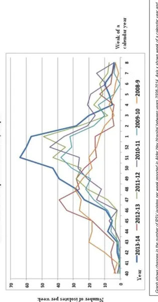
Figure 1.2. Graph presenting number of RSV isolates per week,
recorded in Alder Hey Hospital shows seasonality of the disease…….24
Figure 1.3. Hospital admission rates for bronchiolitis in the UK in
2010/2011………25
Figure 1.4. Degradation of NRF2 in healthy cells……….34
Figure 1.5. Disruption of Keap1-Cul3 ubiquitination system in infected cells………..36
Figure 1.6. NRF2 as a multi organ protector in the body……….40
Figure 1.7. Schematic picture of ubiquitination process………..46
Figure 2.1. Microscopic image of RSV plaques formed during the plaque assay protocol……….58
Figure 3.1. Anti-Nrf2 antibodies comparison on BEAS- 2B cells………79
Figure 3.2. Induction of Nrf2 by sulfurophane in non-infected BEAS 2-B cells. Time course experiment……….83
Figure 3.3. Induction of Nrf2 by sulfurophane in non-infected and RSV infected BEAS 2-B cells. Time course experiment ………84
Figure 3.4. Expression of Nrf2 by BEAS-2B cells with sulfurophane stimulation pre and post RSV infection ………87
Figure 3.5. Comparison of Nrf2 induction by sulfurophane in A549 and BEAS-2B cell lines ………..89
15 Figure 3.7. Design of the experiment. ……….92
Figure 3.8. Induction of Nrf2 expression by BEAS-2B cells using sulfurophane (1)………..93
Figure 3.9. Induction of Nrf2 expression by BEAS-2B cells using sulfurophane (2)………..95
Figure 3.10. Induction of Nrf2 expression by BEAS-2B cells using sulfurophane (3)………..98
Figure 3.11. Suppression of RSV replication by sulfurophane in BEAS-2B cells (1)……….101
Figure 3.12. Suppression of RSV replication by sulfurophane in BEAS-2B cells (2)………..103
Figure 3.13. Percentage RSV expression in comparison to L32 in BEAS-2B cells treated with sulfurophane corresponding to western blot in Figure 3.11……… 105
Figure 3.14. Percentage RSV expression in comparison to L32 in BEAS-2B cells treated with sulfurophane corresponding to western blot in Figure 3.12………...106
Figure 3.15. Percentage RSV expression in comparison to L32 in
BEAS-2B cells treated with sulfurophane and DMSO………108
Figure 3.16. Expression of RSV in BEAS-2B cells treated with
sulfurophane and DMSO……….109
Figure 3.17 Expression of RSV in BEAS-2B cells treated with sulfurophane and DMSO after calculating the average from
16 Figure 4.1. Level of ubiquitin in A549 cells infected with increasing concentrations of RSV and treated with proteasome inhibitor………131
Figure 4.2. Level of Nrf2 in A549 cells infected with RSV and treated with proteasome inhibitor (MG132)………...133
Figure 4.3. Level of DDX21 in A549 cells infected with RSV and treated with proteasome inhibitor………135
Figure 4.4. Level of DDX21 in A549 cells infected with RSV and treated with proteasome inhibitor, harvested at 4 and 24 hours………136
Figure 4.5 Level of DDX21 in A549 cells infected with RSV and treated with proteasome inhibitor and palivizumab………..138
Figure 4.6. Level of RPS10 in A549 cells infected with increasing concentrations of RSV and treated with proteasome inhibitor
(MG132)………....141
Figure 4.7. RSV influence on the expression of ubiquitinated proteins in A459 cells infected with RSV and treated with MG132……….144
Figure 4.8. RSV influence on the expression of ubiquitinated protein in A459 cells infected with RSV and treated with MG132 after
immunoprecipitation with anti-ubiquitin antibody. Western blot probed with anti-ubiquitin antibody………145
Figure 4.9. RSV influence on the expression of ubiquitinated DDX21 in A459 cells with proteasome inhibition after immunoprecipitation with anti- DDX21 antibody. Cell lysates immunoprecipitated with anti- DDX21 antibody. Western blot probed with anti-ubiquitin antibody…………147
17 Figure 4.11. RSV expression in A459 cells infected with RSV and treated with MG132………150
Figure 4.12. RSV influence on the expression of ubiquitinated RPS10 in A459 cells with proteasome inhibition………151
List of Tables
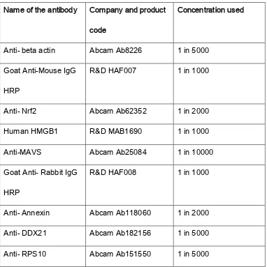
Table 1. Names and product codes of antibodies use for Western blots and immunofluoroscopy……….76
Table 2. Table presenting data exported from the PCR spreadsheet after analysis, corresponding to PCR results from Figure 3. 16……...111
Table 3. Unpaired t-test data………..113
18
1.
Introduction
1.1.
Respiratory Syncytial Virus
Respiratory Syncytial Virus (RSV) is one of the leading causes of
respiratory disease in infants and is a major threat for child health
worldwide, regardless of socioeconomic status. It also contributes to
an increase in mortality and hospitalisation rates in the elderly and
immunosupressed. 1 The virus causes bronchiolitis, lower respiratory
conditions characterised by dry coryza, cough and fever.2 In addition
to the consequences of its acute symptoms, it has been linked to
asthma and recurrent wheeze in later life. All these aspects make the
disease a great burden on both society and the NHS.2
1.1.1.Virology- overview of the virus structure
RSV is a double stranded, enveloped RNA virus from the
Paramyxoviridae family. Although mainly veterinary viruses, there
are two human viruses in this family- RSV and metapneumovirus.3 Its
genome consists of 15,222 nucleotides and encodes for 11 proteins.
Nine of those proteins are structured virion components and 2 are
non- structured proteins 1 and 2 (NS1, NS2) responsible for
opposing the host innate immune response4. Two proteins located on
the surface of the virus- proteins F and G play crucial roles in viral
19
targeting ciliated airway epithelium and protein F for fusing viral and
cellular membranes and allowing entry into the cell. Protein F also
gives the virus its name as it stimulates production of syncytia
enabling direct cell to cell spread.5 Thanks to antigenic determinants
of these two proteins, the host’s body produces neutralising
antibodies.6 Protein F exists in two forms, pre- and post-fusion. The
pre-fusion form (PreF) is the main target for the development of
antiviral drugs due to its superiority in inducing neutralising
antibodies in comparison to its post-fusion form (PostF).7 Proteins G
and F are two out of three integral membrane proteins inserted in a
lipid envelope surrounding the virus. The third protein is the small
hydrophobic (SH) protein but its role is currently unknown.8 The RSV
genome is protected by helical nucleocapsid which also provides a
replication template.9 A mature RSV particle consists of
ribonucleoprotein (RNP) complex created by viral RNA (vRNA), the
nucleocapsid (N) protein, the phosphor (P) protein and the large (L)
protein interacting with one another. The P protein is an essential
component of polymerase complex providing clearance and chain
elongation during transcription. The L protein is responsible for RNA
synthesis stimulation, encoding RNA polymerase, as well as RNA
transcription and replication.10 These proteins are essential for
20
contribute to its efficiency. These proteins are called M2-1, M2-2 and
M and all are required for transcription. M2-1 is a transcription factor
and M2-2 is a regulatory factor responsible for balance between
replication and transcription. M2 mRNA consists of two open reading
frames (ORF1 and ORF2) which overlap. ORF1 promotes chain
elongation during transcription and optimizes mRNA production.
ORF2 plays a role in accumulation of genomic and antigenomic
RNA11. M protein, located on the viral envelope, is a matrix protein
which enables interaction of plasma membrane and
ribonucleoprotein complexes (RNPs) during virion synthesis. As
mentioned earlier, NS1 and NS2, which are not part of the mature
virion structure, are secreted proteins responsible for antagonising
the interferon system. They increase the severity of disease by
blocking production of type I interferon (IFN) and causing rapid
replication of the virus. NS1 has a greater IFN inhibiting effect in
comparison to NS2 but both work synergistically.12 A schematic
21
Figure 1.1. Schematic illustration of RSV particle.
A single particle of RSV showing its envelope and negative single RNA strand. 3 surface proteins (G,F,SH) are shown outside the virus particle and protein M (matrix), L (polymerase), N (nucleocapsid) and P (phosphoprotein), as well as two transcription and termination factors M2-1 and M2-2 inside the cell. Non-structural proteins NS1 and NS2 are not shown in the picture.(4)
1.1.2.History and discovery of RSV
RSV was first described in 1957 as “Chimpanzee Coryza Agent” by
Blount et al, following an outbreak of disease in research purpose
kept group of Chimpanzees.13 Symptoms of bronchiolitis however,
had been described earlier in 1857 by Eberle but at that time, the
cause of the disease was unknown.14 It was the lack of bacteria
detected in the infected sample almost 100 years later (in 1955),
22
infants characterised by cyanosis, cough and dyspnoea might have a
viral cause.15,16 In 1957, Chanock et al discovered that “Chimpanzee
Coryza Agent” cannot be structurally distinguished from the virus
causing bronchiolitis symptoms in infants.17 The name RSV came
from ‘syncytia’, or pseudo large cells, which were observed in
infected human epithelial cells. The same group of scientists
identified the main characteristics of RSV, including its link to
bronchiolitis and pneumonia, its seasonability in winter months and
its propensity to infect young infants.18
1.1.3.Epidemiology
RSV infections are very common. Every winter the virus causes
outbreaks of bronchiolitis in children under the age of 1. The majority
of cases in the Northern Hemisphere are recorded between
November and April19 (Figure 1.2.). Almost all children will have had
a RSV infection before the age of 5, with 70% of children being
exposed to the virus in the first 12 months of life.20 Incidence peaks
during 3rd and 4th month of life.21 There are two major genetic
subgroups, A and B which co-circulate, and their predominance
varies by year and geographic location.22 Worldwide, RSV is
estimated to cause over 30 million lower respiratory tract infections
each year which contribute to more than 3 million hospitalisations.
23
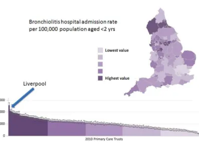
children under 5 years of age.23 Overall, 2-3% of children under 1
who are infected with RSV get admitted to hospital.24 (Figure 1.3.
presents hospital admission rates from bronchiolitis in years
2010-2011 in the UK.) In developing countries, bronchiolitis has been
reported as the second commonest cause of death during the first 12
months of life after malaria.25 In the United Kingdom, mortality rates
from bronchiolitis have decreased from 21.47 in 1979 to 1.82 per
100,00 live-births in 2000.26 Re-infection rates of the virus are also
vey high, with 74-83% in the 2nd year and 46-65% in the 3rd year of
life reported in literature.27 This data shows that human immunity
provides insufficient protection from the virus and highlights the
importance of treatment development.
Clinical research data from both the USA and Britain report high
rates of Intensive Care Unit admissions and need for mechanical
ventilation in children with bronchiolitis.28 A study conducted in 5
London Paediatric Intensive Care Units showed that the average
length of assisted ventilation needed by a child with bronchiolitis was
4.4 days and the average length of hospital stay was 15.9 days.29
RSV infection is an enormous burden not only on a patient’s health
but also on the economy. The predicted annual cost of treating RSV
24
Figure 1.2. Graph presenting number of RSV isolates per week,
recorded in Alder Hey Hospital shows seasonality of the disease.
25
Figure 1.3. Hospital admission rates for bronchiolitis in the UK in
2010/2011.
26
1.1.4.Clinical features and presentation
Presentation of bronchiolitis varies depending on the age of the
patient and the severity of symptoms. Mild RSV infection usually
results in mild upper respiratory tract (URT) symptoms and does not
require medical intervention. However, more severe disease causes
significant lower respiratory tract (LRT) symptoms which manifest as
bronchiolitis or pneumonia and often require hospital admission.
Children with LRT symptoms usually present with cough (98%), fever
(75%), rhinorrhoea, wheezing (65-78%), increased work of breathing
(73-95%) and sometimes hypoxia.31 Symptoms of more severe
disease include: grunting, nasal flaring, subcostal and intercostal
recession. Older children usually present with URT symptoms like
cough, coryza, rhinorrhoea and conjunctivitis.32 Predicting severity
based on symptoms might be misleading because children can
appear disproportionally ill/well.33 However, factors associated with
more severe disease in full term children include age <60 days, male
sex, increased respiratory rate, increased work of breathing, lower
27
1.1.5.Risk factors and prognosis
Even though all infants are susceptible to RSV infection and
bronchiolitis, the following risk factors make the chance of morbidity
much higher: pre-existing chronic lung disease (eg
bronchopulmonary dysplasia), current weight of less than 5 kg,
existing cyanotic heart disease, immune compromise (eg severe
combined immunodeficiency), in utero exposure to tobacco smoke,
low socioeconomic status, neuromuscular disease and premature
birth- before 35 weeks of gestation. Atopy or family history of atopy
have also been reported to be associated with more severe forms of
disease.35
The majority of patients with RSV infection recover uneventfully and
significant disease does not recur. However, 40% of children
hospitalised with bronchiolitis have significantly more wheezy
episodes during the first 5 years of life than age matched controls,
with 10% continuing to have wheezy episodes past the age of 5.36 A
study by Blanken et al, in which healthy pre term infants were
prophylactically treated with palivizumab, showed decrease in
28
control group and proved that RSV infection plays significant role in
the pathogenesis of recurrent wheeze during the first year of life.37
1.1.6.Management and prevention
Despite the importance of the disease, a lack of in-depth knowledge
about the pathogenesis has resulted in inadequate treatment and
vaccination options available. In groups of ex-preterm neonates at
the highest risk of severe disease, palivizumab (a monoclonal
antibody) is administered. However, it is only effective as prophylaxis
not as a therapeutic. Use of the antibody has not been extended to
the general population. The process of vaccine development has
been significantly prolonged due to safety concerns, biological
barriers and practical problems. Extensive research in infected
people and animal models has not yet led to commercially available
effective antivirals or vaccines, with the exception of palivizumab for
immunoprophylaxis in selected high-risk children. Due to the lack of
knowledge about the virus’ intermediate host or animal reservoir, it is
argued that if the vaccine was administered before the first RSV
infection, virus ecology could drastically change and stop the ability
of RSV to continually re-infect humans.38
Currently in the majority of cases, RSV bronchiolitis treatment is
supportive, consisting of close monitoring of the clinical symptoms
29
attitudes and a lack of set treatment protocol, patients often undergo
unnecessary treatment with antibiotics, steroids or inhaled
bronchodilators despite a lack of evidence-based data about their
effectiveness. Bronchodilators (eg nebulised salbutamol) have been
reported to cause modest short term improvement, however, a
definitive benefit with acute symptoms has not been demonstrated.40
Anti-inflammatory medication, such as systemic and nebulised
corticosteroids, have demonstrated no benefits as bronchiolitis
treatment.41
1.2.
Pathogenesis
RSV replicates in the nasopharynx, where epithelial cells are the
primary line of defence, causing URT symptoms.
1.2.1.Viral infection and cytotoxicity
After 2-8 days of incubation, the virus infiltrates the small bronchiolar
epithelium causing LRT symptoms. Viral shedding usually lasts 3-8
days but in some cases can extend up to four weeks.42 In cases
which progress to LRT, pathological changes develop including
oedema, enhanced mucus production and ultimately necrosis and
regeneration of the airway lining. These changes result in small
airway obstruction, air trapping and increased airway resistance.43 In
30
Due to RSV pathogenesis being not fully understood, information
available in the literature is not abundant and remains controversial.
The impact of the host response is reduced because the virus targets
superficial epithelial cells. Lung injury is exacerbated both by the
direct cytotoxic effect of the virus and inflammatory responses
elicited against the virus.45 Some studies show that it is only after
epithelial cells have released inflammatory mediators that apoptosis
occurs.46 RSV has been reported to cause ciliary damage not long
after infection, as well as delayed cell death, even weeks after
infection.47 Numerous studies suggest that damage caused by RSV
is to a great extent immune response mediated.48 Continuous
stimulation of the immune system caused by persistent viral infection, may cause chronic inflammation or changes in the expression of immunoregulatory molecules, which may explain why the clinical
symptoms persist long after the acute viral infection has resolved.49
1.2.2.Immune response to RSV infection
The human immune system can be split into two parts- innate and
adaptive immunity. Both of those components work synergistically to
recognize and remove unwanted matter from the organism and
31
defence is the innate system, which is always present and helps in
the induction of the adaptive system. RSV infects and replicates in
airway epithelial cells. These host cells express toll- like receptors
(TLR), sensors detecting pathogen specific structural motifs. They
recognize virus pathogen associated molecular patterns (PAMP) and
initiate expression of cytokines, soluble protein mediators which
regulate the immune response. 51 One of the receptors (TLR-4),
binds to the RSV F protein (Section 1.1.1.) and together with CD14
starts NF-κB (nuclear factor kappa-light-chain-enhancer of activated
B cells) mediated cytokine production.52 The cytokines produced
during TLR-4 engagement initiate neutrophil and natural killer (NK)
cell migration into the lungs, where they may be themselves further
stimulated by virus or surrounding cytokines.53 Notably neutrophils,
although apparently needed to control RSV infection, have also been
suggested to damage airway tissue.54
Adaptive immunity which includes T cell-mediated immunity and
antibody production by B cells is characterised by immunological
memory and tolerance to the body’s own tissues.55 Studies
conducted on animal models have found that RSV infection
considerably changes host innate immunity, which in turn leads to
32
The two systems are highly integrated and comprised of both
specialised cells and humoral factors.57
One example of cell’s response to viral infection is the interferon
(IFN) pathway. Interferons are a group of cytokines of a pleiotropic
type named after their property of ‘interfering’ with viral replication.58
Studies on murine models stress the importance of reduced IFN
expression in cases with increased RSV spread.59 IFNs can be split
into two groups. Type I can be expressed by the majority of cells and
consists of many IFN- α forms and one IFN-β form. During infection
with virus, this type of IFN is expressed rapidly in response to viral
RNA or DNA recognition. Cells at or around the site of infection are
activated via IFN receptors on the cell surface leading to inhibition of
viral replication through production of endonuclease which destroys
viral DNA/RNA and inhibits translation. Type I IFN also up regulates
major histocompatibility complex (MHC) class I production, which
increases the chance of cytotoxic lymphocytes identifying an infected
cell and increases NK cell activity which up-regulates the production
of proteins such as inflammatory chemokines.60 A study by Spann et
al suggests that expression of IFNs is a very early host reaction to
RSV, and that a major method of RSV inhibiting innate immunity is
33
Type II IFN group consists only of IFN-γ and it is expressed by
macrophages, Natural Killer T and Natural Killer cells. IFN-γ is
recognized as a part of immune response but the exact part it plays
is unknown.62
Another molecule of importance in viral infection is Nrf2, a
transcription factor which has been recognized as an essential
regulator of cellular oxidative stress response caused by viral
infection. Its role is described in greater detail in Section 1.3.1.
1.3.
NRF2
Nrf2 also known as NF-E2-related factor-2, is a transcription factor,
profusely expressed in macrophages.63 It has been recently
recognised as one of the main cellular defence mechanisms against
environmental toxins and carcinogens. Its main role is to stimulate
oxidant response and initiate transcription of genes, which protect the
organism from oxidative stress effects and results in the
re-establishment of homeostasis.64
1.3.1.Molecule overview
Emerging studies suggest that Nrf2 has a major part in the
pathogenesis of various types of cancer, chronic lung disease and
host defenses against viral infection of the respiratory system.65 In a
34
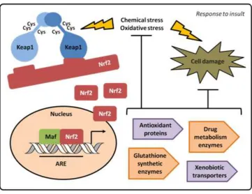
erythroid cell-derived protein with CNC homology ECH-associated
protein 1)66, dependent on another protein- Cullin 3 (Cul3). This
Keap1-Nrf2 pathway is the main method in which cells regulate
protective responses to internal and external stresses resulting from
reactive oxygen species (ROS).67 Keap1 is a substrate adaptor which
keeps Nrf2 in the cytoplasm and helps Cul3 ubiquitinate Nrf2 when
the cell is in redox homeostasis. Ubiquitinated Nrf2 is transported to
proteasome for Cul3-ubiquitin mediated degradation68 (Figure1.4.).
Figure 1.4. Degradation of Nrf2 in healthy cells.
In a non-infected cell Nrf2 is kept in the cytoplasm by Keap1 and Cullin3. Keap1, a substrate adaptor, helps Cull3 degrade Nrf2 by ubiquitination. Nrf2 is marked with ubiquitin and degraded and recycled in the proteasome. This process happens very quickly as Nrf2 half life is only 20 minutes.69
Keap1 has multiple cysteine residues which can be regulated in vitro
by various oxidants. If a cell undergoes oxidative stress, Nrf2 is
released from the complex by a change in Keap1 cysteine disulfide
bonds, undergoes phosphorylation and is translocated to the
35
have been reported to alter the structure of Keap1 which results in
nuclear translocation of Nrf2 and target gene expression.71 Even
though the precise method of cysteine modification in Keap1, which
activates Nrf2, is not fully understood, there are two proposed
models explaining this process. The first one is the “hinge and latch”
model which suggests that Keap1 modification in thiol residues of
Keap1 blocks the interaction with Nrf2. This results in Nrf2 lysine
residue misalignment and inability to polyubiquitnate the transcription
factor. In the second model on the other hand, thiol modification
results in Cul3 dissociating from Keap1. In both models, modified by
inducer and bound to Nrf2 Keap1, is inactive. Newly formed Nrf2
proteins bypass Keap1 and are translocated into the nucleus where
together with the small Maf proteins, attach themselves to antioxidant
response element (ARE) and induce expression of Nrf2 target
genes72 (Figure 1.5.).
Agents which regulate Keap1-Nrf2 pathway have been of recent
interest as therapeutic targets for treatment of oxidative stress
36
Figure 1.5. Disruption of Keap1- Cul3 ubiquitination system in
infected cells.
37
1.3.2.Target genes
The protective mechanism of Nrf2 relies on inducing transcription of
genes which reduce lung injury caused by oxidative stress. A number
of genes have already been identified but modern technical
advances have allowed definition of the transcriptional changes
induced following Nrf2 induction and provided further data about
direct target genes of Nrf2.74 These genes include: 1. Intracellular
redox-balancing proteins involved in heme and iron metabolism like
heme oxygenase-1 (HMOX-1) or glutathione metabolism- glutamate
cysteine ligase (GCL). 2. Phase II detoxifying enzymes involved in
drug metabolism like NAD(P)H quinine oxidoreductase-1 (NQO1) 3.
Transporters (multidrug resistance-associated proteins, MRPs)75 as
well as transcription factors, metabolic enzymes and antioxidants76.
Antioxidant response element (ARE) is necessary for Nrf2 binding
and gene induction and is a specific DNA sequence located on the
promoter region of Nrf2 target genes. There are many other Nrf2
downstream genes which are responsible for other cellular processes
like cell growth and death, inflammatory response, DNA repair and
ubiquitin- mediated degradation pathway (Section 1.4.3.).77 Nrf2
downstream genes are heterogenous in nature, which shows the
38
1.3.3.Existing evidence of Nrf2 importance
Over 200 diseases have been reported to cause oxidative stress in
cells.79 These include Chronic Obstructive Pulmonary Disease,
asthma, various types of cancer and neurological diseases including
multiple sclerosis and Alzheimer’s, cardiovascular and metabolic
disorders such as diabetes, vision disorders and ageing. In this
project the focus is on infection with RSV, however, other viruses
such as Humman Immunodeficiency Virus (HIV), Hepatitis B (HepB)
and C (HepC) have been reported to stimulate reactive oxygen
species (ROS) both in vitro and in vivo.80 One of the characteristics of
Nrf2, which demonstrates its importance, is its polymorphism.
Various studies report its numerous gene variants and haplotypes
appearing in different diseases. For example Arisawa et al described
an Nrf2 gene promoter polymorphism and its relationship with
Helicobacter infection in chronic gastritis.81 Another paper by
Cordova et al describes a particular genotype of Nrf2 (-653G/A)
which plays an important role in nephritis during childhood-onset
systemic lupus erythematosis (SLE).82 Different haplotypes in the
promoter region of Nrf2 have also been found in COPD by Hua et
al.83 Most important for this MPhil project, however, is the study by
39
of RSV. In their experiments, mice deficient in Nrf2 suffered from
much more severe RSV induced disease in comparison to control
mice. The severity was assessed on the basis of higher viral titers,
augmented inflammation, enhanced mucus production and epithelial
injury. It stresses the importance of Nrf2 mediated cellular antioxidant
mechanism in pulmonary anti-RSV activity.84
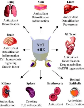
The versatile role of NRF-2 in protecting different systems in the
40
Figure 1.6. Nrf2 as a multi organ protector in the body.85
41
1.3.4.Nrf2- ARE pathway
In a normal cell, a part of aerobic metabolism is the production of
reactive oxygen species (ROS) via respiration and oxidation to create
energy. When the level of ROS is elevated (for example as a
consequence of infection or exposure to toxins, environmental
pollutant or radiation) harmful changes caused by oxidation occur in
a cell. Respiratory viruses such as RSV, human metapneumovirus
(hMPV) or influenza, stimulate production of ROS and decrease
antioxidant enzyme (AOE) efficiency resulting in oxidative injury due
to unbalance oxidative-antioxidants status. Nrf2 controls production
of AOE by binding to antioxidant responsive element (ARE) in AOE
gene promoters. When the cell is exposed to majority of pro-oxidant
stimuli, Nrf2 is induced and AOE expression upregulated. During viral
infections however, AOE expression is inhibited, Nrf2 nuclear
localisation is reduced and transcription of ARE-dependent genes
inhibited. For that reason, agents inducing Nrf2 or antioxidant
mimetics are a possible therapeutic means of treating harmful effects
of respiratory viral infections.86 Nrf2-Are pathway has been examined
in numerous studies, in which pulmonary disorders resulted from
various antioxidant and inflammatory agents. These experiments
42
in which the ARE-driven antioxidant expression is suppressed, have
exacerbated lung inflammation and injury in comparison to control
animals.87
It has been previously reported that Nrf2 expression is significantly
reduced in RSV infection which might be a potential mechanism for
reducing gene expression of AOE. This can be caused by a range of
factors like reduced transcription or increased mRNA degradation. 88
1.3.5.Nrf2 as a clinical drug target
A large number of studies proving how important Nrf2 is in protecting
the human body against an array of diseases, lead to a huge interest
in developing Nrf2 based therapies. Since pathogenesis of
viral-associated lung disease including RSV infection is so strongly related
to oxidative stress, agents with potential to regulate antioxidative
pathways seem like a rational therapeutic approach to these
diseases.89 Antioxidants are known to quench free radicals, which
decreases oxidative damage and enables cells to function normally.
Komaravelli et al. tested two therapeutic approaches: Superoxide
dismutase (SOD) mimetics, which decrease oxidative damage by
interacting with free radicals and Nrf2 inducers which regulate AOE
gene expression. A number of compounds, of both synthetic and
43
influenced transcription. They can be broadly divided into two groups:
Triterpenoids and isothiocyanates.90 Triterpenoids originate from
oleanolic acid, which itself has been reported to have antioxidative
properties.91 Isothiocyanates include Sulforaphane, mainly found in
cruciferous vegetables like broccoli. It has been reported to change a
number of cysteine residues in Keap1 by releasing Nrf2, which
results in elevated nuclear localisation of Nrf2 and ARE
transcription.92 Kesic et al. showed increased levels of Nrf2 in
epithelial cells treated with Sulforaphane before Influenza infection,
which contributed to reduction in viral replication.93 In a different
study, mice treated with sulforaphane were shown to have reduced
numbers of neutrophils and eosinophils after infection.94 These
findings imply that this compound has a big potential for regulating
viral induced oxidative disease process.95
Nrf2 is known to be differentially ubiquitinated and the ubiquitinated
form rapidly degraded by the proteasome to inhibit Nrf2 activity.
Differential regulation of protein activity in a manner similar to Nrf2
occurs for many proteins but has never been studied in relation to
viral infection. This type of modification could lead to both activation
or inactivation of a protein and also translocation and movement of it
in a cell.96 In this thesis I first examined differential ubiquitination of
44
number of candidate genes identified by proteomics. These
molecules are introduced at the beginning of Chapter 4.
1.4.
Ubiquitin
Ubiquitin (Ub) is a highly conserved protein, consisting of 76 amino
acids. In a cell, Ub is linked to target proteins by covalent bonds, in a
process called ubiquitination. Its name comes from its ubiquitous
nature, as it is found in all eukaryotic organisms. 97 Ubiquitination is
one of the best described post-translational alterations which controls
protein expression and function.98
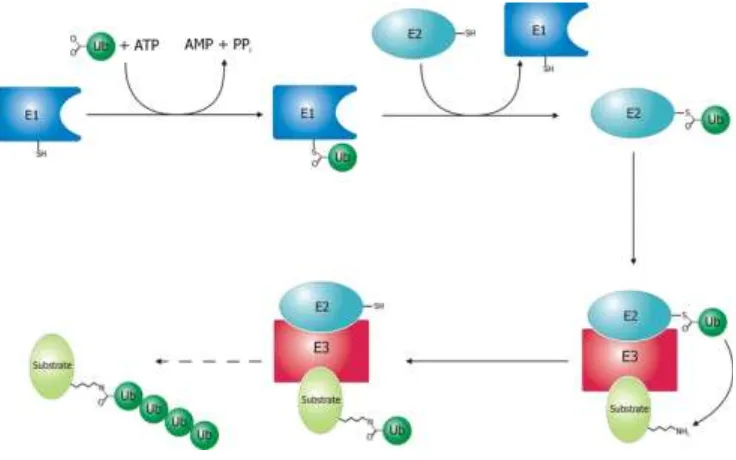
1.4.1.Ubiquitination process
The process is based on an enzymatic cascade. The first enzyme in
the cascade is E1 which hydrolyses ATP, activates ubiquitin and
transfers it to a cysteine of the second enzyme E2- a ub-conjugating
enzyme. The final enzyme is E3 which creates an isopeptide bond
between ubiquitin’s carboxyl terminus and target protein.99 E2 and E3
most often determine substrate selection. So far there are only a few
E1 enzymes known in mammals, about 30 E2 enzymes and
hundreds of E3 enzymes. The final product of the first stage of
ubiquitination is the mono- ubiquitinated protein. Every ubiquitin
molecule has a specific lysine which can be used to initiate
45
process is a target protein with polyubiquitinated chain. Damaged or
misfolded proteins are marked by ubiquitin and are transported to
proteasome and destroyed in the ub-proteasome system (UPS).100
Other ubiquitinated proteins (eg. transmembrane proteins) are
transported to a lysosyme via the multivesicular body pathway
(MVB).101 UPS protein degradation is a key process in DNA damage
repair, cell cycle regulation, cell development and immune system
function.102 It has also been reported that ubiquitin takes part in
protein function and protein interaction with the help of specific
hydrolazes. These structures have similar function as kinases and
phosphatases in the phosphorylation process. The whole process is
very versatile and can be reversed, influencing various properties of
46
Figure 1.7. Schematic picture of ubiquitination process.
Schematic diagram of the ubiquitination system. Created by Roger B. Dodd.The first enzyme in the cascade is E1 which hydrolyses ATP, activates ubiquitin and transfers it to a cysteine of the second enzyme E2. E2 is a ub-conjugating enzyme. The final enzyme is E3 which creates an isopeptide bond between ubiquitin’s carboxyl terminus and target protein, 104
1.4.2.Molecule overview and interaction with viruses
The genes encoding for this protein are grouped in tandem repeats,
due to high demands for transcription of this protein in all cellular
processes. Ubiquitin is a very versatile protein thanks to its seven
lysines and extra residues, used by Ub ligases to create different
kinds of Ubiquitin chains on target proteins. This results in
interactions with different downstream factors.105 An example is the
proteasome-47
mediated protein degradation or K-63 based control of protein
endocytosis, as well as enzyme activity.106
Being obligated intracellular parasites, viruses have to overcome
host cellular machineries at every stage of their life cycle including
entry into the cell, replication and genome transcription, protein
synthesis etc. up until release from the infected cell. Knowing how
important ubiquitination is in those cellular processes, it is expected
that ubiquitin and proteins affected by it, must play a part in viral life
cycle and pathogenesis.107
The first report of viruses being capable of using the UPS for their
own benefit was the Scheffner et al. study of small DNA tumour
viruses and their ability to modulate cell cycle.108 Since then, a
number of other studies have proven that other viral families take
advantage of ubiquitin conjugating system in their life cycle.109 From
this perspective it became obvious that studies involving experiments
with proteasome inhibition are crucial, as such treatment not only
inhibits the UPS but also removes the free ubiquitin from the cell
which would influence all cellular pathways involving ubiquitin.
Proteasome inhibitors have been reported to inhibit many human
viruses like herpesviruses, poxviruses, adenoviruses, influenza
viruses, retroviruses, coronaviruses, paramyxoviruses,
48
herpes simplex virus, influenza virus and adenoviruses, that ubiquitin
modulates the first stage of viral replication- entry to the cell and viral
capsid presentation to the target cell. 111112 Other stages of viral life
cycle such as gene expression in Epstein-Barr113 virus or latency
(property which enables the virus to cause lifelong infection process
in host organism) in Kaposi's sarcoma-associated herpes virus
(KSHV) are also affected by blocking the proteasome.114
Viruses can also take over the ubiquitin conjugating system to
modulate host innate immunity signalling. They stop the induction of
Type I IFN by binding to its receptor.115 Viruses can challenge cellular
ub-conjugating system by adjusting substrate specificity of ubiquitin
ligases, changing which proteins are marked for degradation. Some
viruses , especially large DNA viruses like poxvirus, are able to
encode their own ubiquitinating enzymes, eg KSHV encodes two E3
Ub ligases.116
1.4.3.UPS- Ubiquitin Proteasome System
Ubiquitin Proteasome Pathways are a crucial method of protein
catabolism. Proteins destined for degradation by the proteasome are
marked with ubiquitin in the ubiquitination process but it is not fully
understood how proteins are targeted by the proteasome. In order to
49
a chain of at least 4 ubiquitins attached to it, so it must be
polyubiquitinated.117 Ubiquitin receptor proteins contain N-terminal
ubiquitin like domain (UBL) and one or multiple ubiquitin associated
domains (UBA). Proteasome caps recognise UBL domains and UBA
is bound to ubiquitin by three-helix bundles.118 Because of the large
number of cellular processes that UPS regulates, failing of the
system may result in various diseases. These processes include:
antigen processing, apoptosis, cellular cycle and division, organelles
biogenesis, transcription and repair of DNA, development and
differentiation, inflammation and immune response, response to
stress and extracellular modulators and most importantly for this
thesis- viral infection.119 The process can be divided into two steps:
conjugation- attaching of the ubiqutin molecule, and degradation by
the 26s proteasome consisting of the catalytic 20s core and the 19s
regulator.120 Recently it has become apparent that ubiquitination also
plays a major role in DNA repair and endocytosis. These newly
discovered roles are dictated by the type of ubiqutin chain linkage, as
well as number of ubiquitin molecules attached – mono versus
polyubiquitinated proteins. Linkage of covalent bonds between
ubiquitin and target protein can also be reversed in a process called
de-ubiquitination or de-conjugation by de-ubiquitinising enzymes
50
very dynamic.121 The process has been recognised as crucial and
Avram Hershko, Aaron Ciechanover and Irwin Rose who first
discovered this were awarded the Nobel Prize for Chemistry in 2004.
1.5. Aims and Objectives
The work throughout my MPhil project was divided between two main
objectives. The first set of experiments aimed to determine whether
Nrf2 induction by sulforaphane might reduce replication of RSV in
airway epithelial cells. Detailed description of these experiments
together with results and discussion are included in Chapter 3.
As Nrf2 is known to be differentially ubiquitinated, I first examined
differential ubiquitination of Nrf2 in response to viral infection and
then expanded this work to proteins selected from the proteomics
including DDX21, MAVS, HMGB1, prohibitin, Annexin A1 and
RPS10. These experiments aimed to validate data generated in the
proteomics about whether these proteins change their level of
ubiquitination following RSV infection, as well as answer the
questions whether those changes in ubiquitination might result in
protein turnover by the proteasome as part of host cell defence or
viral manipulation of cellular process to aid viral replication. Detailed
description of these experiments and their results together with
51
2. Methodology
2.1. Cell Culture
Cell types used during the project:
- A549 cells (adenocarcinoma derived human alveolar basal
epithelial cells) normally responsible for substance diffusion in the
alveolar epithelium of the lungs and often used in RSV infection
model.122
- BEAS-2B (non-tumorigenic human bronchial epithelial cells)123
- Hep2 cells (human epithelial type 2 cells) believed to come from
human laryngeal carcinoma, associated with various autoimmune
conditions.124
Cells of each type were grown in Dulbecco’s Modified Eagles
Medium (DMEM, Sigma), supplemented with 10% Foetal Calf Serum
(FCS, Sigma), L-glutamine 200mM (Sigma), Penicillin 10,000units/ml
(Sigma) and Streptomycin 10mg/ml (Sigma). Cells were grown in an
incubator with 5% carbon dioxide (CO2) at 37°C. Every 2-3 days,
dead cells were washed away with Phosphate Bovine Serum (PBS).
Viable cells were harvested from the bottom of the flask and
sub-cultured with fresh media at a concentration of 1x10⁶cells/ml with
15ml of fresh media in T75 flasks. All cell lines used in this study
2.1.1. Seeding the cells.
For each experiment stock cells were seeded as follows:
Media was removed from T75 flasks of cells grown in an incubator.
Cell monolayers were washed once with 5ml of PBS. 3ml of 0.25%
trypsin 0.02% Ethylene-diamine-tetraacetic acid (EDTA, Sigma) was
then added to flasks, which were then incubated at 37°C for 4-5
minutes. Flasks were tapped to allow cells to detach from the bottom
of the container. 7ml of FCS supplemented media (L- Glutamine,
Streptomycin + Penicillin) was subsequently added to neutralise and
deactivate trypsin. Cells were then centrifuged at 1600rpm at room
temperature for 10 minutes and the supernatant removed and
discarded.
The pellet was resuspended in 1ml of supplemented media and the
number of cells measured using a haemocytometer (Section 2.1.2).
Depending on the specific experimental conditions used, cells were
diluted with supplemented media and pipetted into each flask or well
with a correct amount of media.
2.1.2. Counting the cells
Cells were counted using a haemocytometer as follows. Cell
monolayers were washed once with PBS and scraped with a cell
53 centrifuge tube and the dish washed twice with PBS, to make sure
that the maximal number of cells from the dish was in the tube. Cells
were spun down in a centrifuge for 10 minutes at 1600rpm at room
temperature and the supernatant then discarded. Cell pellets were
next resuspended in 1ml of supplemented media and 10µl of this
solution put on the haemocytometer and viewed under a microscope.
Cells in each of the big corner squares consisting of 16 little squares
were counted and the number averaged. This gave a number
equivalent to the cell count in 10⁴/ml.
2.1.3. Harvesting the cells
Depending on the experiment, cells were harvested at 4, 24 or 48
hours, using the following method.
Cells were washed once with PBS. Small amounts of PBS (according
to the surface of the dish, eg. 0,5 ml for a well in a 6 well plate, 3 ml
for a T75 flask) were added to each flask and cells carefully scraped
from the bottom of the dish. The whole surface of the dish was
scraped, in order to maximise the number of cells collected. Scraped
cells in PBS were transferred into a 10 ml universal centrifuge tube
and the dish (flask/well) washed twice with PBS, to maximise the
number of cells collected. Tubes were spun in a centrifuge for 10
minutes at room temperature at 1600 rpm. Supernatant was taken off
54 According to the experiment and desired number of samples, cells
were divided between micro centrifuge tubes in 100µl of solution, and
spun again in a microfuge for 10 minutes at 1330 rpm. Supernatant
was taken off and discarded, leaving dry pellets which were stored in
labelled tubes at -30°C for future use.
2.2. RSV preparation
All virus stock used for the experiments in my project was made
using the following method.
2.2.1. RSV propagation
Day one:
Hep 2 cells were seeded at 3x10⁴ cells/cm in 15 ml of supplemented
media in a T75 flask and incubated at 37°C in 5% humidity for 24
hours (or longer if not 50% confluent after 24 hours).
Day Two:
Once 50% cell confluence was reached, media was removed and
cells were washed twice with 5ml PBS. 500µg of RSV stock was
placed in 3ml of serum-free media and added to the flask with cells.
Cells were then incubated on a rocker for two hours at 37°C to make
55 After two hours, 13ml of supplemented media was added to the flask,
which was then left overnight in the incubator at 37°C.
Day Three:
Flasks were inspected under the microscope and media changed.
Flasks were again stored in the incubator at 37°C overnight.
Day Four:
Forty-eight hours post-infection, cells were harvested as described
above using a cell scraper. These steps were carried out rapidly with
the samples kept on ice to ensure that the virus maintained its
integrity and did not degrade.
In a cooled centrifuge at 4°C, harvested cells were spun down at
1600rpm for 10 minutes in 50ml tubes. Supernatant was taken off
and placed in separate tubes on ice. 2ml of the removed supernatant
was used to resuspend the cell pellet and mixed by vortex. Solution
was evenly split into two micro centrifuge tubes tubes and kept on
ice. Cells were then lysed using a 25 gauge needle and 1ml syringe
for ten passes, to burst cells open and release RSV. Next, 500µl of
solution was transferred to four pre-labelled cryovials and snap
56 2.2.2. RSV Plaque Assay
In order to establish the number of virus Plaque Forming Units (PFU)
per ml of solution, the following method was used for each batch of
virus in this project.
Day One:
Using 27 wells of a 96-well flat bottomed plate, 2 x 10⁴/ml A549 cells
were seeded per well and grown in supplemented DMEM media in
an incubator for 48hrs at 37°C.
Day Three:
After 48 hours, serial dilution of RSV was prepared using the
following method.
Micro centrifuge tubes tubes were placed on ice to prevent virus
degradation. 500µl of serum free DMEM media was placed in one
tube and 250µl in seven others. A water bath was warmed to 37°C. A
vial of RSV was removed from the freezer, and snap thawed in
water. In order to achieve 1:100 dilution in the first tube, 5µl of virus
solution was added to the media and mixed well. 250µl out of this
solution was then transferred to the second tube and doubling
57 Cell monolayers in each well were washed once with PBS, and 50µl
of each dilution of virus added to wells in triplicate going horizontally.
Plates were incubated at 37°C. After two hours, 100µl of
supplemented media was added to each well and plate put in the
incubator until the next morning.
Day Four:
Each well was washed once with 100µl of PBS and cells fixed for 20
minutes at room temperature with 100µl of 100% methanol
containing 2% hydrogen peroxidase. Using a multi-channel pipette,
cells were washed gently with 100µl of PBS per well. Pipetting the
solution directly onto the cell monolayer was avoided, in order not to
disrupt it.
After 20 minutes, 100µl of goat anti-RSV antibody (Bio-rad), diluted
1/200 with PBS/1% Bovine Serum Albumin (BSA), was added to
each well and incubated at room temperature for one hour. Each well
was then washed twice with 100µl PBS/1% BSA. 100µl of extravidin
peroxidise (2mg/ml, Sigma-Aldrich) diluted 1/500 with PBS was
subsequently added and left for 30 minutes at room temperature.
Cells were washed twice with 100µl PBS/1% BSA. A Sigma-Fast
58 of this solution was then added to each well for approximately 10
minutes to stain plaques.
After the plaques appeared, PBS was added in order to stop reaction
and the plaques were counted. Dilutions which produced around
100-200 plaques per well were selected and each replicate was
counted to estimate the average value. In order to decrease the
possibility of counting error, plaques in dilutions above and below
were also counted and the number averaged. The whole process
was repeated by two people separately and the values compared
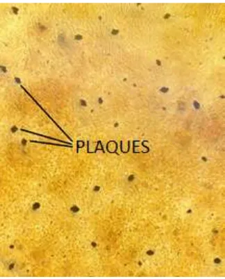
[image:58.595.115.277.475.676.2]and averaged. The RSV plaque assay is shown in Figure 2.1.
Figure 2.1. Microscopic image of RSV plaques formed during the
plaque assay protocol.
59 In order to count the PFU per ml of solution, the following formula
was used:
Number of plaques x dilution (eg. 100, 200, 400 etc) x 20
The dilutions for each well were as follows:
Dilution 1- 1/100
Dilution 2- 1/200
Dilution 3- 1/400
Dilution 4- 1/800
Dilution 5- 1/1600
Dilution 6- 1/3200
Dilution 7- 1/6400
Dilution 8 1/12800
In order to use the right amount of virus for each experiment,
Multiplicity of Infection (MOI) was calculated for each batch. Ratio
between the number of cells in a particular dish and the number of
RSV particles was calculated by dividing the number of RSV particles
60 2.2.3. RSV Infection
BEAS- 2B and A549 cells were seeded in the correct sized plates or
flasks as explained in Section 2.2.1. Cells were usually ready to be
infected after 48 hours in the incubator when they reached 90%
confluence. The correct number of vials was removed from the -70°C
freezer and snap thawed in water bath at 37°C. Those steps were
carried out promptly in order to avoid RSV degradation. Cells were
washed once with PBS and new media added to the flasks. Correct
amount of virus for each plate or flask was then diluted in serum free
media and added to the dishes with cells.
Depending on experiment, cells were left in the incubator at 37°C
until the harvest time (usually 4, 24 or 48 hours).
2.3. BCA protein Assay
The bicinchoninic acid assay (BCA assay) or the Smith’s assay was
carried out on the samples destined for western blotting, in order to
assess protein concentration in each sample and ensure that protein
loading on gel was even. Samples were prepared in the following
micro plate procedure and protein concentration calculated by a
colorimetric scanner.125 Pierce™ BCA Protein Assay Kit produced by
61 Samples for the procedure were prepared by adding 100µl of 1%
protease inhibitor (Sigma) and 99% protein extraction reagent
(CytoBuster™ Protein Extraction Reagent) to each dry pellet taken
out of the freezer. A total of eight standards were made by serial
dilution of 500µl of Albumin Standard Ampules (2mg/mL, 10 x 1 mL)
in each tube, giving the concentrations of 2000, 1000, 500, 250, 125,
62.5, 31.25 and 0. 25µg of each sample, and standard was pipetted
into a 96 microplate well in a working range of 20-2000 . 25µl of
each sample was added in duplicates horizontally on the plate,
diluting the samples as appropriate in sterile distilled water (23µl
H2O+ 2µl sample= 1:12.5 dilution)
Working reagent was made up to 1:50 dilution, 1 part of solution B
(green) and 50 parts of solution A (colourless). 200 µl of this mixture
was carefully added to each well and plate left for 30 seconds on a
plate shaker in room temperature to mix thoroughly. Each plate was
then covered and incubated at 37°C for 30 minutes. After 30
minutes, the plate was left on the bench in room temperature to cool
down for about 5 minutes and read with a plate reader at or near 562
nm.
The amount of sample loaded on the western blot gel was adjusted
62 2.4. RNA extraction
Extraction and isolation of RNA methodology was carried out as
described by the manufacturers of TRIzol® reagent (Life
Technologies) and is described below.
2.4.1. Homogenising sample
Culture supernatants were taken off from the cells and stored at
-20°C for future experiments. Samples were homogenised using
TRIzol® reagent (a monophasic solution made up of phenol and
guanidine isothiocyanate)126.
0,5ml of TRIzol® was used per well containing 5-10x10⁶ cells or per
dry pellet stored in a freezer. This amount of TRIzol® was used to
make sure that there was no DNA contamination within the isolated
RNA. Each sample was carefully pipetted up and down a number of
times to ensure that cells from the whole surface of the well were
taken into the solution. The tubes with dry pellets were mixed using
vortex for 2 minutes and each of the samples were placed in an
63 2.4.2. Phase separation and precipitation
Samples were incubated for five minutes at room temperature, to
allow nucleoprotein complexes to completely dissociate. 200µl of
chloroform was the added to each sample, which were then mixed
thoroughly by vortex for 10 seconds and centrifuged at 13,300rpm for
15 minutes at room temperature.
Samples separated into the following three layers:
1) Bottom pink phenol-chloroform phase
2) Middle interphase
3) Top colourless aqueous phase with the RNA
The top layer was carefully removed using P200 pipette at 45° angle.
Removing the middle or bottom layer was avoided, as that would
result in DNA contamination of the sample. Contents of each tube
were placed in new tubes containing 250µl of isopropanolol with
corresponding labels. Samples were then mixed by vortex and
placed in -70°C freezer for ten minutes, before being centrifuged at
13,300rpm for fifteen minutes. Putting samples in the centrifuge in
the exact same position, allowed for the gel-like pellet formed at the
64 2.4.3. RNA wash
Supernatant was removed from each tube, carefully avoiding
touching the pellet with the pipette tip and discarded. 200µl of 70%
ethanol was added to each tube and mixed by vortex for fifteen
seconds. Samples were microfuged at 13,300rpm for 10 minutes.
Supernatant was then removed carefully with the pipette tip and the
tubes left open to make sure that the pellets air dry for approximately
fifteen minutes. Once the pellets were dry, they were resuspended in
20µl of sterile, nuclease-free water.
2.5. Reverse transcription
Complementary DNA (cDNA) was prepared with High Capacity
cDNA Reverse Transcription Kit (Applied Biosystems) using random
primers. To each 200 µl tube labelled accordingly, the following
components were added from the kit:
2µl 10x RT Buffer
1µl 25x dNTP Mix (100 mM)
2µl 10x RT Random Primers
5.2µl sterile, nuclease-free water
0.8µl of Multiscribe Reverse Transcriptase
65 The contents of the tubes were reverse transcribed at 37°C for one
hour.
2.6. PCR
PCR (Polymerase-chain reaction) is a method used to amplify the
desired region of DNA, with the purpose of making multiple copies of
that sequence. In this project, PCR was used to assess the presence
and amount of RSV in the samples.
2.6.1. Principles of PCR
The process of PCR amplification can be divided into four phases:
1. Baseline - no signal is released during the cycles because the
amplification level is too low to be detected by the quantitative
PCR (qPCR).
2. Exponential - amplicons are quantified, signal is above the
detection level and the product should double exactly every
cycle to produce the number of amplicons if the assay is 100%
efficient.
3. Linear - the efficiency of amplification is reduced to less than 2
per cycle because the amount of reagents goes down with
their use.
4. Plateau - after all the reagents have slowly been used, the