A VAMP7/Vti1a SNARE complex distinguishes a non-conventional traffic route to the cell surface used by KChIP1 and Kv4 potassium channels.
Sarah E. Flowerdew and Robert D Burgoyne*
The Physiological Laboratory,
School of Biomedical Sciences,
University of Liverpool,
Crown Street,
Liverpool,
L69 3BX, UK.
*Corresponding author
E-mail: [email protected]; Tel: +44-51-794-5305; Fax: +44-51-794-5337
Running title: Vti1a/VAMP SNARE complex in ER to Golgi traffic
Summary
The K+ channel interacting proteins (KChIPs) are EF hand-containing proteins
required for the traffic of channel-forming Kv4 K+ subunits to the plasma membrane.
Introduction
Kv4 potassium channels were first identified as homologues of the Drosophila Shal
channels and are now known to have key roles in the regulation of the excitability of neurons and cardiac myocytes. In mammals, four members of the Kv4 family (Kv4.1, 4.2, 4.3 short and 4.3 long) are known [1] and they are highly expressed in the brain and heart [2]. Kv4 potassium channels are responsible for the transient, outward, A-type current in neurons, and its equivalent in myocytes. In the brain, it is thought that this current is responsible for attenuating back propagating action potentials and excitatory post-synaptic potentials [3]. The channels also limit action potentials to individual dendritic compartments [4] and abnormalities in Kv4 channel function have been implicated in the hyperexcitability underlying epilepsy [5].
When Kv4 channels are expressed alone in non-neuronal cells their properties differ from those seen with native channels [6]. A search for interacting partners that could
reconstitute native functions led to the discovery of the K+ channel interacting proteins
(KChIPs) [6] which are members of the neuronal calcium sensor (NCS) family of proteins [7, 8]. The KChIPs have four EF-hand domains of which 3 can potentially
bind Ca2+ and 2 were seen to be occupied by Ca2+ in the crystal structure of KChIP1
[9, 10]. Of the four KChIPs, only KChIP1 has an N-terminal myristoyl group required for it to bind to membranes [11] and uniquely allowing its targeting to a distinct population of intracellular 50-100 nm diameter vesicles (referred from now onwards as KChIP1 vesicles) that partially overlap with markers of the ER-Golgi intermediate compartment (ERGIC) [12]. The KChIPs have two effects on Kv4 channels. Firstly, the KChIPs modulate the properties of the channel, particularly its inactivation kinetics [6]. Secondly, they increase current density by promoting the traffic of the Kv4 channel to the plasma membrane [6, 12-15]. Kv4 proteins alone are able to exit from the ER, but then become trapped in the Golgi complex [11, 12]. Co-expression of Kv4 channels with KChIPs leads to their traffic and co-localization at the plasma membrane[11, 12] where the KChIPs are constitutive channel subunits [9, 10].
triacylglycerol transport vesicles, (pre-chylomicron transport vesicles (PCTVs)) can bud from the ER in a COPII-independent manner, and are lacking in COPI, but need COPII later in the pathway, to fuse with the Golgi [26]. Rab1-independent traffic from the ER has been suggested to occur for some cargos in non-neuronal [27] and neuronal cell types [28]. It has been shown that the traffic of Kv4 channels, with associated KChIP1, also occurs by an alternate pathway that is COPII-independent [12]. KChIP1 vesicles seem to be key intermediates in ER to Golgi traffic but the nature of this pathway and the KChIP1 vesicles involved in traffic are unknown.
Membrane traffic steps can be defined by the nature of the SNARE complex that underlies the fusion of vesicles with their target membrane. SNAREs have been defined as Q or R SNAREs and based on sequence classification of SNARE subfamilies, active complexes appear to have the composition QaQbQcR [29]. Three such complexes have been implicated in ER to Golgi traffic. From analysis of VSVG traffic, the SNARE complex for the step between ER and the ERGIC consists of Sec22b, membrin, rBet1 and syntaxin 5 [30, 31]. The complex of GOS28, Ykt6, rBet1
and syntaxin 5 is thought to be involved in the fusion of intermediates with the cis
-Golgi [32]. Finally, a third complex, comprising VAMP7, Vti1a, rBet1 and syntaxin 5, has been identified in the alternative ER to Golgi trafficking pathway for PCTVs in intestinal cells [4, 33]. The general significance of this latter complex in cargo traffic is unknown. We aimed to further characterise the nature of the KChIP1 vesicles and the traffic route taken by Kv4/KChIP1. The results show that the KChIP1 vesicles and Kv4/KChIP1 traffic are distinguished by a VAMP7/Vti1a containing SNARE complex.
Methods
Cell culture and transfection
HeLa cells were grown in Dulbecco’s modified Eagle’s medium (Gibco, UK), with 5% foetal bovine serum (Gibco, UK), 1% non-essential amino acids (Gibco, UK), and 1% penicillin-streptomycin (10,000 units/ml penicillin, 10,000µg/ml streptomycin,
Gibco, UK) and kept at 37°C, in a 5% CO2 atmosphere. Neuro2A cells were cultured
in DMEM , with 10% FBS and 1% Penicillin/Streptomycin. For transfection, cells were grown in 24-well plates, with glass coverslips for imaging studies. Cells were
plated at ~4 x 10⁵ cells per well, in 1ml supplemented media, and incubated 5-24
hours before transfection. For the transfection reaction mixture, 3µl per µg DNA of GeneJuice (Novagen, USA) was incubated for 5 minutes at room temperature with OptiMEM serum-free media (Gibco, UK) to give a total reaction volume of 100µl. One microgram of each plasmid DNA was then added and the mixture incubated for a further 15 minutes, before being added drop-wise to the cells. Cells were then left for 24-72 hours post-transfection.
Plasmids encoding KChIP1, KChIP1-EYFP, EYFP, KChIP1EF2-4-dsRed, KChIP2-ECFP ARF1-EYFP and Kv4.2 were described previously [11, 12, 34, 35]. The KChIP1EF3-EYFP plasmid bearing the mutations D135A, G140A was prepared in this laboratory by Dr Burcu Hasdemir. The ts045 VSVG-GFP plasmid [17] was provided by John Presley (NIH, Bethesda, Maryland, USA). The plasmid encoding the Rab1 GAP TBC1D20 was a gift from Francis Barr (School of Cancer Studies, University of Liverpool). The EYFP-Golgi plasmid encoding EYFP linked to
the N-terminus of beta 1,4-galactosyltransferase for trans- and medial-Golgi targeting
was obtained from Clontech (California, USA).
Immunocytochemistry
After transfection, cells on coverslips were gently washed three times in phosphate
buffered saline (PBS): 137mM NaCl, 2.7mM KCl, 10mM Na2HPO4, 2mM NaH2PO4.
Cells were then fixed in PBS with 4% formaldehyde for 30 minutes at room
temperature. Coverslips for imaging were then washed again before being air dried, and mounted as described below. For immunofluorescence, cells were washed in PBS after fixation, then permeabilised in PBT (PBS, 0.1% Triton X-100, 0.3% BSA) for 30 minutes. Cells were then incubated with the primary antibody overnight at 4°C.
Antibodies were diluted in PBS with 0.3% BSA. These included mouse monoclonal antibodies to membrin, VAMP7 and GOSR1 (GOS28), obtained from Abcam, UK a mouse monoclonal antibody to Sec22L1 (Sec22b) from Abnova, Taiwan and a mouse monoclonal anti-Vti1a antibody from BD Biosciences, USA. A chicken anti-Ykt6 antibody was obtained from J.C. Hay [36]. A rabbit anti-TI-VAMP (VAMP7) antibody was a gift from T. Galli (Institut Jacques Monod, Paris, France). Rabbit polyclonal antibodies against syntaxin 7 or 8 were obtained from Synaptic Systems, Germany. Anti-lysosomal associated protein 1 (anti-LAMP1) was a rabbit polyclonal obtained from Affinity Bioreagents (Colorado, USA). The primary antibody was removed, cells washed three times in PBT, and the secondary antibody added for one hour at room temperature. Secondary antibodies were diluted in PBS with BSA, at 1:80 dilution for both anti-Mouse and anti-Rabbit IgG conjugated to TRITC (Sigma, USA) or 1:250 for Alexa-488 and Alexa-594-linked secondary antibodies obtained from Molecular Probes/Invitrogen, USA. Cells were washed again in PBT, and then dried, before mounting on ProLong Gold Antifade (Invitrogen, USA).
Confocal microscopy
Transfected cells were viewed using a Leica TCS-SP2-AOBS confocal microscope (Leica, Germany), using a 66.86µm pinhole, and a 63x water immersion objective with a 1.2 numerical aperture. For EYFP constructs, an excitation of 488nm was used, and an emission range of 500-540nm recorded. For TRITC-tagged secondary antibody signals, a 543nm laser was used for excitation, and emission at 570-680nm recorded. GFP was imaged using excitation at 488 nm and light collection at 500-550 nm. CFP was imaged using 405nm excitation and light collections between 500-550nm. HcRed was imaged by excitation at 494 nm and light collected at 610-700 nm. Alexa-488 was excited at 488nm, and emissions recorded from 500-550nm. Alexa-594 was excited at
siRNA knockdown
Expression of SNARE proteins was inhibited using siRNA oligonucleotide duplexes. Three duplexes were obtained against SYBL1, the gene for VAMP7 (Ambion, UK, siRNA ID# 241467, 241468, 241469) and three against Vti1a (Ambion, UK, siRNA
ID# 37855, 37946, 38033). A Silence Negative Control siRNA#1 (Ambion, UK,
AM4611) was also obtained. All siRNAs were resuspended in nuclease free water, to give a final concentration of 100nM when used for transfection. Cells were transfected with untagged Kv4.2, with or without KChIP1-EYFP and the siRNA or VSVG-GFP and siRNA. After 72h the cells were fixed and images were obtained of both KChIP-EYFP and VSVG-GFP or Kv4.2 detected by immunofluorescent staining using an anti-Kv4.2 antibody (Exalpha Biologicals, Inc, USA) at 1:500 dilution with a TRITC-tagged secondary antibody. Using the Leica LCD Lite software (Leica, Germany) regions of interest were drawn around the perimeter of the cell, and within the cell, to give a region of interest at the cell periphery including the plasma membrane that was
1µm wide. The edge of the cell could easily be identified based on either background
fluorescence or that due VSVG/KV4.2. The total level of fluorescence was recorded for each region for both channels and the percentage of fluorescence at the plasma membrane was calculated. For Kv4.2 fluorescence, the background level of fluorescence at the cell periphery in the absence of co-expression of KChIP1 (typically 10% of total [12]) was determined for each experiment and subtracted from values for cells expressing KChIP1. The resulting values were then divided by the mean from each experiment of the percentage of Kv4.2 at the plasma membrane in the presence of KChIP1. These data are shown as the normalised value for Kv4.2 traffic. Values for VSVG were similarly normalised to the control values. All data are expressed as Mean + SEM and statistical analyses (Student’s unpaired t-test) carried out.
Real-time PCR
After 72 hours post-transfection, mRNA was extracted from HeLa cellas using TRIzol reagent (Invitrogen, Paisley, UK) according to manufacturer’s instructions. mRNA was resuspended in 30µl RNase free water (Sigma, UK), and stored at -80°C. cDNA was produced using ImProm-II reverse transcriptase (Promega, USA) following manufacturer’s instructions, and using Oligo (dT)15 primers (Promega USA). The reaction proceeded with 5 minutes annealing at 25°C, 60 minutes at 42°C, and 15 minutes of heat inactivation at 70°C. For real-time PCR, cDNA into a total volume of 15µl, 7.5µl SYBR Green PCR master mix (Applied Biosystems, Warrington, UK) was used with 1µl primer mix (50mM each of forward and reverse primers). Each primer and cDNA combination was repeated in triplicate. Experiments were run on a BioRad iQ5 thermal cycler. After checking each run had completed correctly, the threshold cycle (Ct) was recorded, and averaged for each primer and cDNA set. The relative levels of mRNA for each primer and for each cDNA were calculated by comparison to
Results
KChIP1 EF-hand mutants distinguish between Kv4/KChIP1 and VSVG traffic to the plasma membrane
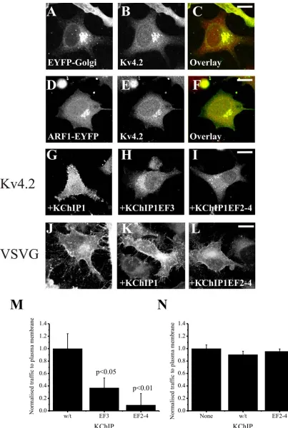
When expressed alone in HeLa cells, Kv4.2 channels do not effectively reach the plasma membrane and remain trapped intracellularly [12]. The intracellular Kv4.2 was highly co-localised with two Golgi complex markers EYFP-Golgi (Figure 1A-C) or ARF1-EYFP (Figure 1D-F) indicating retention in the Golgi complex. In comparison, co-expression with KChIP1 results in traffic of Kv4.2 to the plasma membrane (Figure 1G) where it co-localises with KChIP1 [12]. Mutations of either a single EF hand (EF hand 3 in KChIP1EF3) or all three potentially functional EF hands (in KChIP1EF2-4) to disrupt coordination of divalent ions prevented the stimulatory effect on traffic of Kv4.2 (Figure 1H,I) and instead Kv4.2 was found on punctate intracellular structures that co-localised with the KChIPs (not shown) as seen previously for the triple mutant [12]. To compare this traffic pathway to the conventional pathway followed by VSVG we examined the effect of the inhibitory triple EF hand mutant on traffic of VSVG-GFP to the plasma membrane. Neither wild-type nor mutant KChIP1 had any effect on the levels of VSVG-GFP that reached the plasma membrane (Figure 1J-L). These conclusions were confirmed by quantification of the level of trafficking of proteins (Figure 1M,N) in assays in which fluorescence in peripheral regions of interest were determined [12] and normalised in comparison to control cells to provide a value for relative traffic to the plasma membrane for each experiment. The KChIP1 EF hand mutations appeared, therefore, to specifically impair the traffic of Kv4.2/KChIP1.
Kv4.2/KChIP1 traffic requires Rab1
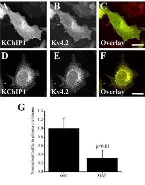
Traffic of VSVG out of the ER requires the function of Rab1 [38]. It has been suggested, however, that some cargo traffic can be independent of Rab1 function [27, 28]. We tested, therefore, whether traffic of Kv4.2/KChIP1 was Rab1-dependent. The approach used was to co-express the identified Rab1 GAP TBC1D20 [38]. Since this converts Rab1 into the inactive GDP-loaded form it acts as an effective and specific inhibitor of Rab1 function. As described previously [38] we found that co-expression of TBC1D20 inhibited traffic of VSVG-EGFP (data not shown) and also effectively inhibited traffic to the plasma membrane of Kv4.2 which remained intracellular but still co-localised with KChIP1 (Figure 2). These results indicate a common Rab1 requirement for the conventional and the KChIP1-dependent trafficking pathways.
KChIP1 vesicles co-localise with the SNARE proteins VAMP7 and Vti1a
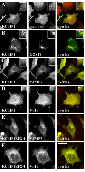
SNARES that are present in only one of the three complexes implicated in ER to Golgi traffic. Membrin, a component of the SNARE complex in the conventional VSVG traffic pathway [31] did not show any significant co-localisation with the KChIP1-EYFP-labelled vesicles (Figure 3A). There was also no co-localisation of KChIP-EYFP with GOS28 (Figure 3B) a component of a SNARE complex in ERGIC to cis-Golgi traffic [32]. Similarly no co-localisation was seen with KChIP1 for Sec22b or Ykt6 (data not shown). In contrast, VAMP7 and Vti1a, two SNAREs implicated in traffic of prechylomicron transport vesicles from ER to Golgi [33], showed extensive co-localisation with KChIP1-vesicles (Figure 3C,D). Using a polyclonal antiserum against VAMP7 [39] similar results were found (data not shown). Furthermore, the EF hand mutants of KChIP1 which also labelled punctate vesicular structures were also co-localised with VAMP7 and Vti1a as shown for the triple EF hand mutant (Figure 3E,F).
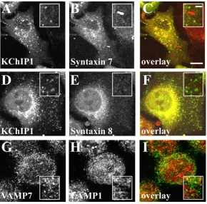
Since VAMP7 has also been shown to be part of a SNARE complex with syntaxins 7 and 8 in late endocytotic traffic [40] we examined possible co-localisation of KChIP1 vesicles with these proteins. Despite some overlapping localisation in the prenuclear region of the cells puncta labelled by KChIP were clearly distinct from those labelled by anti-syntaxin 7 (Figure 4A-C) or syntaxin 8 (Figure 4D-F). We also examined potential overlap with lysosomes. Over-expressed VAMP7 has been found associated with lysosomes [41] but we saw little overlap of staining for endogenous VAMP7 with that for the lysosomal marker LAMP1. The results indicate that the VAMP7-positive KChIP vesicles are not of late endosomal/lysosomal origin.
Functional involvement of VAMP7 and Vti1a in traffic of Kv4.2/KChIP1
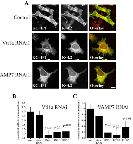
The co-localisation data suggested a potential role for VAMP7 and Vti1a in the unconventional traffic route taken by Kv4.2/KChIP1. In order to test this possibility we used RNA silencing to examine the effect of knock-down of these SNARES on Kv4.2/KChIP1 traffic. Three distinct siRNAs were tested for both Vti1a and VAMP7 and their effect on the localisation of KChIP1-EYFP and Kv4.2 was examined. Cells treated with the siRNAs were compared to cells in the absence of siRNA and also to cells in the presence of a silent siRNA. In control cells KChIP1 and Kv4.2 were co-localised and found at the plasma membrane (Figure 5A). Treatment with a Vti1a or a VAMP7 siRNA blocked traffic of Kv4.2 channels to the plasma membrane and KChIP1-EYFP was also found to be intracellular (Figure 5A). The proportion of Kv4.2 present at the plasma membrane was determined and expressed as a normalised value for plasma membrane traffic. The silent siRNA had no effect on the amount of Kv4.2 traffic but all three Vtila and all three VAMP7 siRNAs had a significant inhibitory effect (Figure 5 B,C). Further studies concentrated on use of the most inhibitory Vti1a or VAMP7 siRNA.
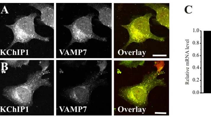
reduced and the effectiveness of the siRNA knock down of VAMP7 was confirmed by real-time PCR to measure relative mRNA levels (Figure 6C). Despite the knock-down, the KChIP1 vesicles did not show any obvious alterations in distribution or morphology and still showed co-localisation with the low levels of VAMP7 (Figure 6B).
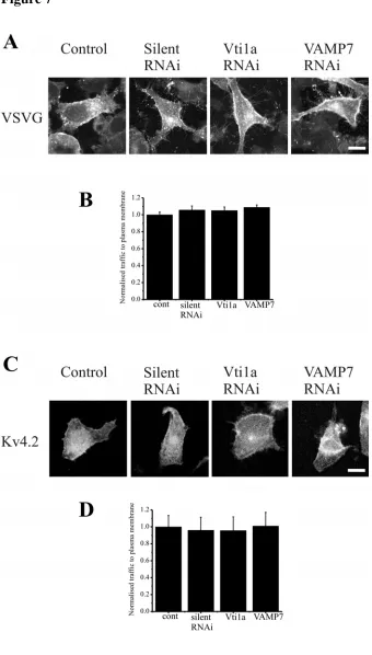
To determine whether the ability of the Vti1a and VAMP7 siRNAs to inhibit Kv4.2 traffic were specific we tested the effect of the selected siRNAs on VSVG traffic to the plasma membrane. The appearance of VSVG-GFP at the plasma membrane was apparent in control cells and also in cells treated with control siRNA or Vti1a and VAMP7 siRNAs (Figure 7A). Quantification of VSVG at the plasma membrane indicated that the siRNAs were without effect (Figure 7B). These finding support the conclusion that Vti1a and VAMP7 are specifically involved in an alternative traffic pathway that is distinct from the conventional pathway from the ER followed by VSVG.
KChIP1 is the only one of the four KChIPs that is myristoylated and the only one that targets to the pool of intracellular vesicles [11, 13, 34]. KChIP2 in contrast is palmitoylated and targets to the plasma membrane when expressed alone [15, 34]. This raised the possibility that its stimulation of Kv4 traffic may involve interaction with the channel in a distinct cellular compartment and via a distinct cellular mechanism to that used by KChIP1. To test this possibility the effect of Vti1a and VAMP7 siRNAs on KChIP2 stimulated traffic of Kv4.2 was examined. No effect of the siRNAs was detected (Figure 7C,D) consistent with distinct pathways being used by KChIP1 and KChIP2 and again confirming the lack of a general disruptive effect of the siRNAs on cellular function.
Discussion
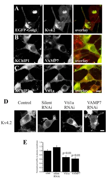
The data presented here support a role for a VAMP7/Vti1a containing SNARE complex in a novel traffic pathway that is used by KChIP1 in its stimulation of Kv4 traffic to the plasma membrane but not by the conventional pathway followed by VSVG protein. This pathway appears to exist in both non-neuronal (HeLa) and neuronal-derived (Neuro2A) cell lines. Where in the secretory pathway are the KChIP1 vesicles placed? KChIP1 is associated with 50-100 nm vesicles and we have previously concluded that the KChIP1 vesicles are an intermediate in ER to Golgi traffic based on their partial co-localisation with ERGIC-53 and the finding that an expressed N-terminal fragment of KChIP1 blocked Kv4.2 traffic and became fully co-localised with an enlarged ERGIC compartment [11]. They were not COPII coated nor was COPII function crucial for Kv4.2/KChIP1 traffic although COPI function was [12]. This pathway is likely to be distinct from the other non-conventional ER to Golgi route, such as the well studied route for procollagen, as this is COPII-dependent and COPI-independent [25]. We now show that the VAMP-7-positive KChIP1 vesicles do not overlap with endosomal or lysosomal markers consistent with the interpretation that they lie in the early exocytic pathway and with previous data suggesting a role for VAMP/Vti1a in ER to Golgi traffic of PCTVs [33].
VAMP7 which is otherwise known as TI-VAMP [42] appears to be involved in multiple SNARE complexes in various traffic steps in different cell types. It has been shown to be involved in endosomal/lysosomal traffic where it can function in at least two distinct SNARE complexes [40, 41, 43]. Despite a focus on VAMP7 in endosomal traffic, VAMP7 is also involved in transport of apical vesicles to the plasma membrane in epithelial cells [39]. In addition, it is clear that many of the vesicular VAMP7 structures identified by immunofluorescence do not overlap with endosomal/lysosomal markers. In PC12 cells, VAMP7 overlaps to only a low extent with endosomal and lysosomal markers and it was noted that in HeLa cells the endogenous VAMP7 does not co-localise with the lysosomal protein LAMP1 [44]. We have now confirmed the latter observation in the present study. These findings suggest that VAMP7 may associate with a variety of other SNARE partners to function in multiple membrane traffic steps which may in part be cell type-specific [42]. It is noteworthy and potentially relevant to the current study that in neurons, VAMP7 is involved in axonal and dendritic outgrowth [45] and is associated with as yet unidentified vesicular structures present in axons and dendrites [44]. In PC12 cells the VAMP7-labelled vesicle seen by immuno-EM [44] are very similar in size and morphology to those labelled by KChIP1 in HeLa cells [12].
alone and where many palmitoyl transferases are located [46]. Interestingly, KChIP2 is expressed at highest levels in the heart [47] where it is the major KChIP and is indispensable for normal Kv4 function [48] and VAMP7 is apparently not expressed in the heart [49].
SNARE complexes suggested to mediate ER to Golgi traffic all contain syntaxin 5 and rBet1 but three distinct complexes have been indentified based on the identity of the other two SNARES. KChIP1/Kv4.2 traffic appears to involve Vti1a and VAMP7 based on co-localisation and functional siRNA data. Vti1a has been shown to interact with syntaxin 5 [50] and both Vti1a and VAMP7 have been found to be quantitatively recovered in a SNARE complex with syntaxin 5 and rbet1 that was implicated in ER to Golgi traffic of PCTVs [33]. Since these organelles are expressed in a tissue specific manner it was possible that Vti1a/VAMP7-dependent ER to Golgi traffic could have been enterocyte specific process. Our data further highlights the distinctive nature of Kv4.2/KChIP1 traffic and now suggest that Vti1a/VAMP7 define a non-conventional pathway that may be used for the traffic of a range of cargo proteins in neuronal and non neuronal cell types.
Acknowledgements
This work was supported by a Wellcome Trust Studentship to SEF. We are grateful to Francis Barr, Burcu Hasdemir, Jesse Hay, Thierry Galli and John Presley for the gift of reagents.
References
1 Birnbaum, S. G., Varga, A. W., Yuan, L.-L., Anderson, A. E., Sweatt, J. D. and
Schrader, L. A. (2004) Structure and function of Kv4-family transient potassium
channels. Physiol. Rev. 84, 803-833
2 Isbrandt, D., Leicher, T., Waldschutz, R., Zhu, X., Luhmann, U., Michel, U.,
Sauter, K. and Pongs, O. (2000) Gene structures and expression profiles of three human KCND (Kv4) potassium channels mediating A-type currents I(TO) and I(SA).
Genomics. 64, 144-154
3 Hoffman, D. A., Magee, J. C., Colbert, C. M. and Johnston, D. (1997) K+
channel regulation of signal propagation in dendrites of hippocampal pyramidal
neurons. Nature. 387, 869-875
4 Cai, X., Liang, C. W., Muralidharan, S., Kao, J. P., Tang, C. M. and
Thompson, S. M. (2004) Unique roles of SK and Kv4.2 potassium channels in
dendritic integration. Neuron. 44, 351-364
5 Bernard, C., Anderson, A., Becker, A., Poolos, N. P., Beck, H. and Johnston,
D. (2004) Acquired dendritic channelopathy in temporal lobe epilepsy. Science. 305,
6 An, W. F., Bowlby, M. R., Bett, M., Cao, J., Ling, H. P., Mendoza, G., Hinson, J. W., Mattsson, K. I., Strassle, B. W., Trimmer, J. S. and Rhodes, K. J. (2000) Modulation of A-type potassium channels by a family of calcium sensors. Nature.
403, 553-556
7 Burgoyne, R. D. and Weiss, J. L. (2001) The neuronal calcium sensor family of
Ca2+-binding proteins. Biochem. J. 353, 1-12
8 Burgoyne, R. D. (2007) Neuronal calcium sensor proteins: generating diversity
in neuronal Ca(2+) signalling. Nat Rev Neurosci. 8, 182-193
9 Pioletti, M., Findeisen, F., Hura, G. L. and Minor, D. L. (2006)
Three-dimensional structure of the KChIP1–Kv4.3 T1 complex reveals a cross-shaped
octamer. Nature Struct. Mol. Biol. 13, 987-995
10 Wang, H., Yan, Y., Liu, Q., Huang, Y., Shen, Y., Chen, L., Chen, Y., Yang, Q.,
Hao, Q., Wang, K. and Chai, J. (2007) Structural basis for modulation of Kv4 K(+)
channels by auxiliary KChIP subunits. Nat Neurosci. 10, 32-39
11 O'Callaghan, D. W., Hasdemir, B., Leighton, M. and Burgoyne, R. D. (2003)
Residues within the myristoylation motif determine intracellular targeting of the
neuronal Ca2+ sensor protein KChIP1 to post-ER transport vesicles and traffic of Kv4
K+ channels. J. Cell Sci. 116, 4833-4845.
12 Hasdemir, B., Fitzgerald, D. J., Prior, I. A., Tepikin, A. V. and Burgoyne, R. D.
(2005) Traffic of Kv4 K+ channels mediated by KChIP1 is via a novel post-ER
vesicular pathway. J Cell Biol. 171, 459-469
13 Shibata, R., Misonou, H., Campomanes, C. R., Anderson, A. E., Schrader, L.
A., Doliveira, L. C., Carroll, K. I., Sweatt, J. D., Rhodes, K. J. and Trimmer, J. S. (2003) A fundamental role for KChIPs in determining the molecular properties and
trafficking of Kv4.2 potassium channels. J. Biol. Chem. 278, 36445-36454
14 Bahring, R., Dannenberg, J., Peters, H. C., Leicher, T., Pongs, O. and Isbrandt,
D. (2001) Conserved Kv4 N-terminal domain critical for effects of Kv channel
interacting protein 2.2. on channel expression and gating. J. Biol. Chem. 276,
23888-23894
15 Takimoto, K., Yang, E.-K. and Conforti, L. (2002) Palmitoylation of KChIP
splicing variants is required for efficient cell surface expression of Kv4.3 channels. J.
Biol.Chem. 277, 26904-26911
16 Beckers, C. J. M. and Balch, W. E. (1989) Calcium and GTP: essential
components in vesicular trafficking between the endoplasmic reticulum and golgi
apparatus. J.Cell Biol. 108, 1245-1256
17 Presley, J. F., Cole, N. B., Schroer, T. A., Hirschberg, K., Zaal, K. J. M. and
Lippincott-Schwartz, J. (1997) ER-to-Golgi transport visualized in living cells.
Nature. 389, 81-85
18 Kuge, O., Dascher, C., Orci, L., Rowe, T., Amherdt, M., Plutner, H.,
Ravazzola, M., Tanigawa, G., Rothman, J. E. and Balch, W. E. (1994) Sar1 promotes vesicle budding from the endoplasmic reticulum but not Golgi compartments. J Cell
Biol. 125, 51-65
19 Gurkan, C., Koulov, A. V. and Balch, W. E. (2007) An evolutionary
perspective on eukaryotic membrane trafficking. Adv Exp Med Biol. 607, 73-83
20 Bannykh, S. I. and Balch, W. E. (1997) Membrane dynamics at the
21 Bednarek, S. Y., Ravazzola, M., Hosobuchi, M., Amherdt, M., Perrelet, A., Schekman, R. and Orci, L. (1995) COPI- and COPII-coated vesicles bud directly from the endoplasmic reticulum in
yeast. Cell. 29, 1183-1196
22 Takida, S., Maeda, Y. and Kinoshita, T. (2008) Mammalian GPI-anchored
proteins require p24 proteins for their efficient transport from the ER to the plasma
membrane. Biochem J. 409, 555-562
23 Muniz, M., Morsomme, P. and Riezman, H. (2001) Protein sorting upon exit
from the endoplasmic reticulum. Cell. 104, 313-320
24 Stephens, D. J. and Pepperkok, R. (2002) Imaging of procollagen transport
reveals COPI-dependent cargo sorting during ER-to-Golgi transport in mammalian
cells. J Cell Sci. 115, 1149-1160
25 Starkuviene, V. and Pepperkok, R. (2007) Differential Requirements for
ts-O45-G and Procollagen Biosynthetic Transport. Traffic 8, 1035-1051
26 Siddiqi, S. A., Gorelick, F. S., Mahan, J. T. and Mansbach, C. M. (2003) COPII
proteins are required for Golgi fusion but not for endoplasmic reticulum budding of
the pre-chylomicron transport vesicle. J. Cell Sci. 116, 415-427
27 Wu, G., Zhao, G. and He, Y. (2003) Distinct pathways for the trafficking of
angiotensin II and adrenergic receptors from the endoplasmic reticulum to the cell surface: Rab1-independent transport of a G protein-coupled receptor. J Biol Chem.
278, 47062-47069
28 Ye, B., Zhang, Y., Song, W., Younger, S. H., Jan, L. Y. and Jan, Y. N. (2007)
Growing dendrites and axons differ in their reliance on the secretory pathway. Cell.
130, 717-729
29 Kloepper, T. H., Kienle, C. N. and Fasshauer, D. (2007) An elaborate
classification of SNARE proteins sheds light on the conservation of the eukaryotic
endomembrane system. Mol Biol Cell. 18, 3463-3471
30 Dascher, C., Matteson, J. and Balch, W. E. (1994) Syntaxin 5 regulates
endoplasmic reticulum to Golgi transport. J Biol Chem. 269, 29363-29366
31 Xu, D., Joglekar, A. P., Williams, A. L. and Hay, J. C. (2000) Subunit structure
of a mammalian ER/Golgi SNARE complex. J Biol Chem. 275, 39631-39639
32 Zhang, T. and Hong, W. (2001) Ykt6 forms a SNARE complex with syntaxin
5, GS28, and Bet1 and participates in a late stage in endoplasmic reticulum-Golgi
transport. J Biol Chem. 276, 27480-27487
33 Siddiqi, S. A., Siddiqi, S., Mahan, J., Peggs, K., Gorelick, F. S. and Mansbach,
C. M. (2006) The identification of a novel ER to Golgi SNARE complex used by the
Pre-chylomicron transport vesicle. J Biol Chem 281, 20974-20982
34 Venn, N., Haynes, L. P. and Burgoyne, R. D. (2008) Specific effects of
KChIP3/calsenilin/DREAM but not KChIPs1, 2 and 4 on calcium signalling and
regulated secretion in PC12 cells. Biochem J. 413, 71-80
35 Handley, M. T. W., Haynes, L. P. and Burgoyne, R. D. (2007) Differential
dynamics of Rab3A and Rab27A on secretory granules J Cell Sci. 120, 973-984
36 Hasegawa, H., Zinsser, S., Rhee, Y., Vik-Mo, E. O., Davanger, S. and Hay, J.
C. (2003) Mammalian ykt6 is a neuronal SNARE targeted to a specialized
37 Livak, K. J. and Schmittgen, T. D. (2001) Analysis of relative gene expression data using real-time quantitative PCR and the 2(-Delta Delta C(T)) Method. Methods.
25, 402-408
38 Haas, A. K., Yoshimura, S., Stephens, D. J., Preisinger, C., Fuchs, E. and Barr,
F. A. (2007) Analysis of GTPase-activating proteins: Rab1 and Rab43 are key Rabs
required to maintain a functional Golgi complex in human cells. J Cell Sci. 120,
2997-3010
39 Galli, T., Zahraoui, A., Vaidyanathan, V. V., Raposo, G., Tian, J. M., Karin,
M., Niemann, H. and Louvard, D. (1998) A novel tetanus neurotoxin-insensitive vesicle-associated membrane protein in SNARE complexes of the apical plasma
membrane of epithelial cells. Mol Biol Cell. 9, 1437-1448
40 Pryor, P. R., Mullock, B. M., Bright, N. A., Lindsay, M. R., Gray, S. R.,
Richardson, S. C., Stewart, A., James, D. E., Piper, R. C. and Luzio, J. P. (2004) Combinatorial SNARE complexes with VAMP7 or VAMP8 define different late
endocytic fusion events. EMBO reports. 5, 590-595
41 Advani, R. J., Yang, B., Prekeris, R., Lee, K. C., Klumperman, J. and Scheller,
R. H. (1999) VAMP-7 mediates vesicular transport from endosomes to lysosomes. J
Cell Biol. 146, 765-776
42 Rossi, V., Banfield, D. K., Vacca, M., Dietrich, L. E., Ungermann, C.,
D'Esposito, M., Galli, T. and Filippini, F. (2004) Longins and their longin domains: regulated SNAREs and multifunctional SNARE regulators. Trends in biochemical
sciences. 29, 682-688
43 Ward, D. M., Pevsner, J., Scullion, M. A., Vaughn, M. and Kaplan, J. (2000)
Syntaxin 7 and VAMP-7 are soluble N-ethylmaleimide-sensitive factor attachment protein receptors required for late endosome-lysosome and homotypic lysosome
fusion in alveolar macrophages. Mol Biol Cell. 11, 2327-2333
44 Coco, S., Raposo, G., Martinez, S., Fontaine, J. J., Takamori, S., Zahraoui, A.,
Jahn, R., Matteoli, M., Louvard, D. and Galli, T. (1999) Subcellular localization of tetanus neurotoxin-insensitive vesicle-associated membrane protein (VAMP)/VAMP7
in neuronal cells: evidence for a novel membrane compartment. J Neurosci. 19,
9803-9812
45 Martinez-Arca, S., Coco, S., Mainguy, G., Schenk, U., Alberts, P., Bouille, P.,
Mezzina, M., Prochiantz, A., Matteoli, M., Louvard, D. and Galli, T. (2001) A common exocytotic mechanism mediates axonal and dendritic outgrowth. J Neurosci.
21, 3830-3838
46 Greaves, J., Salaun, C., Fukata, Y., Fukata, M. and Chamberlain, L. H. (2008)
Palmitoylation and membrane interactions of the neuroprotective chaperone cysteine-string protein. J Biol Chem 283, 25014-25026
47 Pruunsild, P. and Timmusk, T. (2005) Structure, alternative splicing, and
expression of the human and mouse KCNIP gene family. Genomics. 86, 581-593
48 Kuo, H.-C., Cheng, C.-F., Clark, R. B., Lin, J. J.-C., Lin, J. L.-C., Hoshijima,
M., Nguyen-Tran, V. T. B., Gu, Y., Ikeda, Y., Chu, P.-H., Ross, J., Giles, W. R. and Chien, K. R. (2001) A defect in the Kv channel-interacting protein 2 (KChIp2) gene
leads to a complete loss of Ito and confers susceptibility to ventricular tachycardia.
49 Advani, R. J., Bae, H. R., Bock, J. B., Chao, D. S., Doung, Y. C., Prekeris, R., Yoo, J. S. and Scheller, R. H. (1998) Seven novel mammalian SNARE proteins
localize to distinct membrane compartments. J Biol Chem. 273, 10317-10324
50 Xu, Y., Wong, S. H., Tang, B. L., Subramaniam, V. N., Zhang, T. and Hong,
W. (1998) A 29-kilodalton Golgi soluble N-ethylmaleimide-sensitive factor attachment protein receptor (Vti1-rp2) implicated in protein trafficking in the
Figure Legends
Figure 1. Effects of KChIP1 and KChIP1 with EF-hand mutations on traffic of Kv4.2 and VSVG to the plasma membrane. Hela cells were transfected to co-express Kv4.2 and EYFP-Golgi (A-C) or ARF1-EYFP (D-F) or with wild-type KChIP1-EYFP (G), with the single EF hand mutant KChIP1EF3-EYFP (H) or with the triple EF-hand mutant KCHIP1EF2-4-EYFP (I) and Kv4.2 localisation determined by using anti-Kv4.2 immunostaining. Hela cells were transfected to express VSVG-GFP alone (J), or with wild-type KChIP1 (K) or the triple EF-hand mutant KCHIP1EF2-4-HcRed (L) and VSVG-GFP localisation determined by confocal imaging. The scale bars
represent 10 µm. Normalised traffic of Kv4.2 (M) or VSVG-GFP (N) in the presence
of various KChIPs was quantified and the data shown as Mean + SEM from 20 cells for each condition.
Figure 2. Inhibition of Rab1 function prevents traffic of Kv4.2/KChIP1 to the plasma membrane. HeLa cells were co-transfected to express both KChIP1-EYFP and Kv4.2 and immunostained with anti-Kv4.2 so that localisation of KChIP1 (A) and Kv4.2 (B) could be visualised. Alternatively the cells were additionally co-transfected to express the Rab1 GAP TBC1D20 (D-F). The colour overlays (C,F) show co-expressed proteins in green (KChIP1-EYFP) and red (Kv4.2) with co-localisation appearing in yellow. The scale bars represent 10 µm. Normalised traffic of Kv4.2 in the presence or absence of TBC1D20 was quantified and the data shown as Mean + SEM from 20 cells for each condition.
Figure 3. Co-localisation of KChIP1 with VAMP7 and Vti1a. HeLa cells were transfected to express KChIP1-EYFP or KChIP1EF2-4-EYFP as indicated, fixed and immunostained with mouse monoclonal antibodies specific for membrin (A), GOS28 (B), VAMP7 (C,E) or Vti1a(D,F). The images show the localisation of the KChIP1 and the indicated SNARE proteins and the colour overlays show KChIP1-EYFP in green and anti-SNARE staining in red with co-localisation appearing in yellow. The
scale bars represent 10 µm in the main figures and 2 µm in the enlarged inserts.
Figure 4. Lack of co-localisation of KChIP1 with syntaxin 7 and 8 or VAMP7 with LAMP1. HeLa cells were transfected to express KChIP1-EYFP, fixed and immunostained with mouse monoclonal antibodies specific for syntaxin 7 (A-C) or syntaxin 8 (D-F). The images show that KChIP1 punctae are distinct from those stained by syntaxin 7 or 8. In cells immunostained with VAMP 7 adn anti-LAMP1 (G-I) there was little overlap of the stained punctae. The colour overlays show KChIP1-EYFP or VAMP7 in green and syntaxin 7 and 8 and anti-LAMP1 in red with co-localisation appearing in yellow. The scale bars represent 10
Figure 5. VAMP7 and Vti1a siRNA specifically inhibits traffic of Kv4.2/KChIP1 to the plasma membrane in HeLa cells. A, Hela cells were co-transfected to express both KChIP1-EYFP and Kv4.2 and immunostained with anti-Kv4.2 so that localisation of KChIP1 and Kv4.2 could be visualised. Control cells are shown as well as cells transfected with VAMP7 or Vtila siRNA as indicated. The colour overlays show co-expressed proteins in green (KChIP1-EYFP) and red (Kv4.2) with co-localisation
appearing in yellow. The scale bars represent 10 µm. Normalised traffic of Kv4.2 in
control cells and cells co-transfected with Vti1a (B) or VAMP7 (C) siRNAs was quantified and the data shown as Mean + SEM from 20 (Vti1A) or 25 (VAMP7) cells for each condition.
Figure 6. VAMP7 knock-down does not affect the morphology or distribution of KChIP1 vesicles. HeLa cells were transfected to express KChIP1-EYFP, fixed and immunostained with anti-VAMP7 in the absence (A) or presence (B) of VAMP7 siRNA so that localisation of KChIP1 and VAMP7 could be visualised. The colour overlays show co-expressed proteins in green (KChIP1-EYFP) and red (VAMP7)
with co-localisation appearing in yellow. The scale bars represent 10 µm. C. For
quantitative PCR, mRNA was prepared from HeLa cells transfected in the presence of silent or VAMP7 siRNA and relative mRNA levels determined using real-time PCR.
Figure 7. VAMP7 and Vti1a siRNA do not inhibit traffic of VSVG or Kv4.2/KChIP2 to the plasma membrane. HeLa cells were transfected to express VSVG-GFP and imaged. Control cells are shown as well as cells co-transfected with silent, VAMP7 or Vtila siRNA as indicated (A). Normalised traffic of VSVG in control cells and cells co-transfected with silent, Vti1a or VAMP7 siRNAs was quantified and the data are shown from 20 cells for each condition (B). HeLa cells were transfected to express KChIP2 and Kv4.2 and immunostained with anti-Kv4.2 to allow imaging of Kv4.2 localisation. Control cells are shown as well as cells co-transfected with silent, VAMP7 or Vtila siRNA as indicated (C). Normalised traffic of Kv4.2 in control cells and cells co-transfected with silent, Vti1a or VAMP7 siRNAs was quantified and the data shown as Mean + SEM from 30 cells for each condition (D). The scale bars
represent 10 µm.
and cells co-transfected with siRNAs was quantified and the data shown as Mean +