0022-538X/10/$12.00 doi:10.1128/JVI.02590-09
Copyright © 2010, American Society for Microbiology. All Rights Reserved.
HIV-1 Vpr Induces the K48-Linked Polyubiquitination and Proteasomal
Degradation of Target Cellular Proteins To Activate ATR and
Promote G
2
Arrest
䌤
†
Jean-Philippe Belzile,
1Jonathan Richard,
1Nicole Rougeau,
1Yong Xiao,
1and E
´ ric A. Cohen
2*
Laboratory of Human Retrovirology, Institut de Recherches Cliniques de Montre´al,2and Department of Microbiology and
Immunology, Universite´ de Montre´al,1Montreal, Quebec, Canada
Received 10 December 2009/Accepted 8 January 2010
HIV-1 viral protein R (Vpr) induces cell cycle arrest at the G2/M phase by a mechanism involving the
activation of the DNA damage sensor ATR. We and others recently showed that Vpr performs this function by subverting the activity of the DDB1-CUL4A (VPRBP) E3 ubiquitin ligase. Vpr could thus act as a connector between the E3 ligase and an unknown cellular factor whose ubiquitination would induce G2arrest. While
attractive, this model is based solely on the indirect observation that some mutants of Vpr retain their interaction with the E3 ligase but fail to induce G2arrest. Using a tandem affinity purification approach, we
observed that Vpr interacts with ubiquitinated cellular proteins and that this association requires the recruit-ment of an active E3 ligase given that the depletion of VPRBP by RNA interference or the overexpression of a dominant negative mutant of CUL4A decreased this association. Importantly, G2-arrest-defective mutants of
Vpr in the C-terminal putative substrate-interacting domain displayed a decreased association with ubiqui-tinated proteins. We also found that the inhibition of proteasomal activity increased this association and that the ubiquitin chains were at least in part constituted of classical K48 linkages. Interestingly, the inhibition of K48 polyubiquitination specifically impaired the Vpr-induced phosphorylation of H2AX, an early target of ATR, but did not affect UV-induced H2AX phosphorylation. Overall, our results provide direct evidence that the association of Vpr with the DDB1-CUL4A (VPRBP) E3 ubiquitin ligase induces the K48-linked polyubiq-uitination of as-yet-unknown cellular proteins, resulting in their proteasomal degradation and ultimately leading to the activation of ATR and G2arrest.
Viruses have evolved ways to modulate the host cellular environment in order to promote efficient viral replication and to disrupt elements of innate or acquired immunity. One strat-egy particularly favored by viruses to achieve these goals is to hijack components of the host ubiquitin-proteasome system in order to induce the degradation, block the degradation, or mod-ulate the expression and activity of specific cellular factors (4, 8, 16). Human immunodeficiency virus (HIV) is no exception to this precept. HIV harbors two extensively studied accessory proteins, viral protein U (Vpu) and viral infectivity factor (Vif), that are usurping the cellular ubiquitin-proteasome system in order to degrade neosynthesized CD4 and the cytidine deaminases APOBEC3F (apolipoprotein B mRNA-editing enzyme, catalytic polypeptide-like 3F) and APOBEC3G (37), respectively. Recently, we and several other investigators dem-onstrated that a third accessory protein of HIV, viral protein R (Vpr), would also exert its function by usurping the host ubiq-uitination machinery via the recruitment of an E3 ubiquitin ligase complex composed of VPRBP (viral protein R binding protein, also known as DCAF1), damaged DNA binding pro-tein 1 (DDB1), and cullin 4A (CUL4A) (5, 14, 22, 32, 43, 49, 55).
Ubiquitination is a posttranslational modification that in-volves the isopeptidic covalent linkage of the C terminus of a small protein called ubiquitin, most commonly to lysine accep-tor residues. The conjugation of ubiquitin is performed by E3 ubiquitin ligase complexes, which also control specificity via a direct interaction with substrates (24, 33). The cullin-RING E3 ubiquitin ligases (CRL) structured around the scaffold protein CUL4A (CRL4A) interact with the 126-kDa DDB1 adaptor in order to recruit substrate receptors of the WDxR family (31). Mass spectrometric analyses of CUL4A-DDB1 complexes have revealed physical interactions with at least 30 different WDxR substrates receptors (3, 19, 21, 26), suggesting that CRL4A E3 ligases would likely regulate the function of hun-dreds of cellular proteins. Surprisingly, however, relatively few cellular proteins have been shown to date to be regulated by these complexes. CRL4A (DDB2) promotes the ubiquitination of histones 2A, 3, and 4 (28, 53); of xeroderma pigmentosum group C protein (XPC) (48); and probably of xeroderma pig-mentosum group A protein (XPA) (52) to facilitate UV dam-age repair. CRL4A (CSA) and CRL4A (DET1-COP1) induce the proteolysis of Cockayne syndrome type B gene product (CSB) (18) and c-JUN (56), respectively. PCNA (proliferating cell nuclear antigen), via the recruitment of a CRL4A (CDT2) ligase, was previously shown to regulate the degradation of the replication licensing factor CDT1 (20, 35) as well as that of the CDK inhibitor p21 (1, 39). The 180-kDa VPRBP/DCAF1 WDxR substrate receptor was identified more than a decade ago as being a Vpr-interacting protein and was found to be expressed at the mRNA level in most tissues (59, 61).
How-* Corresponding author. Mailing address: Laboratory of Human Ret-rovirology, Institut de Recherches Cliniques de Montre´al, 110, Avenue des Pins Ouest, Montreal, Quebec H2W 1R7, Canada. Phone: (514) 987-5804. Fax: (514) 987-5691. E-mail: eric.cohen@ircm.qc.ca.
† Supplemental material for this article may be found at http://jvi .asm.org/.
䌤Published ahead of print on 20 January 2010.
3320
on November 8, 2019 by guest
http://jvi.asm.org/
ever, its normal cellular functions remained elusive until re-cently. Huang and Chen previously demonstrated that CRL4A (VPRBP) induces the rapid degradation of the tumor suppres-sor Merlin (NF2 [neurofibromin 2]) following serum stimula-tion (23). Moreover, the deplestimula-tion of VPRBP reduced the rate of DNA replication, blocked cells in S phase, and impeded cellular proliferation (22, 38). Consequently, the genetic abla-tion of VPRBP in mouse (38) and in the evoluabla-tionarily distant
Arabidopsis thaliana(60) led to embryonic lethality, suggesting
an essential role for VPRBP in the cell cycle as well as in development.
The small HIV-1 accessory protein Vpr induces cell cycle arrest in the G2/M phase in various cell types, including trans-formed cell lines as well as primary lymphocytes (27, 41, 63). Notably, an abnormal accumulation of infected cells in G2 phase can be observed in tissues from patients infected by HIV-1 (63). Although the function of this cell cycle arrest has remained elusive, its molecular mechanism has recently begun to be elucidated. Several investigators reported that Vpr acti-vates the canonical ATR (ataxia telangiectasia and Rad3 re-lated) DNA damage/stress checkpoint (2, 29, 42, 62, 63). The Vpr-mediated activation of ATR is accompanied by the for-mation of DNA repair foci that include RPA (replication pro-tein A), HUS1, RAD17, BRCA1 (breast cancer 1, early onset), TP53BP1 (tumor protein p53 binding protein 1), and␥-H2AX (phosphorylated histone 2A variant X) (2, 29, 62, 63) and by the activation of the CHEK1 kinase (42). This series of events leads to the inactivation of the CDC2/cyclin B complex, a master regulator of the G2-to-M-phase transition, and ulti-mately prevents entry into mitosis. In addition, Vpr was re-cently found to associate with DDB1 and CUL4A via a direct interaction with VPRBP (5, 14, 22, 32, 43, 49, 55). The deple-tion of VPRBP by RNA interference drastically impaired Vpr-mediated G2arrest. Similarly, mutations of Vpr in the
hydro-phobic leucine-rich core region abrogated binding to CRL4A (VPRBP) and concomitantly impaired G2arrest. In contrast,
mutants of Vpr in the C-terminal arginine-rich domain were not compromised for the association with the E3 ligase but nevertheless failed to induce G2 arrest (5, 14, 22, 32, 43, 49, 55). These later mutants also displayedtrans-dominant nega-tive activity (14), thus indicating that the recruitment of this E3 ubiquitin ligase is essential but not sufficient to induce G2
arrest. The simplest explanation for these observations is that Vpr would recruit cellular substrates to be ubiquitinated by the complex. The ubiquitination of these as-yet-unknown proteins would lead to their degradation or the modulation of their activity, ultimately resulting in ATR activation and G2 arrest (13). This is not the only instance in which Vpr would be implicated in the degradation of a cellular protein. Vpr directly interacts with and induces the proteasomal degradation of the cellular enzyme UNG2 (uracil-DNA glycosylase 2, also known as CCNO) independently of the induction of cell cycle arrest (43, 44). The Vpr-induced degradation of UNG2 was found previously to be mediated by DDB1 (43) but surprisingly did not require VPRBP (55). In addition, viral protein X (Vpx), a paralog of Vpr present in HIV-2 and some simian immunode-ficiency virus (SIV) isolates, is also able to recruit CRL4A (VPRBP) (6, 32, 45, 47). In contrast to Vpr, Vpx does not induce G2arrest but counteracts a putative cellular restriction factor expressed in macrophages and dendritic cells that
tar-gets a postentry step critical for efficient reverse transcription (37). Importantly, the presence of an active proteasome system and the recruitment of CRL4A (VPRBP) by Vpx were previ-ously shown to be required for the inhibition of the restriction factor, suggesting that, similarly to Vpr, Vpx would act via the CRL4A (VPRBP)-mediated ubiquitination and proteasomal degradation of cellular proteins (6, 17, 45, 47).
However, this model, while very attractive, is based solely on the indirect observation that some C-terminal mutants of Vpr retain their interaction with the E3 ligase but fail to induce G2 arrest. Other models for the activity of Vpr have also been proposed and include the inhibition of DDB1 functions (43) or an overall increased activity of CRL4A (VPRBP) (22). More-over, it is unclear whether this association of Vpr with CRL4A (VPRBP) is the result of an overexpression in transformed cell lines and whether this interaction would occur during the in-fection of primary lymphocytes. Therefore, we sought to con-firm the physiological significance of the Vpr-VPRBP interac-tion as well as obtain more direct evidence that Vpr is indeed recruiting cellular proteins and inducing their ubiquitination and proteasomal degradation. Herein, we show that Vpr inter-acts with VPRBP and DDB1 during the infection of primary CD4⫹ T lymphocytes. In addition, using a tandem affinity approach and the overexpression of a dominant negative tagged ubiquitin mutant, we observed that wild-type (WT) Vpr could associate specifically with unknown ubiquitinated cellu-lar proteins and that this interaction required the recruitment of an active CRL4A (VPRBP) E3 ubiquitin ligase complex. Moreover, C-terminal G2-arrest-defective mutants of Vpr dis-played a reduced association with these ubiquitinated proteins. We also provide evidence that Vpr induces the K48-linked poly-ubiquitination of these cellular proteins, leading to their rapid proteasomal degradation. Finally, these Vpr-induced ubiquitina-tion events were necessary specifically for Vpr to induce the phosphorylation of H2AX, an early marker of ATR-mediated DNA damage/stress checkpoint activation.
MATERIALS AND METHODS
Cell lines and antibodies.HEK293T and HeLa cells were cultured as pre-viously described (58). The anti-hemagglutinin (HA) tag and anti-Myc tag monoclonal antibodies were clones 12CA5 and 9E10, respectively. Rabbit polyclonal antibodies against VPRBP and DDB1 were obtained from Accu-rate Chemical and Scientific Corporation (Westbury, NY) and Santa Cruz Biotechnology (Santa Cruz, CA), respectively. Vpr was detected by using a rabbit polyclonal antibody directed against a Vpr N-terminal peptide (12). The anti-green fluorescent protein (GFP) antibody was obtained from Mo-lecular Probes (Invitrogen, San Diego, CA), and the anti-phospho-H2AX (Ser139) antibody was clone JBW301 from Upstate (Millipore, Billerica, MA). The rabbit anti-actin antibody was obtained from Sigma-Aldrich (St. Louis, MO).
Plasmid construction.Plasmids SVCMV-TAP, SVCMV-TAP-Vpr(WT), and SVCMV-TAP-Vpr(Q65R) were described previously (5). Plasmids expressing TAP-Vpr(1-78), TAP-Vpr (1-86), and TAP-Vpr(R87A,R88A) were generated by subcloning the SalI-BamHI fragments from 3HA-Vpr(1-78), SVCMV-3HA-Vpr(1-86), and SVCMV-3HA-Vpr(R87A,R88A) (57), respectively, into SVCMV-TAP-Vpr(WT). Plasmids expressing 84) and TAP-Vpr(1-90) were generated by PCR using a strategy described previously (5). Plasmids expressing Myc-His-tagged ubiquitin K48R (54) and HA-tagged ubiquitin (WT, K0, and K48R) (34) were kind gifts of R. Kopito (Stanford University, Stanford, CA) and T. Dawson (Johns Hopkins University, Baltimore, MD). The plasmid expressing HA-Ub(K63R) was constructed by site-directed mutagenesis using the QuikChange II mutagenesis kit (Stratagene, La Jolla, CA). GFP-expressing plasmid pQBI-25 was obtained from Qbiogene (Carlsbad, CA). Vectors express-ing scrambled small hairpin RNA (shRNA) and VPRBP shRNA were obtained
on November 8, 2019 by guest
http://jvi.asm.org/
from Open Biosystems (Huntsville, AL). The CUL4A dominant negative con-struct (55) was kindly donated by C. de Noronha (University of Albany, Albany, NY). The construction of the infectious molecular clone HxBru(Vpr⫺) was described previously (30). HxBru(HA-Vpr) was generated by PCR by adding an HA tag at the N terminus of Vpr, which resulted in the addition of 9 extra amino acids at the C terminus of Vif. The lentiviral vectors WPI and WPI-Vpr as well as the SVCMV-IN-VSV-G expression construct were described previously (5).
Transfection, TAP, and immunoprecipitation.HEK293T cells were trans-fected by using the calcium phosphate precipitation method. Forty-eight hours later, cells were harvested, washed, and lysed in Triton lysis buffer (50 mM Tris-HCl [pH 7.5], 150 mM NaCl, 0.5% Triton X-100, and complete protease inhibitors) or radioimmunoprecipitation assay (RIPA) buffer (8 mM Na2HPO4
[pH 7.2], 2 mM NaH2PO4, 140 mM NaCl, 1% NP-40, 0.05% SDS, 12 mM
deoxycholate, and complete protease inhibitors). Tandem affinity purification (TAP) was performed as described previously (5). In some experiments, where indicated, IgG pulldown assays were performed instead of full tandem affinity purification, as described previously (5). Immunoprecipitations were performed by using 50l of 50% anti-HA-coupled agarose beads (Sigma-Aldrich, St. Louis, MO) followed by extensive washes in Triton lysis buffer and elution with 100
g/ml HA peptide. Eluted proteins were separated on a 12.5% SDS-PAGE gel, and Western blot analyses of eluted proteins were performed. Ubiquitinated proteins were revealed by using monoclonal antibodies directed at Myc or HA tags depending on the ubiquitin construct used.
Production and titration of viruses and viral vectors.The production and titration of infectious viral particles as well as of the lentiviral vectors expressing GFP (WPI) or coexpressing GFP and Vpr (WPI-Vpr) were performed as described previously (5, 57). Murine leukemia virus-based retroviral vectors expressing VPRBP-targeting shRNA (shVprBP) or scrambled control shRNA (shControl) were produced by the transfection of 15g of vector, 12g of the packaging construct pCIG3-N (9), and 5g of vesicular stomatitis virus G protein (VSV-G)-expressing plasmid pSVCMV-IN-VSV-G in 1.5 million HEK293T cells by using the calcium phosphate precipitation method. Vector-containing supernatants were fil-tered through a 0.45-m filter and used immediately.
Infection of primary lymphocytes. Peripheral blood mononuclear cells (PBMCs) were extracted, by the Ficoll method, from whole blood obtained from consenting healthy adult donors who gave written informed consent under re-search protocols approved by the rere-search ethics review board of the Institut de Recherches Cliniques de Montreal. CD4⫹T lymphocytes were purified from PBMCs by magnetic negative selection using CD4⫹T-cell isolation kit II and the AutoMACS Pro system according to the manufacturer’s instructions (Miltenyi Biotec, Auburn, CA). CD4⫹T cells were cultured as previously described (57). Ten million activated CD4⫹cells were mock infected or infected with HxBru-(Vpr⫺) or HxBru(HA-Vpr) virus at a multiplicity of infection (MOI) of 0.01. Five days after infection, cells were lysed in Triton lysis buffer and subjected to anti-HA immunoprecipitation as described above.
Fluorescence immunohistochemistry.Fifty thousand HeLa cells were seeded onto coverslips in 24-well plates and transfected with Lipofectamine 2000 re-agent according to the manufacturer’s instructions. Twenty-four hours after transfection, cells were transduced with WPI or WPI-Vpr at a multiplicity of infection of 2.5 in the presence of 8g/ml polybrene. Two days later, cells were processed for confocal fluorescence immunohistochemistry as previously de-scribed (15).
Generation of HEK293T cells with stable depletion of VPRBP.HEK293T cells were transduced with shControl and shVprBP retroviral vectors in the presence of 8g/ml polybrene. Two days after transduction, cells were selected for 10 days with 1g/ml puromycin, and single-cell clones were then isolated by the limited-dilution method. Western blots were performed on shControl- and shVprBP-transduced stable clones, and the clone with the most significant decrease in levels of VPRBP expression was selected.
Cell cycle, cell proliferation, and apoptosis assays.Cell cycle profiles were determined by flow cytometry using propidium iodide staining as previously described (5). Apoptosis was assayed by flow cytometry using annexin V and propidium iodide as a dead cell counterstain as described previously (58). Cell proliferation kinetics were monitored by flow cytometry using a standard car-boxyfluorescein succinimidyl ester (CFSE) assay.
RESULTS
Vpr associates with VPRBP and DDB1 in primary CD4ⴙ lymphocytes. The interaction between Vpr and the CRL4A (VPRBP) E3 ubiquitin ligase and its functional importance for Vpr-induced G2 arrest, although strongly established,
were, however, demonstrated solely by overexpression stud-ies of transformed cell lines. In order to determine whether Vpr could recruit the CRL4A (VPRBP) E3 ubiquitin ligase under more physiological conditions, primary CD4⫹ T lym-phocytes were infected with HxBru-derived virus expressing hemagglutinin (HA)-tagged Vpr (HA-Vpr) or deleted for Vpr at a multiplicity of infection of 0.01. Five days after infection, cells were harvested and subjected to immunoprecipitation by using anti-HA-conjugated agarose beads, and immunoprecipi-tated proteins were detected by Western blotting. Endogenous VPRBP and DDB1 could be detected in the lysates of acti-vated primary CD4⫹T lymphocytes, thus confirming their ex-pression at the protein level (Fig. 1). The coimmunoprecipita-tion of VPRBP and DDB1 could be observed for cells infected with HxBru(HA-Vpr) viruses but not for HxBru(Vpr⫺)- or mock-infected cells (Fig. 1). Therefore, the recruitment of VPRBP and DDB1 by Vpr occurs under conditions mimicking those of anin vivoinfection and is thus not the result of an overexpression of Vpr in transformed cell lines.
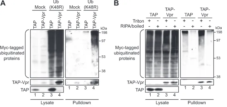
Vpr interacts with ubiquitinated cellular proteins.We next sought to obtain additional data on the mechanisms by which Vpr utilizes this complex to induce G2 arrest. If Vpr indeed acts as a connector between an E3 ubiquitin ligase complex and cellular proteins, and this association leads to the ubiq-uitination of these substrates, we hypothesized that we should be able to observe Vpr interacting with some of these unknown ubiquitinated proteins. To assess this premise, we developed a tandem affinity purification (TAP) procedure (5) that takes advantage of a Myc-tagged ubiquitin K48R mutant with the unusual properties of acting as a weak chain terminator and stabilizing polyubiquitinated products (7, 54). Of note, the TAP-tagged Vpr (TAP-Vpr) construct used for the procedure is able to induce G2arrest although to a lesser extent than that induced by untagged native Vpr (see Fig. S1 in the supplemen-tal material) (5). Under these conditions, we were able to detect an interaction between TAP-Vpr and unknown cellular ubiquitinated proteins, as revealed by the anti-Myc-reacting protein smear in the TAP-Vpr pulldown, but not with the TAP tag alone (Fig. 2A). This association was also detected under
FIG. 1. Vpr interacts with VPRBP and DDB1 during infection of primary CD4⫹T lymphocytes. Ten million activated primary CD4⫹T lymphocytes were mock infected or infected with viruses defective for Vpr [HxBru(Vpr⫺)] or encoding HA-tagged Vpr [HxBru(HA-Vpr)] at a multiplicity of infection of 0.01. Five days after infection cells were harvested in Triton lysis buffer, and immunoprecipitation (IP) against HA was performed as described in Materials and Methods. Coimmu-noprecipitated endogenous VPRBP and DDB1 were detected by Western blotting by using specific rabbit polyclonal antibodies. HA-tagged Vpr was detected by using a monoclonal anti-HA antibody.
on November 8, 2019 by guest
http://jvi.asm.org/
stringent extraction conditions, such as RIPA buffer and high-salt buffer (400 mM NaCl) (data not shown). The Myc-tagged ubiquitination signal detected in the presence of pulled-down Vpr was the result of bound cellular ubiquitinated proteins and not of the ubiquitination of Vpr itself, since the heat denatur-ation of proteins in RIPA buffer prior to the purificdenatur-ation of TAP-Vpr complexes abolished this signal. (Fig. 2B, compare lane 3 to lane 4). For this experiment, we could not perform the full tandem affinity purification procedure because the TAP tag could not bind to calmodulin beads following dena-turation. Consequently, these experiments involved an IgG pulldown step solely, hence explaining why the enrichment of ubiquitination observed with Vpr is less important under this particular condition (Fig. 2B, compare lane 3 to lane 1). Nev-ertheless, the retention of the ubiquitination signal under de-naturing conditions would have indicated that this ubiquitina-tion originated from ubiquitin covalently conjugated to Vpr itself and not to associated proteins. Therefore, in agreement with our model, Vpr is capable of associating with ubiqui-tinated cellular proteins.
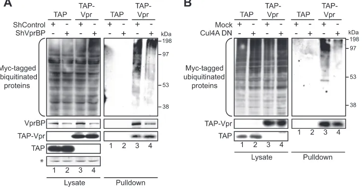
Recruitment of a catalytically active CRL4A (VPRBP) com-plex is required to observe Vpr-associated ubiquitinated pro-teins.This association of Vpr with cellular ubiquitinated pro-teins suggests that these propro-teins might be ubiquitinated by Vpr following the recruitment of the CRL4A (VPRBP) E3 ubiquitin ligase. On the other hand, these cellular proteins might be already ubiquitinated as part of their normal metab-olism before their interaction with Vpr and would thus not constitute substrates of the Vpr ubiquitin ligase complex. To distinguish between these two possibilities, we analyzed the effect of knocking down VPRBP on the association of Vpr with ubiquitinated proteins. The transient depletion of VPRBP with small hairpin RNA (shRNA) resulted in a significant decrease in the association of Vpr with cellular ubiquitinated proteins
(Fig. 3A, compare lanes 3 and 4), suggesting that the asso-ciation of ubiquitinated proteins with Vpr involves the re-cruitment of the CRL4A (VPRBP) E3 ligase. Moreover, the overexpression of a dominant negative construct of CUL4A (Cul4A DN) produced a similar decrease in the association of TAP-Vpr with ubiquitinated proteins, thus corroborating the results obtained with the depletion of VPRBP and indicating that the CRL4A (VPRBP) E3 ligase must be catalytically ac-tive in order to detect these Vpr-associated ubiquitinated pro-teins (Fig. 3B, compare lanes 3 and 4). However, these results do not exclude the possibility that this association of Vpr with ubiquitinated proteins might result from the sole association with ubiquitinated components of the E3 ligase complex with-out necessarily implicating ubiquitinated substrates.
[image:4.585.112.473.69.239.2]To address this issue, we investigated this association in the context of Vpr mutants defective for their interaction with the E3 ligase or for the putative G2arrest substrate protein. Sur-prisingly, the Q65R mutation in Vpr, which results in a strong reduction of the interaction with VPRBP and DDB1 (5, 14, 32, 49), displayed an increased association with ubiquitinated pro-teins (Fig. 4A) independently of its higher level of expression. These unexpected results may be explained by nonspecific in-teractions that might result from the accumulation of this non-functional mutant protein. To test whether the association of Vpr(Q65R) with ubiquitinated proteins was independent of the recruitment of CRL4A (VPRBP), we constructed 293T-based stable monoclonal cell lines expressing shRNA against VPRBP or a nontargeting scrambled shRNA control (see Fig. S2A in the supplemental material). Importantly, we did not detect any significant effect of VPRBP depletion on the cell cycle profile (Fig. S2B). We did, however, observe a slight decrease in the growth kinetics of these cells (Fig. S2C), but it did not result in a significant increase in apoptosis (Fig. S2D). The level of stable VPRBP knockdown achieved with this cell
FIG. 2. Vpr interacts with unknown cellular ubiquitinated proteins. (A) HEK293T cells were transfected with plasmids encoding TAP (lanes 1 and 3) or TAP-Vpr (lanes 2 and 4). Cells were cotransfected with either an empty plasmid (lanes 1 and 2) or a Myc-Ub(K48R)-encoding plasmid (lanes 3 and 4). Two days later, tandem affinity purification was performed on cell lysates as described in Materials and Methods. The levels of ubiquitinated proteins were determined by Western blotting of crude lysates and pulled-down fractions by using a monoclonal anti-Myc antibody. (B) Cells were cotransfected with plasmids expressing TAP-Vpr and Myc-Ub(K48R) (lanes 3 and 4) or with plasmids expressing TAP and Myc-Ub(K48R) as a control (lanes 1 and 2). Forty-eight hours posttransfection, cells were lysed in Triton lysis buffer (lanes 1 and 3) or were heat denatured following lysis in RIPA buffer (lanes 2 and 4). Cell extracts were subjected to IgG pulldowns with precoupled beads, and complexes were eluted following cleavage with tobacco etch virus (TEV) protease. The levels of ubiquitinated proteins were determined by using a monoclonal anti-Myc antibody, and Vpr was detected by using a polyclonal antibody.
on November 8, 2019 by guest
http://jvi.asm.org/
line was sufficient to almost completely abolish Vpr-mediated G2 arrest compared to the scrambled control cell line (Fig.
S2E). We then used this pair of cell lines to assess the levels of cellular ubiquitinated proteins associated with the Q65R mu-tant of Vpr. As expected, the increased association of ubiqui-tinated proteins with Vpr(Q65R) was independent of the re-cruitment of the E3 ligase given that the depletion of VPRBP did not affect the levels of ubiquitinated proteins associated with this mutant of Vpr (Fig. 4A, compare lanes 5 and 6). In comparison, with this system, we observed a significant reduc-tion of the associareduc-tion of wild-type Vpr with ubiquitinated proteins in the VPRBP-knocked-down cell line versus the control cell line (Fig. 4A, compare lanes 3 and 4), corrob-orating results obtained with transient knockdown experi-ments. Therefore, we hypothesize that the Q65R mutation, in addition to its effect on binding to VPRBP, might also result in major conformational defects that would lead to this nonspe-cific association to ubiquitinated proteins. Indeed, we observed that, in contrast to wild-type Vpr, which oligomerizes and displays a nuclear localization, Vpr(Q65R) accumulates in part in the cytoplasm as discrete puncta and also fails to efficiently oligomerize (data not shown). We also investigated the effect of a C-terminal deletion of Vpr, Vpr(1-78), a mutant that fails to induce G2arrest (see Fig. S1 in the supplemental material) while maintaining the interaction with VPRBP (32). Impor-tantly, Vpr(1-78), which should thus not interact with the pu-tative substrate responsible for G2arrest, failed to interact with ubiquitinated proteins (Fig. 4B, compare lanes 2 and 3). Other deletions (starting at positions 84, 86, and 90) or point muta-tions (R87A, R88A) in the C-terminal domain impaired G2
arrest (Fig. S1) and led to a significant decrease in the associ-ation with ubiquitinated proteins (Fig. 4C) while maintaining their association with VPRBP (data not shown). Our data indicate that these proteins are not solely ubiquitinated
UNG2 molecules, given that C-terminal deletions of Vpr retain their interaction with UNG2 (10). Therefore, in agreement with our model, these data further indicate that the putative G2 arrest substrate(s) is ubiquitinated in the
presence of Vpr and that the abrogation of Vpr-substrate or Vpr-CRL4A (VPRBP) interactions impairs Vpr-mediated ubiquitination. Overall, these results provide direct interac-tion-based evidence that Vpr acts as a connector between an E3 ubiquitin ligase complex and substrate proteins.
[image:5.585.114.470.71.256.2]Vpr induces the K48-linked polyubiquitination of cellular proteins, leading to their proteasomal degradation.To deter-mine the fate of proteins that are ubiquitinated by Vpr, we used the tandem affinity purification method in combination with an HA-tagged ubiquitin construct that does not signifi-cantly stabilize polyubiquitinated products and does not pro-tect substrates from proteasome degradation (34). With this system, we detected ubiquitinated proteins associated with Vpr (Fig. 5A), but the ubiquitination signal detected under these conditions did not rely on the recruitment of the E3 ligase complex, since the knockdown of VPRBP did not have any effect on the levels of ubiquitination (data not shown), suggest-ing that in the absence of an interference with polyubiquitina-tion, the Vpr-targeted substrates might be rapidly degraded. In support of this scenario, a 16-h treatment of cells coexpressing HA-Ub and TAP-Vpr with the proteasome inhibitor MG132 resulted in an increased association of Vpr with cellular ubiq-uitinated proteins (Fig. 5A, compare lanes 3 and 4). We also observes an increase in levels of polyubiquitinated proteins with a shorter MG132 treatment (5 h) but to a lesser extent (data not shown). Most notably, this significant increase in levels of HA-polyubiquitinated proteins associated with Vpr following treatment with MG132 required the recruitment of the E3 ligase because the depletion of VPRBP with shRNA drastically reduced the extent of polyubiquitination (Fig. 5B).
FIG. 3. Association of Vpr with ubiquitinated proteins involves the recruitment of an active E3 ubiquitin ligase complex. HEK293T cells were cotransfected with a plasmid encoding Myc-Ub(K48R) and with either TAP-encoding (lanes 1 and 2) or TAP-Vpr-encoding (lanes 3 and 4) plasmids. Cells were transcomplemented with plasmids expressing scrambled shRNA or shRNA targeting VPRBP (A) and with an empty plasmid or a plasmid encoding a dominant negative mutant form of CUL4A (Cul4A DN) (B), as indicated. Two days after transfection, cell extracts were subjected to tandem affinity purification as described in Materials and Methods. The levels of ubiquitinated proteins were determined by Western blotting of crude lysates and pulled-down fractions by using a monoclonal anti-Myc antibody. Vpr and VPRBP were detected by using polyclonal antibodies. An asterisk denotes a nonspecific band detected by the anti-VPRBP antibody used as a loading control.
on November 8, 2019 by guest
http://jvi.asm.org/
Therefore, these results suggest that following polyubiquitina-tion by the Vpr-CRL4A (VPRBP) complex, substrates are rapidly degraded by the proteasome.
To determine whether the observed degradation of Vpr-associated ubiquitinated proteins resulted from classical K48-linked polyubiquitination, we analyzed the effect of the ubiq-uitin K48R mutation on Vpr-associated ubiqubiq-uitination. Cells were cotransfected with plasmids expressing TAP or TAP-Vpr with either HA-Ub(WT) or HA-Ub(K48R). In contrast to Myc-tagged Ub(K48R), which acts as a weak chain terminator and stabilizes polyubiquitin products (7, 54), this HA-Ub(K48R) construct was previously shown to potently block ubiquitin chain elongation through lysine 48 (34). We also used the polyubiquitination-null construct HA-Ub(K0), in which all
[image:6.585.113.471.68.438.2]ly-sines were mutated to arginines, as a control. Following MG132 treatment, the K48R mutation in ubiquitin signifi-cantly reduced the levels of ubiquitinated proteins associated with Vpr compared to wild-type ubiquitin (Fig. 6, compare lanes 6 and 8). However, the reduction in the level of ubiqui-tination did not reach the levels achieved with the K0 mutation (Fig. 6, compare lanes 7 and 8). These data suggest that Vpr induces, at least in part, classical K48-linked polyubiquitination of cellular substrate proteins, leading to their proteasomal degradation. However, given that the levels of ubiquitination observed for the K48R mutant did not reach the levels ob-served for the polyubiquitination-null K0 mutant, we cannot exclude the possibility that other lysine residues in ubiquitin might be involved in the formation of mixed ubiquitin linkages.
FIG. 4. Analysis of the association of Vpr mutants with ubiquitinated proteins. (A) HEK293T monoclonal cell lines stably expressing a control shRNA (shControl) or an shRNA against VPRBP (shVprBP) were cotransfected with a plasmid encoding Myc-Ub(K48R) and plasmids expressing TAP, TAP-Vpr(WT), and TAP-Vpr(Q65R), as indicated. Two days after transfection, cell extracts were subjected to tandem affinity purification as described in Materials and Methods. (B) HEK293T cells were cotransfected with a plasmid encoding Myc-Ub(K48R) and plasmids expressing TAP, TAP-Vpr(WT), and TAP-Vpr(1-78), as indicated. Cell extracts were processed as described above for A. (C) HEK293T cells were cotransfected with a plasmid encoding Myc-Ub(K48R) and plasmids expressing TAP, TAP-Vpr(WT), TAP-Vpr(R87A,R88A), TAP-Vpr(1-84), TAP-Vpr(1-86), and TAP-Vpr(1-90), as indicated. Two days after transfection, cell extracts were subjected to tandem affinity purification as described in Materials and Methods. Pulldown eluates (lanes 7, 8, 10, 12, 14, and 16) and 2-fold dilutions of pulldown eluates (lanes 9, 11, 13, 15, and 17) were resolved by SDS-PAGE for analysis. For all panels, the levels of ubiquitinated proteins were determined by Western blotting of crude lysates and pulled-down fractions by using a monoclonal anti-Myc antibody. Vpr and VPRBP were detected by using polyclonal antibodies. An asterisk denotes nonspecific bands used as loading controls.
on November 8, 2019 by guest
http://jvi.asm.org/
Vpr-induced K48 polyubiquitination is required for phos-phorylation of H2AX.Other investigators demonstrated pre-viously that blocking the activity of the proteasome using small-molecule inhibitors or blocking polyubiquitination via the expression of a dominant negative mutant of ubiquitin [Ub(K48R)] abrogated Vpr-mediated G2arrest (14, 49). How-ever, caution has to be used when interpreting these results given that the inhibition of polyubiquitination or proteasome function might have pleiotropic effects on checkpoint function
without necessarily implicating the direct inhibition of Vpr’s activity. Therefore, to evaluate the direct role of K48-linked polyubiquitination in Vpr’s activity, we instead monitored the phosphorylation of H2AX (␥-H2AX), an early marker of ATR-mediated checkpoint activation previously implicated in Vpr-mediated G2 arrest (29, 62). Following exogenously induced DNA damages, the phosphorylation of H2AX occurs in the absence of any ubiquitination events (36) and should thus not be affected directly by the K48R mutation in ubiquitin. Indeed, the ectopic expression of HA-Ub(K48R) or HA-Ub (K63R) in HeLa cells did not have any effect on the number of cells displaying␥-H2AX foci following UV irradiation (see Fig. S3 in the supplemental material). To analyze the effect of Ub(K48R) on Vpr-induced␥-H2AX focus formation, we tran-siently transfected HeLa cells with a plasmid expressing HA-Ub(K48R) or expressing HA-Ub(K63R) (used as a negative control). Twenty-four hours after transfection, cells were trans-duced with a lentiviral vector expressing GFP alone (WPI) or coexpressing Vpr and GFP (WPI-Vpr). Two days later, cells were processed for immunofluorescence detection by using an anti-HA antibody to detect HA-Ub-expressing cells, anti-GFP (to amplify the GFP signal, a marker of transduced cells), and anti-phospho-H2AX (Fig. 7A). Cells with more than 10 ␥-H2AX foci were considered positive. In mock-transfected cells, transduction with the lentiviral vector expressing Vpr induced a significant increase in the percentage of cells positive for␥-H2AX compared to the control lentiviral vector (83.9% versus 12.3%;P⬍0.0001) (Fig. 7A and B). In the presence of HA-Ub(K48R), there was a drastic decrease in the number of Vpr-expressing cells with␥-H2AX foci (25.9% versus 83.9%;
[image:7.585.112.471.70.255.2]P⬍0.0005) (Fig. 7A and B). In contrast, the overexpression of HA-Ub(K63R) only weakly altered the ability of Vpr to induce ␥-H2AX foci, demonstrating the specific requirement for K48
FIG. 5. Vpr-associated ubiquitinated proteins are degraded by the proteasome. (A) HEK293T cells were cotransfected with a plasmid encoding HA-Ub(WT) and with either TAP-encoding (lanes 1 and 2) or TAP-Vpr-encoding (lanes 3 and 4) plasmids. Twenty-four hours after transfection, cells were treated (lanes 2 and 4) or not (lanes 1 and 3) with 5M MG132 for 16 h. (B) Cells were cotransfected with a plasmid encoding HA-Ub(WT) and with either TAP-encoding (lanes 1 and 2) or TAP-Vpr-encoding (lanes 3 and 4) plasmids. Cells were transcomplemented with plasmids expressing scrambled shRNA or shRNA targeting VPRBP, as indicated. Twenty-four hours after transfection, cells were treated with 5 M MG132 for 16 h. For both panels, cell extracts were subjected to tandem affinity purification. Ubiquitinated proteins were detected by using a monoclonal anti-HA antibody. Vpr and VPRBP were detected by using polyclonal antibodies. An asterisk denotes a nonspecific band detected by the anti-VPRBP antibody used as a loading control.
FIG. 6. Vpr induces the K48-linked polyubiquitination of unknown cellular substrates. HEK293T cells were transfected with a plasmid encoding either TAP (lanes 1 to 4) or TAP-Vpr (lanes 5 to 8). Cells were transcomplemented with plasmids expressing HA-Ub(WT) (lanes 2 and 6), HA-Ub(K0) (lanes 3 and 7), and HA-Ub(K48R) (lanes 4 and 8) or with an empty plasmid as a negative control (lanes 1 and 5). Twenty-four hours after transfection, cells were treated with 5M MG132 for 16 h, and cell extracts were subjected to tandem affinity purification. The levels of ubiquitinated proteins were determined by Western blotting of crude lysates and pulled-down fractions by using a monoclonal anti-HA antibody. Vpr was detected by using a polyclonal antibody.
on November 8, 2019 by guest
http://jvi.asm.org/
[image:7.585.43.283.478.612.2]FIG. 7. K48-linked polyubiquitination is required for Vpr-induced H2AX phosphorylation. (A) HeLa cells were transiently transfected with an empty plasmid or plasmids expressing HA-Ub(K48R) or HA-Ub(K63R). Twenty-four hours after transfection, cells were transduced with a lentiviral vector expressing GFP alone (WPI) or coexpressing Vpr and GFP (Vpr). Two days later, cells were fixed, permeabilized, and stained with antibodies against GFP (green),␥-H2AX (red), and HA (blue). Cells with more than 10␥-H2AX foci were considered to be positive for H2AX phosphorylation. (B) Results depicted in the graphs are the means of data from three independent experiments. Error bars represent standard deviations. Statistical significance was determined with the Studentttest with 95% confidence (P⬍0.05).
on November 8, 2019 by guest
http://jvi.asm.org/
linkages over other types of linkages. Therefore, the overex-pression of the K48R mutant of ubiquitin specifically inhibited the Vpr-induced phosphorylation of H2AX (Fig. 7A and B) without affecting the UV-induced phosphorylation of H2AX (see Fig. S3 in the supplemental material). These observations thus suggest that K48-linked ubiquitination would be espe-cially essential for the activity of Vpr toward early check-point activation but not for exogenously induced DNA dam-ages. Therefore, taken together, our results provide direct evidence that Vpr recruits the CRL4A (VPRBP) E3 ubiquitin ligase complex to induce the K48-linked polyubiquitination of one or several as-yet-unknown cellular proteins, resulting in their proteasomal degradation and ultimately leading to the ATR-mediated phosphorylation of H2AX and G2arrest.
DISCUSSION
The identification of the substrates targeted by the Vpr-CRL4A (VPRBP) complex represents an important aim not only to fully understand how Vpr activates ATR signaling and promotes G2 arrest but also to comprehend the functional relevance of these biological activities. Given that Vpr induces G2cell cycle arrest, it is not conceivable to develop approaches that rely on the differential expression pattern of proteins in the presence or absence of Vpr because Vpr cytostatic activity may affect the expression profiles of numerous proteins with-out necessarily implicating a direct recruitment to the Vpr-CRL4A (VPRBP) E3 ligase. On the other hand, the identifi-cation of substrates of E3 ubiquitin ligases by interaction-based proteomic analyses remains a long-standing challenge due to several different inherent and technical problems. Notably, ubiquitinated proteins are present at a low abundance, display a rapid turnover rate, and are subjected to rapid deconjugation (25, 40). Therefore, special care must be taken to enrich and stabilize ubiquitin conjugates by using tagged ubiquitin con-structs or proteasome inhibitors. To demonstrate a potential interaction of Vpr with its cognate ubiquitinated substrates, we used a combination of both approaches. First, we used a Myc-tagged ubiquitin K48R mutant with the unusual properties of acting as a weak chain terminator and stabilizing polyubiqui-tinated products (7, 54) coupled with a highly specific tandem affinity purification procedure (5) in order to enrich Vpr-inter-acting ubiquitinated proteins. With this method, we were able to show a specific interaction between Vpr and cellular ubiq-uitinated proteins (Fig. 2A and B). Second, using an HA-tagged ubiquitin construct concomitantly with treatment with the proteasome inhibitor MG132, we were also able to reveal a specific association of Vpr with cellular ubiquitinated pro-teins (Fig. 5A). In both cases, a significant part of the Vpr-associated ubiquitinated signal was dependent on the recruit-ment of an active CRL4A (VPRBP) ligase, since the depletion of VPRBP by shRNA (Fig. 3A and 5B) as well as the overex-pression of a dominant negative form of CUL4A (Fig. 3B) drastically reduced this association. Surprisingly, the Q65R mutation in Vpr, which virtually abrogates the interaction with VPRBP and should thus reduce binding to ubiquitinated pro-teins, had the opposite effect: it increased the interaction with ubiquitinated proteins (Fig. 4A). However, we observed that the Q65R mutation led to an accumulation of substantial amounts of Vpr in the cytoplasm and to an inefficient
oligomerization of the protein (data not shown), indicating that this mutation has pleiotropic effects on the functions of Vpr and probably induces conformational defects. Therefore, the knockdown of VPRBP and the use of a dominant negative mutant of CUL4A, both of which reduced binding to ubiqui-tinated proteins, represent a more reliable assessment of the role of the E3 ligase in the association of Vpr with ubiqui-tinated proteins. Importantly, the deletion of the entire puta-tive substrate-interacting C-terminal domain of Vpr resulted in an abrogation of G2arrest (see Fig. S1 in the supplemental material) and of the association with ubiquitinated proteins (Fig. 4B), indicating that these Vpr-associated ubiquitinated proteins are probably not components of the E3 ligase itself. Shorter deletions or point mutations in the C-terminal domain of Vpr also led to an inhibition of G2arrest (Fig. S1) and to a significant reduction in binding to ubiquitinated proteins (Fig. 4C), suggesting that at least a significant fraction of these Vpr-associated ubiquitinated proteins would be substrates forcibly recruited to the E3 ligase by Vpr. Given that C-termi-nal deletions of Vpr retain their interaction with UNG2 (10), our data indicate that these ubiquitinated proteins do not con-tain detectable levels of ubiquitinated UNG2, therefore ex-cluding the possibility that they are solely ubiquitinated UNG2. These are most likely substrates independently recruited by Vpr rather than an increased ubiquitination of VPRBP’s own substrates, because the overexpression of the minimal Vpr-interacting domain of VPRBP was previously reported to in-crease the association of Vpr with DDB1 as well as the effi-ciency of Vpr-mediated G2arrest (32). It is arguably unlikely that this minimal domain, which also contains the WDxR motif responsible for the association with DDB1 (26, 32), would also possess the determinants mediating substrate recognition. Fi-nally, our results do not contradict the previously reported observations that Vpr would increase the neddylation of CRL4A (VPRBP) (22) given that the recruitment of substrates as well as substrate adaptors to CRL complexes, including CRL4A ligases, was shown to markedly promote neddylation (11).
As mentioned above, we found that MG132 stabilized the Vpr-associated HA-tagged ubiquitinated protein (Fig. 5A), suggesting that in the absence of MG132, Vpr’s substrates would be degraded by the proteasome. Indeed, in support of this interpretation, in the absence of proteasome inhibition, the association of Vpr with cellular ubiquitinated proteins was independent of the recruitment of CRL4A (VPRBP) (data not shown), whereas when cells were treated with MG132, the depletion of VPRBP significantly decreased the levels of Vpr-associated ubiquitinated proteins (Fig. 5B). Moreover, Vpr was found to induce, at least in part, the classical K48-linked polyubiquitination of its substrates (Fig. 6), thus further sup-porting our evidence that these substrates are degraded by the proteasome given that this type of homopolymeric ubiquitin chain generally leads to proteasomal proteolysis (50, 51).
Several investigators reported previously that Vpr activates ATR in a variety of cell types, including primary CD4⫹ T lymphocytes. The Vpr-mediated activation of ATR was accom-panied by the formation of DNA repair foci that included RPA, HUS1, RAD17, BRCA1, TP53BP1, and ␥-H2AX. In contrast to data from other investigators, who used the accu-mulation of cells in G2/M phase as a marker of Vpr activity (14,
on November 8, 2019 by guest
http://jvi.asm.org/
49), we reasoned that using an early marker of checkpoint activation would likely constitute a more direct and less am-biguous strategy to assess the role of Vpr-mediated ubiqui-tination in its G2 arrest function. The phosphorylation of
H2AX represents such a marker. Indeed, Mailand et al. re-cently reported that the MG132-mediated depletion of nuclear ubiquitin did not impair the phosphorylation of H2AX in re-sponse to exogenous genotoxic stresses such as DNA double-strand breaks induced by ionizing radiation (36). Moreover, the overexpression of HA-tagged Ub(K48R) or Ub(K63R) in HeLa cells did not inhibit H2AX phosphorylation following UV irradiation (see Fig. S3 in the supplemental material). Therefore, H2AX phosphorylation appears to be independent of ubiquitination. In contrast, in the case of Vpr-induced H2AX phosphorylation, the overexpression of Ub(K48R) but not Ub(K63R) significantly reduced levels of H2AX phosphor-ylation (Fig. 7A and B), suggesting that this effect was most probably due to a direct inhibition of Vpr-induced K48 poly-ubiquitination rather than a pleiotropic inhibition of check-point function. These functional data demonstrating the role of Vpr-induced ubiquitination in its G2 arrest activity are also supported by biochemical evidence whereby G2
-arrest-defec-tive mutants of Vpr in the puta-arrest-defec-tive C-terminal substrate re-cruitment domain, still competent for associations with CRL4A (VPRBP), failed to interact with ubiquitinated cellular proteins (Fig. 4B). Therefore, taken together, our results strongly suggest that the Vpr-mediated K48-linked ubiquitina-tion and degradaubiquitina-tion of one or several putative substrates are responsible for Vpr-induced G2arrest.
Zimmerman and colleagues previously observed that Vpr was unable to induce checkpoint activation in macrophages due to the absence of ATR in these cells, while gamma irra-diation led to ATM (ataxia telangiectasia mutated) activation (63). Importantly, Vpr did not appear to cause DNA double-strand breaks in cycling cells under conditions where ATR was activated (29). Those authors concluded that Vpr likely causes DNA replication stresses rather than direct DNA damages, such as DNA double-strand breaks, that would otherwise ac-tivate ATM (63). Thus, it appears that the cellular substrate(s) targeted by Vpr might have important roles in DNA replication and that its degradation by Vpr would cause DNA replication stress, as demonstrated by the formation of RPA foci (29), ulti-mately leading to ATR activation (42), and the accumulation of cells in G2/M phase. Interestingly, Vpr was previously shown to form nuclear foci that colocalized with DNA repair foci contain-ing␥-H2AX and RPA (29). It would be tempting to speculate that Vpr would recruit CRL4A (VPRBP) onto chromatin to degrade a chromatin-bound component of the DNA replication machinery directly at this site. Alternatively, the degradation of the substrate(s) might not cause DNA replication stresses directly but might somehow mimic signals induced by them. Recently, the forced tethering of DNA repair factors, including ATM, MDC1 (mediator of DNA damage checkpoint 1), and NBS1 (Nijmegen breakage syndrome 1; nibrin), to chromatin was shown to induce the formation of fully competent DNA repair foci in the absence of any DNA damage (46). It is thus conceivable that the ubiqui-tination and degradation of a DNA repair regulator(s) by Vpr might somehow induce the incorrect recruitment of DNA repair proteins to chromatin in the absence of any DNA replication stress.
In conclusion, using a tandem affinity purification approach, we provide additional and more direct evidence that Vpr re-cruits the CRL4A (VPRBP) E3 ubiquitin ligase complex to induce the K48-linked polyubiquitination of one or several putative substrates, resulting in their proteasomal degradation. The proteolysis of these putative substrates would lead to the phosphorylation of H2AX, an early target of ATR activation, and, ultimately, G2 arrest. The identification of the cellular proteins degraded by Vpr will be central for an understanding of how Vpr triggers ATR activation and why Vpr induces cell cycle arrest. The tandem affinity purification procedure pre-sented in this study represents a powerful approach to iso-late and identify cellular ubiquitinated substrates interacting with Vpr.
ACKNOWLEDGMENTS
We thank T. Dawson and R. Kopito for providing us with plasmids expressing Myc-tagged and HA-tagged ubiquitin. We also thank C. de Noronha for the dominant negative construct of CUL4A, D. Trono for the lentiviral vector WPI and the packaging plasmid psPAX2, and J. Stoye for the packaging plasmid pCIG3-N.
J.-P.B. and J.R. are recipients of studentships from the Canadian Institute of Health Research (CIHR). E.A.C. is the recipient of the Canada Research Chair in Human Retrovirology. This work was sup-ported by grants from the CIHR and FRSQ to E.A.C.
REFERENCES
1.Abbas, T., U. Sivaprasad, K. Terai, V. Amador, M. Pagano, and A. Dutta.
2008. PCNA-dependent regulation of p21 ubiquitylation and degradation via the CRL4Cdt2 ubiquitin ligase complex. Genes Dev.22:2496–2506. 2.Andersen, J. L., E. S. Zimmerman, J. L. DeHart, S. Murala, O. Ardon, J.
Blackett, J. Chen, and V. Planelles.2005. ATR and GADD45alpha mediate HIV-1 Vpr-induced apoptosis. Cell Death Differ.12:326–334.
3.Angers, S., T. Li, X. Yi, M. J. MacCoss, R. T. Moon, and N. Zheng.2006. Molecular architecture and assembly of the DDB1-CUL4A ubiquitin ligase machinery. Nature443:590–593.
4.Barry, M., and K. Fruh.2006. Viral modulators of cullin RING ubiquitin ligases: culling the host defense. Sci. STKE2006:pe21.
5.Belzile, J. P., G. Duisit, N. Rougeau, J. Mercier, A. Finzi, and E. A. Cohen.
2007. HIV-1 Vpr-mediated G2 arrest involves the DDB1-CUL4A(VPRBP) E3 ubiquitin ligase. PLoS Pathog.3:e85.
6.Bergamaschi, A., D. Ayinde, A. David, E. Le Rouzic, M. Morel, G. Collin, D. Descamps, F. Damond, F. Brun-Vezinet, S. Nisole, F. Margottin-Goguet, G. Pancino, and C. Transy.2009. The human immunodeficiency virus type 2 Vpx protein usurps the CUL4A-DDB1 DCAF1 ubiquitin ligase to overcome a postentry block in macrophage infection. J. Virol.83:4854–4860. 7.Binette, J., M. Dube, J. Mercier, D. Halawani, M. Latterich, and E. A.
Cohen.2007. Requirements for the selective degradation of CD4 receptor molecules by the human immunodeficiency virus type 1 Vpu protein in the endoplasmic reticulum. Retrovirology4:75.
8.Blanchette, P., and P. E. Branton.2009. Manipulation of the ubiquitin-proteasome pathway by small DNA tumor viruses. Virology384:317–323. 9.Bock, M., K. N. Bishop, G. Towers, and J. P. Stoye.2000. Use of a transient
assay for studying the genetic determinants of Fv1 restriction. J. Virol.
74:7422–7430.
10.Bouhamdan, M., S. Benichou, F. Rey, J. M. Navarro, I. Agostini, B. Spire, J. Camonis, G. Slupphaug, R. Vigne, R. Benarous, and J. Sire.1996. Human immunodeficiency virus type 1 Vpr protein binds to the uracil DNA glyco-sylase DNA repair enzyme. J. Virol.70:697–704.
11.Chew, E. H., and T. Hagen.2007. Substrate-mediated regulation of cullin neddylation. J. Biol. Chem.282:17032–17040.
12.Cohen, E. A., E. F. Terwilliger, Y. Jalinoos, J. Proulx, J. G. Sodroski, and W. A. Haseltine.1990. Identification of HIV-1 vpr product and function. J. Acquir. Immune Defic. Syndr.3:11–18.
13.Dehart, J. L., and V. Planelles.2008. Human immunodeficiency virus type 1 Vpr links proteasomal degradation and checkpoint activation. J. Virol.82:
1066–1072.
14.DeHart, J. L., E. S. Zimmerman, O. Ardon, C. M. Monteiro-Filho, E. R. Arganaraz, and V. Planelles.2007. HIV-1 Vpr activates the G2 checkpoint through manipulation of the ubiquitin proteasome system. Virol. J.4:57. 15.Dube´, M., B. B. Roy, P. Guiot-Guillain, J. Mercier, J. Binette, G. Leung, and
E. A. Cohen.2009. Suppression of tetherin-restricting activity upon human immunodeficiency virus type 1 particle release correlates with localization of Vpu in the trans-Golgi network. J. Virol.83:4574–4590.
on November 8, 2019 by guest
http://jvi.asm.org/
16.Fujimuro, M., S. D. Hayward, and H. Yokosawa.2007. Molecular piracy: manipulation of the ubiquitin system by Kaposi’s sarcoma-associated her-pesvirus. Rev. Med. Virol.17:405–422.
17.Goujon, C., L. Riviere, L. Jarrosson-Wuilleme, J. Bernaud, D. Rigal, J. L. Darlix, and A. Cimarelli.2007. SIVSM/HIV-2 Vpx proteins promote retro-viral escape from a proteasome-dependent restriction pathway present in human dendritic cells. Retrovirology4:2.
18.Groisman, R., J. Polanowska, I. Kuraoka, J. Sawada, M. Saijo, R. Drapkin, A. F. Kisselev, K. Tanaka, and Y. Nakatani.2003. The ubiquitin ligase activity in the DDB2 and CSA complexes is differentially regulated by the COP9 signalosome in response to DNA damage. Cell113:357–367. 19.He, Y. J., C. M. McCall, J. Hu, Y. Zeng, and Y. Xiong.2006. DDB1 functions
as a linker to recruit receptor WD40 proteins to CUL4-ROC1 ubiquitin ligases. Genes Dev.20:2949–2954.
20.Higa, L. A., D. Banks, M. Wu, R. Kobayashi, H. Sun, and H. Zhang.2006. L2DTL/CDT2 interacts with the CUL4/DDB1 complex and PCNA and regulates CDT1 proteolysis in response to DNA damage. Cell Cycle5:1675– 1680.
21.Higa, L. A., M. Wu, T. Ye, R. Kobayashi, H. Sun, and H. Zhang.2006. CUL4-DDB1 ubiquitin ligase interacts with multiple WD40-repeat proteins and regulates histone methylation. Nat. Cell Biol.8:1277–1283.
22.Hrecka, K., M. Gierszewska, S. Srivastava, L. Kozaczkiewicz, S. K. Swanson, L. Florens, M. P. Washburn, and J. Skowronski.2007. Lentiviral Vpr usurps Cul4-DDB1[VprBP] E3 ubiquitin ligase to modulate cell cycle. Proc. Natl. Acad. Sci. U. S. A.104:11778–11783.
23.Huang, J., and J. Chen.2008. VprBP targets Merlin to the Roc1-Cul4A-DDB1 E3 ligase complex for degradation. Oncogene27:4056–4064. 24.Ikeda, F., and I. Dikic.2008. Atypical ubiquitin chains: new molecular
signals. EMBO Rep.9:536–542.
25.Jeram, S. M., T. Srikumar, P. G. Pedrioli, and B. Raught.2009. Using mass spectrometry to identify ubiquitin and ubiquitin-like protein conjugation sites. Proteomics9:922–934.
26.Jin, J., E. E. Arias, J. Chen, J. W. Harper, and J. C. Walter.2006. A family of diverse Cul4-Ddb1-interacting proteins includes Cdt2, which is required for S phase destruction of the replication factor Cdt1. Mol. Cell23:709–721. 27.Jowett, J. B., V. Planelles, B. Poon, N. P. Shah, M. L. Chen, and I. S. Chen.
1995. The human immunodeficiency virus type 1 vpr gene arrests infected T cells in the G2⫹M phase of the cell cycle. J. Virol.69:6304–6313. 28.Kapetanaki, M. G., J. Guerrero-Santoro, D. C. Bisi, C. L. Hsieh, V.
Rapic-Otrin, and A. S. Levine.2006. The DDB1-CUL4ADDB2 ubiquitin ligase is deficient in xeroderma pigmentosum group E and targets histone H2A at UV-damaged DNA sites. Proc. Natl. Acad. Sci. U. S. A.103:2588–2593. 29.Lai, M., E. S. Zimmerman, V. Planelles, and J. Chen.2005. Activation of the
ATR pathway by human immunodeficiency virus type 1 Vpr involves its direct binding to chromatin in vivo. J. Virol.79:15443–15451.
30.Lavalle´e, C., X. J. Yao, A. Ladha, H. Gottlinger, W. A. Haseltine, and E. A. Cohen.1994. Requirement of the Pr55gag precursor for incorporation of the Vpr product into human immunodeficiency virus type 1 viral particles. J. Vi-rol.68:1926–1934.
31.Lee, J., and P. Zhou.2007. DCAFs, the missing link of the CUL4-DDB1 ubiquitin ligase. Mol. Cell26:775–780.
32.Le Rouzic, E., N. Belaidouni, E. Estrabaud, M. Morel, J. C. Rain, C. Transy, and F. Margottin-Goguet.2007. HIV1 Vpr arrests the cell cycle by recruiting DCAF1/VprBP, a receptor of the Cul4-DDB1 ubiquitin ligase. Cell Cycle
6:182–188.
33.Li, W., and Y. Ye. 2008. Polyubiquitin chains: functions, structures, and mechanisms. Cell. Mol. Life Sci.65:2397–2406.
34.Lim, K. L., K. C. Chew, J. M. Tan, C. Wang, K. K. Chung, Y. Zhang, Y. Tanaka, W. Smith, S. Engelender, C. A. Ross, V. L. Dawson, and T. M. Dawson.2005. Parkin mediates nonclassical, proteasomal-independent ubiq-uitination of synphilin-1: implications for Lewy body formation. J. Neurosci.
25:2002–2009.
35.Lovejoy, C. A., K. Lock, A. Yenamandra, and D. Cortez.2006. DDB1 main-tains genome integrity through regulation of Cdt1. Mol. Cell. Biol.26:7977– 7990.
36.Mailand, N., S. Bekker-Jensen, H. Faustrup, F. Melander, J. Bartek, C. Lukas, and J. Lukas.2007. RNF8 ubiquitylates histones at DNA double-strand breaks and promotes assembly of repair proteins. Cell131:887–900. 37.Malim, M. H., and M. Emerman.2008. HIV-1 accessory proteins—ensuring
viral survival in a hostile environment. Cell Host Microbe3:388–398. 38.McCall, C. M., P. L. Miliani de Marval, P. D. Chastain II, S. C. Jackson,
Y. J. He, Y. Kotake, J. G. Cook, and Y. Xiong.2008. Human immunodefi-ciency virus type 1 Vpr-binding protein VprBP, a WD40 protein associated with the DDB1-CUL4 E3 ubiquitin ligase, is essential for DNA replication and embryonic development. Mol. Cell. Biol.28:5621–5633.
39.Nishitani, H., Y. Shiomi, H. Iida, M. Michishita, T. Takami, and T. Tsuri-moto.2008. CDK inhibitor p21 is degraded by a proliferating cell nuclear antigen-coupled Cul4-DDB1Cdt2 pathway during S phase and after UV irradiation. J. Biol. Chem.283:29045–29052.
40.Peng, J.2008. Evaluation of proteomic strategies for analyzing ubiquitinated proteins. BMB Rep.41:177–183.
41.Rogel, M. E., L. I. Wu, and M. Emerman.1995. The human immunodefi-ciency virus type 1 vpr gene prevents cell proliferation during chronic infec-tion. J. Virol.69:882–888.
42.Roshal, M., B. Kim, Y. Zhu, P. Nghiem, and V. Planelles.2003. Activation of the ATR-mediated DNA damage response by the HIV-1 viral protein R. J. Biol. Chem.278:25879–25886.
43.Schro¨felbauer, B., Y. Hakata, and N. R. Landau.2007. HIV-1 Vpr function is mediated by interaction with the damage-specific DNA-binding protein DDB1. Proc. Natl. Acad. Sci. U. S. A.104:4130–4135.
44.Schro¨felbauer, B., Q. Yu, S. G. Zeitlin, and N. R. Landau.2005. Human immunodeficiency virus type 1 Vpr induces the degradation of the UNG and SMUG uracil-DNA glycosylases. J. Virol.79:10978–10987.
45.Sharova, N., Y. Wu, X. Zhu, R. Stranska, R. Kaushik, M. Sharkey, and M. Stevenson.2008. Primate lentiviral Vpx commandeers DDB1 to counteract a macrophage restriction. PLoS Pathog.4:e1000057.
46.Soutoglou, E., and T. Misteli.2008. Activation of the cellular DNA damage response in the absence of DNA lesions. Science320:1507–1510. 47.Srivastava, S., S. K. Swanson, N. Manel, L. Florens, M. P. Washburn, and
J. Skowronski.2008. Lentiviral Vpx accessory factor targets VprBP/DCAF1 substrate adaptor for cullin 4 E3 ubiquitin ligase to enable macrophage infection. PLoS Pathog.4:e1000059.
48.Sugasawa, K., Y. Okuda, M. Saijo, R. Nishi, N. Matsuda, G. Chu, T. Mori, S. Iwai, K. Tanaka, K. Tanaka, and F. Hanaoka.2005. UV-induced ubiqui-tylation of XPC protein mediated by UV-DDB-ubiquitin ligase complex. Cell121:387–400.
49.Tan, L., E. Ehrlich, and X. F. Yu.2007. DDB1 and Cul4A are required for human immunodeficiency virus type 1 Vpr-induced G2 arrest. J. Virol.81:
10822–10830.
50.Varadan, R., M. Assfalg, S. Raasi, C. Pickart, and D. Fushman.2005. Structural determinants for selective recognition of a Lys48-linked polyubiq-uitin chain by a UBA domain. Mol. Cell18:687–698.
51.Vembar, S. S., and J. L. Brodsky.2008. One step at a time: endoplasmic reticulum-associated degradation. Nat. Rev. Mol. Cell Biol.9:944–957. 52.Wakasugi, M., H. Kasashima, Y. Fukase, M. Imura, R. Imai, S. Yamada,
J. E. Cleaver, and T. Matsunaga.2009. Physical and functional interaction between DDB and XPA in nucleotide excision repair. Nucleic Acids Res.
37:516–525.
53.Wang, H., L. Zhai, J. Xu, H. Y. Joo, S. Jackson, H. Erdjument-Bromage, P. Tempst, Y. Xiong, and Y. Zhang.2006. Histone H3 and H4 ubiquitylation by the CUL4-DDB-ROC1 ubiquitin ligase facilitates cellular response to DNA damage. Mol. Cell22:383–394.
54.Ward, C. L., S. Omura, and R. R. Kopito.1995. Degradation of CFTR by the ubiquitin-proteasome pathway. Cell83:121–127.
55.Wen, X., K. M. Duus, T. D. Friedrich, and C. M. de Noronha.2007. The HIV1 protein Vpr acts to promote G2 cell cycle arrest by engaging a DDB1 and Cullin4A-containing ubiquitin ligase complex using VprBP/DCAF1 as an adaptor. J. Biol. Chem.282:27046–27057.
56.Wertz, I. E., K. M. O’Rourke, Z. Zhang, D. Dornan, D. Arnott, R. J. De-shaies, and V. M. Dixit.2004. Human De-etiolated-1 regulates c-Jun by assembling a CUL4A ubiquitin ligase. Science303:1371–1374.
57.Xiao, Y., G. Chen, J. Richard, N. Rougeau, H. Li, N. G. Seidah, and E. A. Cohen.2008. Cell-surface processing of extracellular human immunodefi-ciency virus type 1 Vpr by proprotein convertases. Virology372:384–397. 58.Yao, X. J., A. J. Mouland, R. A. Subbramanian, J. Forget, N. Rougeau, D.
Bergeron, and E. A. Cohen.1998. Vpr stimulates viral expression and in-duces cell killing in human immunodeficiency virus type 1-infected dividing Jurkat T cells. J. Virol.72:4686–4693.
59.Zhang, S., Y. Feng, O. Narayan, and L. J. Zhao.2001. Cytoplasmic retention of HIV-1 regulatory protein Vpr by protein-protein interaction with a novel human cytoplasmic protein VprBP. Gene263:131–140.
60.Zhang, Y., S. Feng, F. Chen, H. Chen, J. Wang, C. McCall, Y. Xiong, and X. W. Deng.2008. Arabidopsis DDB1-CUL4 associated factor1 forms a nuclear E3 ubiquitin ligase with DDB1 and CUL4 that is involved in multiple plant developmental processes. Plant Cell20:1437–1455.
61.Zhao, L. J., S. Mukherjee, and O. Narayan.1994. Biochemical mechanism of HIV-I Vpr function. Specific interaction with a cellular protein. J. Biol. Chem.269:15577–15582.
62.Zimmerman, E. S., J. Chen, J. L. Andersen, O. Ardon, J. L. Dehart, J. Blackett, S. K. Choudhary, D. Camerini, P. Nghiem, and V. Planelles.2004. Human immunodeficiency virus type 1 Vpr-mediated G2 arrest requires Rad17 and Hus1 and induces nuclear BRCA1 and gamma-H2AX focus formation. Mol. Cell. Biol.24:9286–9294.
63.Zimmerman, E. S., M. P. Sherman, J. L. Blackett, J. A. Neidleman, C. Kreis, P. Mundt, S. A. Williams, M. Warmerdam, J. Kahn, F. M. Hecht, R. M. Grant, C. M. de Noronha, A. S. Weyrich, W. C. Greene, and V. Planelles.
2006. Human immunodeficiency virus type 1 Vpr induces DNA replication stress in vitro and in vivo. J. Virol.80:10407–10418.