BCR-ABL kinase inhibitors
using the cyclotide scaffold
Yen-Hua Huang, Sónia T. Henriques, Conan K. Wang, Louise Thorstholm, Norelle L. Daly†, Quentin Kaas & David J. Craik
The constitutively active tyrosine kinase BCR-ABL is the underlying cause of chronic myeloid leukemia (CML). Current CML treatments rely on the long-term use of tyrosine kinase inhibitors (TKIs), which target the ATP binding site of BCR-ABL. Over the course of treatment, 20–30% of CML patients develop TKI resistance, which is commonly attributed to point mutations in the drug-binding region. We design a new class of peptide inhibitors that target the substrate-drug-binding site of BCR-ABL by grafting sequences derived from abltide, the optimal substrate of Abl kinase, onto a
cell-penetrating cyclotide MCoTI-II. Three grafted cyclotides show significant Abl kinase inhibition in vitro in the low micromolar range using a novel kinase inhibition assay. Our work also demonstrates that a reengineered MCoTI-II with abltide sequences grafted in both loop 1 and 6 inhibits the activity of [T315I]Abl in vitro, a mutant Abl kinase harboring the “gatekeeper” mutation which is notorious
for being multidrug resistant. Results from serum stability and cell internalization studies confirm that the MCoTI-II scaffold provides enzymatic stability and cell-penetrating properties to the lead
molecules. Taken together, our study highlights that reengineered cyclotides incorporating abltide-derived sequences are promising substrate-competitive inhibitors for Abl kinase and the T315I mutant.
Chronic myeloid leukemia (CML) is a hematopoietic disease characterized by increased and unregulated growth of predominantly myeloid cells. From onset, CML typically progresses through three clinical phases: the chronic phase, the accelerated phase, and the terminal blastic phase. Philadelphia chromo-some (Ph), the cytogenetic hallmark of CML, results from the reciprocal translocation of chromochromo-somes 9 and 22. This fusion between breakpoint cluster (BCR) gene from chromosome 22 and Abelson (ABL) tyrosine kinase gene from chromosome 91,2 forms the BCR-ABL oncogene. The BCR-ABL gene encodes persistently high levels of cytoplasmic and constitutively active BCR-ABL tyrosine kinase, which is detected in > 90% of CML patients and 25% of adult patients with acute lymphocytic leukemia (ALL)3. Animal studies also provide evidence to support BCR-ABL as the oncogenic cause of CML as it was shown to induce a myeloproliferative syndrome that closely resembles the chronic phase of human CML4.
Philadelphia chromosome-positive (Ph+) patients in chronic phase of CML rely on sustained
admin-istration of small-molecule tyrosine kinase inhibitors (TKIs). The first-line therapy is imatinib mesylate (IM, also known as STI-571 or Gleevec
®
), a TKI that binds to the ATP cleft of the inactive form of BCR-ABL and prevents the conformational change required for kinase activation5. Clinical resistance to TKI therapy is a significant issue in the treatment of CML patients in the advanced stage of the disease1,6, primarily because the induction of point mutations in the BCR-ABL kinase domain impair the interac-tion between IM and the ATP binding cleft7. Two second generation TKIs, dasatinib8,9 and nilotinib9, and one third generation TKI, bosutinib10–12, were developed to overcome IM-resistant BCR-ABL mutants;Institute for Molecular Bioscience, The University of Queensland, Brisbane, Queensland, 4072, Australia. †Current address: Centre for Biodiscovery and Molecular Development of Therapeutics, AITHM, James Cook University, Queensland, 4870, Australia. Correspondence and requests for materials should be addressed to D.J.C. (email: [email protected])
substrates . A large study of kinase specificity using 2.5 billion synthetic peptides and nine tyros-ine kinases19,20 led to the identification of the consensus motif Ile/Val/Leu-Tyr-Xaa-Xaa-Pro/Phe (where Xaa is any amino acid) required for substrate recognition by Abl kinase. As Abl kinase shares the same feature of the catalytic domain of BCR-ABL that is crucial for its oncogenetic activities, abltide (EAIYAAPFAKKK), the optimal substrate of Abl kinase containing the consensus motif, can be used as a starting point for a rational design of a substrate-based inhibitor of the oncogenic BCR-ABL.
Although peptides have high target specificity and low toxicity profiles, their development as ther-apeutics is hampered by their low stability and limited access to intracellular space21. The discovery of cyclotides, peptides that display a head-to-tail cyclic backbone and a cystine knot motif (Fig. 1b), which imparts high stability against physical and enzymatic degradations22, has opened new doors for the devel-opment of peptide-based drug candidates. Some cyclotides, such as MCoTI-I and MCoTI-II (Momordica cochinchinensis trypsin inhibitor-I and II)23, have also been reported to penetrate cells24,25 (and have sub-sequently been classified as cyclic cell-penetrating peptides26), suggesting that they can also be used to deliver bioactive peptides into cells. Indeed, they have been used successfully as scaffolds for the design of stable peptide therapeutics with non-native bioactivities and/or for intracellular delivery27–32. Examples include: a bioactive sequence active against the 3C protease of the foot-and-mouth-disease virus grafted into loop 1 of MCoTI-II32, potent pro-angiogenic peptides inserted into loop 6 of MCoTI-II28, and the introduction of the N-terminal fragment of p53 into loop 6 of MCoTI-I, resulting in an anti-tumor compound31.
In the current study MCoTI-II was chosen as a framework to stabilize linear peptide sequences active against Abl kinase and to deliver them into cells. With this approach we developed novel peptide-based inhibitors with prolonged serum stability and cell-penetrating properties that target the substrate-binding site of BCR-ABL and potentially prevent the binding of the endogenous substrates.
Results
Eight MTAbl peptides with foreign sequences grafted into loop 6 or into loop 1 of MCoTI-II were designed using molecular modeling. Six of these peptides and several variants designed to increase cel-lular uptake were synthesized. Inhibitory activity against native Abl kinase and the [T315I]Abl mutant, as well as serum stability, cytotoxicity and cellular uptake of the lead compounds were evaluated. Molecular modeling of grafted MCoTI-II containing the abltide sequence bound to Abl kinase. A molecular model of the linear abltide bound to Abl kinase was first constructed to facilitate the design of grafted MCoTI-II analogs. This model was built by homology with the crystal structures of an abltide variant bound to an inactive form of Abl kinase5 and of an active form of protein kinase A bound to a peptide substrate, ATP and two magnesium ions (PDB ID: 1atp). Molecular dynamics (MD) simulations of the model were stable over 40 ns, and the backbone root mean square deviation (rmsd) to the crystal structure conformation was only ~2 Å (Supplementary Fig. S1). The conformation of abltide remained almost identical to that in the crystal structure of modified abltide, with a backbone rmsd of ~1.5 Å.
The MCoTI-II scaffold was then “docked/grafted” into the Abl/abltide complex using abltide as an anchor, producing models of peptides MTAbl00–03 and 08–11 in complex with Abl kinase. Each system was studied using triplicate 20 ns MD simulations, and the resulting binding poses suggested that the tested MTAbl peptides mainly interact with the C-lobe of Abl kinase (Supplementary Fig. S2). A total of four MTAbl peptides grafted in loop 6 and four MTAbl peptides grafted in loop 1 were considered for molecular modeling, as detailed below.
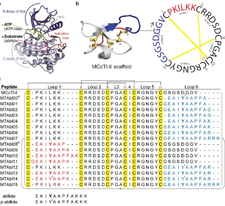
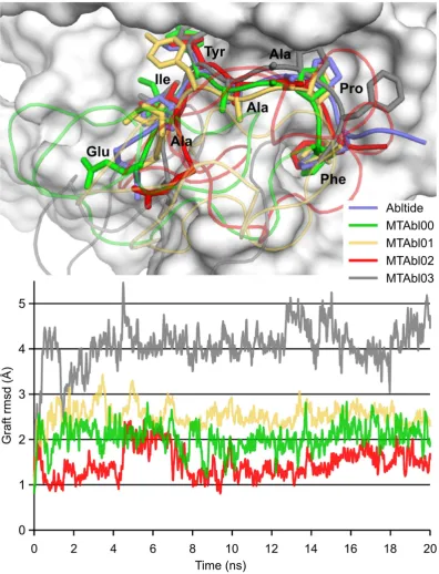
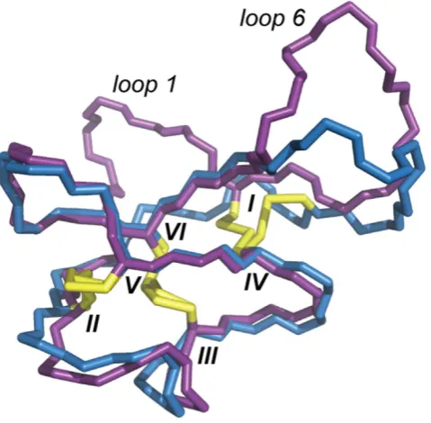
abltide grafted in MTAbl02 can adopt a conformation that is nearly identical to that displayed by linear abltide when in complex with Abl. In contrast, the simulations of three other MTAbl peptides showed that the conformation of their grafted abltide diverged from that of linear abltide, with graft rmsd values of ~2.0 Å, ~2.5 Å and ~3.0–4.0 Å for MTAbl00, MTAbl01 and MTAbl03, respectively (Fig. 2). The mod-els suggest that the C-terminus of grafted abltide sequences is located in a region of negatively charged Figure 1. Three-dimensional structures of Abl kinase and MCoTI-II, and amino acid sequences of MCoTI-II variants considered in this study. (a) Abl kinase with substrate-ATP conjugate bound to the catalytic site (PDB ID: 2g2f). The substrate (abltide, in magenta) binds in the cleft between the N- and C-lobes; the phosphorylation site is oriented towards the ATP binding pocket in the N-lobe.
(b) Three-dimensional structure and amino acid sequence of native MCoTI-II (PDB ID: lib9). The
cysteine-rich peptide has a unique cyclic cystine knot (CCK) motif, comprising a cyclic backbone and three interlocking disulfides (shown in yellow). The starting point of the peptide sequence (G1) is connected to
potential generated by Abl kinase. Consequently, several grafted analogs in loop 6, MTAbl04, MTAbl05, MTAbl06 and MTAbl07, were designed to incorporate additional basic residues at the C-terminus of the abltide sequence.
The engineering of MCoTI-II grafted in loop 1 initially focused on optimizing the length of the graft because loop 1 of MCoTI-II has only six residues whereas linear abltide is 12 residues. The designed peptides incorporated the first seven (MTAbl08), eight (MTAbl09) or ten (MTAbl10) residues of abltide into the scaffold. A nine-residue graft comprising the first eight residues of abltide preceded by a glycine residue (MTAbl11) was also considered. The molecular modeling studies of grafts in loop 6 indicated that adding a glycine at the C-terminus was detrimental to binding, and the addition of a spacer at the N-terminus of the grafted peptide in MTAbl11 could allow the peptide to adopt a conformation compat-ible with binding at the interface. Results from the MD simulations suggested that the grafted segment of MTAbl09 adopts a conformation that is almost identical to that of linear abltide at the interface (graft rmsd ~1.5 Å). The conformation of grafted segments in the three other peptides, MTAbl08, MTAbl10 and MTAbl11, deviated from the optimal abltide conformation, as shown by their graft rmsds reaching 2.5 Å or above (Supplementary Fig. S4). The simulations of MTAbl08 were found to be particularly unstable, leading to very high graft rmsd values. The three simulations of MTAbl09 converged toward a similar binding mode, but those of MTAbl10 and MTAbl11 did not, suggesting that the scaffold does not form strong or specific interactions with the kinase.
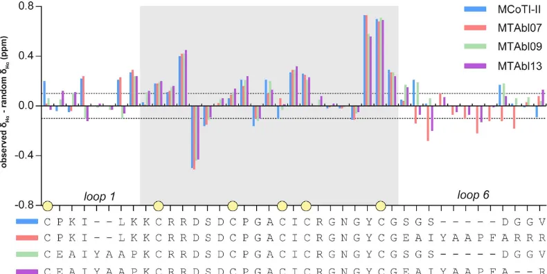
Synthesis and structure of grafted peptides. Fourteen mutant peptides (MTAbl01-07, 09-15) with abltide analogs grafted in loop 1, loop 6 or both loops 1 and 6 were synthesized (Fig. 1c). The one-dimensional 1H NMR spectra of all MTAbl peptides showed well dispersed peaks in the amide region and aligned well with the spectrum of MCoTI-II, suggesting that these peptides adopt native-like folds (data not shown). Two-dimensional homonuclear NMR was used to determine the Hα chemical shifts of three representative MTAbl peptides, namely MTAbl07, MTAbl09 and MTAbl13. A comparison of secondary Hα chemical shifts of these grafted peptides to MCoTI-II is shown in Fig. 3. The secondary Hα chemical shifts of the scaffold positions (gray-shaded region in Fig. 3) were comparable to the equiv-alent positions of MCoTI-II, suggesting that the three-dimensional structures of MTAbl07, MTAbl09 and MTAbl13 are similar to the parent peptide MCoTI-II. As expected, the main differences in Hα chemical shifts between MCoTI-II and MTAbl mutants were observed in the mutated loops (i.e. loop 1 and/or loop 6) or the regions near these two loops.
[image:5.595.156.551.46.244.2]assay was developed for the evaluation of inhibitory activities of kinase substrates, which cannot be fairly evaluated using ADP-Glo
™
.Abltide has inhibitory activity at low micromolar concentrations (IC50: 5.7 μ M) against Abl kinase, as measured using the BacKin assay, and, interestingly, phosphorylated abltide (p-abltide) has a similar activity. This similarity suggests that abltide, either non-phosphorylated or phosphorylated, can bind to Abl kinase and inhibit its activity. The importance of the phosphorylatable tyrosine was further inves-tigated by synthesizing a suite of 17 abltide analogs in which the tyrosine was replaced by an alanine, a phenylalanine or one of 15 unnatural phenylalanine residues (see Supplementary Fig. S5). None of these peptides demonstrated significant inhibition of Abl kinase at concentrations up to 64 μ M; therefore, a tyrosine residue at position 4 of abltide seemed to be optimal for binding and for inhibition of Abl kinase. Moreover, the results indicated that the phosphorylated substrate can also act as inhibitor, suggesting that abltide could serve as an inhibitor of Abl kinase without substituting its phosphorylatable tyrosine.
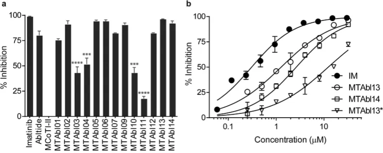
The Abl kinase inhibitory activity of MCoTI-II loop 6 variants MTAbl01–03 (Fig. 1c) was evaluated using the BacKin assay at 32 μ M peptide concentration (Fig. 4a). MTAbl01, MTAbl02 and MTAbl03 inhibited 75%, 90% and 50% of kinase activity, respectively. These values correlated with the similar conformations of the grafted segments to that of the linear abltide bound to Abl kinase, as shown by molecular modeling studies (Fig. 2); therefore, the conformation of the grafted segments at the interface seems to affect activity.
Based on our initial screening and molecular modeling, three analogs, MTAbl04, MTAbl05 and MTAbl06, designed to have better charge complementarity with Abl kinase, were synthesized and tested for inhibitory activity. MTAbl04 was derived from MTAbl03 by substitution of the C-terminal glycine residue in loop 6 with a lysine residue, whereas MTAbl05 and MTAbl06 were derived from MTAbl02 by substitution of the C-terminal glycine residue in loop 6 with a lysine or arginine residue, respectively. MTAbl04 showed similar activity to MTAbl03, inducing ~50% reduction of kinase activity, whereas MTAbl05 and MTAbl06 had similar activity to MTAbl02, inhibiting ~90% of kinase activity. Therefore, the addition of one basic residue at the C-terminus did not cause a dramatic difference in activity at 32 μ M peptide concentration. To appreciate subtle differences between the variants, inhibition dose response curves were evaluated and the IC50 values were calculated (Supplementary Table S1). MTAbl05 and MTAbl06 differed by only the C-terminal residue of loop 6, i.e. lysine and arginine residues, respec-tively, but had a 3-fold difference in activity (IC50: 15.2 ± 7.0 μ M vs 5.5 ± 1.6 μ M). To evaluate the con-tribution of additional positive charges in loop 6 to kinase inhibition, the dose-dependent activity of MTAbl07, which has three arginine residues at the C-terminus of loop 6, was also evaluated. MTAbl07 had comparable activity (IC50: 4.1 μ M) to MTAbl06, indicating that extra arginine residues in loop 6 do not significantly improve binding to Abl kinase. Overall, additional basic residues at the C-terminus of loop 6 only led to small improvements in activity.
[image:6.595.156.551.47.205.2]from linear abltide at the interface with Abl kinase. The incorporation of recognition motif in both loops 1 and 6 (MTAbl13) improved the inhibitory activity, with the IC50 decreasing to 1.3 μ M compared to the equivalent peptides modified only in loop 1 (MTAbl09, IC50: 18.3 μ M) or loop 6 (MTAbl06, IC50: 5.5 μ M). Although MTAbl13 was found to be less potent than IM (IC50: 0.3 μ M), its potency was com-parable to other substrate-based kinase inhibitors from previous studies34,35. Finally, activity of MTAbl12 and MTAbl14 – the phosphorylated versions of MTAbl06 and MTAbl13, respectively – were evaluated and were found to have comparable IC50 values to the non-phosphorylated peptides. This result was in agreement with the measurement of inhibitory activity of phosphorylated abltide, suggesting that cyclic peptides incorporating the non-phosphorylated abltide can function as kinase inhibitors.
Ph+ CML patients harboring the T315I mutation in BCR-ABL do not respond to most drugs currently
available. Therefore, the inhibitory activity of MTAbl peptides against [T315I]Abl kinase is of the utmost relevance and was investigated in vitro. The catalytic activity of [T315I]Abl kinase was first examined by comparing the phosphorylation of 30 μ M abltide catalyzed by 0.2 U/mL wild-type or [T315I]Abl kinase using LC-MS. The time required to phosphorylate half of the abltide molecules by wild-type and [T315I] Abl kinase was 24.2 ± 0.7 and 50.0 ± 1.6 min, respectively (Supplementary Fig. S6). The dose-response curves of IM, MTAbl13 and its phosphorylated version MTAbl14 against wild-type Abl kinase are shown in Fig. 4b. The IC50 values of IM, MTAbl13 and MTAbl14 were 0.3, 1.3 and 2.6 μ M, respectively. The inhibition activity of [T315I]Abl kinase by MTAbl13 was 12.2 μ M, whereas IM showed no inhibitory activity up to 256 μ M (Fig. 4b and Supplementary Table S1).
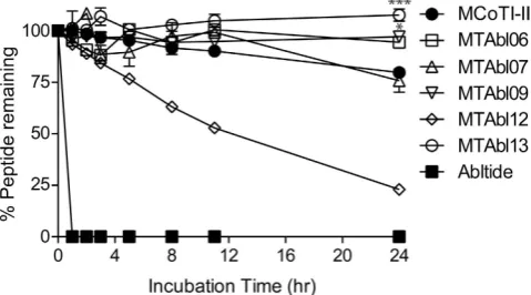
MTAbl peptides are remarkably stable in human serum. The proteolytic stability of MTAbl06, MTAbl07, MTAbl09, MTAbl12 and MTAbl13 in 100% human serum was evaluated, as well as native MCoTI-II and abltide for up to 24 h (Fig. 5). All of the tested MTAbl peptides, with the exception of MTAbl12, were stable, with > 75% of peptide remaining after 24 h of incubation. The lower stability observed for MTAbl12 was due to dephosphorylation of the p-Tyr residue, as suggested by RP-HPLC profile and mass spectrometry analysis in which the retention time and mass of the degradation product was the same as for MTAbl06 (i.e. dephosphorylated MTAbl12). MTAbl09 and MTAbl13 are statistically more stable than MCoTI-II according to a one-way ANOVA test (p< 0.05 and p< 0.001, respectively), whereas MTAbl07 was as stable as MCoTI-II. In contrast to the cyclic peptides, linear abltide was com-pletely degraded in human serum within 1 h. Comparison with control samples in PBS solution showed that the binding of tested compounds to serum proteins or Eppendorf Tubes
®
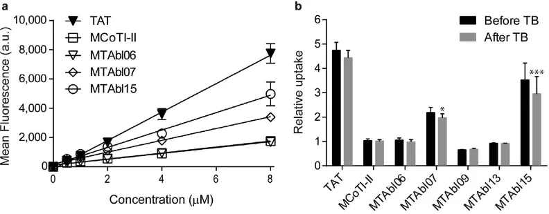
was negligible (< 5%). [image:7.595.156.396.48.181.2]showed statistically higher cellular uptake than A488-MCoTI-II (p< 0.05 and p< 0.001, respectively). Conversely, A488-MTAbl09 and A488-MTAbl13 showed a 25% and 10% decrease in cellular uptake, respectively, compared to MCoTI-II at the same concentration. No significant decrease in fluorescence intensity was observed after addition of trypan blue, a non-permeable quencher able to quench the fluorescence of non-internalized/membrane-bound peptides (Fig. 6b), indicating that the monitored flu-orescence was not from membrane-bound but from internalized peptides.
Solution structure of the double-grafted analog, MTAbl13. MTAbl13 contained a graft in two of its loops and had the most potent inhibitory activity of all MTAbl peptides; therefore, its tertiary structure in solution was elucidated using NMR spectroscopy. Distance and dihedral angle restraints were derived from one- and two-dimensional homonuclear 1H NMR experiments and used in structure calculations (Supplementary Table S2 and Supplementary Fig. S7). The refinement statistics are shown in Supplementary Table S2 and the structure coordinates have been deposited in the Protein Data Bank (PDB ID: 2mt8). As illustrated in Fig. 7, loops 2, 3, 4 and 5 of MTAbl13 are similar in conformation to the corresponding loops of MCoTI-II, in agreement with the secondary Hα chemical shift analysis presented in Fig. 3. The disulfide bond that connects loops 1 and 6 was found to adopt a different con-formation to that of MCoTI-II. This change might result in a small change of concon-formation of the grafted abltide when at the interface with Abl kinase, and we hypothesize that it might be responsible for the observed improved activity compared to singly-grafted peptides, MTAbl06 and MTAbl09.
The secondary Hα chemical shift analysis suggested that both loops 1 and 6 of MTAbl13 are generally disordered. A superimposition of the 20 lowest energy conformations resulting from the structure calculations further suggested that loops 1 and 6 are flexible compared to the rest of the mol-ecule (Supplementary Fig. S8). Loop 1 adopts a more constrained conformation compared to loop 6 (Supplementary Fig. S8), but both loops protrude away from the cystine knot core, and could presumably insert into the substrate-binding pocket of Abl kinase. None of the NMR models of the more constrained conformation of loop 1 corresponded to the kinase-bound conformation. By contrast, loop 6 was more flexible and therefore might be able to adopt the bound conformation.
[image:8.595.156.552.46.201.2]Cell viability assay against K562 cells. The effect of MTAbl peptides on the growth and apoptosis of K562 cells was evaluated using a resazurin-based cell viability assay. K562 is an immortalized cell line derived from a 53-year-old female CML patient in blast crisis38 and has been used extensively as a model for CML studies. Although MTAbl peptides showed potent activity against Abl kinase in vitro, MTAbl06, MTAbl07, MTAbl13 and MTAbl15 did not inhibit the growth of K562 up to 64 μ M (Supplementary Table S1).
Discussion
Although current frontline therapies are initially effective at inhibiting cancer progression, resistance can develop after a few years of continuous treatment because of mutations in the ATP binding site that ren-der these drugs ineffective. All human kinases share similar ATP binding pockets, and designing drugs targeting these sites with high specificity is difficult. The ligand-binding sites of kinases, on the other hand, are more distinct and less prone to mutations than the ATP binding cleft because of the functional requirement to target specific substrates39,40, suggesting that substrate-based inhibitors have the potential to be developed as TKIs with high specificity.
We attempted to turn abltide from being a substrate into an inhibitor by replacing the phosphoryl-atable tyrosine with natural or non-natural amino acids, but none of the resulting peptides were active. Phosphorylated abltide (p-abltide) was then shown to be as active as abltide using the BacKin inhibition assay, suggesting that the substrates of Abl kinase can serve as inhibitors after they have been phosphoryl-ated. Similarly, the phosphorylated MTAbl peptides showed similar activity as their non-phosphorylated form, i.e. MTAbl12 vs MTAbl06 and MTAbl14 vs MTAbl13. Some aspects of the inhibition of Abl kinase by phosphorylated substrates remain unclear; for example, the kinase state to which the phosphorylated peptides bind is unknown. Indeed, phosphorylated peptides cannot possibly bind to the inactive confor-mational state of Abl kinase because the activation loop adopts a conformation incompatible with sub-strate binding. As the charged phosphate group of the phosphorylated tyrosine would create both steric and charge repulsion with the ATP molecule, an alternative state of the kinase in which the activation loop is in an active form but the ATP cleft is empty or occupied by an ADP molecule is possibly the state targeted by the phosphorylated peptides.
In total, 14 synthetic cyclic peptides incorporating one or two grafted abltides were designed and generally showed medium to high inhibitory activity against Abl kinase. The most potent peptides, i.e.
[image:9.595.157.397.47.282.2]MTAbl06, MTAbl07 and MTAbl13, exhibited low micromolar IC50 values, as tested using the BacKin assay. The IC50 values determined using this assay potentially underestimate the ability of the MTAbl peptides to inhibit Abl kinase, as these values are measured based on a competition with abltide, which is the optimal substrate for Abl kinase and has higher affinity for Abl than its physiological substrates. Indeed, the Michaelis-Menten constant Km for phosphorylation of abltide by Abl is between 4 μ M20 and 21 μ M41, but the phosphorylation of the peptide with flanking sequences of the phosphorylatable tyrosine (Tyr-207) derived from the canonical substrate CRKL has a Km of 134 μ M41. The Km of abltide is of the same order of magnitude as the best IC50 values of MTAbl peptides, indicating that the corresponding designed peptides have probably reached the maximal potential of abltide. Molecular modeling studies suggested that the relative potencies of the MTAbl peptides were mainly dependent on the ability of the grafted abltides to adopt an optimal conformation despite the scaffold displaying a different orientation relative to the kinase. This result indicates that the native MCoTI-II scaffold does not contribute signif-icantly to the affinity with the kinase.
on the [T315I] mutant up to 256 μ M, which is > 800-fold when compared to its low IC50 value towards wild-type kinase. The order of magnitude difference in inhibition activity of MTAbl13 against wild-type and mutant kinase might be due to the reported decreased kinase activity of [T315I]Abl towards classical BCR-ABL substrates43.
The stability of MTAbl peptides in human serum was evaluated to provide a guide to their potential stability in vivo. The time course studies indicated that the stability of MTAbl peptides is comparable to or higher than MCoTI-II. Interestingly, MTAbl07 is less stable than MTAbl06 despite high sequence sim-ilarity, and the slightly decreased stability of MTAbl07 might originate from the two additional basic resi-dues (Arg) included in loop 6. A significant increase in serum stability of MTAbl13 (p < 0.001) compared to MCoTI-II after 24 h incubation was observed, and could be explained by fewer positively charged res-idues in MTAbl13 compared to MCoTI-II (Supplementary Table S1). Overall, the results clearly indicate that the cyclic cystine knot motif endow MTAbl peptides with exceptional enzymatic stability (half-life > 24 h) despite the sequence variation.
The cellular uptake efficiency of substrate-based inhibitors show that A488-MTAbl07 internalizes into HeLa cells with a 2.2-fold increase in efficiency compared to A488-MCoTI-II. This finding is in accordance with previous studies demonstrating that the cellular uptake of MCoTI-II can be enhanced by increasing the overall positive charge of the peptide44,45. A488-MTAbl15, an analog of A488-MTAbl07 with the first and second Lys residues in loop 1 replaced with Arg, has a 3.5-fold higher cellular uptake than A488-MCoTI-II, which demonstrates that the cell internalization of MCoTI-II scaffold can be fur-ther improved by Lys to Arg substitution in loop 1 of the molecule.
Despite having high inhibitory activity against Abl kinase, the grafted peptides MTAbl06, MTAbl07, MTAbl13, and MTAbl15 showed no significant effect on the growth of K562 cells when administered at concentrations up to 64 μ M. A possible explanation for their unexpectedly low potency against K562 cells is that only a limited amount is able to reach BCR-ABL within the cytoplasm. Although we showed that the grafted peptides can penetrate cells and, in fact, MTAbl07 and MTAbl15 have improved inter-nalization efficiencies compared with MCoTI-II, a certain amount of the grafted peptides might have been trapped in cell compartments (e.g. endosomes). Our future work will focus on improving the amount of peptide reaching the cytoplasm by optimizing the cell internalization of the MTAbl pep-tides and thereby increase their effective concentration at the interface of protein-protein interactions involved in BCR-ABL signaling network. The molecular dynamics simulation studies suggest potential strategies to increase inhibitory activity by creating specific interactions between the MCoTI-II scaffold and Abl kinase and by trapping the grafted abltide into a conformation compatible with binding to Abl kinase. Whether the substrate-based Abl kinase inhibitors designed in this study can actively inhibit the BCR-ABL kinase, wild-type or with T315I mutation in cytoplasm remains to be determined; however, the kinase inhibitory activity combined with serum stability and improved cell uptake efficiency make these peptides promising candidates as the next-generation TKIs.
Methods
Isolation of MCoTI-II from M. cochinchinensis. Native MCoTI-II was isolated from M. cochinchin-ensis seed extract prepared as described previously26.
Abl kinase bound to MTAbl peptides (MTAbl00, MTAbl01, MTAbl02, MTAbl03, MTAbl08, MTAbl09, MTAbl10, MTAbl11) using the bound peptide as an anchor. The eight complexes were then prepared and studied by 20 ns molecular dynamics simulations using the same molecular modeling protocol described for the linear abltide/Abl system. The molecular dynamics simulations were analyzed using VMD58 and the gromacs tools. The electrostatic potential generated by Abl kinase was computed using the APBS software59.
Peptide synthesis. Linear precursors of 14 MTAbl peptides containing an N-terminal Cys residue were synthesized manually using solid-phase peptide synthesis with Boc chemistry, as described previously28. In brief, precursor peptides were constructed on PAM-Gly-Boc resin via S-tritylmercaptopropionic acid as a thioester linker and cleaved from the resin with hydrogen fluoride. Through a thia zip reaction, the linear precursor peptides with multiple Cys and a C-terminal thioester form an α -amino thiolactone after a series of intramolecular acyl rearrangements and eventually undergo an irreversible S,N acyl migration to form an amide bond between the two ends60,61. Cyclization and oxidation of the MCoTI-II mutants was carried out in 0.1 M ammonium bicarbonate (pH 8.2) at 0.1 mg/mL peptide concentration. The pep-tide mixture was stirred at room temperature for 24 h and purified by reversed-phase high-performance liquid chromatography (RP-HPLC) on a preparative Phenomenex C18 column. A 1%/min gradient of solvent B (90% v/v acetonitrile/10% v/v H2O/0.045% v/v TFA (trifluoroacetic acid)) against solvent A (100% v/v H2O/0.05% v/v TFA) on RP-HPLC was used to separate peptides.
Abltide, [Y4A]abltide, [Y4F]abltide, and p-abltide containing a phosphorylated tyrosine at the fourth position were synthesized along with 15 other abltide analogs (abltide1–15), with the same position substi-tuted with phenylalanine derivatives, as illustrated in Supplementary Fig. S5. The Phe derivatives used in this study were: 1. 4-fluoro-L-phenylalanine, 2. 4-chloro-L-phenylalanine, 3. 4-methyl-L-phenylalanine, 4. 4-nitro-L-phenylalanine, 5. 4-amino-L-phenylalanine, 6. L-homophenylalanine, 7. 3,4-difluoro-L-phenyl alanine, 8. 3,4-dimethoxy-L-phenylalanine, 9. pentafluoro-L-phenylalanine, 10. p-trifluoromethyl-L- phenylalanine, 11. 4-benzoyl-L-phenylalanine, 12. p-phenyl-L-phenylalanine, 13. 4-iodo-L-phenylalanine,
14. 4-cyano-L-phenylalanine, and 15. 4-tert-butyl-L-phenylalanine. Abltide and its 18 analogs were syn-thesized using solid-phase peptide synthesis with Fmoc chemistry on an automatic peptide synthesizer (Symphony
®
, Protein Technologies). Briefly, peptide chains were assembled on 2-chlorotrityl chloride (2-CTC) resin by consecutive addition of Fmoc-protected amino acids. Peptides were then removed from the resin by the treatment with TFA/triisopropylsilane/H2O (95:2.5:2.5, v/v) for 2 h. TFA was removed using rotary evaporation from the mixture and the residual solution was mixed with cold diethyl ether and solvent A/B (1:1, v/v). The aqueous layer containing crude linear peptides was collected and purified using RP-HPLC on a preparative C18 column to 95% purity. The mass of the peptides was confirmed with ESI-MS and the purity was examined by analytical HPLC.Detection of abltide phosphorylation by LC/MS. The time-course of phosphorylation of 30 μ M abltide by 0.2 U/mL human active Abl kinase (1067 U/mg, Merck Millipore) and [T315I]Abl (Abl(T315I) protein, Merck Millipore) was analyzed using Liquid Chromatography-Mass Spectrometry (LC/MS, Shimadzu LCMS-2020). Abltide and Abl kinase were prepared and mixed in kinase buffer (50 mM Tris-HCl, 10 mM MgCl2, 0.1 mM EDTA, 2 mM dithiothreitol and 0.01% Brij 35 (v/v), pH 7.5; NEBuffer for protein kinases, New England Biolabs) at a desired concentration and the reaction was initiated by the addition of 500 μ M ATP (adenosine 5′ -triphosphate disodium salt hydrate, Sigma-Aldrich). The mixture was incubated at 37 °C with gentle shaking and the reaction was terminated with 10% 20 mg/mL dihy-droxybenzoic acid at nine time points (0, 5, 10, 20, 30, 45, 60, 90 and 120 min). Samples were injected onto a C18 analytical column (Jupiter 300 5 μ m 300 Å, 150 × 2.0 mm, Phenomex) and the mass of each component was evaluated using MS. The percentage of phosphorylation of abltide was determined by comparing the peak area of phosphorylated abltide (1417 Da) with the peak area of 30 μ M abltide from LC (1336.6 Da). Triplicate experiments were conducted on three independent days.
One- and two-dimensional 1H NMR. The correct folding of MTAbl peptides was confirmed using 1H NMR. Peptides were dissolved in H
2O/D2O (9:1, v/v) to a concentration of 1–2 mg/mL. NMR spectra were recorded on a Bruker Avance-600 MHz NMR spectrometer at 298 K. The mixing time was 80 ms and 100–300 ms for TOCSY and NOESY experiments, respectively. Spectra were analyzed using Sparky. Spectra were internally referenced to 2,2-dimethyl-2-silapentane-5-sulfonic acid (DSS) at 0.00 ppm. Structure determination of MTAbl13. Structure calculations were carried out for MTAbl13 as described previously for other cyclic disulfide-rich peptides42. Briefly, samples of MTAbl13 were prepared in either 90% H2O/10% D2O (9:1, v/v) or 99.96% D2O (Cambridge Isotope Laboratories, Inc.) at ~1 mM and pH ~3. One- and two-dimensional NMR spectra (1H, 1H TOCSY, NOESY, DQF-COSY and ECOSY, and 1H, 13C HSQC) were acquired using Avance-500 or Avance-600 MHz spectrometers (Bruker). Spectra were processed with TopSpin (Bruker) and analyzed using CCPNMR62. Inter-proton distance constraints were calculated from the relative intensities of NOE cross-peaks. 3J
HN-Hα coupling constants
were measured from one-dimensional spectra or from anti-phase cross-peak splitting in the DQF-COSY spectrum, and 3J
Hα-Hβ coupling constants were measured from the ECOSY spectrum. Backbone amide
protons involved in intramolecular hydrogen bonds were identified by their temperature sensitivities and deuterium exchange rates (Supplementary Fig. 7). Preliminary structure calculations were performed using CYANA 3.063, followed by further structure calculations and refinement in a water shell using CNS64. An ensemble of 20 structures with the lowest energy was selected and the refinement statistics are presented in Supplementary Table S2.
Serum stability assay. The stability of abltide-grafted analogs MTAbl06, MTAbl07, MTAbl12, MTAbl13 and native MCoTI-II was assessed in 100% human male AB serum (Sigma) using a method modified from a previous study27. All peptides were tested at a final concentration of 30 μ M in 100% human serum. Peptides were incubated in human serum at 37 °C for 0, 1, 2, 3, 5, 8, 11, and 24 h. Reactions were stopped at the stipulated times and the serum proteins in each sample were denatured and pre-cipitated, followed by centrifugation at 17,000 g for 10 min to separate the serum proteins from peptide samples. One hundred μ L of supernatant was loaded on an analytical column (150 × 2.0 mm) and run on RP-HPLC using a linear 1%/min gradient of 0–40% solvent B at a 0.3 mL/min flow rate. Equivalent amount of peptides were incubated with PBS and processed in parallel with serum-treated samples at 0 and 24 h time points as negative controls. The elution time for each peptide was determined by the PBS control from 0 time point. The percentage of peptide remaining at each time point was calculated using the height of the serum-treated peptide peak from time 0 as 100% recovery.
Labeling of MCoTI-II and analogs with fluorophore. Native MCoTI-II, MTAbl06, MTAbl07, MTAbl09, MTAbl13 and abltide were labeled with Alexa Fluor
®
488 (abbreviated as A488) using a method described by Cascales et al.26. In brief, peptide solutions prepared in 0.1 M sodium bicarbonate (pH 8.3) were incubated with 2-fold excess (molar ratio) of Alexa Fluor®
488 5-SDP ester (Molecular Probes, Life technologies) for 2 h at room temperature on a roller mixer. The conjugated peptides were purified using RP-HPLC and the mass was confirmed by ESI-MS. Peptides labeled with one A488 mole-cule were collected and used in cellular uptake studies. TAT (NH2-YGRKKRRQRRRPPQG-COOH) was labeled with the same fluorophore and used as the positive control in internalization assays.Cell culture. Human cervical cancer cells (HeLa) were seeded in 175-cm2 tissue culture flasks (Falcon), in Dulbecco’s Modified Eagle’s Medium (DMEM) supplemented with 10% fetal bovine serum, 100 U/mL penicillin and 100 mg/mL streptomycin, until 80% confluent. K562 cells were cultured in Roswell Park Memorial Institute (RPMI) 1640 medium supplemented with 2 mM l-glutamine, 10% fetal bovine serum, 100 U/mL penicillin, and 100 mg/mL streptomycin and grown to 80% confluence.
In brief, 105 cells/well were seeded in a 24-well plate and incubated at 37 °C in a humidified atmosphere (5% CO2) overnight. Peptides labeled with one Alexa Fluor
®
488 were dissolved in sterile water by 2-fold serial dilutions starting at 80 μ M. 100 μ L of serum-free medium in the absence or presence of labeled peptides with final concentrations at 0.5, 1, 2, 4, or 8 μ M was added in each well and incubated with cells for 1 h at 37 °C. After 1 h incubation, peptides were removed and cells were rinsed with Dulbecco’s Phosphate-Buffered Saline (DPBS, Life Technologies). Following that, cells in 24-well plate were treated with 0.25% Trypsin-EDTA (Gibco®
, Life technologies) for 3 min, washed off with DPBS and individually transferred to Eppendorf Tubes®
. Cells were spun down at 500 g for 3 min to remove the supernatant and re-suspended with 1 mL of DPBS. The fluorescence intensity of cells excited at 488 nm was measured using FACS flow cytometry (BD Canto II) using a 530/30nm bandpass filter.Statistical analysis. Results are shown as the mean ± SEM from two or three replicates as indicated. Statistical significance was determined using one-way ANOVA with Bonferroni’s multiple-comparison test within groups.
References
1. An, X. et al. BCR-ABL tyrosine kinase inhibitors in the treatment of Philadelphia chromosome positive chronic myeloid leukemia: a review. Leuk Res.34, 1255–1268 (2010).
2. Lugo, T. G., Pendergast, A. M., Muller, A. J. & Witte, O. N. Tyrosine kinase activity and transformation potency of bcr-abl oncogene products. Science.247, 1079–1082 (1990).
3. Quintas-Cardama, A., Kantarjian, H. & Cortes, J. Flying under the radar: the new wave of BCR-ABL inhibitors. Nat Rev Drug Discov.6, 834–848 (2007).
4. Daley, G. Q., Van Etten, R. A. & Baltimore, D. Induction of chronic myelogenous leukemia in mice by the P210bcr/abl gene of the Philadelphia chromosome. Science.247, 824–830 (1990).
5. Levinson, N. M. et al. A Src-like inactive conformation in the abl tyrosine kinase domain. PLoS Biol.4, e144 (2006).
6. Gorre, M. E. et al. Clinical resistance to STI-571 cancer therapy caused by BCR-ABL gene mutation or amplification. Science. 293, 876–880 (2001).
7. Soverini, S. et al. ABL mutations in late chronic phase chronic myeloid leukemia patients with up-front cytogenetic resistance to imatinib are associated with a greater likelihood of progression to blast crisis and shorter survival: a study by the GIMEMA Working Party on Chronic Myeloid Leukemia. J Clin Oncol.23, 4100–4109 (2005).
8. Talpaz, M. et al. Dasatinib in imatinib-resistant Philadelphia chromosome-positive leukemias. N Engl J Med.354, 2531–2541 (2006).
9. Kantarjian, H. et al. Nilotinib in imatinib-resistant CML and Philadelphia chromosome-positive ALL. N Engl J Med.354, 2542–2551 (2006).
10. Weisberg, E. et al. Characterization of AMN107, a selective inhibitor of native and mutant Bcr-Abl. Cancer Cell.7, 129–141 (2005).
11. Shah, N. P. et al. Overriding imatinib resistance with a novel ABL kinase inhibitor. Science. 305, 399–401 (2004).
12. Golas, J. M. et al. SKI-606, a 4-anilino-3-quinolinecarbonitrile dual inhibitor of Src and Abl kinases, is a potent antiproliferative agent against chronic myelogenous leukemia cells in culture and causes regression of K562 xenografts in nude mice. Cancer Res.
63, 375–381 (2003).
13. O’Hare, T. et al. AP24534, a pan-BCR-ABL inhibitor for chronic myeloid leukemia, potently inhibits the T315I mutant and overcomes mutation-based resistance. Cancer Cell.16, 401–412 (2009).
14. O’Hare, T., Zabriskie, M. S., Eiring, A. M. & Deininger, M. W. Pushing the limits of targeted therapy in chronic myeloid leukaemia. Nat Rev Cancer.12, 513–526 (2012).
15. Zabriskie, M. S. et al. BCR-ABL1 compound mutations combining key kinase domain positions confer clinical resistance to ponatinib in Ph chromosome-positive leukemia. Cancer Cell.26, 428–442 (2014).
16. Levitzki, A. & Mishani, E. Tyrphostins and other tyrosine kinase inhibitors. Annu Rev Biochem.75, 93–109 (2006).
17. Chapelat, J. et al. The Substrate-Activity-Screening methodology applied to receptor tyrosine kinases: a proof-of-concept study.
Eur J Med Chem.57, 1–9 (2012).
18. Eldar-Finkelman, H. & Eisenstein, M. Peptide inhibitors targeting protein kinases. Curr Pharm Des. 15, 2463–2470 (2009). 19. Songyang, Z. et al. Use of an oriented peptide library to determine the optimal substrates of protein kinases. Curr Biol.4, 973–982
(1994).
20. Songyang, Z. et al. Catalytic specificity of protein-tyrosine kinases is critical for selective signalling. Nature.373, 536–539 (1995). 21. Sato, A. K., Viswanathan, M., Kent, R. B. & Wood, C. R. Therapeutic peptides: technological advances driving peptides into
development. Curr Opin Biotechnol.17, 638–642 (2006).
22. Colgrave, M. L. & Craik, D. J. Thermal, chemical, and enzymatic stability of the cyclotide kalata B1: the importance of the cyclic cystine knot. Biochemistry.43, 5965–5975 (2004).
33. Henriques, S. T. et al. A novel quantitative kinase assay using bacterial surface display and flow cytometry. PLoS One.8, e80474 (2013).
34. Parang, K. et al. Mechanism-based design of a protein kinase inhibitor. Nat Struct Biol. 8, 37–41 (2001).
35. Ward, B. C., Kavalukas, S., Brugnano, J., Barbul, A. & Panitch, A. Peptide inhibitors of MK2 show promise for inhibition of abdominal adhesions. J Surg Res.169, e27–36 (2011).
36. Torcato, I. M. et al. The antimicrobial activity of Sub3 is dependent on membrane binding and cell-penetrating ability.
Chembiochem. 14, 2013–2022 (2013).
37. Madani, F., Lindberg, S., Langel, U., Futaki, S. & Graslund, A. Mechanisms of cellular uptake of cell-penetrating peptides. J Biophys. 2011, 414729 (2011).
38. Lozzio, C. B. & Lozzio, B. B. Human chronic myelogenous leukemia cell-line with positive Philadelphia chromosome. Blood.45, 321–334 (1975).
39. Avrahami, L., Licht-Murava, A., Eisenstein, M. & Eldar-Finkelman, H. GSK-3 inhibition: achieving moderate efficacy with high selectivity. Biochimica Et Biophysica Acta. 1834, 1410–1414 (2013).
40. Licht-Murava, A. & Eldar-Finkelman, H. Exploiting substrate recognition for selective inhibition of protein kinases. Curr Pharm Des. 18, 2914–2920 (2012).
41. Hantschel, O. et al. BCR-ABL uncouples canonical JAK2-STAT5 signaling in chronic myeloid leukemia. Nat. Chem. Biol.8, 285–293 (2012).
42. Conibear, A. C. et al. The cyclic cystine ladder of theta-defensins as a stable, bifunctional scaffold: a proof-of-concept study using the integrin-binding RGD motif. Chembiochem. 15, 451–459 (2014).
43. Skaggs, B. J. et al. Phosphorylation of the ATP-binding loop directs oncogenicity of drug-resistant BCR-ABL mutants. Proc Natl Acad Sci USA.103, 19466–19471 (2006).
44. D’Souza, C., Henriques, S. T., Wang, C. K. & Craik, D. J. Structural parameters modulating the cellular uptake of disulfide-rich cyclic cell-penetrating peptides: MCoTI-II and SFTI-1. Eur J Med Chem. (2014).
45. Huang, Y. H., Chaousis, S., Cheneval, O., Craik, D. J. & Henriques, S. T. Optimization of the cyclotide framework to improve cell penetration properties. Front Pharmacol. 6, 17, doi: 10.3389/fphar.2015.00017 (2015).
46. Olsson, M. H. M., Søndergaard, C. R., Rostkowski, M. & Jensen, J. H. PROPKA3: Consistent treatment of internal and surface residues in empirical pKa predictions. J Chem Theory Comput.7, 525–537 (2011).
47. Beglov, D. & Roux, B. An integral equation to describe the solvation of polar molecules in liquid water. J Phys Chem. B.101, 7821–7826 (1997).
48. Kovalenko, A. & Hirata, F. Potential of mean force between two molecular ions in a polar molecular solvent: a study by the three-dimensional reference interaction site model. J Phys Chem. B.103, 7942–7957 (1999).
49. Case, D. A. et al.AMBER 13. (University of California, San Francisco, 2012).
50. Sindhikara, D. J., Yoshida, N. & Hirata, F. Placevent: an algorithm for prediction of explicit solvent atom distribution-application to HIV-1 protease and F-ATP synthase. J Comput Chem.33, 1536–1543 (2012).
51. Hubbard, S. J. & Thornton, J. M. NACCESS, Computer Program. (Department of Biochemistry and Molecular Biology, University College London, 1993).
52. Schmid, N. et al. Definition and testing of the GROMOS force-field versions 54A7 and 54B7. Eur Biophys J.40, 843–856 (2011). 53. Heinz, T. N., Gunsteren, W. F. V. & Hünenberger, P. H. Comparison of four methods to compute the dielectric permittivity of
liquids from molecular dynamics simulations. J Chem Phys.115, 1125–1136 (2001).
54. Bussi, G., Donadio, D. & Parrinello, M. Canonical sampling through velocity rescaling. J Chem Phys. 126, 014101, doi: 10.1063/1.2408420 (2007).
55. Berendsen, H. J. C., Postma, J. P. M., Van Gunsteren, W. F., DiNola, A. & Haak, J. R. Molecular dynamics with coupling to an external bath. J Chem Phys.81, 3684–3690 (1984).
56. Hess, B., Bekker, H., Berendsen, H. J. C. & Fraaije, J. G. E. M. LINCS: A linear constraint solver for molecular simulations.
J Comput Chem.18, 1463–1472 (1997).
57. Pronk, S. et al. GROMACS 4.5: a high-throughput and highly parallel open source molecular simulation toolkit. Bioinformatics (Oxford, England). 29, 845–854 (2013).
58. Humphrey, W., Dalke, A. & Schulten, K. VMD: visual molecular dynamics. J Mol Graph.14, 33–38 27-28 (1996).
59. Baker, N. A., Sept, D., Joseph, S., Holst, M. J. & McCammon, J. A. Electrostatics of nanosystems: application to microtubules and the ribosome. Proc Natl Acad Sci USA.98, 10037–10041 (2001).
60. Dawson, P. E., Muir, T. W., Clark-Lewis, I. & Kent, S. B. Synthesis of proteins by native chemical ligation. Science.266, 776–779 (1994).
61. Tam, J. P. & Lu, Y. A. A biomimetic strategy in the synthesis and fragmentation of cyclic protein. Protein Sci.7, 1583–1592 (1998). 62. Vranken, W. F. et al. The CCPN data model for NMR spectroscopy: development of a software pipeline. Proteins. 59, 687–696
(2005).
63. Guntert, P., Mumenthaler, C. & Wuthrich, K. Torsion angle dynamics for NMR structure calculation with the new program DYANA. J Mol Biol.273, 283–298 (1997).
64. Brünger, A. T. et al. Crystallography & NMR system: a new software suite for macromolecular structure determination. Acta Crystallogr D Biol Crystallogr. 54, 905–921 (1998).
Additional Information
Supplementary information accompanies this paper at http://www.nature.com/srep
Competing financial interests: The authors declare no competing financial interests.
How to cite this article: Huang, Y.H. et al. Design of substrate-based BCR-ABL kinase inhibitors using the cyclotide scaffold. Sci. Rep.5, 12974; doi: 10.1038/srep12974 (2015).