C A S E R E P O R T
Open Access
A case report of a cystic fibrosis patient
with repeated isolation of
Trichosporon
mycotoxinivorans
identified by a novel
short-extraction method
Daniel Goldenberger
1*†, Vladimira Hini
ć
1†, Spasenija Savic Prince
2, Michael Tamm
3, Anna-Maria Balestra
3,4,
Doris Hohler
1and Reno Frei
1Abstract
Background:Trichosporon mycotoxinivoransis a recently described yeast-like fungal organism and its association as a pathogen for patients with cystic fibrosis (CF) was reported previously. We show the clinical course of a CF patient over 9 years as well as the applications of modern molecular and proteomic identification techniques of this rare fungus.
Case presentation:We present the case of a 32-year-old male CF patient with sputum cultures continuously positive with the anamorphic yeastT. mycotoxinivoransduring 9 years. Furthermore, susceptibility testing ofT. mycotoxinivoransto different antifungals were performed. In addition, a rapid identification method of this novel fungal pathogen with matrix-assisted laser desorption ionization-time of flight mass spectrometry (MALDI-TOF MS) was applied using a simple extraction protocol.
Conclusions:Our case presentation confirmsT. mycotoxinivoransas a potential emerging pathogen in patients with CF. However, our CF patient showed mild symptoms over a very long time period of 9 years. A short MALDI-TOF MS procedure allows reliable and rapid identification ofT. mycotoxinivoransand therefore should facilitate further study on the clinical relevance and epidemiology of this unusual fungal organism.
Keywords:Trichosporon mycotoxinivorans, Cystic fibrosis (CF), Internal transcribed spacer (ITS), Emerging pathogen, MALDI-TOF MS
Background
The genus Trichosporon comprises 35 species of this basidiomycetous yeast from which 6 are known to be medically-relevant mainly in the immunocompromised patient [1, 2]. They are ubiquitous yeasts, found in soil and water and known as colonizers of skin and gastro-intestinal tract of humans [1]. Trichosporon mycotoxini-voranswas described for the first time in 2004, when it was isolated from the hindgut of the lower termite Mastotermes darwiniensis (Mastotermitidae) [2]. The name of this species refers to the ability of T.
mycotoxinivorans to degrade certain mycotoxins, an important characteristic which could be used for deacti-vation of the respective mycotoxins in animal feeds [2]. Its association with cystic fibrosis (CF) patients was reported for the first time in the USA in 2009 [3]. A 20-year-old male with CF died with histologically proven T. mycotoxinivorans pneumonia. Another case of a fatal disseminated coinfection with T. mycotoxinivorans, Aspergillus fumigatusandScedosporium apiospermumin a CF patient, recipient of lung and liver transplant, was published recently [4]. Kröner et al. demonstrated the potential association of Trichosporon spp. with severe exacerbations in CF patients [5]. On the other hand, the results of the most recent study by Shah et al. showed that the isolation of T. mycotoxinivorans from CF * Correspondence:daniel.goldenberger@usb.ch
†Equal contributors
1Division of Clinical Microbiology, University Hospital Basel, Petersgraben 4,
CH-4031 Basel, Switzerland
Full list of author information is available at the end of the article
patients is not necessarily associated with significant changes in clinical status [6]. Here we report on a CF patient with repeated isolation ofT. mycotoxinivoransin respiratory specimens during the past 9 years.
Case presentation
A 32-year-old male was regularly followed in our clinic because of CF. His lung function had been stable and is slowly decreasing since the last 7 years with a moderate irreversible airflow obstruction with an FEV1 of 60 % (predicted). He was known to be suffering from a chronic colonization with Staphylococcus aureus, Stenotrophomo-nas maltophilia, and Pseudomonas aeruginosa for which he has been treated repeatedly with oral antibiotics (trimethoprim/sulfamethoxazole, doxycycline and amoxi-cillin/clavulanic acid) as well as rarel intravenous regimens with piperacillin/tazobactam and tobramycin. He was put under permanent treatment with azithromy-cin and intermittent adjunct of amikaazithromy-cin for inhalation. Further he had a history of allergic bronchopulmonary aspergillosis leading to a subacute clinical deterioration. He was successfully treated with oral and inhaled steroids.
In 2007, a basidiomycetous yeast was isolated from sputum of the patient. The colonies grew after 2 days of incubation on Sabouraud agar (Becton Dickinson Diag-nostic Systems, Allschwil, Switzerland), showing typical
“furry” appearance (Fig.1) and histopathology of the bronchoalveolar lavage specimen showed not-specified fungal structures (Fig. 2). The organism was identified as Trichosporon species based on phenotypic features such as the typical morphology of colonies with aerial whitish mycelium (Fig. 1) and characteristic arthroconidia (Fig. 3) as well as the use of the API®ID32C identification system (bioMérieux, Geneva, Switzerland). Identification of the Trichosporonisolate to the species level was performed in 2011, based on PCR amplification and sequencing of the internal transcribed spacers (ITS), as well as the D1/D2 region of the 26S rRNA gene [7]. The investigated
sequence showed 100 % identity to reference sequences of T. mycotoxinivorans. Our 1167-bp-long sequence has been deposited in GenBank under accession no. JQ266092. In 2012, matrix-assisted laser desorption ionization-time of flight mass spectrometry (MALDI-TOF MS) was performed for the first time from colony material using the full ethanol-formic acid extraction protocol as recommended by the manufacturer. The analysis on the MALDI BioTyper system with the BioTyper 3.0 SR software (Bruker Daltonics GmbH, Bremen, Germany) resulted in highly accurate species identification with scores higher than 2.200. The manufacturer recommends ≥2 log score for species identification; 1.7 to 1.99 for genus identification; and <1.7 as unreliable [8]. Subsequent isolates were tested with a simple “short extraction” protocol, consisting of addition of 1μl of 70 % formic acid to the smears on the
Fig. 1Colonies ofT. mycotoxinivoranson sheep blood agar, 40-fold magnification
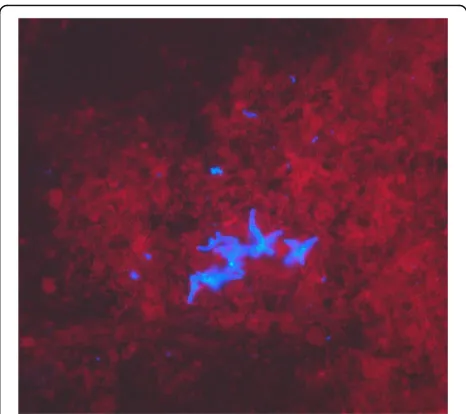
Fig. 2Fluorescence microscopy of bronchoalveolar lavage sample; Fungiqual A, 400-fold magnification
[image:2.595.305.538.86.293.2] [image:2.595.303.540.553.703.2] [image:2.595.57.288.556.703.2]plate before application of the matrix solution [9]. The
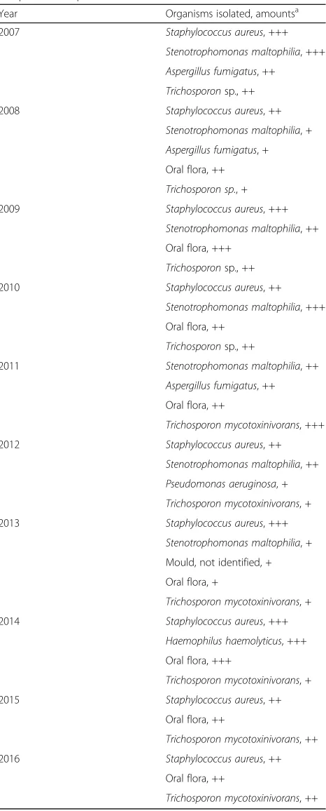
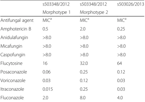
“short extraction” yielded scores ranging from 1.866 to 2.136 (average 1.989, median 1.984). The organism was isolated repeatedly from respiratory specimens during the period from 2007 to 2016. An overview of the microbio-logical results is shown in Table 1. The susceptibility test-ing of the strain was performed in 2012 and 2013 ustest-ing a commercial microtitre system (YeastOne, Trek Diagnostic Systems, Thermo Fisher Scientific Schweiz AG, Reinach, Switzerland). The results of the susceptibility testing are shown in Table 2. The strain exhibited high level resist-ance to the echinocandins (anidulafungin, micafungin, caspofungin) and variable susceptibility to amphotericin B and the triazoles (fluconazole, itraconazole, voriconazole and posaconazole). Due to further clinical deterioration, an antifungal therapy with voriconazole was initiated (200 mg twice a day). Clinical amelioration was observed within 6 weeks. However, a clinical relapse occurred after 9 months later andT. mycotoxinivorans was again identi-fied. After detection of a large quantity of two morpho-types of Trichosporon mycotoxinivorans the therapy was changed to itraconazole with a very good clinical response and lung functional response. Over the years the clinical course was characterized by recurrent worsening of the respiratory situation expressed by a mostly mild symptom-atology with a marked reduction of the detectable T. mycotoxinivoransquantity and satisfying stable clinical re-sults after the treatment with triazoles. It is possible that by using voriconazole, A. fumigatushas been suppressed together withT. mycotoxinivorans. There might have been also a reduction of the antigenic stimulus for bronchial in-flammation as it has been seen in therapies with itracona-zole. Because of the stability of the clinical course after the treatment with voriconazole, a long term suppressive ther-apy with antifungal agents was not applied.
The systematic search forTrichosporoncolonization in our microbiological data sheets of 64 CF patients, 10 patients after lung transplantation, and 32 patients with bronchiectasis did not reveal any other patients with Trichosporonspp.
[image:3.595.306.539.107.685.2]In addition to arthroconidia,T. mycotoxinivorans may sometimes microscopically display large fusiform cells with granular contents called fusiform giant cells [10]. This feature, however, should not be used for species identification, since fusiform giant cells may also appear in other Trichosporon spp. like T. loubieri and T. vanderwaltii[10, 11]. Furthermore, in someT. mycotoxi-nivorans isolates like the one described in this case report or communicated by Hickey et al. [3], the fusiform giant cells may not be present. This feature is probably culture medium or culture age dependent. To date, the only reliable identification of this yeast to spe-cies level could be achieved by sequencing, available only in specialized clinical laboratories. The MALDI-TOF MS
Table 1Chronology of microbiological findings in the sputum samples of the patient
Year Organisms isolated, amountsa
2007 Staphylococcus aureus, +++
Stenotrophomonas maltophilia, +++
Aspergillus fumigatus, ++
Trichosporonsp., ++
2008 Staphylococcus aureus, ++
Stenotrophomonas maltophilia, +
Aspergillus fumigatus, +
Oral flora, ++
Trichosporon sp., +
2009 Staphylococcus aureus, +++
Stenotrophomonas maltophilia, ++
Oral flora, +++
Trichosporonsp., ++
2010 Staphylococcus aureus, ++
Stenotrophomonas maltophilia, +++
Oral flora, ++
Trichosporonsp., ++
2011 Stenotrophomonas maltophilia, ++
Aspergillus fumigatus, ++
Oral flora, ++
Trichosporon mycotoxinivorans, +++
2012 Staphylococcus aureus, ++
Stenotrophomonas maltophilia, ++
Pseudomonas aeruginosa, +
Trichosporon mycotoxinivorans, +
2013 Staphylococcus aureus, +++
Stenotrophomonas maltophilia, +
Mould, not identified, +
Oral flora, +
Trichosporon mycotoxinivorans, +
2014 Staphylococcus aureus, +++
Haemophilus haemolyticus, +++
Oral flora, +++
Trichosporon mycotoxinivorans, +
2015 Staphylococcus aureus, ++
Oral flora, ++
Trichosporon mycotoxinivorans, ++
2016 Staphylococcus aureus, ++
Oral flora, ++
Trichosporon mycotoxinivorans, ++
a
identification is becoming the standard for isolate identi-fication in many laboratories. In contrast to Hirschi et al. [4], we were able to reliably identify theTrichosporon yeast to the species level using the MALDI-TOF MS identification. Our findings are in congruence with suc-cessful identification of this pathogen by Hickey and col-leagues [3]. However, in our study, we describe for the first time the identification of this yeast by using the
“short extraction”on-plate protocol for the routine diag-nostics. As the scores obtained were somewhat lower than those with the full extraction protocol, we propose using lower species cutoff values for identification ofT. mycotoxinivorans. Our proposal is consistent with studies of Vlek and Van Herendael [9, 12], which consider a spe-cies cutoff value of as low as 1.7 as appropriate for routine yeast identification in clinical laboratories. MALDI-TOF MS, as a rapid and reliable identification tool may facili-tate further study on the reservoir and the epidemiologic link ofT. mycotoxinivoransto CF patients. Concerning its antifungal susceptibility, the genusTrichosporonpresents increased in vitro resistance to common antifungal agents such as amphotericin B and echinocandins [1, 2], but also strains resistant to multiple triazoles have been identified [3]. Due to variable susceptibility patterns to triazoles, which are considered as therapy of choice [3], every strain isolated should be tested for antifungal susceptibility. At-tempts to treat our patient with triazoles did not lead to an eradication ofT. mycotoxinivorans, but to an improve-ment of the clinical symptoms.
Conclusions
In summary, our report on a CF patient colonized with T. mycotoxinivoransfor a long period of 9 years suggests that this yeastlike organism may be associated with clin-ical deterioration, and the patient’s condition improved under therapy with voriconazole and itraconazole. T.
mycotoxinivorans could be successfully identified with a simple on-plate MALDI-TOF MS short extraction protocol that may facilitate further study on the reser-voir and the epidemiologic link of T. mycotoxinivorans to CF patients and immunocompromised persons.
Abbreviations
CF:Cystic fibrosis; FEV1: Forced expiratory volume in 1 second; MALDI-TOF MS: Matrix-assisted laser desorption ionization-time of flight mass spectrometry
Acknowledgment
We thank Moritz Grubenmann, Laborgemeinschaft 1, Medizinische Diagnostik, Zürich for providing pictures of the cultured isolate.
Availability of data and materials
All data supporting the presented case report is contained within the manuscript.
Authors’contributions
DG, VH, and RF drafted the manuscript. DG, VH, and DH performed laboratory investigations including sequencing analysis. AMB and MT managed the patient and provided clinical specimens. SSP performed cytologic analysis. All authors read, revised and approved the final manuscript.
Competing interests
The authors declare that they have no competing interests.
Consent for publication
All participants gave written informed consent for publication of their details and images. Approval of an ethics committee was not required for this study.
Ethics approval and consent to participate
Not applicable.
Author details 1
Division of Clinical Microbiology, University Hospital Basel, Petersgraben 4, CH-4031 Basel, Switzerland.2Division of Pathology, University Hospital Basel,
Schönbeinstrasse 40, CH-4031 Basel, Switzerland.3Division of Pneumology, University Hospital Basel, Petersgraben 4, CH-4031 Basel, Switzerland.
4
Present address: Division of Pneumology, St. Claraspital Basel, Basel, Switzerland.
Received: 9 July 2016 Accepted: 11 October 2016
References
1. Chagas-Neto TC, Chaves GM, Colombo AL. Update on the genus
Trichosporon. Mycopathologia. 2008;166:121–32.
2. Molnar O, Schatzmayr G, Fuchs E, Prillinger H.Trichosporon mycotoxinivorans
sp. nov., a new yeast species useful in biological detoxification of various mycotoxins. Syst Appl Microbiol. 2004;27:661–71.
3. Hickey PW, Sutton DA, Fothergill AW, Rinaldi MG, Wickes BL, Schmidt HJ, et al.Trichosporon mycotoxinivorans, a novel respiratory pathogen in patients with cystic fibrosis. J Clin Microbiol. 2009;47:3091–7.
4. Hirschi S, Letscher-Bru V, Pottecher J, Lannes B, Jeung MY, Degot T, et al. DisseminatedTrichosporon mycotoxinivorans,Aspergillus fumigatus, and
Scedosporium apiospermumcoinfection after lung and liver transplantation in a cystic fibrosis patient. J Clini Microbiol. 2012;50:4168–70.
5. Kroner C, Kappler M, Grimmelt AC, Laniado G, Wurstl B, Griese M. The basidiomycetous yeast Trichosporon may cause severe lung exacerbation in cystic fibrosis patients-clinical analysis of Trichosporon positive patients in a Munich cohort. BMC Pulm Med. 2013;13:61.
6. Shah AV, McColley SA, Weil D, Zheng X.Trichosporon mycotoxinivorans
infection in patients with cystic fibrosis. J Clin Microbiol. 2014;52:2242–4. 7. White T, Bruns T, Lee S, Taylor J. Amplification and direct sequencing of fungal ribosomal RNA genes for phylogenetics. In: Innis MA, Gelfand DH, Sninsky JJ, White TJ, editors. PCR Protocols a Guide to Methods and Applications. San Diego: Academic; 1990. p. 315–22.
[image:4.595.56.292.110.276.2]8. Ghosh AK, Paul S, Sood P, Rudramurthy SM, Rajbanshi A, Jillwin TJ, et al. Matrix-assisted laser desorption ionization time-of-flight mass spectrometry
Table 2Antifungal susceptibility testing ofT. mycotoxinivorans
isolated in 2012 (Morphotype 1 and Morphotype 2) and 2013
s503348/2012 s503348/2012 s503026/2013
Morphotype 1 Morphotype 2
Antifungal agent MICa MICa MICa
Amphotericin B 0.5 2.0 0.25
Anidulafungin >8.0 >8.0 >8.0
Micafungin >8.0 >8.0 >8.0
Caspofungin >8.0 >8.0 >8.0
Flucytosine 16 32.0 64
Posaconazole 0.06 0.25 0.12
Voriconazole 0.03 0.12 0.03
Itraconazole 0.015 0.25 0.03
Fluconazole 2.0 8.0 4.0
a
for the rapid identification of yeasts causing bloodstream infections. Clin Microbiol Infect. 2015;21:372–8.
9. Vlek A, Kolecka A, Khayhan K, Theelen B, Groenewald M, Boel E, et al. Interlaboratory comparison of sample preparation methods, database expansions, and cutoff values for identification of yeasts by matrix-assisted laser desorption ionization-time of flight mass spectrometry using a yeast test panel. J Clin Microbiol. 2014;52:3023–9.
10. Padhye AA, Verghese S, Ravichandran P, Balamurugan G, Hall L, Padmaja P, et al.Trichosporon loubieriinfection in a patient with adult polycystic kidney disease. J Clin Microbiol. 2003;41:479–82.
11. Motaung TE, Albertyn J, Kock JL, Lee CF, Suh SO, Blackwell M, Pohl CH.
Trichosporon vanderwaltiisp. nov., an asexual basidiomycetous yeast isolated from soil and beetles. Antonie Van Leeuwenhoek. 2013;103:313–9. 12. Van Herendael BH, Bruynseels P, Bensaid M, Boekhout T, De Baere T, Surmont I,
et al. Validation of a modified algorithm for the identification of yeast isolates using matrix-assisted laser desorption/ionisation time-of-flight mass spectrometry (MALDI-TOF MS). Eur J Clin Microbiol Infect Dis. 2012;31:841–8.
• We accept pre-submission inquiries
• Our selector tool helps you to find the most relevant journal
• We provide round the clock customer support
• Convenient online submission
• Thorough peer review
• Inclusion in PubMed and all major indexing services
• Maximum visibility for your research
Submit your manuscript at www.biomedcentral.com/submit