White Rose Research Online
[email protected]
Universities of Leeds, Sheffield and York
http://eprints.whiterose.ac.uk/
This is a copy of the final published version of a paper published via gold open access
in
Journal of Physiology
.
This open access article is distributed under the terms of the Creative Commons
Attribution Licence (
http://creativecommons.org/licenses/by/4.0/
) which permits
unrestricted use, distribution, and reproduction in any medium, provided the
original work is properly cited.
White Rose Research Online URL for this paper:
http://eprints.whiterose.ac.uk/86845
Published paper
The
Jour
nal
of
P
hysiology
Age-related changes in afferent pathways and urothelial
function in the male mouse bladder
Donna M. Daly
1, Linda Nocchi
1, Marina Liaskos
2, Neil G. McKay
2, Christopher Chapple
3and David Grundy
11University of Sheffield, Western Bank, Sheffield S10 2TN, UK 2Sheffield Hallam University, Howard Street, Sheffield S1 1WB, UK 3Royal Hallamshire Hospital, Glossop Road, Sheffield S10 2JF, UK
Key points
r
The prevalence of bladder conditions such as overactive bladder syndrome and urinaryincontinence significantly increases with age, but how bladder function is altered by ageing is unclear.
r
Sensory nerves together with the epithelial lining of the bladder known as the urothelium playa key role in mediating bladder function.
r
In aged male mice we find a significant increase in natural bladder voiding, augmented afferentnerve firing during bladder filling and a significant increase in urothelial responses to purinergic receptor stimulation.
r
This suggests that with ageing there is increased purinergic transmission in the mouse bladderwhich may lead to increased sensation and result in bladder hypersensitivity.
r
These findings help us better understand how the function of the bladder may be affected by advancing age.Abstract The prevalence of lower urinary tract storage disorders such as overactive bladder syndrome and urinary incontinence significantly increase with age. Previous studies have demonstrated age-related changes in detrusor function and urothelial transmitter release but few studies have investigated how the urothelium and sensory pathways are affected. The aim of this study was to investigate the effect of ageing on urothelial-afferent signalling in the mouse bladder. Three-month-old control and 24-month-old aged male mice were used.In vivonatural voiding behaviour, sensory nerve activity, urothelial cell function, muscle contractility, transmitter release and gene and protein expression were measured to identify how all three components of the bladder (neural, contractile and urothelial) are affected by ageing. In aged mice, increased voiding frequency and enhanced low threshold afferent nerve activity was observed, suggesting that ageing induces overactivity and hypersensitivity of the bladder. These changes were concurrent with altered ATP and acetylcholine bioavailability, measured as transmitter overflow into the lumen, increased purinergic receptor sensitivity and raised P2X3receptor expression in the urothelium.
Taken together, these data suggest that ageing results in aberrant urothelial function, increased afferent mechanosensitivity, increased smooth muscle contractility, and changes in gene and protein expression (including of P2X3). These data are consistent with the hypothesis that ageing
evokes changes in purinergic signalling from the bladder, and further studies are now required to fully validate this idea.
(Received 25 July 2013; accepted after revision 25 November 2013; first published online 2 December 2013)
Corresponding authorProfessor D. Grundy: Department of Biomedical Science, University of Sheffield, Western Bank, Sheffield S10 2TN, UK. Email: [email protected]
Abbreviations ACh, acetylcholine; DRG, dorsal root ganglia; OAB, overactive bladder syndrome; UI, urinary incontinence
C
Introduction
Current demographic forecasts have predicted a worldwide increase in the proportion of people aged 65–80 (United Nations DoEaSA, Population Division, 2013). Understanding how physiological function alters as we age is now vital to ensure the progress of drug discovery and treatment of disease in an ageing society. Previous studies have shown that ageing dramatically affects the function of the urinary bladder. Overactive bladder syndrome (OAB) is a common and distressing disorder of the lower urinary tract which arises due to compromised storage ability of the bladder. Its cardinal symptoms are frequent micturition, urgency (the compelling and urgent sensation to empty the bladder) with or without urinary incontinence (UI, the involuntary leakage of urine from the bladder) and nocturia in the absence of other pathologies. The aetiology of OAB and UI is not fully understood, although studies suggest that the epithelial lining of the bladder, known as the urothelium and sub-urothelium, and the sensory innervation play a key role in normal function. Strikingly, the prevalence of bladder and continence conditions increases considerably with age (Dubeau, 2006).
Normal bladder function is dependent on the integration of autonomic and somatic mechanisms which coordinate a complex cycle of filling and emptying. This process is instigated and ultimately regulated by the sensory signalling pathways, which detect changes in bladder filling (volume and distension) and convey information to the CNS. The sensory innervation from the bladder is carried in the pelvic and hypogastric afferent nerves, the cell bodies of which, lie in the dorsal root ganglia (DRG). They consist of myelinated Aδfibres and unmyelinated C fibres which have polymodal sensitivity responding to a host of mechanical and chemical stimuli. To date, there are relatively few studies that have investigated how bladder function and neural activity are affected by ageing. Morphological studies suggest that there maybe an age-dependent reduction in the numbers of unmyelinated afferents innervating the bladder, although the general pattern of innervation appears to be preserved (Nakayamaet al.1998; Mohammed & Santer, 2002; Aizawa et al. 2010). At the level of the DRG, the sensory neuronal markers calcitonin gene related peptide and substance P are markedly reduced in aged rats (Mohammed & Santer, 2002) but how bladder projecting afferents are specifically affected is unknown. Functional and histological studies have produced contra-dictory reports, with some investigators finding an age-dependent fall in contractile ability, detrusor muscle thinning and collagen deposition and others seeing no functional change and/or increased muscle mass (Lluel
et al. 2000; Smith et al. 2012). This has made it difficult to fully understand how the function of the bladder changes with age and to specifically assess how
afferent signalling is affected. Clinical studies estimate that one-third of elderly patients with UI have overactivity of the detrusor muscle (spontaneous contractile activity) during bladder filling but diminished contractile activity during emptying (Resnick & Yalla, 1987). Potentially, this ‘overactivity’ could generate hypersensitivity of bladder afferents during the storage phase, driving the sensory symptoms associated with OAB (urgency and frequency). In one previous study, bladder afferent nerve responses to increasing volume (but not pressure) were shown to decline in the aged rat, but in another study C-fibre mediated afferent discharge was shown to increase in aged rats (Hottaet al.1995; Aizawaet al.2010).
It is now well established that the urothelium, traditionally believed to act as a barrier to prevent noxious agents in the urine from entering the circulation, can actively modulate sensory nerve firing by releasing a host of excitatory and inhibitory signalling molecules in response to mechanical and chemical stimulation. These mediators act on neighbouring urothelial cells via paracrine and autocrine signalling mechanisms and also act directly at the afferent terminal to excite or inhibit afferent activity. The most well-characterised example is the graded release of ATP from the urothelium, which occurs as the bladder fills. This ATP acts at purinergic receptors (P2X2and P2X3)
located on nerve terminals to initiate afferent nerve firing (Burnstock, 2009). One theory suggests that excitatory and inhibitory agents are released from the urothelium in balance and that bladder dysfunction may be caused by a shift in this balance towards either excitatory or inhibitory pathways (Smith et al. 2008). Yoshida et al.
(2001) demonstrated an age-dependent rise in ATP release from the human urothelium and an age-dependent fall in acetylcholine (ACh) release, suggesting that ageing causes a shift in urothelial function. However, to date, there are few studies that have directly investigated the impact of ageing on the urothelium.
The aim of this study was to investigate how natural ageing affects bladder afferent activity, contractility and the function of the urothelium.
Methods
Ethical approval and animals
Voiding pattern analysis and urine collection
Aged and control mice were housed singly in cages lined with filter paper (Whatman number 1) for 4 h with free access to food and water (similar protocols as previously described by Uvinet al.2013). Urine spots were detected using an ultraviolet transilluminator, photographed and digitised and spot sizes were measured using Image J software. In separate experiments, naturally voided urine samples were collected and osmolality was determined using an osmometer.
Afferent nerve recording
Nerve recording was conducted using an in vitro
model (Daly et al. 2007). The whole pelvic region was dissected and placed in a recording chamber (30 ml), continually perfused with gassed (95% O2, 5% CO2)
Krebs-bicarbonate solution (composition in mmol l−1: NaCl 118.4, NaHCO3 24.9, CaCl2 1.9, MgSO4 1.2, KCl
4.7, KH2PO4 1.2, glucose 11.7) at 35°C. The urethra
and dome were catheterised to enable recording of intra-vesical pressure and enable evacuation of fluid. Multiunit pelvic and hypogastric nerves were dissected into fine branches, and afferent activity was recorded by a neurolog headstage (NL100; Digitimer Ltd, Welwyn Garden City, UK), amplified, filtered (NL215, band pass 300–4000 Hz) and captured by a computer via a power 1401 interface and Spike 2 software (version 7, Cambridge Electronic Design, Cambridge, UK).
Bladder contraction
Following removal of the urothelium, whole detrusor muscle was cut into half and strips were mounted in a 10 ml organ bath (filled with gassed Krebs-bicarbonate solution at 37°C) and connected to a UF1 force trans-ducer. Resting tension was adjusted to 1 g for 30 min and the Krebs solution was changed every 10 min. ATP (10 μM, 100 μM and 1 mM) and KCl (25–65 mM)
were bath applied with a 20–30 min washout period between concentrations. Bethanechol was bath applied cumulatively (1, 10 and 100μM). Tension recordings were
obtained using a PowerLab data acquisition system and Chart software (ADInstruments, Colorado Springs, CO, USA). Contraction amplitude was normalised to tissue weight.
Spontaneous contractions
Whole intact bladders were removed and catheterised via the urethra with a dual-lumen cannula, placed in a 500μl organ bath (filled with gassed Krebs-bicarbonate buffer at 37°C) and distended to 10 mmHg. Preparations were held under isovolumetric conditions and equilibrated for
30 min. Spontaneous contractions were detected as small transient rises in intraluminal pressure, recorded using Spike 2 software and measured using a custom designed script courtesy of Cambridge Electronic Design.
Measurement of ATP, NO and ACh
Using the isolated whole bladder preparation described above, sequential ramp distensions were performed (with 0.9% NaCl, to 50 mmHg). Intraluminal fluids were collected and the amount of ATP, ACh and NO in the samples was determined using the luciferin-luciferase ATP bioluminescent assay (Sigma-Aldrich, St Louis, MO, USA), Amplex Red ACh assay (Molecular Probes, Carlsbad, CA, USA) and the nitric oxide fluorometric assay (BioVision, Cambridge, UK).
Calcium imaging of cultured urothelial cells
Bladders from aged and control mice were dissected and pinned with the urothelium upmost and incubated with minimal essential media containing 2.5 mg ml−1dispase
(2–3 h at 21°C). Cells were collected by gentle scraping and dissociated in 0.025% trypsin EDTA at 37°C (5–15 min) and re-suspended in keratinocyte serum free media and plated on collagen (IV)-coated coverslips. Urothelial cells (20–24 h) were loaded with 2μMfura-2-acetoxymethyl ester (fura-2AM) for 30 min at 37°C and washed with Hepes buffer (Composition: Hepes 10 mM, NaCl 135 mM,
KCl 5 mM, glucose 10 mM, CaCl22 mMand MgCl21 mM,
pH 7.4). Cells were stimulated with ATP, bethanechol,
αβmethylene ATP or βγmethylene ATP for 3 min and changes in intracellular calcium, [Ca2+]
i, were monitored
in real time. Ionomycin (5μM) was applied as a positive
control. Results are expressed as relative fluorescence (RF), and [Ca2+]
iis represented as the ratio between the
fluorescence signal at 350 nm/380 nm (‘n’ numbers are presented asN = number of mice and n= number of cells)
Quantitative RT-PCR (qRT-PCR)
The whole bladder mucosa was dissected from the detrusor muscle under a microscope and both were stored separately in RNA later. Total RNA was extracted (RNeasy mini Kit, Qiagen, Valencia, CA, USA). cDNA was synthesized using a High Capacity cDNA Reverse Transcription kit (Applied Biosystems, Carlsbad, CA, USA). qRT-PCR reactions were performed in duplicate using TaqMan gene expression master mix (Applied Biosystems) and specific TaqMan primer/probe mix (IDT) for the purinergic P2X (P2X1–7) and muscarinic (M1–M5)
Western blotting
The whole bladder mucosa was dissected from the detrusor muscle under a microscope and fixed in 4% paraformaldehyde for 24 h at 4°C. Tissues were washed with PBS and stored at 4°C in PBS-azide. Fixed urothelial tissues were lysed using a Qproteome FFPE Tissue kit (Qiagen) and stored at −80°C. Protein levels were quantified using a Bradford assay (BioRad, Hercules, CA, USA). In total, 20 μg of protein was separated on an 8% SDS-PAGE gel and transferred to nitrocellulose membranes (Protran). After blocking, the membranes were incubated with rabbit polyclonal anti-P2X3 (1:250)
or anti-β-actin (1:1000) (Biorbyt, Cambridge, UK) at 4°C overnight. Membranes were washed and incubated with horseradish peroxidase-conjugated secondary antibody. Protein bands were visualized using the ECL detection system (Pierce, Rockford, IL, USA) and protein expression levels were evaluated by densitometry (Image J), and normalised toβ-actin.
Data analysis
Data are presented as means± SEM. Statistical analysis was carried out using one- or two-way analysis of variance (ANOVA) and Bonferronipost hoctest or Student’sttest, where appropriate, and significance was confirmed at
P<0.05.
Results
Ageing increases voiding frequency and afferent nerve activity
In control mouse urine samples, osmolality was 1662 ± 150.2 mosmol kg−1 (n = 6). This was not significantly altered in aged urine samples (1799 ± 200.6 mosmol kg−1,n=6), suggesting no change in renal
function or fluid intake as a result of ageing. Both control and aged mice tended to urinate in one corner of the cage; voiding pattern analysis found no significant difference in the total area of urine voided, although significantly more urine spots were detected on papers from aged mice than from controls, suggesting an age-related increase in the number of voiding events (P<0.01, Student’sttest,n=6 and 6, respectively, Fig. 1).
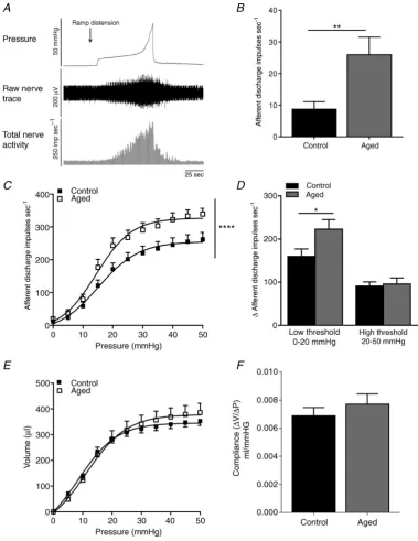
Afferent recordings remained stable for >4 h and repeated distension elicited reproducible response profiles from both control and aged mice. Spontaneous afferent nerve activity in control mice was significantly lower at 8.8 ± 2.3 impulses s–1 (n = 14) compared to 26.0±5.5 impulses s–1in aged mice (P<0.001, Student’s
ttest,n=7, Fig. 2B). As demonstrated previously, ramp distension of the bladder evoked a graded increase in intravesical pressure and afferent nerve discharge (Daly
et al. 2007). The same distension–response profile was
also observed in preparations from aged mice, although the overall magnitude of firing in response to bladder distension was augmented compared to preparations from controls (n = 7 and n = 14 respectively, P < 0.0001, two-way ANOVA, Fig. 2C). This increase in afferent activity affected only the low threshold component of the response (0–15 mmHg, Fig. 2D). Bladder compliance as gauged by the pressure–volume relationship was not significantly affected by age (Fig. 2EandF).
Ageing enhances contractility of the detrusor
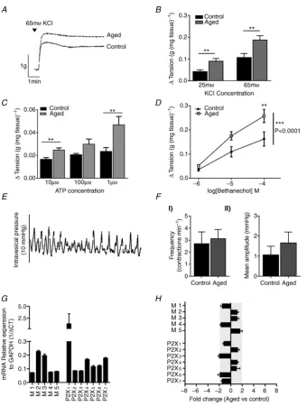
Contractile responses to the non-selective muscarinic receptor agonist bethanechol and the non-selective purinergic receptor agonist ATP were significantly increased in muscle strips from aged mice compared to muscle strips from control mice (n=6 and 6,P<0.001 Student’sttest and two way ANOVA with Bonferronipost hoctest as appropriate, Fig. 3CandD). Moreover, contra-ctile responses to application of KCl (25 and 65 mM) were also significantly greater from aged mice than from control mice (n= 6), in the absence of any change in detrusor weight, suggesting that the overall contractile ability of the detrusor was augmented by ageing (P<0.001, Student’s
ttest, Fig. 3AandB).
Whole bladder spontaneous contractions were observed in 84% of control preparations and 75% of aged pre-parations (Fig. 3E,n = 6 andn = 8) but there was no significant difference in amplitude or frequency of contra-ctions between the two groups (Fig. 3F).
In muscle samples, qRT-PCR identified relative expression of all five muscarinic receptor genes, with the M2 and M3 subtypes exhibiting the greatest expression, and all seven of the P2X receptor genes, with the P2X1
subtype exhibiting the greatest expression. No difference in gene expression was detected between aged and control samples (n=6 andn=9, respectively, Fig. 3GandH).
Altered ATP and ACh overflow with age
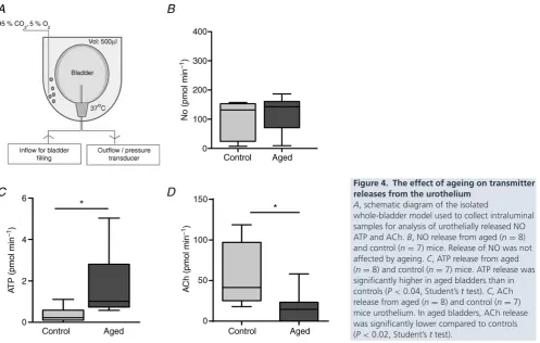
Distension of the bladder evoked release of ATP, ACh and NO, which in this study was detected as overflow of these transmitters into the lumen. No significant difference in NO between aged and control bladders was identified (n = 8 andn = 7, respectively, Fig. 4B), although ATP levels from aged bladder preparations were significantly increased compared to controls (n = 8 and n = 7, respectively). This was concurrent with a reduction in ACh levels and suggests that ageing may alter transmitter release from the urothelium (Fig. 4CandD).
Urothelial purinergic receptor signalling is increased
ageing, calcium imaging experiments were conducted using isolated urothelial cells from aged and control animals. Preliminary studies found an EC50 for ATP
of 10 μM (data not shown). To ensure activation of all purinergic receptors an EC100 concentration
(100 μM) was applied. This application was not
deleterious to cells as viability was tested using an MTT (3-(4,5-dimethylthiazol-2-yl)-2,5-diphenyltetrazolium bromide) assay (data not shown). The number of urothelial cells responding to purinergic receptor stimulation with ATP was significantly greater from aged mice than from controls (N = 4, n = 333 and
N= 5n =241, respectively). Moreover, the magnitude of the response to ATP was also significantly increased, indicating age-related changes in purinergic receptor signalling. In contrast, urothelial cells from aged mice (N=4,n=226) had similar responses to bethanechol as urothelial cells from controls (N=3,n=190), suggesting that muscarinic receptor signalling in the urothelium was unaffected by ageing (Fig. 5CandD).
In separate experiments, the selective P2X1 and P2X3
agonistαβmethyleneATP and the selective P2X1 agonist
βγmethyleneATP were applied. There was no significant difference in urothelial cell responses toβγmethyleneATP
between cells from aged and control mice (N=5n=239 andN= 4n =189, respectively) although significantly more cells from aged mice responded toαβmethyleneATP (Fig. 5EandF) compared to cells from control mice (N=5
n = 218 and N = 4 n = 175, respectively, P < 0.001, Student’sttest). The magnitude of the responding cells was also elevated but did not reach significance.
Gene and protein expression in the urothelium is significantly altered by ageing
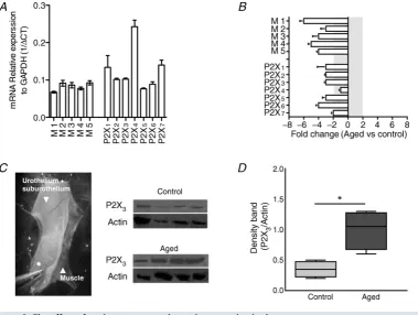
Western blot analysis found a significantly greater P2X3
receptor expression in aged bladder mucosa compared to control mucosa (n =4 and n = 4,P < 0.01, Student’s
t test). However, paradoxically, qRT-PCR experiments showed significantly lower muscarinic and purinergic gene expression in aged samples relative to controls (n=6 and
n=8, respectively, Fig. 6).
Discussion
[image:6.595.118.486.365.652.2]Given the clear correlation between lower urinary tract disorders such as OAB, UI and advancing age, a number of studies have sought to understand how bladder
Figure 1. Urine spot patterns obtained from aged and control mice
A, representative digitised traces showing the urine spot patterns obtained from a control and an aged mouse over a 4 h period.B, total urine spot area was not significantly different between aged (n=6) and control mice (n=6).C, the number of urine spots detected was greater in aged mice than in controls (P<0.05, Student’s
ttest).D, the number of small urine spots (<0.2 cm2) was significantly greater in aged mice than in controls
physiology is affected by ageing. Over the past decade it has become apparent that the sensory innervation of the bladder together with the urothelium plays an integral role in regulating micturition (Birder, 2004, 2009), however it is unclear how these elements are affected by ageing. Afferent
[image:7.595.102.482.141.633.2]innervation from the bladder is conveyed by the pelvic and hypogastric nerves. Distinct subpopulations of these afferents innervate the muscle, urothelial, suburothelial and serosal layers of the bladder and detect changes in mechanical or chemical environment in the bladder wall
Figure 2. Afferent nerve activity was significantly increased in aged mice
(Zagorodnyuk et al. 2007). Changes in the function of any of these components could alter sensory nerve trans-duction. In this study we sought to examine how ageing affects bladder afferent activity and the function of the detrusor and urothelium.
Ageing increases bladder mechanosensitivity
[image:8.595.134.475.142.593.2]There are relatively few studies that have directly investigated the effect of ageing on bladder sensory pathways. One previous study reported an age-related reduction in the afferent response to bladder filling
Figure 3. The effect of ageing on detrusor function and gene expression
A, sample trace showing the contractile response to 65 mMKCl in aged and control muscle strips.B, contractile responses to 25 and 65 mMKCl application in denuded muscle strips from aged and control mice. In aged muscle strips contractile response were significantly greater than controls (n=6,P<0.005, Student’sttest).C, contractile responses to ATP application in denuded muscle strips from aged and control mice (n=6,P<0.005, Student’s
(Hotta et al.1995). However, conversely, a more recent study found increased voiding frequency and higher afferent nerve discharge in aged rats than in younger controls (Aizawa et al. 2012). However, both of these previous studies used paradigms in which animals were anaesthetised. As it is not clear what effect this could have on afferent signalling, interpretation of these data is difficult. In this current study we used both voiding pattern analysis (in awake, unanaesthetised freely moving mice), which has been previously shown to provide an accurate assessment of in vivo bladder function and correspond to cystometry (Hodgeset al.2008), together with direct recordings of pelvic and hypogastric bladder afferentsin vitro. We identified a significant difference in the voiding patterns of aged and control mice, which indicate that aged mice void smaller volumes of urine more frequently than younger controls. Although fluid intake was not recorded, urine osmolality was unchanged, suggesting that the altered voiding patterns relate to changes in micturition rather than increased urine production or altered renal function. Moreover, this increase in voiding activityin vivowas concurrent with a significant increase in spontaneous nerve firing and in low-threshold mechano-sensitivity.
There are a number of components that could contribute to this increased mechanosenstivity. (1) Changes in sensory nerve morphology and function. In
previous studies some moderate changes due to ageing have been described but the general pattern of innervation seems to be conserved with age (Nakayama et al.1998; Mohammed & Santer, 2002; Aizawa et al. 2010). One previous study found a 40% reduction in calcitonin gene-related peptide and substance P containing afferents in the DRG of aged rats (Mohammed & Santer, 2002). However, bladder projecting afferents were not identified specifically, and as yet, there are few data concerning how the exact properties of bladder projecting nerves are affected by age. (2) Altered contractility and changes in detrusor tone. Approximately 30% of hypogastric and 80% of pelvic innervation from the bladder arises from afferents whose terminals lie within the muscle layers (Xu & Gebhart, 2008), and thus altered detrusor activity may indirectly influence bladder mechanosensitivity. (3) Changes in urothelial function: the urothelium has a well-established role in modulating neural excitability, and thus changes in urothelial function could indirectly alter afferent excitability.
Ageing increases contractility of the bladder
[image:9.595.48.546.405.721.2]Urodynamic studies in humans suggest that ageing is associated with overactivity of the detrusor muscle during bladder filling and storage but diminished detrusor contractility during bladder voiding (Resnick
Figure 4. The effect of ageing on transmitter releases from the urothelium
A, schematic diagram of the isolated
Figure 5. The effect of ageing on purinergic receptor signalling in the urothelium
A, example of primary urothelial cells cultured from aged and control mice and loaded with the calcium-sensitive dye Fura-2AM (2μM).B, sample traces showing typical urothelial cell response to ATP (100μM) and the ionofore
ionomycin (5μM).C, the number of cells responding to application of the purinergic receptor agonist ATP (100μM) and the muscarinic receptor agonist bethanechol (100μM). In aged mice (N=4,n=333), the number of urothelial cells responding to purinergic receptor stimulation with ATP was significantly greater compared to controls (N=5,
n=241,P<0.001, Student’sttest). However, responses to the muscarinic receptor agonist bethanechol were unchanged.D, the magnitude of the response to ATP and ACh. Responses to ATP stimulation were significantly higher in aged urothelial cells compared to controls (P<0.001 Student’sttest), indicating age-related changes in purinergic receptor signalling. In contrast, urothelial cells from aged mice (N=4,n=226) had similar responses to bethanechol as urothelial cells from controls (N=3,n=190) suggesting that muscarinic receptor signalling in the urothelium is unaffected by aging.E, the number of cells responding to application of the P2X1and
P2X3agonistαβmethyleneATP (30μM) and the selective P2X1agonistβγmethyleneATP (100μM). There was no significant difference in urothelial cell responses toβγmethyleneATP between cells from aged and control mice (N=5,n=239 andN=4,n=189, respectively) although significantly more cells from aged bladders responded toαβmethyleneATP compared to cells from control mice (N=5,n=218 andN=4,n=175, respectively,
& Yalla, 1987; Madersbacher et al. 1998; Nordling, 2002). Whether this generates hypersensitivity of bladder afferents during the storage phase, driving the sensory symptoms associated with OAB (urgency and frequency), has yet to be established. Moreover, morphological studies seem consistent with age-related changes in the detrusor, showing that with age there is increased collagen deposition but reduced muscle mass and innervation (Lluel et al. 2000; Smith et al. 2012). In this study we did not examine bladder morphology, but no difference in detrusor weight between control and aged tissue was observed, suggesting that in our hands aged mice appear to have no significant difference in detrusor muscle mass. There have been some previous muscle bath studies investigating contractility in ageing, but these studies have used a variety of animal models (guinea pig, rat, mouse, pig and human), different age ranges (2–30 months), different sexes and different experimental protocols, which have led to conflicting results. Some studies indicate decreased in contractility with age, and others suggest increased in contractility or no functional alterations whatsoever (Longhurst et al. 1992; Munro & Wendt,
1993; Saito et al. 1993; Lieu et al. 1997; Pagala et al.
[image:11.595.108.488.346.632.2]2001; Yoshida et al. 2001; Gomez-Pinilla et al. 2011). To examine how muscle function was affected by age in this study, we measured passive bladder compliance in vitro, studied contractility using muscle strip experiments and measured whole-bladder spontaneous contractions. Passive bladder compliance and the amplitude and frequency of spontaneous bladder contractions were not significantly different between control and aged mice. Moreover, cholinergic and purinergic gene expression in the detrusor was also unaltered. Interestingly, stimulation of denuded muscle strips with KCl produced greater contractions in aged tissues, suggesting enhanced contractility. This enhanced contractility was also seen in response to muscarinic and purinergic receptor stimulation, suggesting that in our aged mice detrusor contractility was generally increased but that bladder compliance was unaltered. This is in contrast to a recent study by Smithet al. (2012) that found increased bladder compliance in aged female mice and impaired responses to bladder filling, but no change in detrusor power or contractile force during voiding (Smith et al.2012). As
Figure 6. The effect of ageing on gene and protein expression in the mucosa
A, mRNA expression for muscarinic and purinergic receptors in control mucosal samples relative to the housekeeping gene GAPDH (M1n=4, M2–5n=8, P2X1–7n=8).B, fold change in gene expression between
control and aged mucosal samples. A significant reduction in all muscarinic receptor and all P2X receptor genes (except P2X4) was seen in aged mucosa relative to controls (n=6 andn=8, respectively).C, mucosal layers were separated from the muscle by blunt dissection under the microscope. Western blot showing the expression of P2X3receptors in aged and control mucosal samples.D, relative protein expression compared toβ-actin. There
was significantly more P2X3receptor expression in the mucosa from aged mice (n=4) compared to controls
voiding responses were not measured in the present study, it is difficult to ascertain whether the same was true in our aged mice; however, it is important to remember that the in vivo situation, where the efferent pathways involved in voiding are intact, may be very different to the in vitro where all efferent influences are removed. Moreover the study by Smithet al. was performed under anaesthesia, which may inhibit afferent pathways or affect central control centres. In this study we decided to use male mice to avoid complications arising due to hormonal changes, parturition and fertility senescence, these factors could also contribute to the differences seen between this and the previous study by Smith and colleagues.
Together, these data suggest that aged mice exhibit some moderate changes in agonist-induced muscle contraction; however, spontaneous contractions and bladder tone were not significantly changed with age, suggesting that altered afferent activity during bladder filling may also be driven either by changes in nerve function or by alterations in the urothelium.
Ageing alters purinergic signalling in the urothelium
It is now well established that the urothelium contributes to afferent pathways as it is able to sense and monitor mechanical and chemical changes in the bladder wall and modulate afferent firing via the release of a host of excitatory and inhibitory neurotransmitters. It has also been shown that in disease states such as OAB urothelial transmitter release is altered (Chuang, 2009). Yoshida
et al. (2004) showed that increasing age was associated with a decrease in ACh release and an increase in ATP release from the human bladder. In this study, we saw the same trend in the aged mouse. However, we measured the concentration of these neurotransmitters in the lumen of the intact bladder, and thus it is not clear exactly which cells release these agents, whether it is release or breakdown of the molecules that is changed, or the exact concentration of these mediators in the urothelium/suburothelium and at the nerve terminal. Nevertheless, it is tempting to speculate that this increase in luminal ATP concentration corresponds to changes in transmitter release from the urothelium. Previous studies have clearly shown that ATP released from the urothelium acts at afferent terminals to alter nerve activity, although the mode of action of urothelially released ACh is controversial. It could play a role in detrusor contraction; however, the plexus of blood vessels which lie in the suburothelium are likely to act as a diffusion barrier preventing it from reaching the muscle layers, which would suggest that, instead, urothelially released ACh acts at the urothelium (via autocrine or paracrine mechanism) or on the afferent terminals to alter urothelial-afferent signalling.
In calcium imaging experiments ageing had no effect on urothelial responses to the muscarinic agonist
bethanechol, but clear differences were seen in purinergic receptor-mediated calcium signals. Both the maximal calcium signal and the number of urothelial cells responding to ATP were increased in urothelial cells from aged mice. This altered purinergic signalling is likely to be mediated via the P2X3 receptor as studies with the
selective P2X1and P2X3agonist produced greater signals
in cells from aged mice but the selective P2X1 agonist
did not. These findings may explain the hyperactivity seen in the electrophysiology and voiding pattern studies. Increased urothelial cell excitability could enhance afferent activity either via a direct communication with the afferent terminal or via increased release of excitatory neuro-transmitters such as ATP, which then acts downstream at the afferent terminal to enhance mechanosensitivity and/or sensory excitability.
Western blot analysis identified higher P2X3 receptor
expression in aged mucosa compared to controls. Paradoxically, this increased receptor expression was concurrent with the increased ATP release and may suggest that the feedback mechanisms normally in place to induce receptor down-regulation are aberrant. Moreover, qRT-PCR found a general reduction in purinergic and cholinergic receptor gene expression despite an increased protein expression, suggesting that in ageing, mechanisms involved in P2X3 receptor turnover in the urothelium
could be altered. It is also important to bear in mind that within the mucosal layers there are also afferent and efferent nerve terminals that would express the P2X3receptor protein contributing to the overall mucosal
expression; however, as the cell bodies of these nerves are located elsewhere, gene expression would not be affected. Thus, the increased P2X3 receptor expression in aged
samples could also indicate changes in purinergic receptor expression on nerve terminals in addition to or instead of the other cell types within the mucosa. Unfortunately, it is not possible to identify where P2X3receptor expression is
increased from this study.
In this study we have focused mainly on urothelial signalling but it is important to note that downstream signalling events at the level of the afferent terminal (including receptor expression), neuronal excitability and central processing in the CNS could also be disrupted in ageing. Further characterisation of these pathways may provide greater insight into how bladder physiology alters with age.
Conclusion
It is clear that the incidence of bladder conditions such as OAB increases in line with advancing age for both males and females (Milsom et al. 2001; Irwin
age. In this study we show that ageing causes enhanced bladder activity and peripheral sensory transmission in the mouse. This is associated with altered purinergic signalling in the urothelium via the P2X3 receptor, increased
P2X3 protein expression and increased overflow of ATP.
However, more studies are still required to fully validate this hypothesis. Understanding how the urothelium and afferent mechanisms are affected by ageing may yield a better understanding of normal and aberrant bladder function and could potentially reveal novel targets for the treatment/prevention of bladder conditions in humans.
References
Aizawa N, Iijima K, Rosenbaum JS, Downs TR, Igawa Y, Andersson KE & Wyndaele JJ (2010). Comparison of the effects of oestrogen deficiency and old age on primary bladder afferent activity and voiding behaviour in the ageing female rat.BJU Int108, E10–16.
Aizawa N, Iijima K, Rosenbaum JS, Downs TR, Igawa Y, Andersson KE & Wyndaele JJ (2012). Comparison of the effects of oestrogen deficiency and old age on primary bladder afferent activity and voiding behaviour in the ageing female rat.BJU Int108, E10–16.
Birder L (2004). Role of the urothelium in bladder function.
Scand J Urol Nephrol Suppl215, 48–53.
Birder LA (2009). Urothelial signalling.Auton Neurosci153, 33–40.
Burnstock G (2009). Purines and sensory nerves.Handb Exp Pharmacol194, 333–392.
Chuang Y-C (2009). The role of urothelial dysfunction in pathogenesis of overactive bladder.Incont Pelvic Floor Dysfunct3, 3–4.
Daly D, Rong W, Chess-Williams R, Chapple C & Grundy D (2007). Bladder afferent sensitivity in wild-type and TRPV1 knockout mice.J Physiol583, 663–674.
Dubeau CE (2006). The aging lower urinary tract.J Urol175, S11–15.
Gomez-Pinilla PJ, Pozo MJ & Camello PJ (2011). Aging differentially modifies agonist-evoked mouse detrusor contraction and calcium signals.Age (Dordr)33, 81–88. Hodges SJ, Zhou G, Deng FM, Aboushwareb T, Turner C,
Andersson KE, Santago P, Case D, Sun TT & Christ GJ (2008). Voiding pattern analysis as a surrogate for
cystometric evaluation in uroplakin II knockout mice.J Urol 179, 2046–2051.
Hotta H, Morrison JF, Sato A & Uchida S (1995). The effects of aging on the rat bladder and its innervation.Jpn J Physiol45, 823–836.
Irwin DE, Milsom I, Hunskaar S, Reilly K, Kopp Z, Herschorn S, Coyne K, Kelleher C, Hampel C, Artibani W & Abrams P (2006). Population-based survey of urinary incontinence, overactive bladder, and other lower urinary tract symptoms in five countries: results of the EPIC study.Eur Urol50, 1306–1314; discussion 1314–1305.
Lieu PK, Sa’adu A, Orugun EO & Malone-Lee JG (1997). The influence of age on isometric and isotonic rat detrusor contractions.J Gerontol A Biol Sci Med Sci52, M94–M96.
Lluel P, Palea S, Barras M, Grandadam F, Heudes D, Bruneval P, Corman B & Martin DJ (2000). Functional and
morphological modifications of the urinary bladder in aging female rats.Am J Physiol Regul Integr Comp Physiol278, R964–972.
Longhurst PA, Eika B, Leggett RE & Levin RM (1992). Comparison of urinary bladder function in 6 and 24 month male and female rats.J Urol148, 1615–1620.
Madersbacher S, Pycha A, Schatzl G, Mian C, Klingler CH & Marberger M (1998). The aging lower urinary tract: a comparative urodynamic study of men and women.Urology 51, 206–212.
Milsom I, Abrams P, Cardozo L, Roberts RG, Thuroff J & Wein AJ (2001). How widespread are the symptoms of an overactive bladder and how are they managed? A population-based prevalence study.BJU Int87, 760–766. Mohammed HA & Santer RM (2002). Distribution and
changes with age of calcitonin gene-related peptide- and substance P-immunoreactive nerves of the rat urinary bladder and lumbosacral sensory neurons.Eur J Morphol40, 293–301.
Munro DD & Wendt IR (1993). Contractile and metabolic properties of longitudinal smooth muscle from rat urinary bladder and the effects of aging.J Urol150, 529–536. Nakayama H, Noda K, Hotta H, Ohsawa H & Hosoya Y (1998).
Effects of aging on numbers, sizes and conduction velocities of myelinated and unmyelinated fibers of the pelvic nerve in rats.J Auton Nerv Syst69, 148–155.
Nordling J (2002). The aging bladder–a significant but underestimated role in the development of lower urinary tract symptoms.Exp Gerontol37, 991–999.
Pagala MK, Tetsoti L, Nagpal D & Wise GJ (2001). Aging effects on contractility of longitudinal and circular detrusor and trigone of rat bladder.J Urol166, 721–727.
Resnick NM & Yalla SV (1987). Detrusor hyperactivity with impaired contractile function. An unrecognized but common cause of incontinence in elderly patients.JAMA 257, 3076–3081.
Saito M, Kondo A, Gotoh M, Kato K & Levin RM (1993). Age-related changes in the response of the rat urinary bladder to neurotransmitters.Neurourol Urodyn12, 191–200.
Smith CP, Gangitano DA, Munoz A, Salas NA, Boone TB, Aoki KR, Francis J & Somogyi GT (2008). Botulinum toxin type A normalizes alterations in urothelial ATP and NO release induced by chronic spinal cord injury.Neurochem Int52, 1068–1075.
Smith PP, DeAngelis A & Kuchel GA (2012). Detrusor expulsive strength is preserved, but responsiveness to bladder filling and urinary sensitivity is diminished in the aging mouse.Am J Physiol Regul Integr Comp Physiol302, R577–R586. United Nations DoEaSA, Population Division (2013).World
Population Prospects: The 2012 Revision, Highlights and Advance Tables. Working Paper No. ESA/P/WP.228. United Nations.
Xu L & Gebhart GF (2008). Characterization of mouse lumbar splanchnic and pelvic nerve urinary bladder
mechanosensory afferents.J Neurophysiol99, 244–253. Yoshida M, Homma Y, Inadome A, Yono M, Seshita H,
Miyamoto Y, Murakami S, Kawabe K & Ueda S (2001). Age-related changes in cholinergic and purinergic neurotransmission in human isolated bladder smooth muscles.Exp Gerontol36, 99–109.
Yoshida M, Miyamae K, Iwashita H, Otani M & Inadome A (2004). Management of detrusor dysfunction in the elderly: changes in acetylcholine and adenosine triphosphate release during aging.Urology63, 17–23.
Zagorodnyuk VP, Gibbins IL, Costa M, Brookes SJ & Gregory SJ (2007). Properties of the major classes of mechanore-ceptors in the guinea pig bladder.J Physiol585, 147–163.
Additional information
Competing interests
C.C. is an advisor/consultant for Astellas, Pfizer, Allergan, Recordati, Lilly, ONO and Xention.
Author contributions
All experiments were performed in Professor Grundy’s laboratory at the University of Sheffield except for the muscle
bath experiments which were performed in Dr McKay’s laboratory at Hallam University. D.M.D., L.N. and D.G. were responsible for the conception and design of experiments. D.M.D., L.N. and M.L. were responsible for the collection, analysis and interpretation of data and all authors were involved in drafting the article and revising it critically for important intellectual content. All authors approved the final version of the manuscript and all persons designated as authors qualify for authorship.
Funding
Aged mice were provided by a BBSRC research grant and/or Marie Curie initial training network fellowships (TRUST). M.L. and L.N. were funded by a Marie Curie initial training network fellowship (TRUST).
Acknowledgements
We would like to thank Christopher Keating for providing some of the tissues for this project, Lori Birder for her insightful and constructive comments which greatly improved the manuscript and W. Everaerts for help with urothelial cell culture protocols.
Translational perspectives