CLINICAL AND BIOCHEMICAL STUDIES ON
SENILE CATARACT
Dissertation submitted to
THE TAMIL NADU DR. M.G.R. MEDICAL UNIVERSITY
CHENNAI, INDIA
M.S. DEGREE EXAMINATION
BRANCH – III OPHTHALMOLOGY
ACKNOWLEDGEMENT
I have immense pleasure in expressing my heartfelt gratitude and sincere thanks to Dr.C.A. Nelson Jesudasan, Professor and Director, Institute of Ophthalmology, Joseph Eye Hospital, Trichy, and my guide
for this study for giving me the opportunity to work under his expert tutelage .
His dynamism, patience and competence were an unfailing source of inspiration to me. I wish to thank him for giving me the liberty to utilise all facilities available, for successful completion of my study. His constant encouragement and valuable suggestions have been truly inspiring . I sincerely thank my co-guide, Dr. C.G. Vanila, Professor, Vitreoretinal Services, Joseph Eye Hospital, for he r valuable suggestions and kind support throughout the study.
I owe my sincere thanks to Dr. Amjad Salman, Professor and Head, Vitreoretinal Services,for his extraordinary patience, valuable guidance and advice,throughout my postgraduation.
I am truly indebted to Dr. Saravanan Selvaraj, Assistant Professor, for his steadfast inspiration and constructive comments, and Dr. P. Sukanya, Assistant Professor, Vitreoretinal services,for her constant motivation and valuable support.
Parmar, Professor and Dr. M. Sowjanya, Assistant Professor, Cornea Department, who, inspite of their busy surgical schedules, have helped me in meticulous collection of cataractous lenses for the study on lens proteomics.
My heartfelt thanks to Dr. Isai, Post-Doctoral Fellow, and
Mr. Annadurai and Mr.Murali, Research Scholars, Department of Animal Science, Bharathidasan University, for their timely help and precious suggestions during various phases of this study.
I am indebted to my parents, Dr. Philip A. Thomas and
Dr. P. Geraldine, who have truly been my pillars of support, all these years and not just for this study. Their hard work and dedication in whatever they do, has been a huge inspiration for me.
I cannot adequately express my gratitude to my loving grandmother, Mrs. Emily. P and my uncle Mr. P.C. Selvaraj ,for their unconditional love and unwavering belief in my capabilities.Im also indebted to my dear in-laws, Mr. Theodore and Mrs. Christy for their affection, valuable support and prayers.
Last but never the least, a million thanks is due for my dear husband, Dr. T. Joseph for his loving support , innumerable sacrifices and patient advice, that continues to inspire , encourage and sustain me, especially during difficult times. I lovingly thank my sweet little son,
CLINICAL AND BIOCHEMICAL STUDIES ON
SENILE CATARACT
ABSTRACT
AIM:To determine the frequency of occurrence of different types of senile
cataract and assess the possible putative correlations with lenticular protein
profiles and to compare visual outcomes and factors influencing them, in
patients undergoing small incision cataract surgery (SICS) and
phacoemulsification cataract surgery at a tertiary eye care hospital in India.
PATIENTS AND METHODS:
520 patients-130 in the SICS group and 390 in the phaco group were
enrolled in the study. The putative factors influencing the outcome of surgery,
such as gender, the presurgical presence of systemic co-morbid conditions and
of ocular conditions, and the degree of nuclear sclerosis of the cataractous lens,
was ascertained. Differences in degree of visual improvement between the two
surgical groups were sought .An attempt was made to study the human lens
proteomics , by subjecting varying grades of nuclear cataract to sodium dodecyl
polyacrylamide gel electrophoresis (SDS-PAGE) and 2DE analytical
Results:
Analysis of lenticular profiles revealed that NC3 was the most common
cataract among males and in the SICS group, whereas NC4 was more common
among females and in the phaco group. There was no difference in
post-operative mean visual acuity between SICS and phaco in males, although such
a difference existed in females with better visual acuity in the phaco group.
Proteomic studies showed that the cataractous lens samples showed a
statistically significant reduction in band intensity on SDS-PAGE and
subsequently done two dimensional gel electrophoresis (2-DE) and
MALDI-TOF analysis revealed varying levels of alpha-crystallins in the cataractous lens
Conclusion:
Both SICS and phacoemulsification yield satisfactorily similar visual
outcomes and there are no other obvious factors such as grade of cataract,
associated ocular and systemic comorbid conditions, that would favour
choosing one procedure rather than the other. Additional biochemical studies on
various grades of human cataract may help in identifying factors triggering
CONTENTS
S.No. Title Page No.
1.
INTRODUCTION
1
2.
AIM OF THE STUDY
5
3.
REVIEW
OF
LITERATURE
7
4.
MATERIALS AND METHODS
23
5.
RESULTS
40
6.
DISCUSSION
77
7.
SUMMARY
91
8.
CONCLUSION
95
9. BIBLIOGRAPHY
10. APPENDICES
Proforma
I
1
INTRODUCTION
The eye is a highly specialized organ, which provides a view of the surrounding world by converting incident light into electrical signals that are interpreted by the brain. The human crystalline lens is meant to be transparent to enable it to focus visible light (400-700 nm) on the retina of the eye, providing increased sensitivity and allowing the information contained by that light to be spatially resolved.1,2
The term ‘cataract’ refers to a clouding or opaque area over the transparent ocular lens, which occurs when some of the proteins in the lens begin to aggregate, the end-result being impairment of vision3. Children may occasionally be born with the condition, or cataract may occur secondary to ocular injury or inflammation, to refractive errors such as myopia or to systemic diseases such as hypertension and diabetes mellitus. However, by far, the condition is mostly related to increasing age4. In fact, since cataract most commonly appears in elderly individuals, it is frequently referred to as “senile cataract”; if cataract develops before the age of 60, it may be called a “presenile” cataract.
proportion of the elderly, and enhanced life expectancy in the developing world, suggest that this number will increase dramatically in the coming years. In fact, projections by the World Health Organisation indicate that in 2020, close to 40 million people will be blind due to cataract8,9. In India, cataract has been reported to be responsible for 50-80% of bilateral blindness10,11,12. Thus, it is essential that all possible measures be taken to tackle this condition which may be responsible for a diminution in the quality of life.
At present, the most effective treatment of cataract is the surgical removal of the opacified lens; however, there continues to be a backlog of the services provided in many parts of the world. Globally, the two main techniques of cataract surgery are manual small incision cataract surgery and phacoemulsification; a third technique, microincision cataract surgery, is becoming increasingly popular13.
3
Africa since it is considerably less costly than phacoemulsification while still offering rapid visual recovery and reduced astigmatism. A 6 mm to 6.5 mm scleral incision allows the insertion of a 6 mm intraocular lens (IOL); the posterior capsule of the lens is left intact.
With increasing safety of ocular surgical techniques and improved visual results, it is no longer necessary to wait for the cataract to become sufficiently mature; in fact, surgery can be performed at a much earlier stage when phacoemulsification is used15. Cataract surgery may be performed depending on the patient’s age and visual function demands16.Certain surgical complications can be avoided if the cataract is removed before it becomes too advanced17. Surgery should be considered when the benefits from removal of symptoms outweigh the small risks caused by modern surgery18.
Two-dimensional gel electrophoresis (2-DE) and mass spectrometric (MS) analysis are the tools used in the emerging field of proteomics21; these hold great promise for determining putative modifications in lenticular structure that contribute to cataractogenesis. 2-DE is capable of simultaneously resolving complex mixtures of modified crystallins, which are then quantified by image analysis, and, subsequently, post-translational modifications on excised spots can be determined by MS. There are few studies on proteomic aspects of human lenses, and fewer studies on cataractous lens of varying degrees of nuclear sclerosis.
A
AIMS OF THE STUDY
To compare outcomes in patients undergoing small incision cataract
surgery (SICS) and outcomes in patients undergoing phacoemulsification cataract surgery at a tertiary eye care hospital in India.
Primary outcome measures
1. To determine the frequency of occurrence of different types of senile cataract (based on the Lens Opacities Classification System [LOCS] Version III) in patients about to undergo cataract surgery.
2. To compare the proportions of individuals achieving good functional vision (presenting visual acuity better than or equal to 6/12 in the operated eye) following small incision cataract surgery with the proportions achieved in individuals undergoing phacoemulsification
cataract surgery.
3. To identify putative factors influencing post-operative visual outcome, such as age, gender, types of senile cataract and presence of co-morbid systemic diseases (esp. diabetes mellitus &
6
Secondary outcomes
1. To note the occurrence of intraoperative complications such as
posterior capsular rent and other complications as reported.
2. To note the occurrence of early post-operative complications.
R
7
REVIEW OF LITERATURE
The human lens is a cellular organ, its stability and transparency occurring as a result of tight packing of crystalline proteins into a
glass-like microarchitecture. Senile (age-related cataracts), one of the commonest types of acquired cataracts, affects equally persons of either gender, usually above the age of 50 years. By the age of 70 years, more
than 90% of individuals are likely to have developed a senile cataract. The condition is usually bilateral, but, for the most part, one eye is affected earlier than the other.
Anatomically speaking, the human lens can be divided into two
principal regions, namely, the nucleus and the cortex. While the nucleus is present at birth, the cortex tends to be formed by differentiation of the lenticular epithelial cells throughout life22. Concomitant with the maturation of lenticular fibres, intracellular organelles, such as the
mitochondria, tend to degenerate23. Thus, mature lenticular fibres tend to be devoid of cytoplasmic organelles, so that the bulk of the lens is made up of lipid membranes packed with lenticular proteins. This high
Senile cataractogenesis is essentially related to the ageing process. Although its precise etiology is unknown, various factors are implicated,
such as heredity, ultraviolet radiation, dietary factors, childhood dehydrational crises and smoking22. These factors tend to influence the age of onset, type and maturation of senile cataract. Heredity plays an important role in the age of onset, incidence and maturation of senile
cataract in different families26,27,28,29,30,31. Ultraviolet radiation has been implicated in the early onset and maturation of senile cataract, based on the observations made in several epidemiological studies32,33,34,35,36. Javitt and
Taylor33 found that the latitude of an individual’s residence correlated directly with the UV-B content of sunlight, because the incident angle of the sun determines the atmospheric penetration of ultraviolet radiation. Interestingly, although a consistent dose-dependent association between
ocular UV-B exposure and two common types of cataract (cortical and posterior subcapsular) has been shown by epidemiological studies, the same studies (unlike experimental studies)did not provide sufficient
9
A diet that is deficient in some proteins, vitamins (riboflavin, vitamins C and E) or essential elements has also been blamed for early
onset and maturation of senile cataract37,38.
The incidence of three different cataract types tends to increase with age: nuclear cataracts, which account for approximately 60% of age-related cataracts; cortical cataracts which account for approximately 30%
and posterior subcapsular cataracts, which account for the remaining 10%39.Klein et al.39 described the cumulative incidence of age-related
cataracts and cataract surgery over a 10-year interval through a prospective
epidemiologic study on persons aged 43 to 86 years who were participating in the Beaver Dam Eye Study; there were 4926 individuals at baseline and 2764 individuals for first, five-year and ten-year follow-ups.
Photographs of the lens in each patient were taken using specially
modified cameras and graded per codified rules by trained graders into nuclear, cortical, and posterior subcapsular cataracts. It was found that in right eyes, incident nuclear cataract occurred in 19.4%, cortical cataract in
incidence of cataract surgery. These authors concluded that age-related cataracts are common events in aging, and that age-specific cataract
surgery incidence seemed to be increasing.
With cataract being the leading cause of preventable blindness worldwide, cataract surgery is one of the most frequently performed operations in the world16. Hatch et al.40 sought to estimate cataract surgery
rates for the province of Ontario (Canada’s largest province [state] with a population of about 13 million), by direct standardization of age- and sex-specific cataract surgery rates, obtained from the Ontario Health Insurance Plan Database, to Ontario’s population projections; they found that the need for cataract surgery in this region was likely to more than double in the ensuing 25 years (the study was done in 2012).
11
and ability to safely place multiple corneal sutures. In addition, such microscopes provide the advantages of leaving the posterior capsule intact, leading to a reduced risk of potentially blinding complications, such as vitreous loss or retinal detachment, and also permitting the implantation of an IOL in the posterior chamber.
Currently, phacoemulsification is the standard of care for cataract
extraction in the western world. The major advantage of phacoemulsification is that it has reduced the morbidity from cataract surgery by reducing the incision size with subsequent faster recovery and
decreased risk of complications including endophthalmitis. In addition, the learning curve for this procedure seems to be remarkably small. Meeks et al. (2013)41 reported on a retrospective cohort evaluation of the safety and
efficacy of phacoemulsification cataract extraction and manual
extracapsular cataract extraction performed by beginning resident surgeons in a medical centre in Dallas, Texas, USA. A review was performed of each resident's (post-graduate student) series of initial cataract surgery
Complications (vitreous loss or dropped nucleus) occurred in six (2.5%) of 244 cases in which phacoemulsification was performed by a beginner
resident primary surgeon and in seven (4.1%) of 172 cases in which extracapsular cataract extraction was performed. Posterior chamber IOLs were placed in all but two phacoemulsification cases and four patients in whom extracapsular cataract extraction was peformed. Moreover, three
cases in the phacoemulsification group and one in the group that had undergone extracapsular cataract extraction required a reoperation within 90 days. These authors concluded that phacoemulsification cataract
extraction can be taught safely and effectively to residents with no cataract surgery experience as a primary surgeon.
Mithal et al. (2012)42 evaluated the outcome of manual SICS under
topical anesthesia with lignocaine 2% jelly in a prospective interventional
case series involving 128 patients with senile cataract; intra-cameral anesthesia was not used, and there was only a single operating surgeon. More than 55% of patients did not experience pain, while more than 90%
13
200443 on 120 patients surveyed just before and one month after surgery found that there was a wide gap between expectations and what was
actually achieved. While 60% of patients expected to achieve a perfect Visual Function Index score, and the average expected score was 96.1, the
score actually achieved was 89.8; it was found that patients harboured unrealistic expectations regarding driving at night, reading small print, and
doing fine handiwork. Surprisingly, improvement in visual function did not correlate with satisfaction in vision. The author of the study felt that the results highlighted the highly unrealistic expectations harboured by
patients with cataract and underscored the need for physicians to attempt to control the expectations of their patients; the author even went so far as to say that controlling patient expectations was probably more effective than improving patients’ postoperative outcome in terms of maximizing patient
satisfaction43.
Several studies have tried to evaluate visual outcomes of SICS versus that in phacoemulsification Gogate et al. (2007)44 sought to
Prior to this, a single masked randomized controlled clinical trial had been conducted to compare the safety and efficacy of the two techniques for
rehabilitation of the cataract patient. The participants included 400 patients and four surgeons.The average cost of a phacoemulsification surgery for the hospital was Indian rupees (INR) 1978.89 while that of the SICS surgery was INR 720.99, with INR 500.99 being the fixed-facility cost
common to both. The cost of phacoemulsification cataract surgery was higher due to the cost of the foldable lens used. Moreover, phacoemulsification required additional expenditure to cover the financial
depreciation of the instrument, the replacement of faulty or old parts, and cost of the annual maintenance contract. These authors concluded that manual SICS is far more economical than phacoemulsification, safe and yields a visual result that is comparable with that of phacoemulsification.
In a prospective randomised study in Nepal, Ruit et al. (2007)45
sought to compare the efficacy and visual results of phacoemulsification with that of manual sutureless SICS in 108 consecutive patients with
15
months, 89% of the SICS patients had uncorrected visual acuity of 20/60 or better and 98% had a best-corrected visual acuity of 20/60 or better
versus 85% of the patients in the phacoemulsification group with uncorrected visual acuity of 20/60 or better and 98% with best corrected visual acuity of BCVA of 20/60 or better; this difference was not statistically significant. Interestingly, the surgical time for SICS was found
tobe significantly shorter than that taken to perform phacoemulsification. These authors concluded that both phacoemulsification and SICS achieved excellent visual outcomes with low complication rates. However, they also
opined that since SICS is significantly faster, less expensive, and less dependent on high-end technology than is phacoemulsification, SICS might be the more appropriate surgical procedure for the treatment of advanced cataracts in the developing world.
Singh et al. (2009)46 sought to compare the safety and efficacy of
different types of surgical procedures (phacoemulsification versus SICS) for cataract surgery in immature cataract.
two- thirds of patients in the phacoemulsification group and more than three quarters of the patients in the SICS group had good visual outcome
(6/6-6/18) on the first postoperative day; this difference was not statistically significant. Poor outcome (<6/60) was recorded in 6% (phacoemulsification group) and 1% (small incision cataract surgery group). Mean visual acuity was 0.43 +/- 0.27 in the phacoemulsification
group and 0.47 +/- 0.24 in the SICS group. Interestingly, the mean time for surgery was statistically significantly shorter in the SICS group than in the phacoemulsification group. These authors concluded that SICS cataract
surgery with implantation of a rigid polymethmethylacrylate lens is a suitable surgical technique to treat immature cataract in developing countries.
Wong47, who examined rates of cataract extraction in Singapore,
found that the average rate was highest for Indians (about 396.5 per 100, 000/year), followed by Chinese (371.2 per 100, 000/year) and Malays (only 237.2 per 100, 000/year); it was also found that women had higher
17
determine the prevalence of cataract surgery as well as factors in association with post-surgical outcomes of vision in migrant Indians living
in Singapore, conducted a population-based study in 3, 400 Indian immigrants residing in Singapore. Post-operative visual impairment was defined as best-corrected or presenting visual acuity of 20/60 or worse. The age- and gender-standardized prevalence of cataract surgery was 9.7%
in Singapore resident Indians, while the post-operative visual impairment, as defined by best corrected visual acuity, was found to occur in 10.9% of the eyes studied. The main causes of this impairment were found to be
diabetic retinopathy, posterior capsular opacification, and age-related macular degeneration. These authors48 concluded that socioeconomic variables and migration did not significantly contribute to the prevalence of cataract surgery, and that diabetic retinopathy was a major cause of
postoperative visual impairment in migrant Indians living in Singapore. A study conducted in Fukui (Japan) echoed similar findings (Morikubo et al.
2004)49. No significant differences were observed in any preoperative
parameter between diabetic and nondiabetic individuals. However, there
was a significantly higher increase in corneal thickness one month post-
surgery, and significantly greater losses of corneal endothelial cells at one
Lam et al. (2007)50 sought to evaluate the visual acuity and
astigmatism of individuals undergoing manual cataract extraction by local
surgeons in rural China over a 4 month period.
Among 313 eligible subjects, 242 (77%) could be contacted, of
whom1 176 (73%) were examined. Of those examined, mean ± SD age
was 69.3±10.5 years, and 66.5% were female; 35 had been operated on
bilaterally at Sanrao, and 85.2% had a preoperative presenting visual
acuity of 6/60 or worse. Presenting and best-corrected postoperative acuity
in the eye that was operated on were 6/18 or better in 83.4% and 95.7%,
respectively.
Skiadaresi et al. (2012)51 found poor correlations between cataract
morphology and visual symptoms with the exception of the correlation found between blurred vision and posterior subcapsular cataracts.
Although excellent outcomes are frequently reported for cataract surgery in urban areas of China and India, the same cannot be said for rural areas of Asia, wherein poor visual outcomes, low visual function and
19
undergoing cataract surgery in rural China. Interestingly, although visual
function was high in this group of individuals and there was substantial
potential benefit if refraction and second-eye surgery services were
utilized, actual uptake of services was modest because individuals did not
feel the need to do so. The authors concluded that programs to improve the
utilization of services needed to focus on provision of reading glasses and
on strategies to reduce costs52.
Lai et al. (2013)53 investigated the clinical outcomes of cataract
surgery in 207 individuals aged 90 and older who underwent cataract
surgery for primary senile cataracts.
Interestingly, 79.7% of the 207 participants (mean age 92.0 ± 2.1), 79.7% achieved visual improvement after cataract surgery in spite of a high incidence of systemic comorbidities such as hypertension (66.2%),
diabetes mellitus (25.1%), and myocardial infarction (19.8%), as well as ocular comorbidities such as age-related macular degeneration (15.9%), glaucoma (10.6%), and myopic degeneration (5.3%). The most common
Crystallins are structural proteins which possess the ability to form soluble oligomers in high concentrations; specific, short range interactions
are also possible, which increase refractive power of the lens and maintain transparency54.Crystallins are divided into three major classes, termed as alpha-, beta- and gamma-crystallins55.
Alpha crystallin, a member of the small heat shock protein family of
molecular chaperones, is an aggregate of two polypeptides, namely, alpha-A and alpha-B crystallins, that share 55% aminoacid sequence identity56. Alpha crystallin has chaperone- like properties, being capable of binding to
unfolded or denatured proteins and suppressing non-specific aggregation57.The chaperone activity of alpha crystallin helps to prevent the formation of large light- scattering aggregates, inactivation of enzymes, and, possibly, cataract formation58,59. Thus, alpha A crystallin is of much
importance in development of the lens and in the development and maintainenance of lenticular transparency.
In the Beta crystallin superfamily, beta-crystallins have seven
21
assembly of beta crystallins and the potential role of truncated versions of the protein in cataract formation have also been reported62.
Cataract-specific forms of alpha-, betaB1 and B2-crystallins have been observed to lack from 5 to 22 residues in their termini. Truncated forms of alpha- and beta- crystalline were also observed to be more abundant in the water insoluble fraction, suggesting that their truncation
may contribute to the formation of insoluble protein in cataractous lenses. Loss of 11 or more C-terminal residues in alpha-crystallin dramatically decreases its chaperone activity63.
Two-dimensional electrophoresis (2-DE) and mass spectrometric analysis (MS) are the tools in the emerging field of proteomics21; these hold a great promise in determining the modifications in lenticular crystalline proteins that contribute to cataractogenesis. 2-DE is capable of
simultaneously resolving complex mixtures of modified crystallins. These resolved crystallins can be then quantified by image analysis, and then, subsequently, post- translational modifications on excised spots can be
determined by MS.
been made of patients undergoing small incision cataract surgery (SICS) and phacoemulsification cataract surgery at a tertiary eye care hospital.
Previous studies have not reported significant differences in final visual outcome between these two types of surgery, so, in the current study, differences in degree of visual improvement between the two surgical groups were sought. Different authors have reported on various factors that
may possibly influence the outcome of therapy. Hence, in the present study, putative factors influencing the outcome of surgery, such as gender, the presurgical presence of systemic co-morbid conditions and of ocular
conditions, and the degree of nuclear sclerosis of the cataractous lens, were sought. Increasingly, proteomic studies of cataractogenesis are being performed in a bid to unravel the mechanisms leading to the development of cataract. There have been many studies on experimental cataracts, but
comparatively fewer on human cataracts. Hence, in the present study, human lenses with varying grades of nuclear cataract were subjected to sodium dodecyl polyacrylamide gel electrophoresis (SDS-PAGE) and 2DE
M
MATERIALS AND METHODS
Clinical aspects of the present dissertation were studied in patients presenting with diminished vision due to lenticular opacification at the
Institute of Ophthalmology, Joseph Eye Hospital, Tiruchirapalli, Tamilnadu, over a period of 11 months (December 1, 2011 to October 31, 2012). Biochemical aspects were studied concurrently. Clinical and
biochemical data were analysed and interpreted subsequently (March 2013 to September 2013). The study had the approval of the Institutional Review Board and conformed to the tenets of the Declaration of Helsinki ; written informed consent was also obtained from each patient.
CLINICAL ASPECTS
Over an 11-month period, 604 patients with diminished vision due to cataract were seen. Of these, 22 individuals did not provide consent to participate while 30 individuals could not be enrolled due to the presence
of one, or multiple, exclusion criteria. During data analysis, it was found that records of an additional 32 patients were incomplete. Hence, analysis could be performed for the clinical aspects of the study only on 520
patients.
11-24
month period, the available interventions being small incision cataract surgery (SICS) or phacoemulsification cataract surgery. Provided that there
was no specific indication to perform one particular intervention (SICS or phacoemulsification), the selection of the intervention was based on the patient’s choice.
Sample size calculation: A key parameter to be investigated in the current study was the visual acuity following SICS or phacoemulsification cataract surgery. Based on published data, it was hypothesized that phacoemulsification surgery would yield a marginally better post-operative
mean visual acuity than that achieved by SICS, and that this difference could be shown to be statistically significant by a Student `t’ test. To calculate the minimum total sample size (two-tailed hypothesis) and minimum sample size per group (two-tailed hypothesis), the parameter
values defined were:
a) Anticipated effect size (Cohen’s d): 0.5
b) Desired statistical power level (β-error): 0.8 (80%)
c) Probability level (α –error): 0.05
Minimum total sample size (two-tailed hypothesis): 128 Minimum sample size per group (two-tailed hypothesis): 64.
In actual fact, a total of 520 patients were enrolled in the clinical study (130 in the SICS group; 390 in the phacoemulsification group)
Inclusion Criteria: Patients were considered for inclusion in the study if: - they provided written informed consent, and were 40 to 70 years of
age;
- exhibited normal anterior segment morphology (other than for the presence of senile cataract in one or both eyes);
- showed normal intraocular pressure (IOP) or senile cataract with primary open angle glaucoma under medical management (with or without control of IOP), or intumescent lens with narrow angles on gonioscopy (without any peripheral anterior synechiae);
- exhibited a normal fundus or ultrasound B-scan evidence of a normal posterior segment;
- exhibited patent naso-lacrimal ducts on syringing; or
- had systemic hypertension, diabetes mellitus or
26
Exclusion criteria: Patients were not considered for the current study if they exhibited any one of the following exclusion criteria:
- declined to undergo surgery; - were less than 45 years of age;
- suffered from uncorrected high refractive errors or amblyopia ;
- had a conjunctival swab report showing significant growth of
potentially pathogenic bacteria;
- had nasolacrimal duct obstruction due to untreated acute or chronic nasolacrimal duct infection;
- suffered from nystagmus or congenital anomalies of the eye;
- suffered from external ophthalmoplegia with or without multiple cranial nerve palsies;
- suffered from corneal opacities (nebula, leucoma), dystrophies and
degenerations;
- exhibited active allergic, infective or inflammatory conditions such as episcleritis, scleritis, conjunctivitis, keratitis, anterior,
intermediate or posterior uveitis, optic or retrobulbar neuritis or infective foci anywhere in the body;
- suffered from traumatic cataract, complicated cataract or polar cataract;
- suffered from intraocular, intraorbital or systemic malignancies; - had a B-scan showing posterior segment abnormalities other than
normal age-related changes;
- suffered from uncontrolled systemic hypertension, diabetes mellitus, ischemic heart disease, chronic kidney disease, asthma or tuberculosis were receiving topical or systemic steroids or
immunosuppressive agents reported a recent history of a cardiovascular or cerebrovascular accident.
Clinical examination: All patients were subjected to visual acuity measurement objectively by auto-refractometer and subjectively by Snellen chart and the best corrected visual acuity (BCVA) was recorded after Snellen to decimal conversion.
The patient’s head position and eye position were assessed before
starting the ocular examination. Extraocular movements were recorded in all nine cardinal positions of gaze. This was followed by examination of each eye separately.
28
Slit-lamp biomicroscopic examination was done for all patients. The status of all the corneal layers, namely the epithelium, stroma and
endothelium, was noted. The presence of guttae, keratic precipitates, pseudoexfoliative material and pigments on the corneal endothelium was recorded.
The central and peripheral anterior chamber depth was assessed
using Shaffers system of grading of anterior chamber depth. The presence of flare or cells was also looked for in the anterior chamber.
The colour and pattern of the iris, and the presence of peripheral
anterior synechiae, iris atrophic patches, or pre-existing surgical or non-surgical peripheral iridotomy wounds were noted. The pupillary size, shape, regularity, presence of synechiae, reaction to direct and consensual light reflex and presence of pseudoexfoliative material along the papillary
ruff were also noted.
In cases of mature cataract, the macular function was assessed by the presence of a normal pupillary reaction and the absence of any relative
afferent pupillary defect (RAPD).
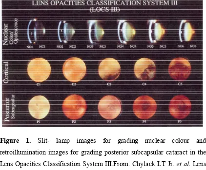
1993)64. The LOCS III contains an expanded set of standards (Fig. 1) selected from the Longitudinal Study of Cataract slide library at the
Center for Clinical Cataract Research (Boston, MA, United States of America). It consists of six slit lamp images for grading nuclear colour (NC) and five retroillumination images for grading posterior subcapsular cataract (PSCC) (Fig. 1).
Nuclear cataract was graded by comparing the colour of the lens to be graded with the standard colour photographs defined by the LOCS III as NC grades 1 to 6.While grading nuclear colour, two regions of the nucleus,
30
Figure 1. Slit- lamp images for grading nuclear colour and retroillumination images for grading posterior subcapsular cataract in the Lens Opacities Classification System III.From: Chylack LT Jr. et al. Lens
Opacities Classification System III [LOCS III]. Arch Ophthalmol 1993;
This grading is different from that followed in LOCS II in that the colour of the entire nucleus is considered, in addition to the posterior
reflex. This method thus avoids overestimation of the brunescent stage, which occurs occasionally when the reflex alone is used.
Posterior subcapsular cataracts were graded by using only posteriorly focused retro-illumination images.The area of the opacity in the
lens being graded was compared with the standards 1 to 5.The posterior segment status was assessed using the slit-lamp and a 90D lens.In patients in whom there was no view of the posterior segment, ultrasound B-Scan
was done to exclude any underlying ocular pathology.
Pre-operative workup: The patency of the nasolacrimal duct was assessed.
Keratometry was performed on both eyes manually with a Super
KMS-6™ keratometer, (Bausch and Lomb, USA) and with an automated keratometer (KM-500 NIDEK™) Autokeratometer, and the average keratometry reading was then calculated.
32
using the immersion technique using the Ocuscan ™ P/N 683-3001-505 (Alcon Surgicals, USA) was resorted to. The SRK-II formula (A-2.5L-0.9K) was used to calculate IOL power, taking into account the axial
length (L) and average keratometry (K) reading. ‘A’ constant was calculated as provided by the manufacturer.
Pre-operative preparations: On the day of surgery, a thorough preoperative assessment of the patient was performed. This was followed by pre-operative preparation of the patient. The patients were instructed to wash their face with soap and water. Appropriate head and foot gear were
provided before entry into the operation theatre complex. The skin around the eye was cleaned with povidone-iodine 5%. Following this, peribulbar block was given to the eye to be operated. This was again followed by cleaning of the conjunctival cul de sac and the skin around the eye with
povidone iodine.
Operative techniques
Surgery (both SICS and phacoemulsification) was performed under
the magnification of an operating microscope (Leica Microsystem™
In patients undergoing SICS65, following conjunctival peritomy, bleeders were cauterized.The expected size and density of the nucleus
determined the size of the tunnel, with the extraction of earlier stages of nuclear cataracts requiring only a small tunnel (sufficient for the IOL optic to pass through) while very big, brown nuclei required a larger tunnel size. A 3 plane sclerocorneal tunnel incision extending at least 1 to 2 mm into
the clear cornea was made. Following continuous curvilinear capsulorhexis, hydrodissection was performed, followed by use of a vectis or viscoexpression. A 360 degree cortex aspiration was done using a
Simcoe cannula, followed by placement of the posterior chamber IOL.
In the phacoemulsification technique 66,67,68, the same pre-operative protocol for patient preparation was followed. After conjunctival peritomy, a 5 mm corneo-scleral tunnel incision was made. Continuous curvilinear
capsulorrhexis not exceeding 5mm was also performed. The phaco probe (an ultrasonic handpiece with a titanium or steel needle) was used (Millenium ™ Ref CX6200 S721305, Bausch and Lomb Inc., Rochester,
34
steel instrument, called a "chopper", was used from a side port to help with chopping the nucleus into smaller pieces. Either a `stop and chop’
technique or a `divide and conquer’ technique was followed to break the nucleus. Thus, the cataract was broken into two or four pieces and each piece was emulsified and aspirated out with suction. After removing all hard central lenticular nuclear pieces by phacoemulsification, the softer
outer lens cortex was removed by suction only. An irrigation-aspiration probe or a bimanual system was used to aspirate out the remaining peripheral cortical matter, while leaving the posterior capsule intact.
An IOL was placed in the capsular bag. For implanting a polymethylmethacrylate IOL, the incision had to be enlarged while this was not necessary to implant a foldable IOL.
All patients were examined the day after surgery as a routine
post-operative day one follow-up. The status of the cornea was noted and presence of striate keratopathy, Descemet’s membrane folds, Descemet’s membrane stripping, and epithelial or stromal edema was also noted. The
including centration, was noted. The fundus was examined for vitreous clarity and presence of any underlying posterior segment pathology. IOP
was recorded by non contact tonometry.
The patients were started on topical corticosteroids for hourly use and were advised to come for followup after one week. At the follow-up visit, the patient’s uncorrected visual acuity was measured. The anterior
segment was evaluated for presence of corneal edema, flare and cells. The posterior chamber IOL position and centration were noted.
BIOCHEMICAL STUDIES
These were performed on representative cataractous lenses from patients undergoing cataract surgery, and on transparent lenses obtained during eye donation.
Sodium-Dodecyl-Sulfate Polyacrylamide Gel Electrophoresis (SDS-PAGE) analysis of total lenticular protein (soluble and insoluble fractions) pattern : The total soluble and insoluble protein pattern was analyzed by subjecting the sample to 4-20% gradient SDS-PAGE
36
constituted and the gradient was prepared by mixing these two solutions using a gradient mixer. The mixed solution was poured into a sealed glass
plate setup (8 cm high and 1.5 mm thick) for polymerization. After the polymerization was complete, the 3% stacking gel was poured over the separating gel and a Teflon comb was inserted to form wells. Later, the Teflon comb was removed and the wells were rinsed with distilled water.
The basal strip was then removed and the glass plate with polymerized gel was fixed to the electrophoretic apparatus. When the setup was ready, the samples were mixed with equal amounts of gel-loading dye and loaded
into the well. Electrophoretic separation was allowed for 180 minutes at a constant voltage of 50V for the stacking gel and 100V for the separating gel. The electrode solution used was 25 mM Tris, 0.193 M glycine buffer containing 0.1% SDS. Broad range protein markers were simultaneously
run for comparison. The gel was stained using Coomassie Brilliant Blue R-250 and destained with 7% acetic acid and 40% ethanol. The bands that developed, after staining and destaining, were scanned in a gel
documentation system (Bio-Rad, USA) to determine the intensity of the bands. The concentration of proteins in each band was determined from the standard graph plotted against the intensities of the bands corresponding to the known standard marker proteins. The program Quantity OneSW
the gels. Density profiles of each lane of the gels were used to calculate the peak intensity of the selected protein bands corresponding to soluble and
insoluble proteins.
Two dimensional (2D) gel electrophoresis analysis of the pattern of total lenticular protein (soluble and insoluble fractions): Four lenses from each group were homogenized in 200 mL lysis solution containing
protease inhibitors, followed by centrifugation, as described earlier. The supernatant containing the soluble protein was removed, and the pellet (insoluble protein) was washed twice with homogenizing buffer. The
insoluble protein was then resuspended in 8M Urea, and the protein content in both the soluble and insoluble fractions were measured by the
Bradford method (1976)70, using bovine serum albumin as a standard. Both fractions of lens proteins were aliquoted into 400-mg
portions and stored at -70°C.
Immobilized pH gradient (IPG) strips (linear pH 5-8, 7 cm) (Bio-Rad, USA) were rehydrated overnight with 160 μg of total protein derived
38
in Protean IEF Cell (Bio-Rad, USA) at 20°C, using stepwise mode to reach 10,000 Vh. After completion of the isoelectric focusing, the separated
proteins were equilibrated with another buffer for a further 15 min. The equilibrated IPG strips were then transferred onto 12% acrylamide slab gels (8 × 9.5 cm) and the second-dimensional separation was performed in Mini-Protean Tetra Cell (Bio-Rad, USA) with the current of 200 v/gel for
approximately 40 min. The resolved protein spots were then visualized using Coomassie Brilliant Blue R-250 stain.
PD Quest (Bio-Rad), was used for matching and analysis of protein
spots on 2-D gels. A reference gel was created by combining into one image all of the spots that had appeared in the individual gels. The reference gel was then used for matching of corresponding protein spots among different gels. Background subtraction was performed and the
intensity volume of each spot was normalized with the total intensity volume (summation of the intensity volumes obtained from all spots within the same 2-D gel). Protein spots of particular interest, based on pI and
acetonitrile and 0.1% (v/v) TFA, and the spots were allowed to dry completely. Mass spectra of positively-charged ions were recorded on an
Ultraflex MALDI TOF mass spectrometer (Bruker Daltonik) under the control of FlexControlTM 2.2 software (Bruker Daltonik GmbH).
Statistical Analysis
SPSS™ statistical software version 16 (Statistical Package for the
Social Sciences for MS Windows, SPSS Inc., Chicago, IL, USA) and Microsoft 2007 Office Excel™ software were used for statistical calculations. Differences in age, mean visual acuity and other continuous
variables were calculated by independent sample Student `t’tests. Yates’s corrected chi-square tests were used to analyse differences between categorical variables, using two by two tables, when appropriate. All statistical tests were two-sided (two-tailed), and P ≤ 0.05 was considered to
R
RESULTS
Clinical aspects of the present dissertation were studied in patients presenting with diminished vision due to lenticular opacification at the Institute of Ophthalmology, Joseph Eye Hospital, Tiruchirapalli, Tamilnadu, over a period of 11 months (December 1, 2011 to October 31, 2012). Biochemical aspects were studied concurrently. Clinical and
biochemical data were analysed and interpreted subsequently (March 2013 to September 2013).
1. CLINICAL ASPECTS
Over an 11-month period, 604 patients with diminished vision due
to cataract were seen. Of these, 22 individuals did not provide consent to participate while 30 individuals could not be enrolled due to the presence of one, or multiple, exclusion criteria. During data analysis, it was found
41
Using an A-priori sample size calculator for Student `t’ tests, the
minimum total sample size (two-tailed hypothesis) was calculated to be 128 while the minimum sample size per group (two-tailed hypothesis)was calculated to be 64 (details in Materials and Methods section). In actual fact, a total of 520 patients were enrolled in the clinical study (130 in the SICS group; 390 in the phacoemulsification group).
Hence, the requirements for sample size were met in the present study.
Patients who were enrolled in the study underwent monoocular cataract surgery either by small incision cataract surgery (SICS) or by
phacoemulsification, as described earlier (Materials and methods). Patients who underwent SICS constituted the “SICS group” while those who underwent phacoemulsification constituted the “Phaco group”. There were 130 patients in the SICS group and 390 patients in the Phaco group
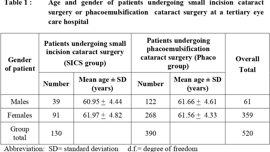
Table 1 : Age and gender of patients undergoing small incision cataract surgery or phacoemulsification cataract surgery at a tertiary eye care hospital
Gender of patient
Patients undergoing small incision cataract surgery
(SICS group)
Patients undergoing phacoemulsification cataract surgery (Phaco
group) Overall Total
Number Mean age ± SD
(years) Number
Mean age ± SD (years)
Males 39 60.95 + 4.44 122 61.66 + 4.61 61 Females 91 61.97 + 4.82 268 61.56 + 4.33 359
Group
total 130 390 520
Abbreviation: SD= standard deviation d.f.= degree of freedom
Statistical Analysis
a. Chi-square test (2X2 table) b, c, d. & e. Student ‘t’ test
a. Gender distribution in SICS group versus Phaco group
χ2 (d. f. = 1) = 0.048 ; P > 0.05
b. Mean age of males in SICS group versus mean age of males in Phaco group ( unpaired) ` t’ (d. f. = 159) = 0.08 ; P= 0.93
c. Mean age of females in SICS group versus mean age of females in Phaco group (unpaired) `t’ (d. f. = 357) = 0.05 ; P= 0.96
d. Mean age of males versus mean age of females in SICS group (unpaired) `t’ (d. f. = 128) = 0.13 ; P= 0.9
43
1.1 Age and gender distribution
There were 39 males and 91 females in the SICS group and 122
males and 268 females in the Phaco group (Table 1); these differences were not statistically significant (χ2 (degree of freedom [d.f]=1) = 0.05 ; p> 0.05). The mean ages of males and females were 60.95 + 4.44 years and 61.97 + 4.82 years, respectively, in the SICS group and 61.66 + 4.61 years
and 61.56 + 4.33 years, respectively, in the Phaco group (Table 1); these differences were not statistically significant (SICS group males versus females, `t’ = 0.13 [d.f. = 128]; p = 0.9 ; Phaco group males versus
females, `t’ = 0.01 [d.f.=388]; p=0.99; males in SICS versus Phaco groups, `t’ = 0.08 [d.f.=159], p=0.93; females in SICS versus phaco groups, `t’ = 0.05 [d.f.= 357] p = 0.96). Thus, patients in the SICS group and phaco group were, essentially, age-and gender-matched.
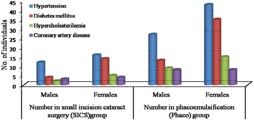
1.2 Co-morbid systemic conditions
Co-morbid systemic conditions noted in the patients in the two groups were hypertension, diabetes mellitus, hypercholesterolemia and
Figure 2 : Presence of undergoing phacoemulsi hospital
Abbreviations:d.f.= degre
Statistical Analysis
a. Proportions of patient (60/130 = 46%) versus 1.04 ; P > 0.05
b. Proportion of males (7 total 359 females) with P = 0.03
c. Proportions of males ( total 359 females) with d. Proportions of males (1
total 359 females) with
f systemic co-morbid conditions in small incision cataract surg ification cataract surgery at a tertiary
ee of freedom
ts with systemic co-morbid conditions in S s (vs.) Phaco group (158/390 = 41 %) : χ2 (d
78[48%] of total 161 males) vs. females (14 h systemic co-morbid conditions: χ2 (d. f. =
39 [24%] of total 161 males) vs. females (59 h hypertension: χ2 (d. f. = 1) = 4.4; P=0.036
17 [10.6%] of total 161 males) vs. females (49 h diabetes mellitus: χ2 (d. f. = 1) = 0.96; P=0.33
patients gery or y eye care
SICS group d. f. = 1) =
0[39 %] of = 1) = 4.47;
[16.4%] of
45
1.2.1 In SICS group versus Phacoemulsification group
Overall, co-morbid conditions occurred in 60 (46%) of 130 patients
in the SICS group and in 158 (40.5%) of 390 patients in the Phaco group (Figure 2); this difference was not statistically significant (χ2 [d.f.=1]=1.04; p > 0.05). Hypertension was, by far, the most frequent co-morbid condition in males in both groups, affecting 57% in the SICS group and 47% in the
phaco group (Figure 2).
1.2.2. In Males versus Females
Interestingly, co-morbid conditions occurred significantly more
frequently in males (78 [48.4 %] of 161) than in females (140 [39%] of 359) studied (χ2 [d.f.=1] = 4.47 ; P = 0.03) (Figure 2). Hypertension and diabetes mellitus were the most frequent co-morbid conditions in females in both groups, affecting 41% and 36%, respectively, in the SICs group,
and 43% and 35%, respectively, in the Phaco group (Figure 2). Interestingly, hypertension was seen significantly more frequently in males (39 [24%] of 161 males) than in females (59 [16.4%] of 359 females) in
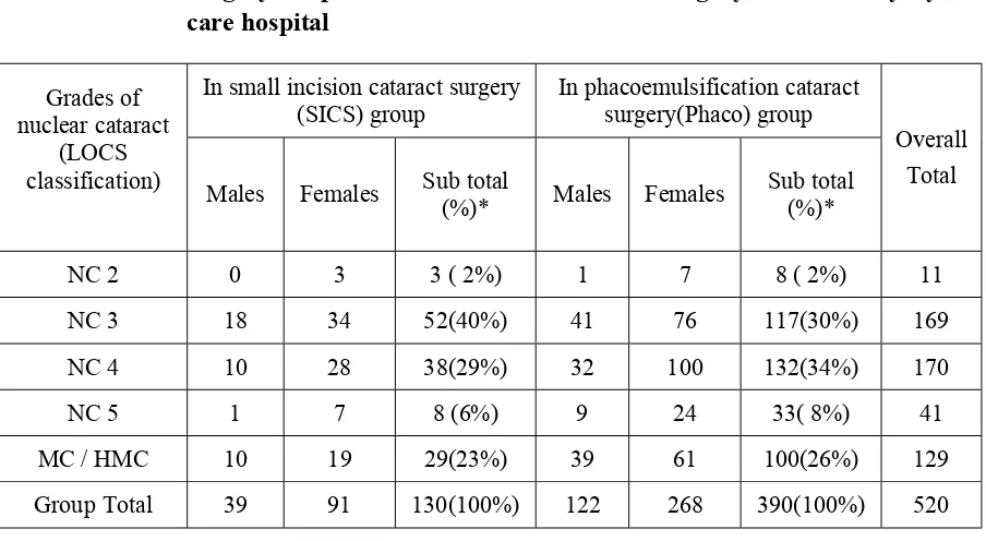
1.3 Types of cataracts
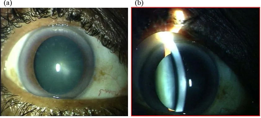
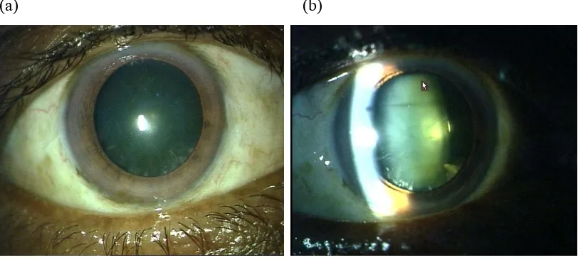
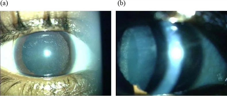
Various grades of nuclear (Figures 3, 4 and 5), posterior subcapsular
(Figures 7, 8 and 9) and end-stage cortical (mature and hypermature) cataracts (Figures 10 and 11) were seen in the patients. These cataracts were graded according to the LOCS classification III (Fig.1 in Materials and Methods section); these details are listed in Table 2 and Figures 6, 12
and 13.
1.3.1 In SICS group versus Phacoemulsification group
In the SICS group (total 130 patients), three patients (2%) had NC2,
52 (40%) had NC3, 38 (29%) had NC4, eight (6%) had NC5 and 29 (23%) had mature / hypermature cataracts ; corresponding percentages in the Phaco group (total 390 patients) were 2%, 30%, 34%, 8% and 26%, respectively (Table 2). These differences were not statistically significant
(χ2 [d.f.=4]=4.8 ; P=0.31).
Sixty-six (12.7%) of 520 patients in the current study suffered from diabetes mellitus.
47
Table 2: Grades of cataracts in patients undergoing small incision cataract surgery or phacoemulsification cataract surgery at a tertiary eye care hospital
Grades of nuclear cataract
(LOCS classification)
In small incision cataract surgery (SICS) group
In phacoemulsification cataract surgery(Phaco) group
Overall Total Males Females Sub total (%)* Males Females Sub total (%)*
NC 2 0 3 3 ( 2%) 1 7 8 ( 2%) 11
NC 3 18 34 52(40%) 41 76 117(30%) 169
NC 4 10 28 38(29%) 32 100 132(34%) 170
NC 5 1 7 8 (6%) 9 24 33( 8%) 41
MC / HMC 10 19 29(23%) 39 61 100(26%) 129
Group Total 39 91 130(100%) 122 268 390(100%) 520
* Percentage of group total
Abbreviations: LOCS =Lens Opacities Classification System III
NC= nuclear cataract ; MC/HMC= mature cataract/hypermature cataract
Statistical Analysis
a. Proportions of patients with different grades of cataracts in SICS group versus(vs.) Phaco group: χ2 (d. f. = 4) = 4.8 ; P =0.31
b. Proportions of males vs. females with different grades of cataracts:
NUCLEAR CATARACT GRADE 2
(a) (b)
[image:62.595.95.516.141.332.2](C)
Figure 3. Anterior segment photographs of the right eye showing Grade 2 nuclear cataract
49
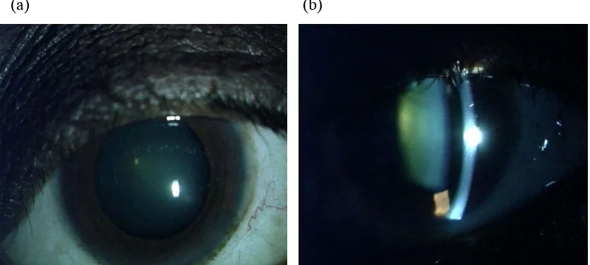
NUCLEAR CATARACT GRADE 3
(a) (b)
(c)
[image:63.595.98.516.152.340.2]
Figure 4. Anterior segment photographs of the right eye showing Grade 3 nuclear cataract
NUCLEAR CATARACT GRADE 4
(a) (b)
[image:64.595.94.511.130.319.2](c)
Figure 5. Anterior segment photographs of the right eye showing Grade 4 nuclear cataract
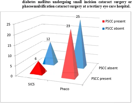
Figure 6. Occurrence o diabetes melli phacoemulsific
Percentage of diabetics in Percentage of diabetics cataracts= 50%
Percentage of diabetics cataracts=47.9%
Posterior subcapsular cata 43.9%, in the current study
0 5 10 15 20 25 SICS 6 51
f posterior subcapsular cataracts in pat itus undergoing small incision cataract s cation cataract surgery at a tertiary eye care
this study : 66 out of 520 = 12.7% in SICS group with posterior su
in Phaco group with posterior su
POSTERIOR SUBCAPSULAR CATARACT GRADE 1
(a) (b)
[image:66.595.97.503.153.336.2](c)
Figure 7. Anterior segment photographs of the right eye showing posterior subcapsular cataract Grade 1 on
(a) – Diffuse illumination (8X magnification) (b) – Slit beam illumination
53
POSTERIOR SUBCAPSULAR CATARACT GRADE 2
(a) (b)
[image:67.595.96.491.150.318.2](c)
Figure 8. Anterior segment photographs of the right eye showing posterior subcapsular cataract Grade 2 on
(a) – Diffuse illumination (8X magnification) (b) – on Slit beam illumination
POSTERIOR SUBCAPSULAR CATARACT GRADE 3
(a) (b)
[image:68.595.94.504.148.319.2](c)
Figure 9. Anterior segment photographs of the right eye showing posterior subcapsular cataract Grade 3 on
(a) – Diffuse illumination (8X magnification) (b) – on Slit beam illumination
55
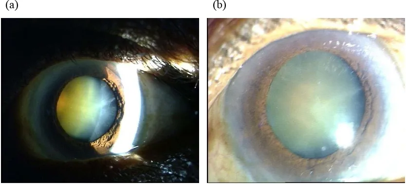
MATURE CATARACT
(a) (b)
(c)
Figure 10. Anterior segment photographs of the left eye showing mature cataract on
[image:69.595.94.506.149.342.2]HYPERMATURE CATARACT
(a) (b)
(c)
[image:70.595.94.517.117.324.2](
Figure 11. Anterior segment photographs of the right eye showing hypermature cataract (sclerotic type) on
Figure 12 : Association of of nuclear cat surgery at a te
* Posterior subcapsular catarac
57
f posterior subcapsular cataracts with vari aracts in patients undergoing small incisio rtiary eye hospital
ct (grade 2) not associated with nuclear catarac
ous grades on cataract
Figure 13 : Association of of nuclear cata cataract surge
* Posterior subcapsular catara ** Posterior subcapsular catara
posterior subcapsular cataracts with variou aracts in patients undergoing phacoemulsific
ry at a tertiary eye hospital
act (grade 1) not associated with nuclear catara act (grade 2) not associated with nuclear catara
us grades cation
59
The percentage of diabetics in the Phaco group with posterior subcapsular cataracts was 47.9%. Posterior subcapsular cataracts occurred
in 23 out of total 66 diabetics= 43.9%, in the current study (Fig. 6).
In the SICS group (total 130 patients), 54 patients (41.5 %) had different grades of posterior subcapsular cataract (PSSC) in association with nuclear cataract (Fig.11); 29 had PSSC1, 21 had PSSC 2 and six had
PSSC 3. In the Phaco group (total 390 patients), 195 (50%) had PSSC cataracts in association with nuclear cataracts (Fig. 12); 95 had PSSC 1, 78 had PSSC 2 and 21 had PSSC 3 (Fig. 12). These differences were not
statistically significant (χ2 with Yates’ correction [d.f.=1]=2.5 ; P=0.11).
1.3. 2 In Males versus Females
In males, NC2 cataracts were noted in 0.4 %, NC3 cataracts in 37%, NC4 cataracts in 26%, NC5 cataracts in 6.2% and mature / hypermature
cataracts in 30.4% ; in female patients, the percentages were 3%, 31%, 35%, 9% and 22%, respectively. Interestingly, these gender differences approached statistical significance (χ2 [d.f.=4] = 10.6, P = 0.03 : with
Yates’ correction, χ2 = 8.63, P = 0.07) (Table 2).
PSSC 3 in eight (11.2% of 71 males); in females the corresponding numbers (percentages) were 89 (49.7% of 179 females), 69 (38.5%) and 19
(10.6% of 179 females (Fig. 11 and 12). These differences were not statistically significant (χ2 with Yates’ correction [d.f.=1] = 1.26, P = 0.26).
1.4 Pre-operative and post-operative visual acuity
In the 130 patients who had undergone SICS, the post-operative mean visual acuity (decimals), 0.58 + 0.02, was significantly better than the pre-operative mean visual acuity of 0.07 + 0.01 (unpaired `t’ [d.f.=258]
= 21.6 ; P < 0.0001) (Table 3). This significant improvement was seen in both males and females (Table 3).
Similarly, in the 390 patients who underwent phacoemulsification surgery, the post-operative mean visual acuity (0.64 + 0.01) was
significantly better than the pre-operative mean visual acuity of 0.07 + 0.01 (unpaired `t’ [d.f.= 776) = 38.7 ; P < 0.0001) (Table 4). This significant improvement was seen in both males and females (Table 4).
61
versus Phaco [0.07 + 0.01] ; t = 0.28 [d.f.=519, P = 0.78), males (SICS [0.08 + 0.01] versus Phaco [0.06 + 0.01]; t = 1.25 [d.f. = 159], P=0.21) or
females (SICS [0.07 + 0.01] versus Phaco [0.07 + 0.01]) were compared. However, when post-operative mean visual acuities (decimal units) were analysed and compared, the following interesting observations emerged (Tables 3 & 4):
a) there was no significant difference between the post-operative mean visual acuity in the SICS and Phaco groups in males (SICS [0.67 + 0.04] versus Phaco [0.63 + 0.03]; t = 0.88 (d.f.=159), P = 0.38);
b) in females, the post-operative mean visual acuity in the Phaco group [0.64 + 0.02] was significantly higher (better) than that in the SICS
group [ 0.55 + 0.03] (t = 2.79 [d.f.=357] ; P = 0.0055).
Table 3 : Mean pre-operative and mean post-operative visual acuity in patients undergoing small incision cataract surgery at a tertiary eye care hospital Gender of patients Number of patients
Mean (+ SEM) visual acuity
(decimals) Statistical Analysis (Student `t’[paired]) Pre-operative Post-operative
Male 39 0.08 + 0.01 0.67 + 0.04 t = 12.6 (d. f.=38); P < 0.0001 Female 91 0.07 + 0.01 0.55 + 0.03 t= 17.8 (d. f.=90);
P < 0.0001 All 130 0.07 + 0.01 0.58 + 0.02 t = 21.6 (d. f.=129);
P < 0.0001 Statistical
Analysis(Student `t’ [unpaired]) Males versus Females
t=0.82 (d.f.=128)
P=0.42
t=2.52 (d.f.=128) P=0.01
Abbreviation SEM = Standard error of mean, d.f.= degree of freedom
Table 4 : Mean pre-operative and mean post-operative visual acuity in patients undergoing phacoemulsification cataract surgery at a tertiary eye care hospital
Gender of patients
Number of patients
Mean + SEM visual acuity
(decimals) Statistical Analysis (Student `t’[paired]) Pre-operative Post-operative
Males 122 0.06 + 0.01 0.63 + 0.03 t = 20.7 (d. f.=121); P < 0.001 Females 268 0.07 + 0.01 0.64 + 0.02 t= 33.1 (d. f.=267);
P < 0.0001 All 390 0.07 + 0.01 0.64 + 0.01 t = 38.74 (d. f.=389);
P < 0.0001 Statistical Analysis
(Student ‘t’[unpaired]) Males versus Females
t=1.03 (d.f.=388) P=0.30
t=0.41 (d.f.=388) P=0.68
[image:76.595.90.527.455.671.2]63
1.5 Surgical and post-operative complications, pre-existing pathology and combined surgery
The frequency of surgical and post-operative complications, pre-existing pathology and combined surgery was compared between the SICS and Phaco groups (Table 5). Complications were encountered in five (4%) of 130 patients in the SICS group and 17 (4.3%) of 390 patients in the
Phaco group, this difference was not statistically significant (χ2 [d.f.=1] = 0.06 ; P = 0.8) (Table 5). Pre-existing pathology was present in six (4.6%) of 130 SICS group patients and nine (2.3%) of 390 Phaco group patients
(Table 5). This difference was not statistically significant (χ2 [d.f.=1] =1.9; P = 0.17). Combined surgery was needed for 13 (10.0 %) of 130 SICS group patients and 17 (4.4 %) of 390 phaco group patients (Table 5); this difference was statistically significant (χ2 with Yates’ correction [d.f.=1] =
4.72 ; P=0.03).
When differences between males and females in frequency of occurrence of these parameters were compared, no statistically significant
Table 5: Complications, pre-existing pathology and combined surgery in
small incision or phacoemulsification cataract surgery at a tertiary eye care hospital
Parameter
In small incision cataract surgery (SICS) group
(total n = 130)
In phacoemulsification cataract surgery
(Phaco)group(total n= 390) Overall Total Males Females total Sub Males Females Sub
total
Surgical complications
2
3 5 7 10
17 22 Pre-existing
pathology 2 4 6 6 3 9 15 Combined
surgery 4 9 13 5 12 17 30 Group total 8 16
24 18 25 43 67
Statistical Analysis (Chi-square with Yates’ correction)
a) Combined surgery needed for 13 (10.0 %) of 130 SICS group patients and 17 (4.4 %) of 390 phaco group patients; χ2 with Yates’ correction [d.f.=1] = 4.72 ; P=0.03.
b) Phacogroup ; Pre-existing pathology in 6 of 122 males and 3 of 268 females
65
1.6 Post-operative visual outcome in relation to pre-operative cataract grade
1.6.1 In SICS group versus Phacoemulsification group
Different degrees of post-operative (PO) visual acuity (Snellen’s units) in relation to pre-operative cataract grade were compared in patients who had undergone SICS and in those who had undergone
phacoemulsification cataract surgery (Table 6). Of three patients in the SICS group who had had preoperative NC2 cataracts, two achieved PO visual acuity of 6/9 and one achieved 6/12, while of eight patients in the
Phaco group who had had preoperative NC2 cataracts, two each achieved PO visual acuities of 6/6, 6/9, 6/12 and ≥ 6/24 (Table 6).
Fifty-one patients in the SICS group who had had preoperative NC3 grade cataracts achieved PO visual acuities of 6/6 (13 [25.5%] of 51
patients), 6/9 (21 [41.2%]), 6/12 (seven [13.2%]), 6/18 (four [7.8%]) and 6/24 or worse (six [11.8%] of 51 patients) whereas 117 patients in the Phaco group who had had preoperative NC3 grade cataracts achieved PO
Table 6: Post-operative visual acuity versus preoperative nuclear cataract grades* in small incision or phacoemulsification cataract surgery at a tertiary eye hospital.
Grade of nuclear cataract (LOCS)
Post-operative (PO)visual acuity (Snellen) PO visual acuity(Snellen) Phaco group (total
no. = 390) Overall Total SICS group (total no. = 130)
6/6 6/9 6/12 6/18 6/24 ≥ Group total 6/6 6/9 6/12 6/18 ≥6/24 Group total
NC 2 - 2 1 - - 3 2 2 2 - 2 8 11
NC 3 13 21 7 4 6 51 30 51 14 8 14 117 168
NC 4 8 14 8 5 3 38 41 45 20 10 16 132 170
NC 5 1 3 1 1 2 8 11 8 7 2 5 33 41
MC/
HMC 4 9 9 2 6 30 28 41 11 11 9 100 130
Group
total 26 49 26 12 17 130 112 147 54 31 46 390 520
Abbreviations: LOCS= Lens Opacities Classification System; no. = number; SICS =
small incision cataract surgery; NC=nuclear cataract; MC/HMC= mature/hypermature cataract;
Statistical Analysis (χ2 [degree of freedom {d.f.}= 4] with Yates’ correction where
necessary)
1. NC 2 cataract : No. of patients in SICS group vs. Phaco group with PO visual acuities (statistical analysis not done)
2. NC 3 cataract : No. of patients in SICS group vs. Phaco group with PO visual acuities; χ2 = 0.19; P= 0.99
3. NC 4 cataract : No. of patients in SICS group vs. Phaco group with PO visual acuities; χ2 = 3.23; P= 0.5
4. NC 5 cataract: No. of patients in SICS group vs. Phaco group with PO visual acuities; χ2 = 2.36; P= 0.67
67
Thirty-eight patients in the SICS group who had had preoperative NC4 grade cataracts achieved PO visual acuities of 6/6 (eight [21%] of 38
patients), 6/9 (14 [36.8%]), 6/12 (eight [21%]), 6/18 (five [13.2 %]) and 6/24 or worse (three [8%] of 38 patients) whereas 132 patients in the Phaco group who had had preoperative NC4 grade cataracts achieved PO visual acuities of 6/6 (41 [31.1%] of 132 patients), 6/9 (45 [34.1%]), 6/12 (20
[15.2%]), 6/18 (10 [7.6 %]) and 6/24 or worse (16 [12 %] of 132 patients) (Table 6); these differences were not statistically significant (χ2
[d.f.=4]=3.23; P=0.5). Similarly, of eight patients in the SICS group and 33
patients in the Phaco group who had had