ANALYSIS OF IMMUNOHISTOCHEMICAL EXPRESSION
OF p
16INK4aIN PRENEOPLASTIC AND NEOPLASTIC
SQUAMOUS CELL LESIONS OF CERVIX
Dissertation submitted in
Partial fulfillment of the regulations required for the award of
M.D. DEGREE
in
PATHOLOGY – BRANCH III
THE TAMILNADU
DR. M.G.R. MEDICAL UNIVERSITY
CHENNAI
DECLARATION
I hereby declare that the dissertation entitled “Analysis of
Immunohistochemical Expression of p16INK4a in preneoplastic and neoplastic squamous cell lesions of cervix” was done by me in the
Department of Pathology, Chengalpattu Medical College from June
2014-August 2015 under the guidance and supervision of Dr.S.Ravi, M.D.,
Professor and Head, Department of Pathology, Chengalpattu Medical College.
This dissertation is submitted to the Tamilnadu Dr.MGR Medical
University, Chennai towards the partial fulfillment of the requirement for the
award of M.D. Degree in Pathology.
I have not submitted this dissertation on any previous occasion to any
University for the award of any degree.
Place:
CERTIFICATE FROM THE GUIDE
This is to certify that the dissertation entitled “Analysis of
Immunohistochemical Expression of p16INK4a in preneoplastic and neoplastic squamous cell lesions of cervix” submitted by the candidate
Dr.P.Sakunthala in partial fulfillment for the award of the degree of Doctor of
Medicine in Pathology by The Tamilnadu Dr.M.G.R .Medical University,
Chennai is a record of original work done by her under my guidance and
supervision in the Department of Pathology, Chengalpattu Medical College,
Chengalpattu during the academic year 2013-16.
Place:
Chengalpattu
Date:Dr.S.Ravi, M.D.,
Professor and Head,
Department of Pathology,
CERTIFICATE
This is to certify that the dissertation entitled “Analysis of
Immunohistochemical Expression of p16INK4a in preneoplastic and neoplastic squamous cell lesions of cervix” is a record of bonafide work
done by Dr.P.Sakunthala in the Department of Pathology, Chengalpattu
Medical College, Chengalpattu under the supervision of Dr. S. Ravi, M.D.,
Professor and Head , Department of Pathology and submitted in partial
fulfillment of the requirements for the award of M.D. Degree in Pathology by
The Tamilnadu Dr. MGR Medical University, Chennai. This work has not
previously formed the basis for the award of a degree or diploma.
Dr.K.Muthuraj, M.S ., Dr.S.Ravi, M.D.,
Dean, Professor and Head,
Chengalpattu medical college, Department of Pathology,
Chengalpattu. Chengalpattu Medical College,
ACKNOWLEDGEMENT
To think with, I thank the almighty GOD in making this project a
successful one.
I express my deep gratitude to Dr.K. Muthuraj, M.S., Dean,
Chengalpattu Medical College, for granting me permission to undertake this
study.
I profusely thank and express my sincere gratitude to Dr.S.Ravi, M.D.,
Professor and Head, Department of Pathology, Chengalpattu Medical College,
for having suggested this topic for dissertation and for having rendered his
valuable support and encouragement without which this project work would
not have been feasible.
I wish to record my sincere thanks to Dr.I.Vijay sathish kumar,M.D.,
Dr.S.Sasikala, M.D., Dr.K.R.Mohan, M.D., Dr.S.Premalatha, M.D.,
Associate Professors, Department of Pathology, Chengalpattu Medical
College, for their constant support and encouragement throughout my work.
I express my heartfelt thanks to Dr.G.Selvambigai, M.D., Associate
Professor, Department of Pathology, Government omanthoorar Medical
College, chennai for her support and valuable advice during the course of this
study.
I also wish to record my sincere thanks to Dr. M.Kuzhalmozhi, M.D.,
Dr. G.Devi Priya., Dr. S.Rohini Priya., Dr. V.Dhamotharan, MD.,
Assistant Professors Dr. T.Renuga Sarojini, DCP., Tutor of the
pathology,Department of Pathology, Chengalpattu Medical College, for their
constant support and encouragement throughout my work.
I extend my heartfelt thanks to all my colleagues for their timely help,
comments and support.
I thank all the technical staff in the Department of Pathology,
Chengalpattu Medical College, for their sincere and timely technical
assistance.
Also, I am indebted to my husband Mr.R.Pradeep kumar for his
constant support, encouraging words and source of strength all the way
through this endeavour.
To my lovable family members, I express my gratitude for their
extreme patience and tireless support while pursuing this study.
Last, but not the least I am indebted to all the patients who made it
possible for me to carry out this study. I extend all my support and wishes for
their healthy lives.
CONTENTS
Sl. No Particulars Page no.
1 Introduction 1
2 Aims and Objectives 5
3 Review of Literature 6
4 Materials and Methods 50
5 Observation and Results 56
6 Discussion 77
7 Summary 88
8 Conclusion 90
9 Bibliography 92
10 Annexures
I Proforma 110
II Master Chart 112
LIST OF TABLES
Sl. No. Tables
1 Types of Human papilloma virus with oncogenic risk
2 Risk factors for cervical cancer.
3 WHO histological classification of tumors of uterine cervix
4 Classification of HPV associated intraepithelial lesions of cervix
5 Natural history of CIN depending upon lesion grade
6 Bethesda system of cytological classification(2001)
7 TNM and FIGO classification of carcinoma of cervix uteri
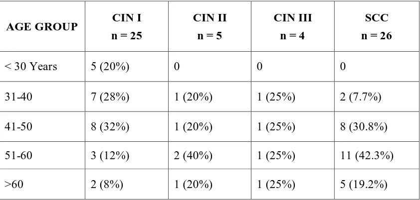
8 Age wise distribution of cervical squamous lesions
9 Incidence of preneoplastic and neoplastic lesions of cervix in relation to parity
10 Incidence of various symptoms in cervical squamous lesions
11 Correlation between visual screening methods and biopsy diagnosis
12 Degree of distribution of cervical squamous intraepithelial lesion in all cervical biopsies
13 Distribution of various histological subtypes of squamous cell carcinoma of cervix
14 Results of p
16INK4a
immunostaining in cervical squamous cell lesions
15 Grading of p16
INK4a
expression in preneoplastic and neoplastic squamous cell lesions of cervix
16 Correlation between histopathological diagnosis and reaction intensity in P16 staining
18 Comparison of incidence of parity among preneoplastic and neoplastic lesions of cervix
19 Comparison of incidence of various symptoms in CIN and SCC
20 Comparison of incidence of CIN with other studies
21 Comparison of distribution of various histological subtypes of SCC
22 Comparison of P16 positivity in CIN and SCC with other studies
23 Comparing Grade of P16 expression in squamous cell lesions of cervix
LIST OF FIGURES
Figure
No. TITLE
1 Development of cervix
2 Anatomy of cervix showing angles of antevertion and ante flexion
3 Vascular supply and the lymphatic drainage of the cervix
4 Schematic diagram of transformation zone
5 Transformation zone - HPE
6 Phylogenetic tree of Alpha – Human papilloma virus
7 Genomic organisation of HPV
8 Molecular basis for cervical neoplasia
9 Human papilloma virus life cycle
10 Koilocytic changes in the cervical squamous epithelium
11 Progression model of cervical carcinoma
12 Schematic diagram of cervical intraepithelial neoplasia
13 Age wise Distribution of cervical squamous lesions
14 Incidence of preneoplastic and neoplastic lesions of cervix in relation to parity
15 Incidence of various symptoms in cervical squamous lesions
16 Correlation between visual screening methods and biopsy diagnosis
17 Degree of distribution of cervical squamous intraepithelial lesion in all cervical biopsies
18 Distribution of various histological subtypes of squamous cellcarcinoma of cervix
19 Results of p
16INK4a
immunostaining in cervical squamous cell lesions
20 Grading of p16
INK4a
expression in preneoplastic and neoplastic squamous cell lesions of Cervix
21 Condition between histopathological diagnosis and reaction intensity in P16 staining
Figure
No. TITLE
23 Cervical intraepithelial neoplasia I (CIN I) . Negative p16 IHC staining
24 Cervical intraepithelial neoplasia I (CIN I) . p16 IHC staining Grade 1 staining
25 Cervical intraepithelial neoplasia I (CIN I) .p16 IHC staining Grade 2 staining.
26 Cervical intraepithelial neoplasia II H&E
27 Cervical intraepithelial neoplasia II (CIN II) . p16 IHC staining Grade 2,3 staining
28 Cervical intraepithelial neoplasia III (CIN III) . H&E
29 Cervical intraepithelial lesion III (CIN III).P16 IHC immunostaining Grade 3
30 Early invasive squamous cell carcinoma. H& E.
31 Early invasive squamous cell carcinoma.P16 immunostaining shows grade 3 immunostaining
32 Large cell keratinizing squamous cell carcinoma H& E.
33 Large cell keratinizing squamous cell carcinoma.P16 immunostaining shows grade2& 3 immunostaining
34 Large cell non keratinizing squamous cell carcinoma. H& E.
35 Large cell non keratinizing squamous cell carcinoma.P16 immunostaining shows grade 3 immunostaining
36 Small cell non keratinizing squamous cell carcinoma. H& E.
37 Small cell non keratinizing squamous cell carcinoma.P16 immunostaining shows grade 3 immunostaining
38 p16 immunostaining showing weak reaction intensity
39 p16 immunostaining showing moderate reaction intensity
ABSTRACT
BACKGROUND
Cervical cancer is the fourth most common cancer affecting women worldwide. Pap
smear screening and histopathological interpretation of cervical biopsy has significantly
reduced the number of deaths due to cervical cancer. However, they provide little or no
information regarding the association of HPV in precancerous lesion and invasive
cervical cancer. Hence the need to use biomarker to know the association of HPV in
those lesions in order to predict the risk of progression or regression and prognosis.
P16INKa is a surrogate marker of HPV which fulfils all the above criteria. OBJECTIVES
1. To evaluate the results of expression of p16INK4A in preneoplastic and neoplastic
lesions of cervix in order to assess the association of HPV infection in those
lesions.
2. To study the pattern of expression of p16 in various histological types of cervical
squamous cell lesions by p16 immunohistochemistry.
3. To compare p16 expression in various histological types of cervical squamous cell
MATERIALS AND METHODS
Immunohistochemical analysis of p16expression was performed on 60 paraffin
embedded tissue samples, obtained from cervical biopsy including 25CIN I, 5CIN II,
4CIN III and 26 SCC. Two parameters were evaluated in p16 expression: Percentage
ofp16 positive cells and reaction intensity of p16 immunostaining. The p16 expression
was graded as Negative, Grade 1, 2, 3 and its reaction intensity was graded as Negative ,
Weak, Moderate, Strong.
RESULTS
In the present study out of 60 cases, the incidence of squamous cell carcinoma
constituted majority (44%).Among CIN group , CIN I constituted majority of the
preneoplstic lesions of cervix.p16 expression was seen in 28% of CIN I, 80% of CIN II,
all CIN III and all SCC cases. Only one CIN I case show showed grade3 staining and
strong reaction intensity, but most of the CIN II(60%), CIN III(100%) and SCC(96.15%)
cases showed grade 3 staining. In our study there was a statistically significant correlation
CONCLUSION
In the present study out of 60 cases, 68.33% of cases showed p16 positivity. In
preneoplastic lesions, totally 44.12% of cases showed p16 positivity. In invasive
squamous cell carcinoma cases, 100% cases showed p16 positivity. So p16 may be useful
as an adjunct in histological sections to know the association of HPV in preneoplastic
lesions lesions to predict the risk of progression of the disease and to plan proper
treatment and in neoplastic lesions to predict the prognosis, since HPV negative SCC
showed poor prognosis in literatures.
In our study p16 expression was correlated well with increasing grade of CIN. So
p16 has significant implication in diagnostic, prognostic and preventive aspects of
cervical cancer.
KEY WORDS
P16INK4A, Cervical intraepithelial neoplasia, Immunohistochemistry, Human
1
INTRODUCTION
Cervical cancer is the fourth most common cancer and seventh overall
among women worldwide, with an estimated incidence of 5,28,000 cases and
2,66,000 deaths in 2012 and it is most frequent among women between 15 and
44 years of age.1Screening by pap smear has reduced the incidence of cervical cancer in developed countries, but implementation of this screening technique
has not been successful in developing countries. Developing countries carry
major burden of cervical cancer cases (85%) and deaths (88%) worldwide.
The incidence of this disease in India is around 1,23,000 cases and death
around 67,000 cases every year.2 So cervical cancer is considered as a public health problem and World Health Organization(WHO) gives priority to
cervical cancer control programmes.
Before the development of invasive squamous cell carcinoma of cervix,
there are certain stages of premalignant changes that occur in the cervical
epithelium which are described previously as dysplasia. They are now divided
into cervical intraepithelial neoplasia (CIN) I, II and III. Bethesda system
classifies these abnormalities into low grade squamous intraepithelial lesion
(LSIL) and High grade squamous intraepithelial lesion (HSIL) based on the
morphology. The LSIL encompasses condyloma and CIN I, whereas HSIL
encompasses CINII and CIN III. All LSIL cases will not directly progress into
invasive squamous cell carcinoma. Most cases of LSIL regress spontaneously.
But all HSIL cases are considered to be at high risk for progression to cervical
2
It is well known that the main causative factor for both precancerous
and invasive cervical cancer is persistent infection with one or more oncogenic
types of Human papilloma virus(HPV).3In addition to the infection with HPV, there are several cofactors have been associated with the increased risk of
persistent infection of high-risk HPVs and progression to preneoplastic and
neoplastic lesions of cervix, including Viral infections like HIV, Herpes
simplex virus–2 (HSV-2)4, Smoking5, Dietary deficiencies6, Immunosupression7, Hormonal contraceptives, family history and sexually associated factors like multiple sexual partners, Early sexual activity, Sexually
transmitted diseases, Multiple pregnancies.
Experimental studies have identified nearly 200 types of Human
papilloma viruses, of those more than 40 have been identified in the genital
tract.8 These are divided into those with low risk and high risk categories based on the association with invasive cervical carcinoma.HPV16, 18, 31, 33
and 45 are examples of high-risk types, while HPV6 and 11 belong to the
low-risk types. In a large epidemiological study conducted in India showed that
genotypes 16 and 18 either alone or together were detected in 76.3% of
cervical cancer cases followed by genotype 33.9
The two viral oncoproteins in HPV namely E6 and E7 are mainly
responsible for the progression of neoplasm. The E6 oncoprotein of high risk
HPV causes degradation of p53, a tumor suppressor gene thus preventing cell
cycle arrest or apoptosis. Similarly HPV E7 oncoprotein bind and inactivates
the tumor suppressor protein pRB ( Retinoblastoma protein), which normally
3
P16INK4a (henceforth referred to as p16) is a tumor suppressor protein that inhibits cyclin dependant kinase 4 and 6, which phosphorylate the RB
protein. A reciprocal relation between p16 and pRB expression has been seen,
suggesting the presence of negative feedback loop allowing pRB to limit the
concentration of p16. So functional inactivation of pRB by the HPV E7
oncoprotein results in over expression of p16.
p16 protein is detectable immunohistochemically, over expression of it
may serve as a surrogate biomarker of HPV infection which makes it useful in
evaluating HPV- associated preneoplastic and neoplastic lesions of cervix.
Many literatures have given evidence that p16 may be a very useful
marker for preneoplastic, neoplastic squamous lesions and glandular dysplasia
of cervix. Moreover, expression of p16 appears to correlate with degree of
cervical neoplasia.10
Many countries have started vaccination against HPV 16 and 18 and
mainly targeted towards adolescent girls. Current vaccines provide excellent
efficacy not only against HPV16and 18, but also provide cross protection
against non vaccinated types. However HPV vaccines do not protect against
all invasive forms of cervical cancer.11, 12 In developing countries like India, it is necessary to vaccinate all adolescent girls especially in HPV high
prevalence area.
Although Pap smear screening test is the easily available test used
widely, the gold standard for diagnosis of cervical neoplasm is
4
adjunct to morphological examination, we can recognize the high risk type of
HPV infection in those lesions which may progress to high grade lesion.
p16 Immnostaining is a new and cost effective and easily available
method which gives valuable information regarding the HPV infection without
the need for molecular techniques such as Polymerase chain reaction( PCR ),
Southern blotting, or Insitu hybridisation ( ISH ).
Although there are several previous reports on the role of p16 in
cervical cancer, there is paucity of them in Indian literature in spite of the fact
that cervical cancer is one of the most common cancers among females in
India.
This study is an attempt to analyze the association of HPV infection in
and around Chengalpattu by using p16 immunostaining in preneoplastic and
neoplastic squamous cell lesions of cervix and evaluate its etiological and
5
AIM AND OBJECTIVES
1. To evaluate the results of expression of p16 in preneoplastic and neoplastic
lesions of cervix in order to assess the association of HPV infection in
those lesions.
2. To study the pattern of expression of p16 in various histological types of
cervical squamous cell lesions by p16 immunohistochemistry.
3. To compare p16 expression in various histological types of cervical
6
REVIEW OF LITERATURE
Epidemiology
Cervical cancer has become a serious public health issue, being the
fourth most common cancer in women worldwide .1There is a drastic difference in incidence rate and prevalence of cervical cancer between
developed and developing countries. Many developed countries have become
successful in reducing the cancer burden over the past six decades through
screening programme and other diagnostic workup. Because of the lack of
proper health facilities, cervical cancer is leading in developing countries like
India.
The age-standardized incidence and mortality rate of cervical cancer in
India are 27.0 and 15.2, respectively.13 An estimated incidence of 1,23,000 cases and 67,000 deaths due to cervical cancer occurred in India in 2012,
contributing 23.2% and 25.2% to the global cervical cancer incidence and
mortality respectively.14It has been estimated that there will be around 205496 new cases and 119097 deaths due to cervical carcinoma by 2020 in India,
contributing to 29% and 30% respectively of the global burden of cervical
cancer cases and mortality.15Cytological screening by pap smear is a very useful test but false positive (15 – 50%) and false negative rates (30%) are
high. So histopathological examination of cervical biopsies is regarded as a
7
Embryology of Cervix:-
Female reproductive tract organs including uterus, cervix, uterine tubes,
and upper part of vagina are developed from mullarian duct, otherwise called
as paramesonephric ducts, are a pair of ducts which are present in the
intermediate mesoderm. They are formed by invagination of coelomic
epithelium. The paramesonephric duct consists of upper vertical part, middle
horizontal part and lower vertical part. The upper vertical part lies lateral to
the Wolffian duct. Middle horizontal part crosses in front of the Wolffian duct.
Both upper and middle part forms the fallopian tube. Lower vertical part fuses
with the similar part of the opposite side to form uterovaginal canal in which
upper part forms the body of the uterus and cervix, while the lower part forms
the upper 4/5 th of vagina. The mullarian ducts meets the endoderm derived
urogenital sinus at mullarian tubercle which meet a pair of endodermal
[image:25.595.210.425.483.698.2]sinovaginal bulbs which forms the lower 1/5 th of the vagina.16
8
Gross Anatomy:-
The uterus is divided into body of the uterus (corpus uteri) which forms
the upper two-third, and the cervix (cervix uteri) which forms the lower third.
In the adult nulliparous state the cervix tilts forwards relative to the axis of the
vagina, called as ante version, and the body of the uterus tilts forward relative
to the cervix called as ante flexion. The cervix measures 2.5 cm in length in
the adult nulligravida. The lower part of the cervix projects into vagina which
divides it into supravaginal and vaginal parts. The parametrium separates the
supravaginal portion of the cervix anteriorly from the bladder and also passes
laterally between the anterior and posterior layers of the broad ligaments. The
vaginal part of the cervix projects into the anterior vaginal wall. The spaces
between this part and the vaginal wall are called the vaginal fornices. Through
the internal os the upper end of the cervix communicates with the uterus and
through external os the lower end of the cervix opens into the vagina. The
vaginal portion of the cervix is called as ectocervix and the portion related to
the endo cervical canal is known as endo cervix. In nulliparous women, the
external os is small and circular, whereas after childbirth become a transverse
9
Figure 2: Anatomy of cervix showing angles of antevertion and ante
flexion
VASCULAR SUPPLY OF THE CERVIX
Arterial supply
The descending branches of the uterine arteries, reaches the lateral wall
of the cervix along the upper margin of the paracervical ligaments and
supplies the cervix
Venous drainage
The veins from the cervix form a cervical plexus and run along the
lateral border of the uterus. The cervical plexus drains through the uterine,
10
LYMPHATIC DRAINAGE
Lymphatics from the cervix pass laterally in the parametrium and drain
into four efferent channels running toward the external iliac and obturator
nodes, the hypo gastric and common iliac nodes, the sacral nodes, and the
[image:28.595.188.475.249.498.2]nodes of the posterior wall of the urinary bladder.
Figure 3:Vascular supply and the lymphatic drainage of the cervix.20 NERVE SUPPLY
The cervix is supplied by nerves from the pelvic autonomic system, the
superior, middle and inferior hypo gastric plexuses. Pain sensation from the
cervix passes along the parasympathetic nerves. The nerve supply is mainly
restricted to the endocervix and peripheral deep portion of ectocervix. This
11
LIGAMENTS OF CERVIX
The paracervical ligaments, otherwise called as mackenrodt’s ligament
and the uterosacral ligaments, attach the supra vaginal portion of the cervix to
the second vertebrae through fourth sacral vertebrae, are the greatest sources
of fixation and support of the cervix.
HISTOLOGY OF CERVIX
The cervix has a covering epithelium and an underlying stroma. The
stroma is an admixture of fibrous, muscular, and elastic tissue. Most of the
ectocervix is covered by non keratinizing stratified squamous epithelium, and
is composed of three layers of squamous cells: Basal/ parabasal, intermediate,
superficial cells. The basal cell layer is one cell thick, with scant cytoplasm
and oval nuclei oriented perpendicularly to the basement membrane. The
nuclear cytoplasmic ratio decreases progressively from the basal to superficial
cells during normal maturation. The parabasal cells are larger than basal cells
and have more cytoplasm. The cells in the midzone are called as intermediate
cells. The superficial cells have abundant cytoplasm and a pyknotic nuclei
than the intermediate cells, and they orient with their longest axis parallel to
the basement membrane. The morphological appearance of this various layers
varies with age. The cells are become atrophic and exhibit high nuclear
cytoplasmic ratio during post menopausal period.
The Endocervix and the endocervical glands are lined by mucinous
columnar epithelium. The endocervical glands, represents infoldings of the
12
The junction between squamous and mucinous epithelia is known as
squamocolumnar junction (SCJ) 22.At birth, SCJ is located on the endocervix which is called as original squamocolumnar junction. Under the influence of
estrogen at puberty and pregnancy, the endocervix everts to expose the
columnar epithelium, glycogenisation of the epithelium takes place,
lactobacilli colonize the epithelium and the PH becomes acidic. These changes
stimulate the columnar epithelium to undergo metaplasia and convert into
immature squamous and later mature squamous epithelium. With these
changes the histologic squamocolumnar junction moves to the external os and
this is called as functional or new columnar junction. The area between the
original SCJ and the new SCJ is the transformation zone where columnar
epithelium is slowly replaced by active metaplasia and this the area where
most cervical preneoplastic and neoplastic lesions develop.23
13
Figure 5 : Transformation zone of uterine cervix.
25ETIOLOGY AND PATHOGENESIS
Human papilloma virus
It is now well accepted that cervical cancer is caused by Human
papilloma virus. Almost all cervical cancer are directly associated with
infectivity with one or more of the oncogenic types of HPV. Approximately
7.9% of women in the general population are estimated to harbor cervical
HPV infection at a given time. About 82.5% of invasive cervical cancers are
attributed to HPV16 or HPV18. All cervical squamous cell cancers as well as
distinct subsets of vulvar, vagina, anal and oral cancers among women and
penile anal and oral cancers among men are causally associated with HPV
14
Epidemiology and Natural history of Human papilloma virus infection
Papilloma viruses are classified as members of papovaviridae family.
These are circular double-stranded DNA viruses with approximately 8000
base pairs in length and measures 45 – 55nm in diameter. Its icosahedral
capsid composed of 72 capsomers.26Papilloma viruses are epitheliotrophic
viruses, means it predominantly infect skin and mucous membrane.27
Many lines of evidence have demonstrated that the association between
HPV and many types of cervical diseases ranging from the innocuous
condyloma acuminatum to fatal invasive squamous cell carcinoma.28,29,30 At present about 200 types of HPV have been identified and it can be further
divided into high- and low-risk types depending on their carcinogenic
potential.31,32,33.
A large epidemiologic study by Munoz et al observed data from nine
countries has identified15 high risk HPV types (16, 18, 31, 33, 35, 39, 45, 51,
52, 56, 58, 59, 68, 73, 82),three probably high risk types(26,53,66) and low
risk types (6, 11, 40, 42, 43, 44, 54, 61, 70, 72, 81 and cp6108).34Of the HPV types infecting the anogenital mucosa, 12 types have been classified as group
1 carcinogens to humans and one is probably of carcinogenic (Group 2A)
type. All these 13 HPV types, and also several other possibly carcinogenic
types (Group 2B), belong to the same evolutionary branch of the alpha genus
15
Figure6 : Phylogenetic tree of Alpha – Human papilloma virus.HPV types in red clade are associated with CIN 3 and cervical cancer.HPV types in blue clade cause genital warts. HPV in green clade cause commensal infections.38
International Agency for Research Cancer (IARC) classified thirteen
anogenital HPV as oncogenic, based on their association with cervical and
anogenital cancer which are HPV16,18,31,33,35,39,45,51,52,56,58,59 and
66.39In a study by Bosch et al, it was concluded that majority of HPV infections are transient and clear within few months. Persistent high risk HPV
infection of the cervical epithelium triggers neoplastic proliferation.40, 41
A meta analysis of HPV types in women with LSIL found that HPV was
detected in 80% of the LSIL from North America and approximately 70% of
LSIL from other regions of the world.42
16
TABLE 1: Types of Human papilloma virus with oncogenic risk. 43 Low oncogenic risk 6, 11, 42, 43, 44, 53
High oncogenic risk 16, 18, 31, 33, 35, 39, 45, 51, 52, 56, 58, 59, 66
Unclear oncogenic risk 26, 68, 73, 82
A number of studies have demonstrated that prevalence of HPV 16
from different regions of the world range from 30% to 70%.42, 44 HPV 16, 18 and 31 have been mostly associated with invasive cervical cancer.31,45A large epidemiological study conducted by L loveras et al , evaluate the distribution
of HPV types in invasive cervical cancer and showed HPV types 16,18,31,33
and 45 strains cause 85% of the invasive cervical carcinoma worldwide.46
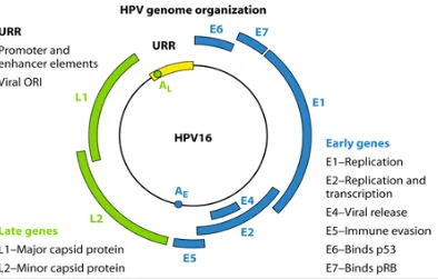
Genomic organization of Human papilloma virus
HPV – DNA consist of distinct three different regions. They are early
region (ER), late region (LR), upstream regulatory region (URR).The Early
region is composed of seven genes,E1 – E7,which play a significant role in
viral replication and have oncogenic properties. Late region is composed of
two genes, namely L1, the major capsid protein, which can self assemble into
virus like particles which are used for the generation of the currently available
VLP based HPV vaccines, and L2, the minor capsid protein that is thought to
facilitate encapsidation of viral DNA and viral infectivity, The URR is the
regulatory region which contains binding sites for both viral and cellular
17
Figure 7 : Genomic organisation of HPV.47
In preneoplastic lesions of cervix the HPV DNA is not integrated into
the host DNA, rather it is found in circular or episomal form. The episomal
HPV produces mostly the E2 protein. This E2 protein encodes for a DNA
binding protein that binds to a specific nucleotide motif found in E6 and E7
region. 48,49 E2 regulates the expression of E6 and E7.So that only minimal amount of E6 and E7 is produced. When the episomal form integrates into the
host chromosome at E1/E2 region, causing break in this region, results in
uncontrolled production and expression of E6 and E7 proteins. This E6 protein
forms a complex with p53 tumor suppressor protein leading to degradation of
p53.50The viral E7 oncoprotein binds to the Retinoblastoma protein (pRb),a tumor suppressor protein and inactivates it. Inactivation of pRb mediates
release of transcription factor E2F,which activates the genes necessary for
18
an increase in INK4A gene transcription, the INK4A gene product, the p16
INK4A protein (p16).
P16, a tumor suppressor gene, located on chromosome 9p21, is belongs
to the inhibitors of cyclin dependant kinase (CDK) 4 family. p16INK4ais named
after its molecular weight (15,845) and its role in inhibiting CDK4.Normally
CDK4 and CDK6 binds cyclin D and forms an active protein complex ,which
phosphorylate retinoblastoma protein(pRb) .The phosphorylation of pRb
induces release of transcription factor E2F from its bound state allowing it to
enter into the nucleus. Once in the nucleus E2F promotes the transcription of
target genes that are essential for cell cycle progression. p16 binds to the
CDK4 and CDK6 and preventing its interaction with cyclin D. This interaction
ultimately inhibits the downstream activities of transcription factors, such as
E2F and arrest G1-S transition. So inactivation of pRB by E7 causes over
expression of p16, because p16 is regulated by negative feedback of pRB.51 p16 expression is not associated with proliferation, but it is associated with
senescence and cell cycle arrest. So p16 expression is not seen in normal cells
19
Figure 8 : Molecular basis for cervical neoplasia . This figure shows three pathways involved in HPV related tumorigenesis including alteration in the cell cycle activity induced by E7, up regulation of telomerase via E6 with loss of replicative senescence, and induction of centrosome instability by E6 and E7 with HPV 16 as a model. Progression is also associated with promoter methylation of tumor suppressor gene.52
In a recent study by Iana Leniskova et al concluded that, p16
expression was not seen in normal cervical tissue, but its expression was
increased in following frequency : CIN 1 (180/249; 72.3%),CIN2 (212/233;
20
Life Cycle of Human papilloma virus
HPV initially infects the basal cells or immature squamous cells. It
enters into the basal cell layer through defects in the epithelium and remains
within the cell in two distinct biological states. In one form HPV continues to
remains in the basal cells, without producing virions, this is referred as latent
infection. In other form viral DNA replication occurs independently of host
chromosomal DNA synthesis, results in large amount of viral DNA formation.
Viral DNA replication mainly occurs in intermediate and superficial squamous
cells which show the distinct cytological and morphological abnormality,
including acanthosis, koilocytosis, multinucleation and nuclear pleomorphism.
21
Figure10: Koilocytic changes in the cervical squamous epithelium.54
HPV E7 protein is entrapped inside the nucleus. So it is not exposed to
the antigen presenting cell, moreover it inhibits the function of interferon
and . HPV viral proteins which are recognized by dentritic cell are carried to
the lymph nodes and also presented to the T cells. The CD4 and CD8 T cells
are activated and reach the infected site to destroy the virus. But the Human
papilloma virus expose only few viral particles to immune surveillance
mechanism, therefore it resides for many years without clinical recognition
Other Risk Factors
Infection with high risk HPV virus is necessary factor for the
development of cervical cancer, it is not sufficient for the development of
cervical cancer. Because only a small proportion of women exposed to HPV
develop cervical cancer, suggest that additional cofactors are necessary in the
pathogenesis of cervical neoplasia. These factors may modify the risk in
22
Smoking
Szarewski et al concluded that there is a positive association between
cigarette smoking and the development of cervical cancer. Some of the studies
demonstrated that smoking may be a risk factor only for squamous cell
carcinoma, not for adenocarcinoma of cervix.55, 56
Oral Contraceptives
A Meta analysis of 28 studies concluded that the relative risk of
invasive cervical cancer increased with increasing duration of contraceptive
use. There is no associated risk for intraepithelial lesion if it is used less than 5
years but the risk increases to 3 times and 4 times higher if they use oral
contraceptives for 5 – 9 yrs and more than 10 years respectively. A large
reanalysis of epidemiological studies conducted in more than 50,000 women
has confirmed that oral contraceptive use increases the risk of cervical
cancer.57, 58.
Infections other than HPV
Sexually transmitted infections, especially Chlamydial infection is
found to be one of a risk factor. Viral infections like Human
immunodeficiency virus (HIV) ,Herpes simplex virus -2also plays an
important role in the development of cervical neoplasm. The risk of
developing cervical cancer is 9.2 times more in women infected with HIV than
23
Immunosuppression
Immunity determines whether the patient is cleared of HPV infection or
whether it is persist and progress into malignancy. Studies showed a relative
risk of 13.6 for the development of cervical carcinoma in situ in renal
transplant recipients compared to women in the general population.60 Because of the immunosupression in HIV status ,they are more prone to develop
cervical cancer than women in general population.
Sexually associated factors
Multiple sexual partners, early age at first intercourse, early marriage,
male sexual behavior, concurrent penile cancer in males are also an important
sexually associated factors in pathogenesis of cervical neoplasm.
Other factors
There are various other risk factors associated with development of
cervical cancer. They are dietary deficiency, early age at first pregnancy,
24
Table2: Risk factors for cervical cancer: HPV infection vs. persistence
and Malignant transformation
Risk factor HPV infection HPV persistence And transformation
Multiple sex partners + n.e.
Partner’s multiple partners + n.e
Poor hygiene + n.e
Absence of male
circumcision + +
Immunodeficiency, HIV + +
High parity n.e +
Oral contraceptives n.e +
Smoking n.e +
STDs other than HPV n.e +
Poor nutritional status n.e +
STDs = Sexually transmitted diseases (especially C, trachomatis). n.e = No evidence for being a risk factor at this time.
Studies in transgenic K14E7 mouse models showed that estrogen
receptor is required for the initiation and maintenance of cervical cancer .63 Another study demonstrated that HPV oncogenes promote squamous
25
TABLE 3:WHO Histological classification of tumors of the uterine cervix
I EPITHELIAL TUMORS
:-1. Squamous Tumours and precursors
1A Squamous cell carcinoma, not otherwise specified Keratinizing Non -Keratinizing Basaloid Verrucous Warty Papillary
Lymphoepithelioma – like Squamotransitional
1B. Early Invasive squamous Cell Carcinoma 1C. Squamous intraepithelial neoplasia
Cervical intraepithelial neoplasia
Squamous Cell Carcinoma insitu
1D. Benign Squamous Cell lesions
Condyloma acuminatum Squamous Papilloma Fibro Epithelial polyp
II Glandular tumours and precursors
III Other epithelial tumours
IV Mesenchymal tumours and tumour like conditions
V Mixed epithelial and mesenchymal tumours
VI Melanocytic tumours
VII Miscellaneous tumours
VIII Lymphoid and haematopoietic tumours
26
PRENEOPLASTIC LESIONS OF CERVIX
Natural history of cervical intraepithelial neoplasia
Genital HPV lesions are more common among women in reproductive
age group, but most of them are asymptomatic. On average 50% of the
infections cleared within 8 months and 90% of the HPV infections cleared
within two years. Persistent high risk HPV type infection is the major risk
factor for the development of both squamous cell carcinoma and
adenocarcinoma of the cervix.33However, the natural history of squamous cell carcinoma of cervix is well understood than adenocarcinoma.66 The development of cervical cancer is a multistep process in which precancerous
lesions persist, progress and regress overtime except the last step leading to
invasive lesion is not reversible.
The precancerous lesions of cervix are usually described as cervical
intra epithelial neoplasia. 67The cellular changes in precancerous lesions of cervix involves nuclear atypia, increased nuclear cytoplasmic ratio, mitotic
activity limited to the surface epithelium and do not extend beyond the base
ment membrane. There are different classification systems for cervical
precursor lesions which are used interchangeably overtime. Older
Papanicolaou classification used the term as ‘atypical cells with abnormal
features’. Another classification system grouped the lesions into mild,
moderate, severe dysplasia and carcinoma in situ. The cellular changes are
limited to lower one third of epithelium in mild dysplasia, extend to middle
one third in moderate dysplasia and to upper one- third in severe dysplasia.
27
cervical intraepithelial neoplasia (CIN) classification. In this classification
mild dysplasia were termed CIN I, moderate dysplasia CIN II, and severe
dysplasia and carcinoma insitu termed CIN III. This three tier classification
system has been recently simplified into two tier system , with CIN I ,
condyloma acuminatum, Exophytic condylomas and squamous papilloma are
coming under low grade squamous intraepithelial lesion (LSIL) and CIN II
,CIN III and carcinoma insitu combined into one category called as high grade
squamous intraepithelial lesion (HSIL).68
Table4 : Classification of HPV associated intraepithelial lesions of
cervix.69
Term HPV risk
category
Comparison of classification systems
Two-tiered
CIN Dysplasia/CIS SIL
Exophytic
condyloma Low risk _______ _______ LGSIL
Squamous
papilloma Low risk _______ _______ LGSIL
Flat
condyloma
Low and
high risk _______ _______ LGSIL
CIN 1 Low and
high risk
Low grade
CIN Mild dysplasia LGSIL
CIN 2 High risk High grade
CIN
Moderate
dysplasia HGSIL
CIN 3 High risk High grade
CIN
Severe
dysplasia/ CIS HGSIL
CIN = Cervical intraepithelial neoplasia SIL = Squamous intraepithelial lesion CIS = Carcinoma in situ
28
Generally more than 80% of LSIL lesions and 100% of HSIL lesions
are associated with high risk HPV types. A study conducted by Moscicki et al
,showed in terms of CIN lesions of any grade, up to 90% regress
spontaneously in women aged 13 to 22 years , whereas among women 34
years and older, the estimated risk of regression is only 40%. In Boyes et al
study, 77% of the most severe preinvasive lesions, carcinoma in situ, regressed
spontaneously among women younger than 40 years-old whereas the
estimated rate of regression is 61% among women aged 40 and older.
McCredie et al undertook study in New Zeeland reported that 20% of women
with untreated CIN3 lesions developed cancer cervix within 10 years and 31%
within30years. Generally, the median time from initial exposure to HPV to the
development of carcinoma in situ is at least 7 to 12 years.70
Table5: Natural history of CIN depend upon lesion grade.71
% Regression % Persist Progress to CIS
CIN 1 57 32 11
CIN 2 43 35 22
29
Figure 11 : Progression Model of Cervical Carcinoma
Figure 12 : A classic schematic diagram of cervical intraepithelial
neoplasia (lower) defines the cytopathologic (A -E) and histopathologic
[image:47.595.167.475.392.657.2]30
Low grade squamous intraepithelial lesion (LSIL)
Normal squamous cell nuclei become smaller when they mature and
move towards the surface. In low grade squamous intraepithelial lesion also
cells are mature squamous cells with polygonal shape, but nuclei are enlarged
at least 3 – 4 times, that of normal intermediate cell nucleus. The cells become
smaller and the nuclei become smaller and pyknotic, irregular nuclear contour
sometimes with bi/multinucleation and cytoplasmic halos when HPV changes
are evident. These pyknotic nuclei may also exhibit features like increased size
that of the normal superficial squamous cell and a mild nuclear atypia, fine to
coarsely granular and evenly distributed chromatin, hyperchromasia. Bi
nucleated cells are present in 90% of the LSIL and when they surrounded by
cytoplasmic halo are termed koilocytes.52
Condyloma acuminatum, Immature condyloma / squamous papilloma,
Flat condyloma are the three morphological subsets of LSIL.
Moreover, LSIL do not progress directly to invasive cervical cancer,
because most cases regress spontaneously, only few cases progress to HSIL.
So LSIL is not treated like a premalignant condition.
High grade squamous intra epithelial lesion
High grade squamous intraepithelial lesion is characterized by presence
of atypical cells with nuclear pleomorphism, irregular nuclear contours and
31
mitosis in the upper half of the epithelium and abnormal mitotic figures. If
these abnormalities involve one third to two third of mucosal thickness, it is
said to be Cervical intra epithelial lesion 2(CIN2),and more than two- thirds ,it
is called as cervical intraepithelial lesion 3(CIN3).Apart from the above,
nuclear cytoplasmic ratio is considered as a important criterion to diagnosis of
HSIL. The nuclei in LSIL can be markedly enlarged and pleomorphic but have
low nuclear cytoplasmic ratio, whereas the nuclei in HSIL are more uniform
but with irregular nuclear contours and high nuclear cytoplasmic ratio. LSIL
involves superficial layers of the mucosa, whereas in HSIL the atypical cells
extend upwards from the basal layer to at least one third of the mucosal
thickness.
Early invasive (micro invasive) squamous cell carcinoma
It is defined as stromal invasion of malignant squamous cells by less
than or equal to 3mm in depth and 7mm in length. But assessment of this early
stromal invasion is very difficult. The criteria for the diagnosis of early
invasion include
1. Desmoplastic response in the adjacent stroma
2. conspicuous maturation of malignant squamous epithelium
3. Blurring of epithelial stromal interface
32
Invasive squamous cell carcinoma
The most common malignant tumor of female genital tract in both
developed as well as developing countries is invasive squamous cell
carcinoma of cervix. The role of HPV in the pathogenesis of all squamous cell
carcinoma has become obvious, which was discovered by Harald zur Hausen,
for that he was awarded the Noble prize in 2008.
Invasive squamous cell carcinoma of cervix can be classified by grade (
well differentiated, moderately differentiated and poorly differentiated) and/or
by morphology ( Large cell keratinizing, Large cell non keratinizing and
small cell non keratinizing) . WHO now recommend two tiered classification
as keratinizing and non keratinizing tumors to avoid confusion with small cell
carcinoma. But the grade and type have not found to be prognostically
significant, instead, the depth of invasion, size, lymphatic or vascular invasion
are the important prognostic variables.
MORPHOLOGY
Gross
Grossly, invasive cervical carcinoma may be polypoid or fungating or
deeply infiltrative. Infiltrative carcinomas invade adjacent structures more
33
MICROSCOPY
Keratinizing
These tumors are considered as well differentiated tumor , shows
conspicuous evidence of keratinization in the form of keratin pearls,
keratohyaline granules, individual keratinized cells and nests of squamous cell
with central keratinization.The nuclei are large, hyper chromatic with coarse
chromatin. Mitotic figures are not commonly seen.
Non keratinizing
These tumors are composed of large squamous cells which are
polygonal in shape with eosinophilic cytoplasm but lack the evidence of
keratin pearls. Cellular and nuclear pleomorphism is more obvious with
numerous mitotic figures. Non keratinizing carcinomas are typically
moderately differentiated.
Basaliod
Some invasive squamous cell carcinoma have nests of basal type
squamous cells having scant eosinophilic cytoplasm and peripheral palisading
of nuclei with variable amount of squamous differentiation. These carcinomas
34
OTHER RARE VARIANTS OF SQUAMOUS CELL CARCINOMA
Verrucous
These tumors are exophytic and composed of broad based papillae
lined by squamous epithelium with little or no atypia. These tumors have
pushing margin.
Warty or condylomatous
These tumors are exophytic squamous cell carcinoma that have
koilocytic surface epithelial changes characteristic of HPV infection.
Papillary
This type of tumor is characterized by a papillary growth pattern and it
is subdivided into three histological subtypes.
1. Papillary undifferentiated carcinoma: - In this carcinoma, the tumor cells
lining the papillae do not show histological evidence of specific type of
differentiation.
2. Papillary transitional cell carcinoma: - Has a similar histologic appearance
to lesions that occur in the urinary tract.
3. Papillary squamotransitional carcinoma:-which has a combination of
35
Lymphoepithelial – like
It resembles undifferentiated nasopharyngeal carcinoma and consists of
poorly defined aggregates of nonkeratinizing tumor cells with large vesicular
nuclei, prominent nucleoli and moderate amount of eosinophilic cytoplasm,
syncytial appearance and heavy lymphocytic infiltration.73
DIAGNOSIS AND AIDS TO DIAGNOSIS
Diagnosis is done by
- History
- Physical examination
- Investigations
History
Any women of reproductive age presenting with abnormal uterine
bleeding, post coital bleeding, white discharge, pelvic pain, mass per vaginum,
urinary or bowel symptoms should suggest the possibility of cervical cancer.
Physical Examination
General examination – Cachexia, pallor , supraclavicular and inquinal
nodes
Systemic examination
Abdominal examination – Ascites, Enlarged uterus, Hepatomegaly
Speculum examination – Growth on the cervix (Type of growth, Bleeds
36
Investigations
- Pap smear
- HPV testing
- Cervical biopsy
- Cystoscopy / proctoscopy/IVP
- USG/CT / MRI
- Complete blood count, liver function test, Renal function test. 74
PREVENTION AND EARLY DETECTION OF CERVICAL CANCER
Prevention of cervical cancer consists of creating awareness about the
risk factors through health education, promoting practice of safe sex, use of
condoms to prevent STDs, lifestyle modification, screening and early
treatment of premalignant lesions and HPV vaccines.
Cervical cancer screening
Several screening methods are available, but cytology (Pap smear) is
the most widely used method.
Methods used for cervical cancer screening
Cytology
Conventional cytology ( Pap smear )
Liquid based cytology (LBC)
37 Automated screening
Visual inspection after acetic acid(VIA )
Visual inspection after acetic acid with magnification(VIAM )
Visual inspection after Lugol’s iodine(VILI)
Cervical biopsy
HPV testing
Investigational strategies
Polar probe
Laser – induced fluorescence
HPV detection techniques
1. Immunohistochemistry
2. Southern Blot
3. Dot Blot assays
4. In situ Hybridization
5. Hybrid capture 2 assay (HC2)
6. Polymerase chain reaction
7. HPV genotyping
38
The only test presently approved by U.S.Food and Drug Administration
is the Hybrid capture 2 assay (HC2) test..But it is not widely available. Among
the above mentioned tests , immunohistochemistry, In situ Hybridization,PCR
are the most commonly used methods.
CERVICAL CYTOLOGY
The abnormal cells of cervical neoplastic exfoliate which can be
collected by scraping the cervix and staining the smear. Based on the severity
of abnormality, it is possible to diagnose cervical pre-neoplastic and neoplastic
lesions . This method of screening was first introduced by Papanicolaou and is
known as Pap test or Pap smear.
Screening guidelines
Begin at age 21
Screen every 2 years till age 30
Screen every 3 years from age 30 if
Three consecutive negative smears
No CIN II or III / HIV infection in the past
Not immunocompromised
No DES exposure in utero
Stop screening at 65 – 70 if previous three smears negative, except when
39 No screening after hysterectomy
Conventional cytology or LBC can be used
For women > 30 years ,combined cytology and HPV testing recommended
Table 6: The Bethesda System of Cytologic Classification (2001)
Specimen type
Indicate conventional smear (Pap smear) versus liquid based versus other
Specimen adequacy
Satisfactory for evaluation
Unsatisfactory for evaluation (specify reason) Specimen rejected / not processed (specify reason) Specimen processed and examined, but unsatisfactory for evaluation of epithelial abnormality because of (specify reason) General categorization (optional)
Negative for intraepithelial lesion or malignancy
Epithelial cell abnormality. (See Interpretation/result [specify 'squamous' or 'glandular' as appropriate))
Other: See Interpretation/result (e.g., endometrial cells in a woman over 40 years of age)
Automated review (specify) Ancillary testing (specify) Interpretation/result
Negative for intraepithelial lesion or malignancy Organisms (specify)
Other non-neoplastic findings (optional to report; list not inclusive) Other (specify)
Epithelial cell abnormalities Squamous cell
Atypical squamous cells of undetermined significance (ASCUS) cannot exclude HSIL (ASC-H)
Low-grade squamous intraepithelial lesion (LSIL) encompassing: HPV/miki dysplasia/CIN-1
High-grade squamous intraepithelial lesion (HSIL) encompassing: moderate and severe dysplasia, CIS/CIN-2 and CIN-3 (with features suspicious for invasion (if invasion is suspected)
40
MANAGEMENT OF ABNORMAL SMEARS
The risk of developing CIN II or III after ASC-US is approximately 5-
10%.So aggressive management is not required. HSIL is found in 25% of
women with ASC-H, therefore immediate colposcopy is recommended.
Colposcopy is usually the course of action in LSIL since CIN II or III may be
found in 15 – 20%.All women with HSIL must have immediate colposcopy
evaluation.
COLPOSCOPY
Colposcopy is performed in all women with abnormal cytology.
Colposcopy helps in localization of the lesion and taking a directed cervical
biopsy
MANAGEMENT OF LSIL
Since rate of progression of LSIL to invasive cancer is low ,aggressive
management is not indicated. Repeat smear 6 - 12 months later.HPV DNA
testing may be performed at 12 months, and if negative, routine screening is
recommended.
MANAGEMENT OF HSIL
HSIL lesions are treated by excision or ablation.76
Treatment modalities
41
- Thermo ablation
Cryotherapy
- Carbondioxide laser
Excisional procedures
- Loop electroexcision procedures
- Cold knife conisation
- Carbon dioxide laser
42
Table 7:TNM and FIGO Classification of carcinoma of the uterine cervix
TNM Classification T- Primary Tumour
TNM Categories
FIGO Stages
Tx Pnmary tumour cannot be assessed
To No evidence of pnmary tumour
Tis 0 Carcinoma in situ, (preinvasive carcinoma)
T1 1 Cervical carcinoma confined to uterus (extension
to corpus should be disregarded)
T1a IA
Invasive carcinoma diagnosed only by microscopy. All macroscopically visible lesions even with superficial invasion are Tlb/ Stage 1B
T1a1 IA1 Stromal invasion no greater than 3.0 mm in depth and 7.0 mm or less in horizontal spread
T1a2 IA2
Stromal invasion more than 3.00 mm and not more than 5.0 mm with a horizontal spread 7.0 mm or less
T1b 1B Clinically visible lesion confined to the cervix or microscopic lesion greater than Tla2/1A2
T1b1 1B1 Chnical visible lesion 4.0 cm or less in greatest Dimension.
T1b2 1B2 Clinically Visible lesion more than 4 cm in greatest Dimension.
T2 II
Tumour invades beyond uterus but not to pelvic wall
or to lower third of the vagina.
T2a IIA Without parametrial invastion
T2b IIB With parametrial invasion
T3 III
43
T3a IIIA Tumour involves lower third of Vagina no
extension to pelvic wall.
T3b IIIB Tumour extends to pelvic wall or causes
hydronephrosis or non-functioning kidney
T4 IVA Tumour invades mucosa of the bladder or rectum
or extend beyond true pelvis.
N- Regional Lymph Nodes
NX Regional lymphnodes can not be
assessed
N0 No regional lymphnode metastasis
N1 Regional lymphnode metastasis
M - Distant metastasis
44
TREATMENT OF INVASIVE CERVICAL CARCINOMA
Treatment of invasive squamous cell carcinoma involves surgery,
Radiotherapy, Chemotherapy depending on the extent of tumor involvement
and general condition of the patient. FIGO stage IA1 tumors can be treated
with simple hysterectomy or large loop excision in women who wish to
preserve fertility.77 Patients with FIGO Stage IA2 tumors and above are treated with modified radical hysterectomy and regional lymph node dissection.
Patients with FIGO stage IB to IIA tumors can be treated with radical
hysterectomy or with radiation therapy.78 FIGO IIB to IVB tumors are treated with radiation therapy and concurrent chemotherapy .In case of post
irradiation relapse of cervical carcinoma, pelvic exentration should be
considered.79
Stage IA1 – Conisation or simple hysterectomy
Stage IA2 – Modified radical hysterectomy with pelvic
lymphadenectomy
Stage IB1 – Radical hysterectomy with pelvic lymphadenectomy(or)
chemo radiation
Stage IB2 – Radical hysterectomy with pelvic lymphadenectomy and
postoperative radiation
Stage III – Chemo radiation
Stage IVA – Chemo radiation
45
SPREAD AND METASTASIS
Direct spread
The squamous cell carcinoma of cervix spreads to uterus, vagina,
parametrium, Utrosacral ligaments and lower urinary tract by direct extension.
Lymphatic spread
Lymph node metastasis proceeds in a sequential fashion. The
paracervical, hypogastric, obturator and external iliac groups are the first
station, and the second station is represented by the sacral, common iliac,
aortic and inguinal groups. The nodal involvement is directly related to the
stage of the disease.80
Hematogenous spread
Distance metastasis may be seen in lungs (9%),bones (4%),liver and
other structures.81, 82
Prognosis
The prognosis of invasive squamous cell carcinoma of cervix is related
to the following parameters. 54
1. Clinical staging :- most important prognostic determinator
2. Nodal status, size and number of positive nodes
46
4. Endometrial extension, parametrial involvement and blood vessel
invasion
5. Microscopic type
6. Microscopic grade
7. Tumor associated tissue eosiniphilia;-
This feature is regarded as good prognostic sign.
8. Cell proliferation index
9. HPV:- Lombard I, Vincent – Salomon et al study shows ,patients with
intermediate risk HPV ,the 5 year disease free survival was 100%, 58%
for patients with HPV16 positive tumors and 38% for patients with
HPV18 positive tumors. Also absence of detection of HPV in the tumor
cells indicate poor prognostic sign.
10. Expression of HER2/neu, RAS oncogene, Tn antigen, allelic loss of
chromosome1,stromal infiltration by S-100 protein positive langerhans
cells are associated with poor prognosis.
MARKERS COMMONLY USED IN PRENEOPLASTIC AND
NEOPLASTIC SQUAMOUS LESIONS OF CERVIX:-
p16INK4a :-
47
is a cellular correlate of the increased expression of the viral oncoprotein E7
which distrupt the tumor suppressor protein, pRb. The disturbance of the Rb
pathway leads to a compensatory over expression of p16 through a negative
feedback loop.83Furthermore , p16 over expression is correlate well with the degree of cervical neoplasia.10,84 The role of p16 INK4A has been reviewed in many previous articles85,86 These reviews showed significant heterogeneity in methods used for defining p16 positivity, so there is wide range in the
sensitivity(59% – 96%) and the specificity (41% – 96%) reflecting
heterogeneity in interpretation and analyzed population. Over expression of
p16 is seen only when HPV has integrated into the genome of the host and this
does not occur in low risk types of HPV.p16 is generally a nuclear protein, but
overproduction of it force it into the cytoplasm.
Murphy et al in 2005 analyzed and compared the expression patterns of
three potential biomarkers p16, CDC6 and MCM5.Among three markers they
found that the p16 as a reliable marker of cervical dysplasia and its expression
was closely linked with high risk HPV infection.
In some of the studies, p16 was used as a prognostic marker. A four
year follow up study conducted by Negri et al in 2004 assessed the role of p16
in predicting the progression of CIN I to CIN III. They concluded that p16 was
seen in low grade lesions of cervix which may undergo spontaneous
regression, but cases with diffuse p16 staining had a significantly higher
chance to progress to high grade lesion than p16 negative cases.(109) A 2 year
follow up study by omori et al in 2007 observed that CIN 2 lesions with
48
MIB – 1 as a proliferation marker in cervical neoplasia :-
MIB -1 (Molecular immunology Borstel) is an important proliferation
marker for CIN. Gerdes et al in 1990 demonstrated that MIB -1 antibody
detects Ki – 67 antigen in the G1,S, G2 and M phase, but it is absent in G0
phase. Ki – 67 is an antigen expressed in proliferating cells. Several studies
have demonstrated that this antibody may be a useful marker of proliferative
activity of preneoplastic and neoplastic lesions of cervix.
Keating et al in 2001 analyzed the staining patterns of p16 and MIB- 1
in normal, reactive epithelial changes, LSIL and HSIL. Expression of these
markers are closely linked to the grade of the SIL .Positive scores for Ki -67
and p16 were seen in 68.4% and 100% of LSIL and 94.7% and 100% of HSIL
respectively.
Markers of aberrant S-phase induction :-
The cell cycle activation mediated by HPV oncogene in transforming
infections is characterized by aberrant S – phase induction. Topoisomerase IIA
(TOP2A) and minichromosome maintenance protein 2 (MCM2) are the two
proteins detected by an assay which is commercially available ( proEx C ) .
There are only few literatures that studied these markers and that too on
minimal samples and their result showed sensitivity of 67% - 99% and
49
Other biomarkers undergoing clinical validation :-
Other cellular makers such as CK13 and CK1489, MCM5 and CDC690, Survivin91and CEA.92, Telomerase /TERC and Ki – 67 have also been evaluated in various stages of development. But most of them are marked by
non-uniformity in determination of end points and limited sample sizes. Other
viral markers like HPV L1 capsid protein93and E6 oncoprotein94detection are also under study, but further evidence is needed to confirm their utility.
Among all of the immunomarkers, p16 INK4A expression is
considered as a valuable and cost effective marker for cervical intraepithelial
neoplasia and squamous cell carcinoma of cervix.95According to different studies, immunohistochemistry (IHC) has a sensitivity of 52-87% for the
50
MATERIALS AND METHODS
Study place:
Departement of Pathology, Chengalpattu Medical College,
Chengalpattu
Study design:
The present cross-sectional study was a prospective study conducted in
the Department of Pathology during the period of June 2014 to August 2015.
Ethical clearance for the study was obtained from the Ethics Committee of
Chengalpattu medical college, Chengalpattu.
A total sample of 60 cases, including both preneoplastic and neoplastic
squamous cell lesions of cervix, were analyzed during the period of June 2014
to August 2015
Inclusion criteria
Tissue blocks of patients who are diagnosed as CIN (Cervical
Intraepithelial neoplasia) I, II, III and squamous cell carcinoma of cervixon
histopathological examination were included in this study.
Exclusion criteria:
Tissue blocks of squamous cell carcinoma patients who underwent
51
Methodology and Technique Used:
Materials needed:
Hematoxylin & Eosin stain
p16 INK4a immunohistochemical marker kit
Formalin fixed & paraffin embedded blocks which were reported as
CIN I, II, III and Squamous Cell Carcinoma of Cervix
Methods :
All blocks and slides of 60 patients in whom Cervix biopsy was
reported as CIN I, II, III and Squamous Cell Carcinoma of Cervix as
per standard protocol were taken for study.
Immunohistochemistry was performed on the 60 study sections.
4-micrometer thin sections were cut & placed on charged slides and
incubated at 60 – 70 degree Celsius for 1 hour.
Sections were deparaffinized in xylene for 15 minutes x 2 changes and
rehydrated through graded alcohols as follows:-
Absolute alcohol – Two changes for 5 minutes each.
90% alcohol - fo