“
FUNCTIONAL OUTCOME OF PROXIMAL HUMERUS PLATING IN DISPLACEDPROXIMAL HUMERUS FRACTURES”
Dissertation submitted in
Partial fulfilment of the regulations required for the award of M.S. DEGREE
In
ORTHOPAEDIC SURGERY – BRANCH II
THE TAMILNADU
DR. M.G.R. MEDICAL UNIVERSITY CHENNAI
APRIL 2016
CERTIFICATE
This is to certify that the dissertation entitle “Functional outcome of Proximal Humerus Plating in Displaced Proximal Humerus Fractures”is a record of bonafide work done by Dr.R.RAGAVANANDAM in the Department of Orthopaedics, Coimbatore Medical College, Coimbatore under the
guidance and supervision of Dr.S.ELANGOVAN, M.S.,D.Ortho., Professor, Department of Orthopaedics, Coimbatore Medical College and submitted in partial
fulfilment of the requirements for the award of M.S. Degree (Branch II) in
Orthopaedic Surgery by The Tamilnadu Dr. MGR Medical University, Chennai.
Guide: Head of the Department
Dr.S. ELANGOVAN, M.S.,D.Ortho., The Professor & Head of the Department,
Department of Orthopaedics, Coimbatore medical college,
Coimbatore.
Dr.S.EDWIN JOE M.D.,B.L., The Dean,
DECLARATION
I hereby declare that the dissertation entitled "Functional outcome of Proximal Humerus Plating in Displaced Proximal Humerus Fractures" is a bonafide research work done by me in the Department of Orthopaedics, Coimbatore Medical College during the period from July 2014 to July 2015 under the guidance and supervision of Dr.S.ELANGOVAN, MS., D.Ortho., Professor, Department of Orthopaedics & Traumatology, Coimbatore Medical College.
This dissertation is submitted to The Tamilnadu Dr.MGR Medical University, Chennai towards the partial fulfilment of the requirement for the award of M.S., Degree (Branch II) in Orthopaedic Surgery. I have not submitted this dissertation on any previous occasion to any University for the award of any Degree.
Place: Coimbatore
ACKNOWLEDGEMENT
To begin with, I thank the Almighty in making this project a successful one.
I express my deep gratitude to Dr.S.EDWIN JOE M.D.,B.L., Dean, Coimbatore Medical College, for permitting me to undertake this study.
I express my sincere gratitude to Prof Dr. S. ELANGOVAN, M.S.,D.Ortho., Professor & Head of the Department of Orthopaedics & Traumatology, Coimbatore Medical College, for having suggested this topic for dissertation and for having rendered his valuable support and encouragement without which this project work would not have been feasible.
I also wish to record my sincere thanks to all Associate and Assistant Professors of Department of Orthopaedics & Traumatology, Coimbatore Medical College, for their constant support and encouragement throughout the work.
I express my heartfelt thanks to Department of Anaesthesiology, Coimbatore Medical College, for their constant support throughout the course of this study.
CONTENTS
S.NO TITLE Page No.
1. INTRODUCTION 1
2. AIMS AND OBJECTIVES 3
3. REVIEW OF LITERATURE 4
4. PROXIMAL HUMERUS FRACTURES
A. Anatomy
B. Vasculrity of Proximal Humerus C. Nerve supply
D. Mode of Injury & Fracture Mechnism
E. Radiological Evaluvation F. Treatment
G. Implants & Methods of Fixation
8 16 19 21 23 25 28
5. MATERIALS AND METHODS 36
6. CASE ILLUSTRATION 54
7. EVALUATION 70
8. RESULTS 73
9. COMPLICATION 76
10. DISCUSSION 78
11. CONCLUSION 84
12 . ANNEXURE
A. BIBLIOGRAPHY
B. PROFORMA
C. MASTER CHART
LIST OF TABLES
SI.NO TABLE
PAGE NO
1. Sex Incidence 38
2. Age Distribution 40
3. Type of Fractures 44
4. Evaluation 72
5. Consant Score vs Neer's parts of Fracture 73
6. Mean Constant Score Vs Age Distribution 75
7. Complication of Philos Plate 77
8.
Functional scores achieved with different treatment options for proximalhumeral fractures in the current literature
81
LIST OF CHARTS
SI.
NO. TITLE
PAGE NO
1. Sex Incidence 39
2. Age Distribution 39
3. Side Involvement 40
4. Mode of Injury 41
5. Type of Fracture - Neer's Classification 44
6. Co-Morbidities 45
7. Evaluation 72
8. Mean Constant Score as per Neers Classification 74
INTRODUCTION
Fractures of the proximal humerus comprise nearly 4% of all fractures
and 26% of fracture of humerus.(1)They are the commonest fractures in elderly population, which ranks the third and the first and second being, hip and distal radius fractures respectively. Proximal humerus involves head, greater tuberosity , lesser tuberosity and proximal one fourth of the shaft. Mostly
common in elderly patients due to osteoporosis and less frequently in young
adults due to high energy trauma.(2) Usually high energy trauma associated with dislocation .These fractures challenge the treating orthopaedician because
of its osteoporotic quality in the elderly people and the deforming forces of
the muscles attached. Most of proximal humerus fractures, in younger as well
as in the elderly patients , are stable & slightly or non displaced , can be
treated non operatively.(3) These comprise nearly 80% of proximal humerus
fractures. The rest of 20% requires surgical fixation either because they needs
better shoulder mobility or because their fracture is more severe. Neer's
classification distinguishes between the number of displaced fragments with
displacement defined as greater than 45 ° of angulation or > 1 cm of
separation . These type of fractures require stable fixation. There are different
types of fixation for proximal humerus fracture like k-wires, screw fixation,
T-buttress plate , conventional plate , locking plate and prosthetic replacement.
Every fixation has its own complication. The proximal humerus with poor
failure of fixation with conventional plating system.(4,5,6) The PROXIMAL
HUMERUS INTERNAL LOCKING SYSTEM (PHILOS) plate has been
introduced to reduce these complications especially in older osteoporotic
individual. Even minimally displaced fracture can be treated with philos plate
to early mobilise the fracture thereby to avoid shoulder stiffness. Highly
communited 3 & 4 parts fractures can be reconstructed with rotator cuff sutural
ties with plate and thereby enhance the functional out come. This study
enlightens the functional outcome of management of the fracture of humerus
AIM & OBJECTIVES
AIM OF THE STUDY
The aim of study is to analyse the functional outcomes of patients with
proximal humerus fracture with philos plate fixation.
OBJECTIVES
1. To evaluate the functional outcome of Proximal Humerus Locking Plate for
displaced fracture of proximal humerus .
2. To improve stability in osteoporotic humeral bones.
3 To preserve the biological integrity of the humeral head and to secure an
REVIEW OF LITERATURE
1. Jin-Qi Song & colleagues in 2015 compared the effect of
operative vs. nonoperative management for comminuted proximal humerus
fractures in elderly persons. Out of 287 patients, 144 patients (50.17%) were
managed nonoperatively, 20 patients (6.97%) underwent tension band fixation,
55 patients (19.16%) were treated with locked plate, and 68 patients (23.69%)
had undergone hemiarthroplasty. Mean follow-up range varied from 12–50
months. Results showed no marked difference in post-operative Constant
scores and DASH scores, but conservative treatment showed superior results
compared to operative treatment.
2. Rizwan SHAHID & colleagues in 2008 evaluated the final
outcome of proximal humeral fractures (2part-11, 3part-11 and 4part -8) treated
with the PHILOS plate.Out of fifty patients, 5 patients died and four were lost
to follow-up. Mean follow-up duration was 22 months. Radiological union was
achieved within 8 weeks in 40 patients . Complication in < 1%. Younger &
male patients gave better result than female & elderly patients.
3. Moonot et al showed in 32 patients with displaced 3 or 4-part
fractures of the proximal humerus were treated by PHILOS plate. There was
no significant difference in final outcome when comparing patients above 60
years (56%) with below 60 years (44%). They concluded that early
4. Murray et al in 2011 stated that the management of the smaller &
more communited and unstable fractures is more difficult to fix and new
locking plate have greatly being used in fracture and assured the benefit.
5. Sivanandha & colleagues in 2014 tested the efficacy and functional
outcome of locking compression plate in proximal humerus fractures in 30
patients for one year and concluded that the locking compression plate is
mechanically and biologically an advantageous implant in proximal humeral
fractures to mobilise early in comminuted fractures and in elderly patients.
6. AA Martineza & colleagues in 2009 evaluate the final outcome of
Philos plate fixation for proximal humerus fractures in 31 men & 21 women
with follow up of 18 months . All fractures healed with good functional
outcome, except in one (malunion) with mean constant score of 80. They
concluded that the Philos plate is good device of fixation in treating proximal
humeral fractures.
7. MA Fazal & colleagues assessed the clinical outcome of Philos plate
fixation for displaced fracture of proximal humerus. Out of 27 patients,
followed up 13 months. All have satisfactory outcome & union except one who
had fracture collapse and screw penetration of the humeral head at one & half
month. Later development of non-union and avascular necrosis were evidented.
The mean score was 70. They concluded that this locking plate provided
stable fixation of fracture , less metal work problems and enabled early
8. Yong Girl Rhee et al in 2014 , 24 patients were evaluvated for final
clinical outcome and its complications. Anatomical reduction of medial cortex
buttress obtained in 16 patients were compared with non anatomical reduction
in 8 patients. They concluded that indirect reduction & locking plate internal
fixation for acute fractures result in good bony union and better fuctional
outcome.
9. PATIL et al in 2012, 50 patients (18 - 3parts ; 32 - 4 parts) were
treated with Philos plate. The mean score was 80 (range, 41 – 100).
Complications of this study were osteonecrosis - percent, malunion - 0.5
percent, Axillary nerve njury - 0.5 percent and impingement syndrome - 0.5
percent. The most significant factor for better outcome is proper acceptable
anatomical reduction, which is easily obtained by locking plate because of its
multiaxial screw alignment. It is a good and promising tool for all ages with
proximal humerus fracture without complications..
10.Manjeet et al in 2015, 20 patients (8- 2parts:12 -3parts) were
managed by Philos plate and were followed for 23.2 months. Average Constant
Murley Score at final follow up was 84.75 ± 11.6. 85% patients had very good
and good fuctional results. No patient had poor functional results. They
concluded that the Proximal humeral locking plate is an excellent implant in
Neer’s fractures of the proximal humerus. Complications can be minimized by
case of medial comminution, use of PHILOS with placement of medial support
screws and bone grafting should be preferred to prevent varus collapse.
11.Rather et al in 2014, case series of 25 cases ( 21 male and 4 female ;
4 - 4part fracture, 12- 3 part fracture and 9 - 2 part fractures) of displaced
proximal humerus fractures operated with the proximal humerus locking plate.
Mean union time was 15.6 weeks. Results were excellent/good in 20, fair
results in 2 and poor results in 2 patients.
12. Dr.K.Venkateswarlu & colleagues , concluded that locking plate is
superior to other implant fixation in 2 & 3 part fractures of Neer's
classification. however it has doubtful result in 4 part fracture. In 20 patients
they had good result in 2 & 3 part fractures. Functional evaluation was based
on pain relief , range of motion & functional outcome.
13. Bansal et al in 2015, in 25 patients (11- 2 parts, 11-3 parts and 3- 4
parts) with outcome of excellent in 16%, good in 44%, fair in 16% while poor in
24%. With their experience, they concluded that the chance of complications
and re-operation is relatively high. Steep learning curve and surgeons
experience are considered to be more essential for successful operative
treatment.
14. Sharma et al in 2015, concluded that with well advancement of
technology, philos plate gives good result in displaced proximal humerus
PROXIMAL HUMERUS FRACTURES
Anatomy ( 7,8)
The proximal humerus consists of
1.Head,
2.Neck - anatomical & surgical
3.Tubercles - the greater and lesser tubercles &
4.Shaft
Head :
The humeral head is a spheroidal bony structure (strictly ovoidal) which
has smooth articular surface lined by hyaline cartilage. In neutral position, it is
The surface of the joint is larger than the glenoid cavity, and only a portion of
it is in contact with the cavity in any one position of the arm.
The humeral head is constricted below and is called as the anatomical
neck, in contrast to a part constriction below the greater & lesser tubercles
called the surgical neck where fracture is more common.
Anatomical Neck :
It is an oblique part of proximal humerus, which forms an angle with
the shaft. It is a groove directed downwards from medial to lateral just below
the circumference of humeral head.
Surgical Neck :
The narrow line separating the upper end of the humerus from the shaft
is called the surgical neck. It is a diaphyseal expand ends in a metaphyseal flare
just below the greater and lesser tuberosities. Common site for fracture.
Greater Tuberosity :
The greater tuberosity is a prominence that forms the lateral part of
upper end of humerus. Its upper surface being round has three impressions for
the insertion of Supraspinatus, Infraspinatus, Teres minor from above
downwards. The lateral surface of the greater tuberosity is convex, rough,
presents with numerous vascular foramina and is covered by the thick, bulky
deltoid, which presents the normal spherical contour of the shoulder. A part of
the subacromial bursa may cover the upper part of this area and separate it from
Lesser Tuberosity :
The lesser tuberosity is a prominence, situated just anterior and beyond
the anatomical neck. It is directed medially and forward. An impression over it
provides the insertion for Subscapularis. In it's lateral margin it gives
attachment for the transverse ligament of the shoulder joint.
The tuberosities are separated from each other by a deep impression
called the intertubercular groove. It is also called the bicipital groove, which
contains the long tendon of the Biceps brachii and anterior humeral circumflex
artery which supplies the shoulder-joint. It runs obliquely distally, and ceases
near the junction of the upper with the middle third of the bone. It gives
insertion to Latissimus dorsi. The elevation on either side of the bicipital
groove is called the bicipital ridge, which attaches the pectoralis major
laterally and teres major medially.
Bone Density of Humeral Head
Anatomic relationship
The proximal humerus is anatomically related to the shaft and it's
tuberosities as follows:(11)
a Retroversion, of head
b Inclination angle of head
c Translation of the head relative to the shaft
The articular segment is retroverted 30° relative to the arm. The
range is from 0-69° and can vary from one side to the other.
Inclination of the articular segment also can vary. It range from
120-142°. (12)
The head segment can lie directly over the medullary canal but
Proximal Humerus Anatomy
Proximal Humerus
-Anterior view bony anatomy
1. Humeral head 2. Anatomic neck 3. Lesser tuberosity 4. Intertrubercular groove 5. Greater tuberosity 6. Surgical neck
Proximal humerus - Anterior view muscular attachments
1. Supraspinatus 2. Subscapularis 3. Teres major 4. Latismus Dorsi 5. Pectoralis major
Proximal Humerus - Lateral view bony anatomy
Proximal humerus - Lateral view muscular attachments
1. Subscapularis 2. Supraspinatus 3. Infraspinatus 4. Teres minor 5. Pectoralis major 6. Latissmus Dorsi
Proximal Humerus - Posterior view bony anatomy
1. Humeral head 2. Anatomic neck 3. Greater tuberosity 4. Surgical neck
Proximal humerus - Posterior view
muscular attachments
1. Supraspinatus 2. Infraspinatus 3. Teres minor
Proximal Humerus - Medial view bony anatomy
1.Humeral head 2.Lesser tuberosity 3.Anatomic neck
Proximal humerus Superior view-muscular attachments
1.Humeral head
2.Supraspinatus: anatomic footprint of the supraspinatus is 25mm from anterior to posterior and 12mm from medial to lateral.
Vascular Anatomy of Proximal Humerus(14,15)
The main blood supply from the axillary artery through its
1.anterior circumflex humeral artery (85%)
2.posterior circumflex humeral artery (15%)
which anastamose in the following regions:
a.medially in th quadrilateral space
b.laterally in the area of the greater tuberosity and
c.in the humeral head through the rich network of
Anterior Circumflex Humeral Artery feeds the following region
through
1) Lesser tuberosity and the
2) Majority of humeral head
through
a) Anterolateral ascending artery (lies in bicipital groove)
b) Intraosseous arcuate artery (just below articular surface)
Posterior Circumflex Humeral Artery
1)greater tuberosity and
2)posteromedial aspect of the head
It enters the head along the line of the capsular insertion in the
anatomic neck posteriorly and inferiorly.
The prime source of vascularity to head is arcurate artery on medial
aspect once major plexus of blood vessel are disturbed in four part
fracture. The plexus included anterior circumflex humeral artery,
The blood supply is usually compromised in case of four -part
fractures. But in some special instances where posteromedial cortex is
intact, such as impacted head of humerus and in valgus, the blood supply
from posteromedial vessels is being retained. This is because the
posteromedial cortex forms a bridge through which the vascularity of the
head is maintained. Therefore avascular necrosis of head of humerus
becomes a rare incidence. But when there is a discontinuity in the medial
aspect of the neck, the chances of avascular necrosis is higher.(16,17,18)
Perfusion angiograms of the humeral head showing : the arcuate artery (A)
the metaphyseal anastomosis (M) the posteromedial anastomosis (P) and the
Nerve Supply
The main innervation from branches of the brachial plexus (C5-T1). It
may be damaged by displaced fracture fragments or through traction injury.
Conjoined tendons of the short head of biceps and coracobrachialis protects
the trunks, divisions, cord & branches of brachial plexus during surgery.
Conjoint tendon forms the medial extent of surgical exposure through the
deltopectoral approach.
Musculocutaneous nerve can be injured by prolonged traction
during surgery. This pierces the conjoined tendon approximately 6 to 8
cm distal to the tip of the coracoid process.(19)
The axillary nerve (C5-C6) is the main structure at risk during
operative treatment of proximal humeral fractures. The nerve lies
posterolateral to the lower subscapularis to enter the quadrilateral space,
where it is an immediate inferior relation of the glenohumeral joint
capsule.
Its posterior branch supplies the posterior deltoid and teres minor
Axillary Nerve - Muscular Innervation
The anterior branch winds around the surgical neck deep to the
deltoid muscle and has a somewhat variable course.(20) It innervates the
Mode Of Injury :
The common mode of injury is fall on an outstretched,
typically in an elderly osteoporotic female.(21)
In young patients, frequent cause is violent trauma
associated with increased energy and the ultimate impact is very severe.
Other mechanisms include:
1.Abduction of shoulder beyond the limit in an
osteoporotic individual, in which the further rotation is
prevented by greater tuberosity.
2.Direct trauma, over proximal humerus (22)
3.Electrical shock or seizure.
4.Pathologic fracture of proximal humerus
Fracture Mechanism
The deforming forces of the muscular attachments to the
fragments of the proximal humerus determines the fracture pattern. 1.Supraspianatus ,infraspinatus, and teres minor tendons inserted
onto the greater tuberosity contribute to the typical posterior and superior
displacement of this fragment. The rotator interval functions as a
two-part fractures and most three-two-part fractures. Functionally significant tears
of the rotator interval are uncommon.
2.The pull of the subscapularis muscle tends to retract lesser
tuberosity fragments medially. When the lesser tuberosity remains
attached to the head fragment, the head fragment is rotated internally.
Although the bone at the tendinous insertion tends to be very dense and
strong, thus providing a potential site for fracture fixation, it is important
when using suture fixation to remember that the tendons are even
stronger than the bone.(23)
Deforming Muscle Forces
3. Pectoralis inserted on lip of bicipital groove hence fractured shaft
displaced medially.
4. Deltoid insertion causes adbuction of the fractured proximal
It is difficult to fix osteoporotic bone with rigid implant because of
poor quality of bone. Several studies have pointed out age is the most
notable prognostic factor for implant failure and poor outcome after rigid
surgical fixation of proximal humerus fractures. This is due to the
comparison of bone quality and age with increasing degrees of
osteoporosis. (24)
In general, two specific types of patients can be identified based
on bone quality. In type 1, the patients are younger, as a result of greater
trauma , there may be minimally displaced fractures or more
comminution of dense bone. These individuals are better suited for rigid
fixation due to good-quality bone.
In type 2 patients, the bone is more osteoporotic due to advanced
age and decreased bone density, and usually less trauma is required to
generate a fracture. These fractures are more often displaced than
impacted, and for this reason appropriate reduction and fixation can be a
challenge for the osteoporotic bone.
Radiological Evaluation
Radiological evaluation is necessary for classification of fractures
and planning of operative treatment.
Routine views include Antero posterior, Lateral and Scapula Y
Special view like velpeau view(25) or modified axillary view can be easy
to take and comfortable for patient , that can be taken with arm in the
sling.
The best fracture geometry is depicted if the orthogonal views are
taken in the plane of the glenoid. Since the scapula is protracted on the
chest wall, the anteroposterior view is usually obtained by tilting the
x-ray beam approximately 45 degrees medial to the normal anatomic plane.
True anteroposterior view - the beam to be angled 45 ° from the sagittal plane.
Computed tomography scans provide the most reliable
information and are helpful in evaluating of intraarticular fractures to
assess the degree and nature of damage to the joint surface and in
evaluating of fracture displacement, particularly the greater tuberosity
TREATMENT (26, 27)
Non Operative Treatment
Non Operative treatment is mostly preferable for
1. Elderly patients with osteoporosis
2. severe co-morbid conditions
3. minimally displaced fractures
4. Impacted fractures
5. one part fracture
Closed reduction of highly comminuted or displaced fractures
are difficult to reduce and manage often results in poor functional
results.(28,29)
Conservative treatment is maintained with a triangular sling or U
slab/cast for 3 to 6 weeks. Wrist & Elbow movement is encouraged
immediately to minimise the risk of stiffness and edema. Passive
mobilisation is allowed after 2 weeks when the pain is reduced and when
there is a evidence of radiological union.(30)
Operative Treatment
Surgery is indicated if more than one of the fracture fragments is
Indications for surgery:
1) More than 1 cm displacement of a fracture fragment, 2) Angulation of fracture fragments is 45° or greater,
3) Greater tuberosity avulsion fracture if displacement is 5 mm or
more.
4) Two part surgical neck fracture, displaced 3 or 4 part fractures
However, other factors like quality of bone, orientation of
fracture, and soft tissue injuries, the age of the patient, co morbid
condition and the surgeon's skill in treating these injuries also have
a tremendous effect on indications of surgical treatment.
Preoperative Planning
Preoperative planning and evaluation include patient history and clinical examination, radiographic evaluation and planning.
Key components of the history include the mechanism of injury as well as the patient’s age, handedness, pre-injury shoulder function, occupation of patient, functional demands and co-morbidities.
Radiological evaluation includes trauma series x ray, standard
plain x rays of shoulder like AP view, axillary view and scapula view
and special views like west point view & velpeau view.
Thin slice coronal and sagittal CT scans of the shoulder may be
helpful when intra-articular involvement is doubtful, including articular
comminution of the humeral head or suspected glenoid involvement, and
when plain x rays do not show the fracture geometry clearly. The
information obtained from both plain radiographs and CT regarding the
characteristics of the fracture is vital in developing a surgical plan, which
includes determining intraoperative reduction maneuvers and choosing
the appropriate method of internal fixation.
Before planning for operative procedure , it is necessary to
determine the vascularity of head, bone quality , choice of implant and
method of fixation.
Humeral head ischemia can be predicted by Hertel radiographic
criteria (31) - 1) <8mm of metaphyseal extension of the humeral head,
2) > 2mm of medial hinge distruption . Combination of both plus
Bone quality can be predicted by cortical thickness of humeral
diaphysis - cortical thickness of less than 4mm does not allow to have
good screw purchase . Hence conservative treatment, transosseous
suturing or hemiarthroplasty may be the better choice of treatment.
Implants and fixation methods
Minimally invasive techniques
i. Percutaneous Pinning or Screw fixation
ii. Minimally invasive Plating and intramedullary
Nailing
iii. External Fixation
Open reduction internal fixation techniques
i. Transosseous suture fixation
ii. Plate - conventional T plate or LCP
iii. Intramedullary Nail - Polarus os polyaxial nail
Replacement Arthroplasty
i. Conventional Arthroplasty
Percutaneous Pinning
This method has an advantage of minimal injury to soft tissue and
vascularity to humeral head. It is cheap and less expensive. But it
demands adequate close reduction, satisfactory bone stock, lesser
comminution and an intact medial cortex.
complications like loss of purchase, pin site infection and
neurovascular damage are common. Only contraindication is
metaphyseal comminution of fracture.
Percutaneous pinning
Minimally Invasive Plating and Intramedullary Nailing
This technique is similar to percutaneous pinning with same
advantages and complications. Newer model of locked-plating and
intramedullary nailing systems now for percutaneous insertion through
percutaneously using custom-made jigs. This technique is reserved for
minimally displaced two-part surgical neck fractures with good bone
stock, where the reduction is easily obtained.(32)
Transosseous Suture Fixation
Ideal for isolated greater tuberosity fractures with displacement of
>5mm. Also advocated for two-part surgical neck fractures and
three-part proximal humerus fractures.( 33,34,35)
Technique - Flatow et al
Advantages
Low soft tissue damage
Less chance of avascular necrosis
Early passive joint mobilisation
Avoidance of bulky & expensive implant
In lateral approach, strong nonabsorbable sutures are inserted
under the supraspinatus tendon, and drill holes are created in the
Intramedullary Nailing
It provides less chance of unstable fixation than percutaneous
screw/pinning. Polarus nail and polyaxial nail are availble in market with
various type of jigs to provide easy fixation and polyethylene bushings to
provide stable construct and less chance of backing out of screws. It injuries
the rotator cuff while inserting. Absolute contraindication is fracture involving
medial cortex and tuberosities.
Polarus Nail
Locking Compression Plate
Advantages are
- Since it is a fixed-angle implant, it makes the
fracture fragment to be more stable,
particularly in more comminuted fracture
patterns and in osteoporotic bone;
- early mobilisation makes rehabilitation to happen
soon;
- less chance of soft tissue dissection especially
rotator cuff;
- chance of implant removal is highly unlikely;
- reduced hardware complications &
- in patients with more complex fractures, the
potential to avoid the use of hemiarthroplasty.
LCP Design
The 3.5 mm LCP Proximal Humerus Plate is part of the Small
Fragment LCP System.
Pre contoured plate proximal humerus
Ten small holes for suture around the border of the proximal end
Proximal locking holes accept 3.5 mm Locking Screws
Head of the plate consists of 9 holes which are arranged in five rows as follows;
A & E - 4 holes aligned in centre at 95° B - 2 holes that are convergent D - 2 holes that are divergent
C - one hole in the centre aligned at 45°
Arrangement of screws in PHILOS plate
Distal shaft consists of three or five locking compression holes in
the shaft, including one elongated hole to aid in plate positioning.
These holes accept 3.5 mm Locking Screws in the threaded
portion, and 3.5 mm Cortex Screws, 4.0 mm Cortex Screws, and
“
Diverging” screw patternMATERIALS & METHODS
This is a prospective study , conducted at Coimbatore Medical
College & Hospital, Coimbatore in Department of Orthopaedics &
Traumatology on those who were admitted with displaced fracture of
Proximal Humerus from July 2014 to September 2015. Before including
them in this study, informed consent was obtained from them in the
language in which they were well versed , and ethical committee
clearance was obtained for the same.
MATERIALS
Twenty two patients were admitted with displaced fracture of
Proximal Humerus to Orthopaedic ward in the DEPARTMENT OF
ORTHOPAEDICS AND TRAUMATOLOGY, COIMBATORE MEDICAL
COLLEGE & HOSPITAL, COIMBATORE and involved in this study
prospectively based on the following criteria.
Inclusion criteria:
1.Patients with displaced proximal humerus fracture, on basis
of Neer's classification.
2. Open and closed fractures of proximal humerus.
4. Associated dislocation of shoulder.
5.Patients undergoing revision surgery for failure of other
implants.
6.Patients who have given consent to this study.
Exclusion criteria
1. Metastatic & pathological fractures
2. Children ( 0-14 yrs)
3. Undisplaced fractures
4. Those who not will for surgery
Age, profession and sex of the patient, mode of injury, severity of
the injury, associated injuries, time since injury and their function
demands were noted down. Confirmed with radiographic evaluation
including standard & special view. Intra-articular extent of fracture
geometry were assessed with thin slice of CT scan in doubtful cases.
Fracture was classified using NEER'S Classification and planned
pre-operatively according to it. Patient was treated with analgesics ,
U-slab till surgery. Co-morbidities were treated accordingly.
Intra-operative events, difficulties and complications, post
operative radiological evaluations and bony union were noted. Patients
radiographical evaluation and clinical examination and outcome. All
patients at their final assessment, underwent radiological and functional
evaluation using the CONSTANT score.
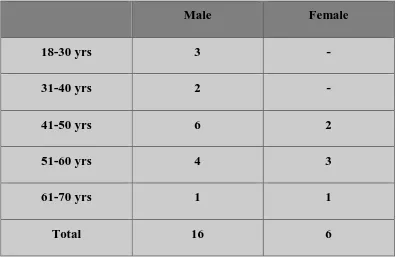
This study compises the sample of 22 patients, in which 6 were
females and 16 were males. The age distribution was varied from
18years to 66 years with an average age of 42 years. Out of 22 patients,
10 patients were victim of road traffic accident in which one associated
with fracture neck of femur on ipsilateral hip, 7 patients had self fall, 4
patients were fell from height (minimum 10 feet) and one was victim of
an animal attack. Longest duration of follow up was 21 months with a
mean follow up of 12 months. 3 patients lost follow up in this study.
In our study all were right handed persons and our study 14
patients had a fracture of the right proximal humerus and 8 patients had a
fracture of the left proximal humerus. This because of right handedness
and can be attributed to the left side driving in the roads and subsequent
[image:49.595.90.489.640.733.2]RTAs.
Table 1 : Sex Incidence
Male Female
Number 16 6
Chart 1: Sex Incidence
Chart 2: Age Distribution
Male
Female
0 1 2 3 4 5 6
18-30 yrs 31-40 yrs 41-50 yrs 51-60yrs 61-70 yrs
Female
Table 2 : Age Distribution
Male Female
18-30 yrs 3 -
31-40 yrs 2 -
41-50 yrs 6 2
51-60 yrs 4 3
61-70 yrs 1 1
Total 16 6
Chart 3 : Side Involvement
Sales
Right
Chart 4: Mode of Injury
METHODS
After hemodynamic stabilization, detailed clinical history and
clinical examination is undertaken from the patient who have been
admitted in department of Orthopaedics & Traumatology, Coimbatore
medical college & hospital.
Patients were treated with appropriate analgesic & antibiotics(if
necessary) . Then splinted with U-slab and cuff & collar was given. AP,
lateral and axillary view radiographs were taken preoperatively. These
were reviewed by to determine the Neer’s classification of the fracture.
In selected cases CT scan / special views were taken in order to know the
extent of articular surface involved.
Numbers
FALL INJURY
RTA
FALL FROM HEIGHT
FRACTURE CLASSIFICATION ( 36 , 37 )
The Neer classification system is based on displacement criteria of
1 cm or fragment angulation of 45°. The type of fracture then is divided
into segments. Four segments are possible, including the articular
segment, the lesser tuberosity, the greater tuberosity, and the surgical
neck.
These four parts are separated by epiphyseal lines (bone growth
plates) during the early developmental years. When the proximal
humerus is broken, the fracture line predictably occurs along one or
more of these planes.
More recently, displacement of greater tuberosity more than 5
mm is an indication of fixation.
NEER CLASSIFICATION
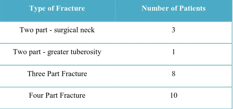
The fracture of all 22 patients were classified using NEER'S
Classification. Out of 22 patients, 10 were had Neer's 4 part fracture
( one - non union ) , 8 were had Neer's 3 part fracture and 4 had Neer's 2
Table 3 : Type of Fractures
Type of Fracture Number of Patients
Two part - surgical neck 3
Two part - greater tuberosity 1
Three Part Fracture 8
Four Part Fracture 10
Chart 5: Type of Fracture - Neer's Classification
4 part
3 part
2 part - neck
Co Morbidities
Out of 22 patients, 2 patients with diabetes, one is hypertensive,
one is heart disease patient and one is suffering from Rheumatoid
Arthritis.
Chart 6 : Co-Morbidities
All patients were treated operatively with proximal humerus
locking plate. 15 patients were operated under c-arm guidance and rest
without it. 17 patients were operated through deltopectoral approach and
5 were through deltoid splitting approach. The average duration of
surgery is about 103 minutes ranging from 50 minutes to 155 minutes.
The average blood loss is about 202 ml ranging from 50 ml to 300 ml.
The average day of surgery from incident of injury is about 28 days
ranging from 2 days to 51 days. No intra operative anaesthetic
Co-Morbidities
Nil
DM
HTN
CAHD
complications. No neurological deficit due to anaesthetic complications
and surgical complications.
SURGICAL TECHNIQUE FOR PLATE OSTEOSYNTHESIS –
DELTOPECTORAL APPROACH (38)
With the patient in supine position in fracture table with 30 - 45°
angulation at head end,with a sandbag behind the operating scapula , a
deltopectoral/ deltoid splitting approach were used.
Locate the deltopectoral groove percutaneously. In an obese
patient, the groove is located by abduction and external rotation of the
shoulder with pressure behimd the scapula. Start the incision at coracoid
proces, and extend it distally along the deltopectoral groove to the deltoid
insertion for approximately 15 cm
Develop skin flaps to expose the deep fascia. Open the fascia over
the deltopectoral groove with blunt scissors, looking for the cephalic
vein. This vein serves as an important landmark for identifying the
avascular interval between the deltoid and pectoralis major muscles.
Bluntly develop this interval, and retract the deltoid laterally and the
pectoralis major medially. The vein can be ligated or retracted with the
The anterior circumflex artery lies in the middle of the wound, just
superior to the pectoralis major muscle; they may need to be isolated,
clamped, and coagulated.
Wider exposure is possible if the muscle origins from the coracoid
are transected. If more proximal exposure is needed, it may be necessary
to transect the origin of the pectoralis minor muscle. In such cases,
release the origins of the coracobrachialis and the short head of the
biceps from the tip of the coracoid, leaving a cuff on the tip of the
coracoid for repair.
It is better to avoid devascularization of the fracture fragment by
meticulous dissection of tendino osseous attachments. The osseous
attachments of the rotator cuff are pull together to reduce the fracture.
If reduction is difficult, insert a k-wire as a joystick in the
humeral head to rotate the head into a reduced position. or place sutures
under the rotator cuff tendon (supraspinatus) also can be helpful for
mobilization & reduction.
For 3-part or 4-part fractures or osteoporotic fragments, place
sutures into the rotator cuff tendons attached to fractured fragments to
Place the plate onto the greater tuberosity, just posterior to the
biceps tendon, and temporarily fix it with Kirschner wires; confirm
correct plate position in c-arm both in ap view in adduction and
abduction. If plate placement is too proximally , it may cause
impingement and If plate placement too close to the biceps tendon may
damage the anterior humeral circumflex artery.
If plating is preferred, plate is placed at least 8 mm distal to the tip
of the greater tubercle and fixed to the humeral shaft with screws.
If there is fractures with medial comminution, first fix the plate to
the head with screws, and reduce the shaft segment to the plate. This
helps avoid varus malposition, which is associated with higher failure
rates. Screw insertion into the inferomedial humeral head adds stability
for fractures without medial calcar support.
Confirmation with c-arm on anteroposterior and lateral views is
INTRA OPERATIVE IMAGES
Position of patient with bump under ipsilateral shoulder & drapped
Cephalic vein in deltopectoral groove
Fracture reduced and provisionally fixed with k wires, reduced and fixed to distal fragment with locking screw.
Final construct with philos plate for proximal humerus fracture
POST OPERATIVE PROTOCOL(39,40)
Postoperatively, the arm was immobilized in a sling. The drain was
removed on 2nd post operative day. The time for commencement of shoulder
rehabilitation was determined by stability of fixation , quality of bone, and
compliance of patient. Passive ROM exercises (ie, pendulums, passive forward
elevation, external rotation) generally were begun on the first postoperative
day provided that a stable reduction was achieved. Active ROM of the elbow,
wrist, and hand was also begun immediately after surgery. The patient then
progressed through a three-phase rehabilitation program, consisting of passive
assisted exercises early, active exercises starting at approximately 6 weeks
postoperatively, and strengthening or resisted exercises beginning 10 to 12
weeks after surgery. Early passive assisted exercises help to avoid adhesion
formation. No limitation of exercises within the pain-free ROM was necessary
during this time provided that bone stock was good and medial buttressing
adequate. Shoulder strengthening and resistance exercises were initiated only
after bony consolidation was confirmed on plain radiographs and adequate
coordination of the extremity had been achieved.
Standard AP, axillary, and scapular Y radiographic views were taken
immediately after surgery. Routine follow-up radiographs were taken 2, 6
weeks, 3,6 & 12 months postoperatively to ensure that no pin has migrated, no
loss of reduction has occurred, evidence of callus formation and consolidation
CASE ILLUSTRATIONS
Case – 1
53 years old female
Fall by herself
Neer 3 parts fracture Right side
Open Reduction and internal fixation with Locking plate.
Duration of surgery – 90 minutes
Radiological fracture union: 12 weeks
Range of Motion:
Constant score – 79
Comment – Good
Case 1- Radiographs
Pre-operative picture
Case – 2
24 years old male
Fall from 20 feet height
Neer 4 parts grade IIIb open fracture Right side
Wound debridement & Open Reduction and internal fixation with Locking plate.
Duration of surgery – 150 minutes
Radiological fracture union: 12 weeks
Range of Motion:
Constant score – 75
Comment – Good
Flexion - 90 Abduction - 90
Case 2 - Radiographs
Pre op radiograph Immediate post op radiograph
Case 3
18 years old male
Fall from 20 feet height
Neer 2 parts surgical neck fracture Right side
Open Reduction and internal fixation with Locking plate.
Duration of surgery – 90 minutes
Radiological fracture union: 6 weeks
Range of Motion:
Constant score – 92
Comment – Excellent
Case 3 - Radiographs
Pre op radiograph
CT Scan - 3 D shows angulation more than 30°
Case 4
65 years old female
Fall on out stretched hand
Neer 3 parts fracture Left side
Open Reduction and internal fixation with Locking plate.
Duration of surgery – 90 minutes
Radiological fracture union: 12 weeks
Range of Motion:
Constant score – 60
Comment – Moderate
Flexion - 120 Abduction - 90
Case 4 - Radiographs
Anteroposterior view Axillary View
Scapula Y View West Point View
Case 4 -Radiographs
Immediate Post op xray
Case 5
44 years old male
Road Traffic Accident
Neer 4 parts fracture Right side
Open Reduction and internal fixation with Locking plate.
Duration of surgery – 90 minutes
Radiological fracture union: 6 weeks
Range of Motion:
Constant score – 91
Comment – Excellent
Case 5 - Radiographs
Pre op Xray - 4 part fracture CT shoulder shows 4 part Fracture
Immediate post op 12 weeks follow up - fracture consolidated
EVALUATION
A physical examination was performed, the Constant score was
calculated, and radiographs of the proximal part of the humerus were
made and evaluated for bony healing, signs of malunion, nonunion or
avascular necrosis.
The Constant score assigns points for Pain, Range of movements,
Power and Activities of daily living. Muscle strength was measured with
use of a 1 kg weight in the patient’s hand and the shoulder in 90° of
abduction, or, if 90° could not be reached, in maximum active abduction
as described by Constant.(41)
The Constant score was graded as
Poor (0 to 55 points),
Moderate (56 to 70 points),
Good (71 to 85 points),
Table 4 : Evaluation
Result - Outcome Numbers Percentage (%)
Excellent 3 14
Good 4 18
Moderate 8 36
Poorer 3 14
Lost follow up 4 18
Chart 7 : Evaluation
Excellent 14%
Good 18%
Moderate 36%
RESULT
Out of the 22 patients followed up, 3 patients had excellent scores,
4 had good scores , 8 had moderate scores and 3 had poor outcome
scores. Mean constant score is 67.28 (range 38-92 points). Mean constant
score for Neer two part fracture was 75 (range 56 – 92), for Neer's three
parts fracture was 66.71(range 38 – 91) and for Neer's four parts fracture
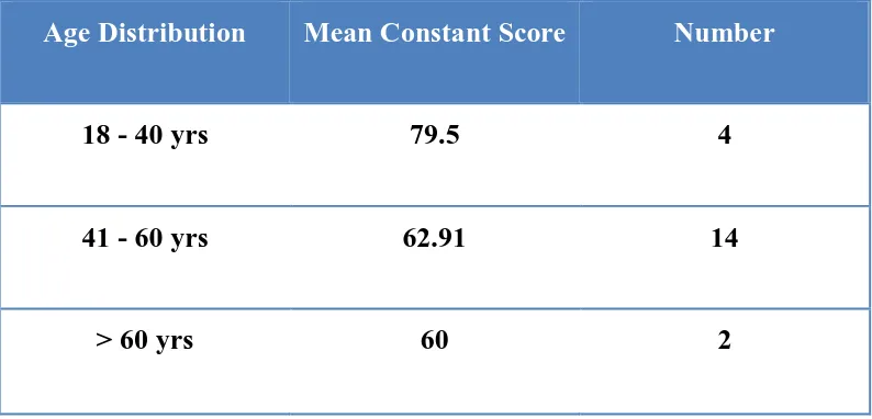
was 60.14 (range 40 – 81). Mean constant score for middle age
group(18-40) was 79.5 (range 75 – 92), for old age group(41-60) was
62.91(range 38 – 91) and for very old age group(>60) was 60.0 (one case
[image:84.595.95.492.509.695.2]- 60).
Table 5 : Consant Score vs Neer's parts of Fracture
Neer's Classification Constant Score Number
Two Part 75 (range 56 – 92) 4
Three Part 66.71(range 38 – 91) 8
Chart 8: Mean Constant Score as per Neers Classification
Chart 9 : Mean Constant Score as per Neers Classification
0 10 20 30 40 50 60 70 80
2 part 3 part 4 part
Mean Constant Score
Mean Constant Score
0 10 20 30 40 50 60 70 80
18 - 40 years 41 - 60 years 61 - 70 years
NUMBER
Table 6 : Mean Constant Score Vs Age Distribution
Age Distribution Mean Constant Score Number
18 - 40 yrs 79.5 4
41 - 60 yrs 62.91 14
> 60 yrs 60 2
Eighteen patients out of twenty two patients have gone for union
at around 9 weeks follow up (88%) , except those complicated by screw
COMPLICATION
The common complications after fixation of fractures of
proximal humerus are restricted movements, restrictive pain, wound
infection, failure of fixation, avascular necrosis of humeral head and late
rupture of the rotator cuff.(42,43)
Two patients, one with Neer 2 part fracture later leads to
osteonecrosis of the humeral head. One who with 4 part fracture
encountered backing out of screw with failure of fixation and finally
leads to Osteonecrosis who undergone implant exit and planned for
hemiarthroplasty of shoulder. Unfortunately Patient not willing for
further procedure.
Avascular necrosis is not in itself a clinical problem. However,
it may end up in partial or total collapse of the humeral head with
incongruency. This may result in malfunction and pain, although the
x-ray appearance frequently does not correlate with the clinical picture.
One patient with Neer 4 part fracture treated with locking
compression plate had failure of fixation with collapse of fractured
fragments on second post operative month and patient was on lost follow
up.
Postoperative wound or bone infection is one of the common
(between 21 and 56 days) or chronic (>56 days). Once the purpose of
implant is over, it can be removed.
One patient with Neer 4 parts open fracture leads to chronic
osteomyelitis for which iv antibiotic followed by oral antibiotic
according to culture & sensitivity, waited till bone union and finally
[image:88.595.94.493.339.564.2]undergone for implant exit.
Table 7: Complication of Philos Plate
COMPLICATIONS NO OF PATIENTS
Perforation of screw 1
Chronic Osteomyelitis 1
Failure of Fixation 1
Osteonecrosis 2
DISCUSSION
The treatment of complex humeral 3- or 4-part fractures represents
a challenge. The surgeon must obtain an exact anatomical reduction and
stable fixation, and at the same time minimise the iatrogenic risk of
screw penetration and avascular necrosis of humeral head by maximal
protection of the periarticular soft tissues.
Poor results in these complex fractures are due to following
causes:
1) Inadequate fracture reduction especially medial
cortex
2) Unstable fixation
3) Incorrect positioning of the fixation devices .
There is consensus in the literature that, regardless of the
procedure and the implant chosen, a good functional final result depends
mainly on anatomical reduction of the fracture combined with a stable
fixation, and early initiation of functional rehabilitation of the shoulder.
But in this study, age of the patient, minimal part of fractures and early
fixation of fracture , directly increase the functional outcome.
In recent decade, rigid internal fixation of fracture have been
Inspite of an early and secure functional postoperative therapy, it was
believed that these implant would reduce the risk of secondary reduction
loss in osteoporotic patients.
In the very old age group with osteoporosis, functional outcome
after conventional plate osteosynthesis was poor.(44) In order to obtain better and reproducible results, the AO/ASIF has developed a special
locking compression plate (Philos) for fractures of the proximal humerus. (45)
Patients with good bone quality have previously been treated
successfully with the conventional plate osteosynthesis.(46)
In this study, most of the patients (i.e;14 out of 22) lie in the group
of 41- 60 years , a group highly prone for osteoporosis.
In normal conventional plates, the chance of backing out or cutting
out of screws is more. It is difficult to hold the bony fragments as they
are highly fragile due to osteoporosis, thereby affecting proper reduction.
The normal screws are highly prone for soft tissue dissection, and all
these accounts for the high rate of failure in procedures using
conventional plates in an osteoporotic bone.
With advent of locking plates, the fraction of backing out or
cutting out of screws are reduced due to the locking head and fixed angle
present in fixed angle screws.
Due to multidirectional nature of screws in the locking plate,
which spans through sphericity of head and not the centre alone, reduces
Suturing of tendons with eyelets of plate is possible in locking
plates which reduces the risk in fixation of small fragments of
osteoporotic bone which was otherwise hard, and also reduces the
possibility of collapse.
Soft tissue dissection rates are similar in both conventional and
interlocking plates, but with the skills of surgeon and meticulous surgical
procedures this negativity can be overcome.
In bone plate interface, the reduced compression effect of locking
plates when compared to conventional plates, play a high role in
reducing avascularityof the bony fragments and head of humerus.
The average clinical result obtained in our study, with a mean
Constant-Murley score of 67.28 points is satisfactory.
Comparable studies of internal fixation of Proximal humerus
fractures demonstrate similar short term results. Although the follow-up
period of our series was short, studies have shown that early function is
comparable to final long term outcome. The outcome seems to correlate
with fracture severity, anatomic reduction, etiology, bone quality, length
of time elapsed from injury to surgery, concomitant injuries and the
Table 8: Functional scores achieved with different treatment
options for proximal humeral fractures in the current literature.(48
to 54)
Study Type of fixation Constant score Neer's
classification
Kuchle et al
(2006)
Cloverlaef plate 72.4 2,3& 4 part
fracture
Ketter et al
(2006)
Angle stable
humerus plate
70.0 2,3&4 part
fracture
Lill et al (2003) Angle stable
humerus plate
72.5 2,3&4 part
fracture
Kollig et al
(2003)
T palte, screws
& k wires
72.1 3 & 4 part
fracture
Wijgman et al
(2002)
Classic T Plate
cerclage
80.0 3 & 4 part
fracture
Gerber et al Internal fixation 78 2,3,& 4 part
fracture
Hessman et al T plate 69 2,3,& 4 part
fracture
Our study Locking plate 67.28 2,3,& 4 part
[image:92.595.95.489.157.767.2]A meticulous anatomical reduction with appropriate plate
positioning led to a significantly better result. The Constant-Murley
score was significantly lower if anatomical reconstruction did not
succeed or a nonanatomical reconstruction was accepted
intraoperatively, and/or when the plate was not correctly positioned on
the shaft at the proper height to avoid subacromial impingement.
In our study, three cases (14%) with poor outcome scores include
one case of osteonecrosis of humeral head, one case of improper
reduction , one case of shoulder stiffness due to delay in surgery. There
is no significant poorer result in perforation of screws in joint and in
chronic infection
The 4.5 % ( 1 / 22 patients) infection rate in our series is
comparable to the 2.5% ( 2 / 41 patients) patientsof Paavolainen et al
(1983).
The development of aseptic humerus head necrosis (2 patients or
9%) significantly affected the clinical result ; these patients only
achieved a mean Constant-Murley score of 45.0. In the literature the rate
of necrosis for 3-and 4-part fractures has been between 0% and 50%,
depending on the osteosynthesis procedure. The rate of aeptic necrosis
Table 9 : Aseptic Necrosis rate in various studies
Study Type of Fracture Method of
Fixation
Incidence
Hessmann et al 2,3& 4 parts T plate 4%
Fankhauser et al AO - A,B,C Locking plate 10%
Gerber et al 2,3 & 4 parts Locking plate 12%
Our study 2,3 & 4 parts Locking plate 9%
Early fixation, exact anatomical repositioning of the fracture
fragments and rigid internal fixation was associated with a significantly
better functional result. The results attained in our patients gains less
importance of the restoration of the correct anatomical relationship
between the individual fragments.
The functional results after rigid fixation of three- and four-part
fractures using a locking plate were shown to be better than conservative
treatment or semi-rigid fixation without anatomical reduction of the head
fragment. Shoulder function continued to improve as the strength and
CONCLUSION
Although our study was relatively short and it was not a
randomized controlled study, the results are comparable with other
published journals.
Accurate anatomical reduction gains and early fracture fixation are
more important than the implant used, to get a good final functional
outcome, and this factor is independent from the implant design and
procedure selected.
The options as to the surgical approach or the type of implant used
depend on the pattern of the fracture, the quality of the bone encountered,
the patient’s goals and the surgeon’s familiarity with the techniques. The
learning curve with the implants chosen certainly also plays a role. An
adequate surgical technique will minimise complications and an
aggressive rehabilitation regime will ensure the best possible result.
There is no much difference among 2,3 & 4 parts of fracture with
locking plate. All are nearly more or less with good function outcome.
In general, 2- and 3-part fractures can be treated with open
reduction and internal fixation (a plate with screws is the choice).
Four-part fractures in the younger, active patient also can be treated