Copyright © 2003, American Society for Microbiology. All Rights Reserved.
Survey of Human Genes of Retroviral Origin: Identification and
Transcriptome of the Genes with Coding Capacity for
Complete Envelope Proteins
Nathalie de Parseval,
1Vladimir Lazar,
2Jean-Franc¸ois Casella,
1Laurence Benit,
1and Thierry Heidmann
1*
Unite´ des Re´trovirus Endoge`nes et Ele´ments Re´troïdes des Eucaryotes Supe´rieurs, UMR 8122 CNRS,1and
Unite´ de Ge´nomique Fonctionnelle,2Institut Gustave Roussy, 94805 Villejuif Cedex, France
Received 19 March 2003/Accepted 7 July 2003
Sequences of retroviral origin occupy approximately 8% of the human genome. Most of these “retroviral” genes have lost their coding capacities since their entry into our ancestral genome millions of years ago, but some reading frames have remained open, suggesting positive selection. The complete sequencing of the human genome allowed a systematic search for retroviral envelope genes containing an open reading frame and resulted in the identification of 16 genes that we have characterized. We further showed, by quantitative reverse transcriptase PCR using specifically devised primers which discriminate between coding and noncoding elements, that all 16 genes are expressed in at least some healthy human tissues, albeit at highly different levels. All envelope genes disclose significant expression in the testis, three of them have a very high level of expression in the placenta, and a fourth is expressed in the thyroid. Besides their primary role as key molecules for viral entry, the envelope genes of retroviruses can induce cell-cell fusion, elicit immunosuppressive effects, and even protect against infection, and as such, endogenous retroviral envelope proteins have been tentatively identified in several reports as being involved in both normal and pathological processes. The present study provides a comprehensive survey of candidate genes and tools for a precise evaluation of their involvement in these processes.
Completion of the sequencing of the human genome has led to the conclusion that a significant fraction (approximately 8%) of our genome is of retroviral origin, with thousands of proviral sequences disclosing similarities with the integrated form of infectious retroviruses (23). These elements—also called human endogenous retroviruses (HERV)—are most probably the traces of “ancient” infections of the germ line by active retroviruses, which have thereafter been transmitted in a Mendelian manner. According to sequence homologies, these elements can be grouped into distinct families, with copy num-bers ranging from a few to several hundred per haploid ge-nome. Families have been tentatively named according to the tRNA normally used to prime reverse transcription in a retro-viral replicative cycle (reviewed in references 25 and 43). In agreement with the proposed evolutionary scheme for the presence of these proviral elements, strong similarities be-tween HERV and the present-day infectious retroviruses can be observed at the sequence level and in several instances at the functional level. Actually, phylogenetic analyses based on either the highly conserved reverse transcriptase (RT) domain of the pol gene or the transmembrane (TM) moiety of the envelope gene reveal interspersion of HERVs and infectious elements, suggesting a common history and shared ancestors (4, 40). HERV-encoded retrovirus-like particles have been
detected in some tissues by electronic microscopy, revealing structural similarities to exogenous retroviruses (10). It has also recently been demonstrated that the HERV-K(HML-2) family can encode a regulatory protein (called Rec or cORF) which is functionally homologous to the Rev protein encoded by the human immunodeficiency retrovirus (26, 44) and that the coding envelope protein of the HERV-W family can still interact with the cellular receptor of the present-day D-type retroviruses (8). Finally, it has been shown for endogenous retroviruses of other species that their replicative cycle is closely related to that of exogenous retroviruses, with evidence for retrovirus-like recombination in the course of the reverse transcription step (20). As a consequence of the close relation-ship between HERVs and exogenous infectious retroviruses, it can be proposed that HERVs may still possess some of the functions of infectious retroviruses and as such have patho-genic effects, provided that they are transcriptionally active. Conversely, it is plausible that HERV proteins may have been subverted by the host for its benefit. Along this line, it has been proposed that the HERV envelope proteins could play a role in several processes, including (i) protection against infection by closely related exogenous retroviruses via receptor interfer-ence (5, 9, 37), (ii) protection of the fetus from the mother’s immune system via a domain (the immunosuppressive domain) located in the TM subunit of most retroviral envelope proteins and known to inhibit immune effector functions (12, 28), and (iii) placenta formation via envelope protein-mediated fuso-genic effects that could be involved in the generation of the syncytiotrophoblast (8, 31). An assessment of these putative roles of HERV is rendered extremely difficult as a result of the
* Corresponding author. Mailing address: Unite´ des Re´trovirus En-doge`nes et Ele´ments Re´troïdes des Eucaryotes Supe´rieurs, UMR 8122 CNRS, Institut Gustave-Roussy, 39 rue Camille Desmoulins, 94805 Villejuif Cedex, France. Phone: 33/1-42-11-49-70. Fax: 33/1-42-11-53-42. E-mail: [email protected].
10414
on November 8, 2019 by guest
http://jvi.asm.org/
multicopy nature of these elements, which precludes classical genetic approaches. However, it has to be taken into account that a large fraction of endogenous retroviral genes are no longer coding genes, due to the accumulation of mutations, frameshifts, and deletions. Accordingly, we have made an ex-haustive survey of the human genome for complete proviral elements, and specifically those containing a complete and codingenvgene. Interestingly, this survey has led to the iden-tification of a limited number of genes that we have character-ized. To get insight into their potential functions, we devised a quantitative RT-PCR assay using primers allowing amplifica-tion of the coding copies specifically and provide the transcrip-tome of these human genes of retroviral origin in a large panel of healthy tissues. This analysis results in the unraveling of a series of new transcriptionally active human genes whose func-tions can now be appraised by classical molecular genetic ap-proaches.
MATERIALS AND METHODS
Computer sequence analyses.Homology searches were performed using the BLASTN program at the National Center for Biotechnology Information (http: //www.ncbi.nlm.nih.gov/BLAST), screening the finished (nr [nonredundant]) and unfinished (htgs [high-throughput genomic sequence]) databases with consensus envelope genes for each family. Hydropathy of the envelope proteins was cal-culated by the Kyte-Doolittle method implemented by the DNA Strider program (29). Alignments were performed with the CLUSTALW multialignment pro-gram at Infobiogen (http://www.infobiogen.fr). Chromosomal localizations of the codingenvgenes were performed at the Ensembl web site (http://www.ensem-bl.org/Homo_sapiens).
In vitro transcription-translation assay.The codingenvgenes for in vitro transcription-translation assays were amplified with the Expand long-template PCR system (Roche, Indianapolis, Ind.). PCRs were carried out for 35 cycles (1 min at 94°C, 30 s at 58°C, 2 min at 68°C) using 1 ng of bacterial artificial chromosome (BAC) DNA. Primers were as follows: envHT7.3 (GCTAATACG ACTCACTATAGGAACAGACCACCATGCACCACAGTATCAACCTTAC) and flR2B1 (TTCTGTTTCAGCTACAACTCTGT) forenvH1; envHT7.3 and flRB2 (AGCAATAGTTTGTTAAATTC) forenvH2; envHT7.2 (GCTAATAC GACTCACTATAGGAACAGACCACCATGAGGGCACCCTCCAATAC TTC) and flRB3 (ACCCCATGTTCTAGTCTTCC) forenvH3; envKT7 (GCT AATACGACTCACTATAGGAACAGACCACCATGGAGATGCAAAGAA AAGCA) and LTRenvsK (GTGAACAAAGGTCTTTGCATCATAG) forenvK1, envK3,envK5, andenvK6; envKT7 and 3⬘fl51C12 (GAATTAGGCTTTCGGGA CTTGAA) forenvK2; envKT7 and KLTR3⬘(C/TTTAAC/G/AG/AAGCATGC TGC/AC) forenvK4; envTT7.2 (GCTAATACGACTCACTATAGGAACAGA CCACCATGTTGGATTCATCACTCCCA) and envTflanq2 (CTGAAGGGAG TTCCTCCTAGG) forenvT; envWT7 (GCTAATACGACTCACTATAGGAA CAGACCACCATGGCCCTCCCTTATCAT) and envW64flR2 (ACAGCCAA GCAGGTACAG) forenvW; envFRDT7.2 (GCTAATACGACTCACTATAGG AACAGACCACCATGCTCCTGCTGGTTCTCATTC) and envFRDfl3⬘(CTGC AGCAGACTCCATCCTTG) for envFRD; enverv3T7 (GCTAATACGACT CACTATAGGAACAGACCACCATGACTAAAACCCTGTTGTATCA) and enverv3AS (GTTAATACTTAGTTAGGGCC) forenvR; HS89F2T7 (GCTAA TACGACTCACTATAGGAACAGACCACCATGGATCCACTACACAC GATTGA) and HS89R3fl (TGTTTTGGGACACCACGAAT) for envR(b); envF(c)2T7 (GCTAATACGACTCACTATAGGAACAGACCACCATGAAT TCTCCATGTGAC) and envF(c)2flR3 (GACACTTAATAGTTGCGACA) for envF(c)2; and envF(c)1T7 (GCTAATACGACTCACTATAGGAACAGACCA CCATGGCCAGACCTTCCCCACTATGC) and envF(c)1fl3⬘(GCCTTGGCA ACTAAACCATTC) forenvF(c)1. T7 promoter-containing PCR products were ethanol precipitated, and 200 ng of the amplification products was used in the TNT coupled reticulocyte lysate system (Promega Corp., Madison, Wis.) accord-ing to the manufacturer’s instructions, with [3H]methionine (ICN Biomedicals
Inc., Irvine, Calif.) for protein labeling. After electrophoresis of the translation products, sodium dodecyl sulfate-polyacrylamide gels were impregnated with Amplify (Amersham Biosciences, Piscataway, N.J.), rinsed with water, dried, and autoradiographed.
Tissue samples, RNA calibration, and reverse transcription.Human BAC clones containing the identified coding envelope genes were purchased from
BACPAC Resources (Oakland, Calif.). RNAs from various human tissues were purchased from Clontech (Palo Alto, Calif.), Stratagene (La Jolla, Calif.), and Ambion (Austin, Tex.). Quality of the RNA was assessed on an RNA LabChip (Agilent 2100 Bioanalyzer), and RNA concentration was quantified spectropho-tometrically. Five micrograms of each RNA sample was subjected to DNase treatment (DNA-free; Ambion) to eliminate DNA contaminants. One micro-gram of RNA from each sample was reverse transcribed in a 20-l reaction using 50 U of Moloney murine leukemia virus RT and 20 U of RNuclease inhibitor (Applied Biosystems, Foster City, Calif.) per reaction, 1 mmol of dA/T/G/C (Amersham-Pharmacia Biotech, Uppsala, Sweden) per liter, 5 mmol of MgCl2
per liter, 10 mmol of Tris-HCl (pH 8.3) per liter, 10 mmol of KCl per liter, and 2.5mol of random hexamers (Applied Biosystems) per liter. The cDNAs were then diluted 1/25 in nuclease-free H2O (Promega Corp.).
Real-time quantitative PCR.Oligonucleotides were designed with the com-puter program Oligo (MedProbe, Oslo, Norway). The special requirements were a melting temperature of 60°C and an amplicon length between 80 and 350 bp. Oligonucleotides were purchased from MWG (Ebersberg, Germany).
Real-time quantitative PCR was achieved by using a cDNA equivalent of 20 ng of total RNA. The reaction was performed in 25l using SYBR green PCR core reagents (Applied Biosystems) according to the manufacturer’s instructions. PCR was developed with the ABI PRISM 7000 sequence detection system (Applied Biosystems). Amplification was performed using a 2-min step at 50°C and then a 10-min denaturation step at 95°C, followed by 40 cycles of 15 s of denaturation at 95°C, 1 min of primer annealing, and a polymerization step at 60°C. To normalize for differences in the amount of total RNA added to the reaction, amplification of 18S total rRNA was performed as an endogenous control (variation was less than a factor of 3.5). The primers and probe from 18S RNA were purchased from Applied Biosystems. The relative expression in each sample was calculated with respect to a standard calibration curve (the dilution series of genomic DNA). Each sample was analyzed at least twice.
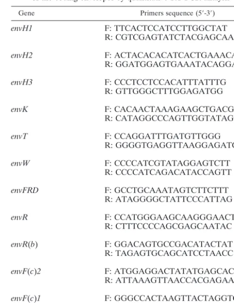
[image:2.603.299.541.87.399.2]The control plasmid (p11env) containing the 11 envelope amplicons obtained with the primers listed in Table 1 was constructed first by cloning each amplicon into the pGEMT vector and then by successive three-fragment ligations using fragments from these 11 plasmids and from construct intermediates. A control TABLE 1. Sequences of the primers used to detect the expression
of the coding envelopes by quantitative RT-PCR analysis Gene Primers sequence (5⬘-3⬘)
envH1 F: TTCACTCCATCCTTGGCTAT
R: CGTCGAGTATCTACGAGCAAT
envH2 F: ACTACACACATCACTGAAACAAA
R: GGATGGAGTGAAATACAGGAC
envH3 F: CCCTCCTCCACATTTATTTG
R: GTTGGGCTTTGGAGATGG
envK F: CACAACTAAAGAAGCTGACG
R: CATAGGCCCAGTTGGTATAG
envT F: CCAGGATTTGATGTTGGG
R: GGGGTGAGGTTAAGGAGATGG
envW F: CCCCATCGTATAGGAGTCTT
R: CCCCATCAGACATACCAGTT
envFRD F: GCCTGCAAATAGTCTTCTTT
R: ATAGGGGCTATTCCCATTAG
envR F: CCATGGGAAGCAAGGGAACT
R: CTTTCCCCAGCGAGCAATAC
envR(b) F: GGACAGTGCCGACATACTAT
R: TAGAGTGCAGCATCCTAACC
envF(c)2 F: ATGGAGGACTATATGAGCACAA
R: ATTAAAGTTAACCACGAGAAGC
envF(c)1 F: GGGCCACTAAGTTACTAGGTC
R: AGTTAGGAGGGAGTTACTGGG
on November 8, 2019 by guest
http://jvi.asm.org/
series of p11env plasmid dilutions ranging from 1 to 0.008 ng was amplified with each primer set to measure relative yield (variation of less than a factor of 2). A control tube containing 1 ng of the p11env plasmid was included in each real-time PCR assay as a reference.
RESULTS
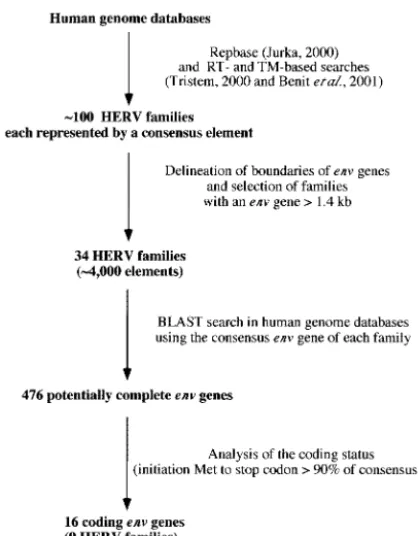
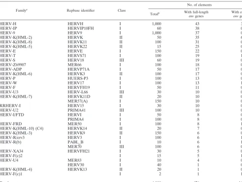
Screening of human databases for coding envelope genes. The rationale for the screening procedure to identify the cod-ing envelope genes in the human genome is illustrated in Fig. 1. Basically, we used the Repbase database (final control search with update 8.2.1, Oct. 2002) (22), in which each family is represented by a “consensus” element, built from an align-ment of all the proviruses belonging to the same family (i.e., copies which cluster together in phylogenetic trees). We im-plemented it by using RT- and TM-based searches as described in references 4 and 40, which revealed two additional families, the F(c)1 and F(c)2 families. Overall, approximately 100 HERV families were identified. For each family, the envelope gene was tentatively delineated, with thepolgene (which was easily positioned due to its high degree of conservation among retroviral elements) at its 5⬘end and with the retroviral long terminal repeat (LTR) at its 3⬘ end. When the distance be-tween the pol gene and LTR was higher than 1.4 kb (the average length of an envelope gene being 1.9 kb), the envelope gene was considered potentially complete and the HERV fam-ily was selected. The resulting list of the 34 corresponding HERV families is given in Table 2. For each family, we then performed a BLAST query on human genome databases using the envelope gene as a probe. Overall, it yielded 476 poten-tially complete envelope genes (see Table 2 for their distribu-tion among the HERV families). The coding status of each
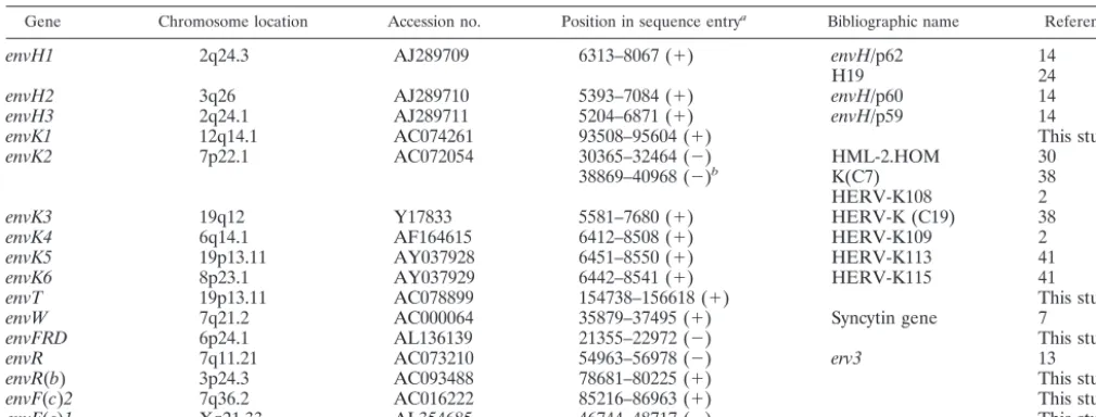
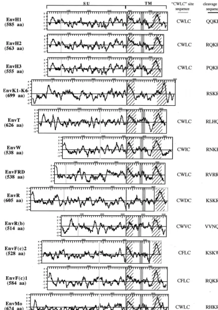
identifiedenvgene was finally assessed, and only those with an open reading frame (ORF) beginning at the first Met codon of the consensus envelope gene and uninterrupted over⬎90% of this gene were retained. Among the 476 envelope genes that were individually analyzed, only 16 genes were found to po-tentially encode envelope proteins. These 16 genes are listed and described in Table 3 and Fig. 2. Ten of these genes had previously been identified (2, 7, 13, 14, 24, 30, 38, 41), and six new genes emerged from this screen, including genes from the FRD, T, R(b), F(c)1, and F(c)2 families and one supplemen-tary gene from the HERV-K(HML-2) family. The chromo-somal localization was determined or confirmed by using the Ensembl web site and the corresponding accession numbers. It is noteworthy that one of these genes,envK2, can be found in duplicate in some individuals, since the corresponding provirus is organized as a tandem repeat (35). The twoenvsequences are 100% identical at the nucleotide level and are both re-ferred to here as theenvK2gene. As can be observed in Fig. 2, which describes the 16 putative envelope proteins in compar-ison with the Moloney leukemia virus envelope protein, the overall length of the proteins is variable, with a minimum of 514 amino acids for EnvR(b) and maximum of 699 amino acids for EnvK. This size variability is also found among exogenous retroviral envelope proteins since, for example, human T-cell leukemia virus type 1 and human immunodeficiency virus type 1 envelope proteins are 488 and 861 amino acids long, respec-tively.
To ascertain the existence of the ORFs inferred from the nucleotide sequences, an in vitro transcription-translation as-say was performed with human BAC clones containing the identified coding envelope genes. The 16 BACs were obtained from BACPAC Resources, except for the H1-, H2-, and H3-containing BACs that had been cloned previously (14). The in vitro transcription-translation assay was performed directly on theenvPCR products (generated by using a forward T7-con-taining primer at theenv5⬘end and a reverse primer down-stream of the stop codon). In all cases, translation products of the size expected from the sequences in the database were obtained (Fig. 3). For some envelope proteins, additional bands of lower molecular weight were observed, compatible with initiation at internal sites, and interestingly, in one case [F(c)2], additional bands at higher molecular weights were detected, most probably associated with the presence of a frameshift signal at the gene’s 3⬘end (3).
[image:3.603.55.264.68.336.2]The predicted hydrophobic profiles of the 16 proteins rep-resented in Fig. 2 allow the identification of characteristic domains of these envelope proteins, namely, the fusion pep-tide, located just downstream of the proteolytic cleavage site between the Surface (SU) and TM moieties of the envelope proteins, and the transmembrane domain of the TM subunit, which permits the anchorage of the envelope protein in the membrane. Noteworthy, this analysis revealed that one enve-lope protein (EnvR) seems to be devoid of a fusion peptide and that two envelope proteins [EnvR and EnvF(c)2] disclose a premature stop codon just upstream of their transmembrane hydrophobic domain. Two other domains are delineated in Fig. 2, which are characteristic features of the envelope pro-teins belonging to the C-type and D-type retroviruses: the CWLC domain, involved in the interaction between the SU and TM moieties (34), and the CKS17-like immunosuppressive
FIG. 1. Flow chart for the identification of the 16 coding envelope genes of the human genome (see Results).
on November 8, 2019 by guest
http://jvi.asm.org/
domain (12). It is noteworthy that, like B-type retrovirus, len-tivirus, and spumavirus envelope proteins, the six EnvK pro-teins lack these two domains.
Transcriptome of the coding envelope genes. (i) Strategy.To get insight into the expression profile of these genes in a quan-titative manner, we devised a real-time RT-PCR strategy which uses specific primers designed in such a way that only envelope genes with an ORF should be amplified among all the enve-lope genes of a given family. To do so, for each HERV family containing a member with a coding element, env nucleotide sequences were aligned with the CLUSTALW program and primers were designed within domains of maximal divergence between the coding copy and the other copies. Primers for Sybr green amplification were devised with their 3⬘ends forced at nucleotide positions with, again, maximum divergence between the coding sequence and the others. For the HERV-K(HML-2) family, which contains six codingenvgenes, this strategy could not be applied due to the too-high sequence conservation between copies. In this case, specific primers were devised that matched all six coding genes, tentatively excluding most other
HERV-K(HML-2) envelope genes. The complete list of de-vised primers is given in Table 1.
[image:4.603.46.541.81.452.2]To determine whether the resulting primers fulfilled the requirements for both efficiency and specificity, a first series of assays was performed by PCR amplification of human genomic DNA. As expected, in all cases a single band was observed, thus excluding nonspecific amplifications or amplifications of elements of unusual size (data not shown). Then, PCR prod-ucts for each couple of primers were cloned into a pGEM-T vector, and at least six clones per amplicon were sequenced for each envelope gene [26 clones for the HERV-K(HML-2) fam-ily; see below]. In all cases, the six clones for a given envelope gene were identical and unambiguously corresponded to the coding sequence, being different from all the other aligned sequences within each HERV family. For the HERV-K (HML-2) family, 26 clones were sequenced, of which 21 had an identical sequence corresponding to the sequence of the six coding envelope genes, and 5 corresponded to two other HERV-K(HML-2) envelope genes. Despite this parasitic am-plification of noncoding envelope genes, the HERV-K(HML-2)
TABLE 2. HERV families containing full-length envelope genes
Familya Repbase identifier Class
No. of elements
Totalb With full-length
envgenes With codingenvgenes
HERV-H HERVH I 1,000 43 3
HERV-IP HERVIP10FH I 60 38 0
HERV-9 HERV9 I 1,000 37 0
HERV-K(HML-2) HERVK II 50 35 6
HERV-K(HML-8) HERVK11 II 100 31 0
HERV-K(HML-5) HERVK22 II 15 25 0
HERV-E HERVE I 150 22 0
HERV-T HERVS71 I 100 19 1
HERV-S HERV18 III 60 19 0
HERV-Z69907 MER66 I 100 18 0
HERV-ADP HERVP71A I 50 17 0
HERV-K(HML-6) HERVK3 II 100 17 0
HERV-P HUERS-P3 I 100 13 0
HERV-W HERV17 I 100 13 1
HERV-F HERVFH19 I 50 11 0
HERV-U3 HERV-L66 III 30 10 0
HERV-K(HML-7) HERVK11D II 20 10 0
MER57(A) I 150 10 0
RRHERV-I HERV15 I 30 10 0
HERV-U2 PRIMA41 III 100 10 0
HERV-I/FTD HERVI I 50 8 0
PRIMA4 I 100 8 0
HERV-FRD MER50 I 100 8 1
HERV-K(HML-10) (C4) HERVK14 II 20 7 0
HERV-K(HML-3) HERVK9 II 150 6 0
HERV-R/erv3 HERV3 I 100 6 1
HERV-R(b) PABL_B I 10 6 1
MER70 III 100 6 0
HERV-XA34 HERVFH21 I 30 5 0
HERV-F(c)2 I 15 5 1
HERV-U4 MER83 I 10 4 0
HERV30 I 40 1 0
HERV-K(HML-4) HERVK13 II 20 1 0
HERV-F(c)1 I 2 1 1
Total ⬃4,000 476 16
aNames are those given in original publications, mostly following primer binding site-related nomenclature (reviewed in reference 4 and 40). bEstimates based on the number of hits obtained in BLAST searches with the consensus provirus in human databases.
on November 8, 2019 by guest
http://jvi.asm.org/
coding envelope primer set was retained since it allowed the amplification of all the coding envelope genes.
A second series of assays was then performed to determine the yield of each pair of primers to normalizeenvexpression levels. To do so, we constructed a single plasmid (see Materials and Methods) which contained the complete set of 11 ampli-cons [the sixenvK(HML-2) genes being amplified by the same primer set]. This plasmid, used as a “control” matrix for each couple of primers, actually allowed a refined normalization for possible differences in the yield of the various primers and was used in each real-time amplification below.
(ii) Survey of human tissues.A systematic screening of the expression level of the 16 coding env genes present in the human genome was achieved with the primers listed above, on a series of 19 healthy human tissues (Fig. 4). RNA were sub-jected to DNase treatment and reverse transcribed using mu-rine leukemia virus RT, and a first real-time quantitative PCR was performed using primers for 18S rRNA to normalize for differences in the amount of total RNA added to the reaction mixture (less than a 3.5-fold variation among samples). Real-time PCR was then performed for each pair of specific primers. Control PCRs performed on RNA without the RT step never resulted in any amplification, as expected. The expression lev-els represented in Fig. 4 using a logarithmic gray scale point to several interesting features. Firstly, there is an enormous vari-ation in the level of expression (up to 4 log) among the differ-ent tissues for a given gene as well as among the differdiffer-entenv
genes. Secondly, and at a more refined level, it appears that all genes are transcribed at a significant level in the testis, still with variations (over a 100-fold) depending on the gene tested. Thirdly, the placenta is the organ where maximal expression can be observed forenvR,envW, andenvFRD, but the otherenv
genes are expressed very poorly or not at all in this organ. Fourthly, thyroid exhibits a specific and high-level expression of theenvTgene not observed for the otherenvgenes in this organ. Fifthly, there is no expression of coding envgenes in heart and liver, except forenvR(the lowest level of expression
for this gene). Finally, two groups of coding envelope genes can be inferred from these transcriptional data: those which display severe tissue specificity together with an overall low expression level, namely, the threeenvHgenes,envR(b),envF(c)1, andenvF(c)2, and those which are transcribed in the majority of the tissues, namely,envK,envT,envW,envFRD, andenvR. The latter gene is singular, as its level of expression is extremely high in all tissues tested, with the highest value among all codingenvgenes (with the exception of the thyroid forenvT).
DISCUSSION
Limited number of retroviral coding envelope genes.The extensive survey of the human genome performed here reveals that among the ⬎10,000 retroviral elements clustered into approximately 100 families, only 16 possess a coding retroviral envelope gene (Table 2 and Fig. 2). The most important con-tributor to the coding envelope genes is the HERV-K(HML-2) family, since it comprises six coding envelope genes out of 35 full-length envelope gene copies. The status of the HERV-K(HML-2) family is unique among the codingenv-containing families in several respects. Although elements of this family first entered the primate genome more than 30 million years ago (36), new proviral copies have been generated in the re-cent past. Actually, the date of entry into the human genome of four of the six HERV-K(HML-2) proviruses with a codingenv
gene (K1 [data not shown] and K2, K3, and K4 [2]) has been estimated to be less than 5 million years ago. In addition, the proviruses K5 and K6 are polymorphic in humans with allele frequencies of 0.04 and 0.19 (41). Furthermore, the six HERV-K(HML-2) proviruses with coding env have ORFs in other genes [all six have ORFs in theirgaggenes, four have ORFs in theirprogenes, and four have ORFs in theirpolgenes, result-ing in three completely codresult-ing HERV-K(HML-2) proviruses], and among the 29 otherenv-containing proviruses of this fam-ily, 10 additional retroviral coding genes can be found.
[image:5.603.42.547.81.273.2]In comparison with the HERV-K(HML-2) family, the other
TABLE 3. The 16 coding envelope genes of the human genome
Gene Chromosome location Accession no. Position in sequence entrya Bibliographic name Reference
envH1 2q24.3 AJ289709 6313–8067 (⫹) envH/p62 14
H19 24
envH2 3q26 AJ289710 5393–7084 (⫹) envH/p60 14
envH3 2q24.1 AJ289711 5204–6871 (⫹) envH/p59 14
envK1 12q14.1 AC074261 93508–95604 (⫹) This study
envK2 7p22.1 AC072054 30365–32464 (⫺) HML-2.HOM 30
38869–40968 (⫺)b K(C7) 38
HERV-K108 2
envK3 19q12 Y17833 5581–7680 (⫹) HERV-K (C19) 38
envK4 6q14.1 AF164615 6412–8508 (⫹) HERV-K109 2
envK5 19p13.11 AY037928 6451–8550 (⫹) HERV-K113 41
envK6 8p23.1 AY037929 6442–8541 (⫹) HERV-K115 41
envT 19p13.11 AC078899 154738–156618 (⫹) This study
envW 7q21.2 AC000064 35879–37495 (⫹) Syncytin gene 7
envFRD 6p24.1 AL136139 21355–22972 (⫺) This study
envR 7q11.21 AC073210 54963–56978 (⫺) erv3 13
envR(b) 3p24.3 AC093488 78681–80225 (⫹) This study
envF(c)2 7q36.2 AC016222 85216–86963 (⫹) This study
envF(c)1 Xq21.33 AL354685 46744–48717 (⫺) This study
a⫹and⫺, orientation in the sequence entry.
bThe HML-2.HOM/K(C7)/HERV-K108 locus is organized as a tandem repeat in some individuals, with 100% nucleotide identity for the two envelope genes.
on November 8, 2019 by guest
http://jvi.asm.org/
FIG. 2. Structural organization and characteristic features of the 16 identified HERV envelope proteins. Hydrophobicity profiles and charac-teristic domains of the 16 HERV envelope proteins (the six HERV-K HML2 envelope proteins that are almost identical are represented by a single protein) and of the Moloney murine leukemia virus envelope protein (EnvMo) are shown. The black frames delineate the ORFs, and the gray frames at the end of the R and F(c)2 envelope proteins represent the short reading frames present downstream of the stop codon. Hatched areas correspond to hydrophobic regions associated with the fusion peptide and the transmembrane region, respectively; the CWLC motifs (consensus, C-X-X-C) and the proteolytic cleavage sites (consensus, R/K-X-R/K-R) are represented by light and dark gray vertical bars, respectively, and their sequences are indicated on the right; the putative immunosuppressive domain is represented with a gray box.
10419
on November 8, 2019 by guest
env-containing families can be considered “old families,” with no human-specific integrations. The date of entry into the primate genome of the codingenvgene-containing proviruses varies from 10 million years (for the HERV-Hp62 provirus [15]) to more than 45 million years [for the coding env
-con-taining HERV-R(b) provirus (data not shown)]. Furthermore, no entirely coding gene besides theenvgenes has been found so far among those families.
[image:7.603.137.447.73.250.2]Transcriptional status of the coding envelope genes. We have devised an efficient method to detect the expression of
FIG. 3. SDS-PAGE analysis of the radiolabeled proteins produced upon direct in vitro transcription-translation assays of the amplification products of the coding retroviral envelope genes obtained from the corresponding BAC clones. Arrowheads point to the translation products of the expected size for each envelope gene. Bands of higher molecular weight are observed for EnvF(c)2, most probably associated with the presence of a frameshift signal at theenvgene 3⬘end (3).
FIG. 4. Transcript levels of the coding retroviral envelope genes of the human genome in a panel of 19 healthy human tissues, as determined by real-time quantitative PCR. Each value is expressed as the mean⫾standard deviation relative to the value for a reference control plasmid assayed in each real-time PCR experiment (see Materials and Methods), after correction for total RNA content in each tissue extract (using the 18S transcript as an internal control). The code for the logarithmic gray scale is indicated on the right. PBL, peripheral blood lymphocytes.
on November 8, 2019 by guest
http://jvi.asm.org/
specific genes belonging to large multigenic families with high homology between their members. It allowed us to quantita-tively and specifically monitor the expression level of these endogenous retroviral genes, a task which would be impossible using the Northern blot or classical RT-PCR method, which detect the overall expression of the complete set of elements among each family. The first important outcome in the ob-served transcriptional pattern is that all genes are transcribed, at least in the testis. Despite the low (although unambiguous) transcriptional level of some of theenvgenes in this organ [e.g.,
envR(b), envF(c)2, and the three envHgenes], this indicates that all promoters are active. The fact that testis is an organ in which all coding envelope genes are transcribed is not a totally unexpected result, since germ line expression is a common feature of transposable elements in other species, including mice (17, 39) andDrosophila(reviewed in references 11 and 18). In these species, expression in the germ line is associated with a high transpositional activity which may result in stably inherited mutations and most probably plays a role in the generation of genetic diversity in the course of evolution. For the placenta, expression of the codingenvgenes is much more heterogeneous than in the testis, with an extremely high ex-pression level detected in this organ for three envelope genes, i.e., for the newly identifiedenvFRD gene and forenvR and
envW(for the latter two, high-level expression had previously been observed at the protein level by using antibodies [8, 42]). Conversely, for five coding envelope genes [namely, theenvH
genes, envF(c)2, and envF(c)1], expression is undetectable. Several studies on HERV transcription have been performed by Northern blot analyses (reviewed in reference 25), which revealed a preferential expression of all HERV families in the placenta (except for the HERV-K family). It was suggested that a release of retroelements expression might take place in this organ without deleterious consequences for the individual due to the “provisional” status of this organ. Our results sug-gest that such a “generalized” expression is likely not to stem from all HERV members among each family but rather results from the activation of a limited number of proviral copies (possibly noncoding), for instance via position effects (for an example, see reference 17). Finally, the high-level expression of the newly identified envT gene detected in the thyroid is intriguing because it is one of the rare codingenvgenes highly expressed in a “permanent” healthy tissue and it is highly expressed only in this organ. This expression might be relevant to the hormone-producing status of the thyroid gland (and this interpretation might similarly hold for envR in the adrenal gland) and to specific sequences in the HERV LTR or in the vicinity of the proviral insertion site.
Biological relevance of env expression in human tissues. Detection of an active transcription of the coding envelope genes in healthy tissues together with the probable positive selective pressure responsible for the conservation of ORFs throughout millions of years raises the question of a putative role of these genes in normal cell physiology. The envelope proteins of exogenous counterparts of HERVs possess several functions besides their primary role as key molecules for viral entry: most of them elicit immunosuppressive effects, and some of them have the capacity to create cell-cell fusion. Conse-quently, the high-level expression of three endogenous coding
envgenes observed in the placenta (namely,envR,envW, and
the newly identifiedenvFRD) could be implicated in two major physiological processes of this organ: fusion of the cytotropho-blast cells to form the syncytiotrophocytotropho-blast layer and local im-munosuppression at the materno-fetal barrier. Along the first line, it has been shown that theenvWgene product is a highly fusogenic protein (8). Furthermore, inhibition ofenvW expres-sion in primary cultures of human villous cytotrophoblasts leads to a decrease in trophoblast fusion and differentiation, suggesting a role in syncytiotrophoblast layer formation (19). Interestingly, the protein encoded byenvFRD(also highly ex-pressed in the placenta) might have a similar role, as it can also generate cell-cell fusion (S. Blaise, personal communication). In the case ofenvR, in contrast, we had previously ruled out its possible implication in fundamental processes of placenta for-mation by the discovery of a premature stop mutation present in 16% of Caucasians in the heterozygous state and in 1% in the homozygous state (16). Another property of retroviral en-velope proteins is to inhibit infection by retroviruses sharing the same receptor—i.e., receptor interference (reviewed in references 5 and 37). It is therefore plausible, though again difficult to demonstrate, that some of the identified endoge-nous envelope proteins, as demonstrated in the mouse for the Fv4 locus (21), protect cells against infections by exogenous retroviruses. Interestingly, it has been demonstrated that EnvW interacts with the type D mammalian retrovirus recep-tor (8), and this envelope protein could therefore play a pro-tective role at the placental barrier.
HERV-encoded proteins have also been tentatively involved in several human pathologies, including cancer, immune dis-orders, and neurological diseases (reviewed in references 25 and 32). By analogy with animal models, it is particularly tempting to implicate endogenous retroviral envelope proteins in tumorigenesis as a result of a transforming (1, 33) or immu-nosuppressive (6, 27, 28) effect. Numerous studies report on expressed endogenous retroviral sequences in pathological tis-sues, but in most cases the techniques used (Northern blot or RT-PCR using degenerate primers) did not allow the specific detection of coding sequences. Accordingly, the significance of these expressions remains elusive, and an association with dis-eases is only speculative. With the help of the primers devised in this study, a systematic and quantitative evaluation of the transcription levels of coding envelope genes in pathological versus healthy tissues should now be possible.
In conclusion, the present survey of codingenvgenes of the human genome provides a comprehensive list of candidate genes for the possible involvement of HERVs in human phys-iology and physiopathology. Furthermore, we show that all of the identified genes can be expressed at least in some tissues, and we provide tools to specifically evaluate their transcrip-tional status in physiological and pathological conditions. The identified genes should allow further studies of their function via classical genetic approaches (identification of susceptibility loci and searches for polymorphisms among the human popu-lation as previously performed for the HERV-R locus [16]), while the limited number of unraveled coding sequences re-duces a hitherto insoluble multigenic analysis to a simpler one.
ACKNOWLEDGMENTS
This work was supported by the CNRS and by grants from the Ligue Nationale contre le Cancer (Equipe Labellise´e).
on November 8, 2019 by guest
http://jvi.asm.org/
REFERENCES
1. Alberti, A., C. Murgia, S. L. Liu, M. Mura, C. Cousens, M. Sharp, A. D. Miller, and M. Palmarini.2002. Envelope-induced cell transformation by ovine betaretroviruses. J. Virol.76:5387–5394.
2. Barbulescu, M., G. Turner, M. I. Seaman, A. S. Deinard, K. K. Kidd, and J. Lenz.1999. Many human endogenous retrovirus K (HERV-K) proviruses are unique to humans. Curr. Biol.9:861–868.
3. Benit, L., A. Calteau, and T. Heidmann.2003. Characterization of the low copy HERV-Fc family: evidence for recent integrations in primates of ele-ments with coding envelope genes. Virology312:159–168.
4. Benit, L., P. Dessen, and T. Heidmann.2001. Identification, phylogeny, and evolution of retroviral elements based on their envelope genes. J. Virol.
75:11709–11719.
5. Best, S., P. R. Le Tissier, and J. P. Stoye.1997. Endogenous retroviruses and the evolution of resistance to retroviral infection. Trends Microbiol.5:313– 318.
6. Blaise, S., M. Mangeney, and T. Heidmann.2001. The envelope of Mason-Pfizer monkey virus has immunosuppressive properties. J. Gen. Virol.82:
1597–1600.
7. Blond, J.-L., F. Bese`me, L. Duret, O. Bouton, F. Bedin, H. Perron, B. Mandrand, and F. Mallet.1999. Molecular characterization and placental expression of HERV-W, a new human endogenous retrovirus family. J. Vi-rol.73:1175–1185.
8. Blond, J. L., D. Lavillette, V. Cheynet, O. Bouton, G. Oriol, S. Chapel-Fernandes, B. Mandrand, F. Mallet, and F.-L. Cosset.2000. An envelope glycoprotein of the human endogenous retrovirus HERV-W is expressed in the human placenta and fuses cells expressing the type D mammalian ret-rovirus receptor. J. Virol.74:3321–3329.
9. Boeke, J. D., and J. P. Stoye.1997. Retrotransposons, endogenous retrovi-ruses, and the evolution of retroelements, p. 343–436.InJ. M. Coffin, S. H. Hughes, and H. E. Varmus (ed.), Retroviruses. Cold Spring Harbor Labo-ratory Press, Cold Spring Harbor, N.Y.
10. Boller, K., H. Ko¨nig, M. Sauter, N. Mueller-Lantzsch, R. Lo¨wer, J. Lo¨wer, and R. Kurth.1993. Evidence that HERV-K is the endogenous retrovirus sequence that codes for the human teratocarcinoma-derived retrovirus HTDV. Virology196:349–353.
11. Bucheton, A., C. Vaury, M.-C. Chaboissier, P. Abad, A. Pe´lisson, and M. Simonelig.1993. I elements and theDrosophilagenome, p. 173–188.InJ. F. McDonald (ed.), Transposable elements and evolution. Kluwer Academic Publishers, Dordrecht, Netherlands.
12. Cianciolo, G. J., T. Copeland, S. Orozlan, and R. Snyderman.1985. Inhibi-tion of lymphocyte proliferaInhibi-tion by a synthetic peptide homologous to ret-roviral envelope protein. Science230:453–455.
13. Cohen, M., M. Powers, C. O’Connell, and N. Kato.1985. The nucleotide sequence of the env gene from the human provirus erv3 and isolation and characterization of an erv3-specific cDNA. Virology147:449–458. 14. de Parseval, N., J. F. Casella, L. Gressin, and T. Heidmann.2001.
Charac-terization of the three HERV-H proviruses with an open envelope reading frame encompassing the immunosuppressive domain and evolutionary his-tory in primates. Virology279:558–569.
15. de Parseval, N., H. Alkabbani, and T. Heidmann.1999. The long terminal repeats of the HERV-H human endogenous retrovirus contain binding sites for transcriptional regulation by the myb protein. J. Gen. Virol.80:841–845. 16. de Parseval, N., and T. Heidmann.1998. Physiological knock-out of the envelope gene of the single copy ERV-3 human endogenous retrovirus in a fraction of the Caucasian population. J. Virol.72:3442–3445.
17. Dupressoir, A., and T. Heidmann.1996. Germ line-specific expression of intracisternal A-particle retrotransposons in transgenic mice. Mol. Cell. Biol.
16:4495–4503.
18. Engels, W. R.1989. P elements inDrosophila melanogaster, p. 437–484.InD. Berg and M. Howe (ed.), Mobile DNA. American Society for Microbiology, Washington, D.C.
19. Frendo, J. L., D. Olivier, V. Cheynet, J. L. Blond, O. Bouton, M. Vidaud, M. Rabreau, D. Evain-Brion, and F. Mallet. 2003. Direct involvement of HERV-W Env glycoprotein in human trophoblast cell fusion and differen-tiation. Mol. Cell. Biol.23:3566–3574.
20. Heidmann, O., and T. Heidmann.1991. Retrotransposition of a mouse IAP sequence tagged with an indicator gene. Cell64:159–170.
21. Ikeda, H., and H. Sugimura.1989. Fv-4 resistance gene: a truncated endog-enous murine leukemia virus with ecotropic interference properties. J. Virol.
63:5405–5412.
22. Jurka, J.2000. Repbase update, a database and an electronic journal of repetitive elements. Trends Genet.16:418–420.
23. Lander, E. S., L. M. Linton, B. Birren, C. Nusbaum, M. C. Zody, J. Baldwin, K. Devon, K. Dewar, M. Doyle, W. FitzHugh, et al.2001. Initial sequencing and analysis of the human genome. Nature409:860–921.
24. Lindeskog, M., D. Mager, and J. Blomberg.1999. Isolation of a human endogenous retroviral HERV-H element with an openenvreading frame. Virology258:441–450.
25. Lo¨wer, R., J. Lo¨wer, and R. Kurth.1996. The viruses in all of us: character-istics and biological significance of human endogenous retrovirus sequences. Proc. Natl. Acad. Sci. USA93:5177–5184.
26. Magin, C., R. Lower, and J. Lower.1999. cORF and RcRE, the Rev/Rex and RRE/RxRE homologues of the human endogenous retrovirus family HTDV/HERV-K. J. Virol.73:9496–9507.
27. Mangeney, M., N. de Parseval, G. Thomas, and T. Heidmann.2001. The full-length envelope of an HERV-H human endogenous retrovirus has im-munosuppressive properties. J. Gen. Virol.82:2515–2518.
28. Mangeney, M., and T. Heidmann.1998. Tumor cells expressing a retroviral envelope escape immune rejectionin vivo. Proc. Natl. Acad. Sci. USA95:
14920–14925.
29. Marck, C.1988. “DNA Strider”: a “C” program for the fast analysis of DNA and protein sequences on the Apple Macintosh family of computers. Nucleic Acids Res.16:1829–1836.
30. Mayer, J., M. Sauter, A. Racz, D. Scherer, N. Mueller-Lantzsch, and E. Meese.1999. An almost-intact human endogenous retrovirus K on human chromosome 7. Nat. Genet.21:257–258.
31. Mi, S., X. Lee, X. Li, G. M. Veldman, H. Finnerty, L. Racie, E. LaVallie, X. Y. Tang, P. Edouard, S. Howes, J. C. J. Keith, and J. M. McCoy.2000. Syncytin is a captive retroviral envelope protein involved in human placental mor-phogenesis. Nature403:785–788.
32. Nakagawa, K., and L. C. Harrison.1996. The potential roles of endogenous retroviruses in autoimmunity. Immun. Rev.152:193–236.
33. Palmarini, M., and H. Fan.2001. Retrovirus-induced ovine pulmonary ad-enocarcinoma, an animal model for lung cancer. J. Nat. Cancer Inst.93:
1603–1614.
34. Pinter, A., R. Kopelman, Z. Li, S. C. Kayman, and D. A. Sanders.1997. Localization of the labile disulfide bond between SU and TM of the murine leukemia virus envelope protein complex to a highly conserved CWLC motif in SU that resembles the active-site sequence of thiol-disulfide exchange enzymes. J. Virol.71:8073–8077.
35. Reus, K., J. Mayer, M. Sauter, D. Scherer, N. Muller-Lantzsch, and E. Meese.2001. Genomic organization of the human endogenous retrovirus HERV-K(HML-2.HOM) (ERVK6) on chromosome 7. Genomics72:314– 320.
36. Reus, K., J. Mayer, M. Sauter, H. Zischler, N. Muller-Lantzsch, and E. Meese.2001. HERV-K(OLD): ancestor sequences of the human endoge-nous retrovirus family HERV-K(HML-2). J. Virol.75:8917–8926. 37. Rosenberg, N., and P. Jolicoeur.1997. Retroviral pathogenesis, p. 475–586.
InJ. M. Coffin, C. L. Hughes, and H. E. Varmus (ed.), Retroviruses. Cold Spring Harbor Laboratory Press, Cold Spring Harbor, N.Y.
38. To¨njes, R. R., F. Czauderna, and R. Kurth.1999. Genome-wide screening, cloning, chromosomal assignment, and expression of full-length human en-dogenous retrovirus type K. J. Virol.73:9187–9195.
39. Trelogan, S. A., and S. L. Martin.1995. Tightly regulated, developmentally specific expression of the first open reading frame from LINE-1 during mouse embryogenesis. Proc. Natl. Acad. Sci. USA92:1520–1524. 40. Tristem, M.2000. Identification and characterization of novel human
en-dogenous retrovirus families by phylogenetic screening of the human ge-nome mapping project database. J. Virol.74:3715–3730.
41. Turner, G., M. Barbulescu, M. Su, M. I. Jensen-Seaman, K. K. Kidd, and J. Lenz.2001. Insertional polymorphisms of full-length endogenous retrovi-ruses in humans. Curr. Biol.11:1531–1535.
42. Venables, S., M. Brookes, W. Fan, E. Larsson, R. N. Maini, and M. T. Boyd.
1995. Abundance of an endogenous retroviral envelope protein in placental trophoblasts suggests a biological function. Virology211:589–592. 43. Wilkinson, D. A., D. L. Mager, and J.-A. C. Leong.1994. Endogenous human
retroviruses, p. 465–535.InJ. A. Levy (ed.), The Retroviridae, vol. 3. Plenum Press, New York, N.Y.
44. Yang, J., H. P. Bogerd, S. Peng, H. Wiegand, R. Truant, and B. R. Cullen.
1999. An ancient family of human endogenous retroviruses encodes a func-tional homolog of the HIV-1 Rev protein. Proc. Natl. Acad. Sci. USA
96:13404–13408.