i
A Study of the Extensor Tendons
of the Human Hand
Dissertation submitted for
M.D Anatomy Branch V
Degree Examination,
The Tamil Nadu Dr.M.G.R. Medical University
Chennai, Tamil Nadu.
October – 2015
ii
DECLARATION
I hereby declare that the dissertation entitled
“A study of the extensor
tendons of the human hand”
is a bonafide research work done by me
under the supervision of Dr. Bina Isaac, Professor and Head,
Department of Anatomy, Christian Medical College, Vellore, in partial
fulfillment of the requirements for the MD Anatomy examination
(Branch V) of the Tamilnadu Dr. M.G.R. Medical University, Chennai
to be held in October 2015.
iii
CERTIFICATE
This is to certify that
“A study of the extensor tendons of
the human hand”
is a bonafide work of
Dr. Rex Joe Max J
in
partial fulfillment of the requirements for the M.D. Anatomy
examination (Branch V) of The Tamil Nadu Dr. M. G. R. Medical
University to be held in October 2015.
Dr. Bina Isaac, M.S,
Professor and Guide,
iv
CERTIFICATE
This is to certify that
“A study of the extensor tendons of
the human hand”
is a bonafide work of
Dr. Rex Joe Max J
in
partial fulfillment of the requirements for the M.D. Anatomy
examination (Branch V) of The Tamil Nadu Dr. M. G. R. Medical
University to be held in October 2015.
Dr. Bina Isaac, M.S.,
Professor and Head,
Department of Anatomy,
Christian Medical College,
Vellore, Tamil Nadu.
Dr. Alfred Job Daniel M.S.,
Principal,
v
vi
ACKNOWLEDGEMENTS
I sincerely thank God and,
Dr. Bina Isaac, my guide, for her constant guidance, encouragement, and
for her meticulous attention to detail.
Dr. Binu Thomas and Dr. Srikanth, Department of HLRS, for their
valuable suggestions, help and giving me access to hand surgery journals.
To Dr. Sunil Holla and Dr. Suganthy, Professors, Department of Anatomy
for their valuable suggestions, help and encouragement
To Dr. Ivan and Dr. Tripti, Associate professors, Department of Anatomy
for their valuable suggestions, help and encouragement
Mr. Silambarasan, Department of Biostatistics for his availability and
statistical expertise offered during this project.
To all Assistant professors, Co-PGs and Non-PGs for their valuable
suggestions, help and encouragement.
To all technical staff Mr. Gopinath, Mr. Balakrishnan, Mr. K.R.Gopi, and
Mr. Rajkumar for their timely help.
To all non-teaching staff Mr.Shanmugam, Mr.Antony, Mr.Dhandapani,
Mr.Babu, Mr.Narayanan, and Mr.Gopi for their help with arrangement of
cadavers and support all time.
To the Institutional Review Board (IRB) of Christian Medical College
Vellore for giving me permission and for funding this project.
To all my family members for their support and encouragement
vii
CONTENTS
1
Introduction
1
2
Aim
3
3
Objectives
4
4
Review of literature
5
5
Materials and methods
24
6
Results
29
7
Discussion
75
8
Conclusion
91
9
Limitations
94
10
References
95
1
1. Introduction
Hand is a unique structure in the human body making everyday tasks in
life easy and simple. It has got fine movement skills and high sensitivity.
Because of its function it has got a relatively great representation in the brain.
The invaluable use of hands is not realized unless it is disabled. Injury of hands
is commonly encountered in the emergency department (1,2). Patients with
tendon injuries form 29% of all patients treated for hand injuries (1). The
extensor tendons are more prone for injury due to their superficial location (3,4).
The extensor tendons of thumb are commonly injured (25.7%), followed by
middle finger (24.8%), and the small finger was least affected (10.5%) (5).
The extensor muscles whose tendon inserts into fingers function to extend
them mainly at the metacarpophalangeal joint and proximal and distal
interphalangeal joints (6–8). The pattern of arrangement of tendons is not as
simple as depicted by most Anatomy textbooks such as Snell (9), Moore (10) ,
O’Rahilly (11), Romanes (12), Gray (13) and Last (14). There are studies
describing various patterns, knowledge of which is important for surgeons for
planning treatment (15). Treatment of the injured tendon is to be done
immediately, failure of which can lead to life lasting severe malformation and
functional disturbance.
The extensor tendons most of the time are linked to each other by narrow
connective tissues bands called intertendinous connections or juncturae tendinum
2
extension (16,17) and stabilization of metacarpophalangeal joints (18). Injury to
tendons are masked by these juncturae (18,19). Injury of juncturae can lead to
snapping or subluxation of tendons (18). These intertendinous connections can
be used to repair the subluxation by centralizing the tendon (20,21) and it is also
used to repair the extensor expansion (22) and aid in recognition of tendons (21).
The dorsum of hand is classified into several zones (21). Depending on
the severity and zone of injury, the treatment option can vary from simple
conservative management like splints to surgery involving tendon transfers (23).
The choice of tendons for transfer depends on moderate size of the tendons and
also the presence of accessory tendons or slips from adjacent fingers (17). This
study has been undertaken to study in detail the zonal anatomical variations and
the morphometry of tendons and the intertendinous connections in the Indian
3
2. AIMS
1. To determine the anatomical variations and morphometry of extensor tendons of hand.
2. To determine the morphology and morphometry of intertendinous
4
3. OBJECTIVES
- To identify the individual extensor tendons and to look for
arrangements and variations of each tendon under extensor
retinaculum, middle of metacarpals and over metacarpophalangeal
joint.
- To measure the thickness and width of each tendon at the level of
extensor retinaculum and middle of metacarpals.
- To identify and classify the attachment of each type of intertendinous
connections in all intermetacarpal spaces.
- To measure the length, thickness and width of each intertendinous
connection.
- To measure the distance of the origin and insertion of each
intertendinous connection from radiocarpal joint.
- To determine the ratio between the origins of each juncturae
tendinum from the radiocarpal joint to the distance of 3rd metacarpal
head from radiocarpal joint.
5
4. Review of Literature
Tendon anatomy
Tendons are cord or band like flexible regular dense connective tissue
structures, which connect the muscle belly to periosteum of bone (13). The word
tendon means ‘stretch out’ (24). Tendons are usually attached at roughened bone
surfaces. They are made of bundles of type I collagen fibers which are arranged
parallel to each other (13) and densely packed to provide maximum strength. To
a very little extent they are interwoven. Tendinocytes are fibroblasts situated in
between the collagen fibers and they are arranged in rows. Small blood vessels
and nerves reach inside the tendon through the loose connective tissue called
endotendinum (25). But in places where greater degree of movement is required,
the tendons are lined by synovial sheath (13) which also reduces friction (9).
These synovial sheaths are like longitudinal bursae with visceral layer lining the
tendon and separated from the parietal layer by synovial fluid (10). The mesentry
of the synovial sheath is called mesotendon through which blood vessels are
transmitted to the tendon (9).
Extensor tendons of hand
The tendons on the dorsum of the hand are from the muscles of the
posterior compartment of forearm. These tendons pass through the extensor
retinaculum of the hand. Some of these tendons insert into the carpal bones
6
the proximal or distal phalanges of the fingers through the dorsal digital
expansion (13).
The tendons of these muscles can be single or double. At times it can be
tripled especially for the middle or index finger. Occasionally, some anomalous
muscles when present can give rise to tendons which gain insertion into the
fingers through the dorsal digital expansion. The space between the tendons and
the bones beneath the extensor retinaculum is converted into six tunnels through
which the extensor tendons enter onto the dorsum of the hand from the forearm.
As the tendons pass through these tunnels, each is covered by a separate synovial
sheath. The tendons passing through each compartment is as follows (13):
1st compartment – Abductor pollicis longus
Extensor pollicis brevis
2nd compartment – Extensor carpi radialis longus
Extensor carpi radialis brevis
3rd compartment – Extensor pollicis longus
4th compartment – Extensor digitorum communis
Extensor indicis
5th compartment – Extensor digiti minimi
6th compartment - Extensor carpi ulnaris
The tendon of first compartment lies lateral to styloid process and second
lies behind the styloid process. Tendon of third compartment lies immediately
7
Tendon of fifth compartment is in the interval between the radius and ulna and
sixth compartment is in the groove between the head of the ulna and its styloid
process (13).
At the dorsum of metacarpophalangeal joint, each tendon is held in central
position by a flat fibrous expansion called dorsal expansion or extensor
expansion (10). This extends over the dorsum and sides of head of metacarpal
bone and proximal phalanx of each digit. This expansion is triangular in shape
with its base being proximal. This extensor expansion trifurcates into a median
slip and two lateral slips proximal to the proximal interphalangeal joint. The
central slip gains attachment in to the base of middle phalanx and both the lateral
slips join together and get attached to the base of distal phalanx. The extensor
tendons act mainly on the proximal phalanx to extend and secondarily on the
proximal and distal interphalangeal joints via this extensor expansion. This hood
moves distally when the joint is flexed and proximally when it is extended. The
tendon at this level blends with the extensor expansion and a small bursa
separates this from the underlying joint (13).
Juncturae tendinum
The adjacent extensor digitorum tendons present on the dorsal aspect of
hand are connected distally by juncturae tendinum or intertendinous connections
(10,13). The word ‘juncturae’ is a Latin word meaning joining and ‘tendinum’
represents tendons (13). The exact function of these intertendinous connections
are not known but they may prevent the independent extensions of the fingers
8 Studies in different population
Indian population
Dass et al. (2011), in their study of 100 upper limbs found variations of
the extensor tendons especially in the middle and ring fingers. In 98% of the
specimens, the extensor indicis proprius (EIP) was a single tendon with a single
insertion, whereas in two right upper limbs there were two EIP tendons with two
insertions. In 77% of the specimens the extensor digitorum communis (EDC)
distally had tendons to the middle three fingers (EDC-index, EDC-middle and
EDC-ring). The most common types of juncturae tendinum seen in the 2nd, 3rd
and 4th intermetacarpal spaces were Type 1, 2 and 3r, respectively (26).
Agarwal et al. (2011), studied 120 hands and found one tendon of the
extensor digitorum communis (EDC) present for the second finger in 88.33% of
cases, one for the third finger in 55.8%, two for the fourth finger in 48.33%, and
the absence of the extensor digitorum communis tendon to the fifth finger in
26.66% of cases. The extensor indicis had only one tendon for the second finger
in 96.66%, two tendons in 0.83% and none in 2.5% cases. The extensor digiti
minimi (EDM) to fifth finger was present in 75.83%, and in 23.33% of the hands
two tendons were present. The most common intertendinous connections were
seen between the EDC tendons of the fourth and third fingers in 96.66% cases
9 Japanese population
In the Japanese population, Hirai et al. (2001) studied extensor tendons
and its intertendinous connections in 548 hands of which 276 were right and 272
were left. The index finger, middle and ring fingers had a single EDC tendon,
and the small finger had a single EDC tendon or a common EDC tendon
distributed to the ring and small finger. A single extensor indicis proprius tendon
ran along the ulnar side of the EDC, and the extensor digiti minimi tendon
consisted of 2 slips. The most common types of juncturae tendinum seen were
Type 1 in the second intermetacarpal space, Type 3r in the third intermetacarpal
space and Type 3y in the fourth intermetacarpal space (8).
Arabian population
El badawi et al. (1995) studied the pattern of extensor tendons in 181
hands. Extensor digitorum had multiple tendons for the middle and ring fingers.
Its contribution to the small finger was usually by a bifurcating tendon common
with that of the ring finger. The index finger received a single tendon.
Intertendinous connections between the various tendons of the extensor
digitorum were variable but were most frequent between ring and middle fingers.
Extensor indicis had one tendon in most of the specimens and it was always on
the ulnar side of the extensor digitorum tendon to the index finger. Extensor
digiti minimi had two tendons in most cases. It was invariably linked to extensor
digitorum either by receiving one or part of its tendon or by an intertendinous
10 Turkish population
Celik et al. (2008) did a study on the extensor tendons to find out which
can be used as donor tendons in the event of trauma to the extensor tendons.
Fifty four male hands belonging to the Turkish population were dissected. Their
findings were – a single extensor indicis proprius (EIP) tendon which inserted
ulnar to the ED-index; a single ED-index; a single ED-middle; a single ED-ring;
an absent small, a double extensor digiti minimi (EDM), and a single
ED-ring to the small finger. The frequencies of the tendons found were a single
(87.03%) EIP, a single ED-index (100%), a single (92.6%) ED-middle, a single
(75.9%) ED-ring, and an absent (68.5%) or a single (24.1%) ED-small. Double
(88.9%) EDM tendons were seen. The thickest juncturae tendinum (JT) were
found mainly between the ring and small fingers (90%). Tendons which can be
used as donor tendons were found in the fourth intermetacarpal space (29).
Govsa et al. (2011) did a study to investigate the similarities between the
extensor tendon and the thickest juncturae tendinum seen in the 4th
intermetacarpal space. In this space, Type 3 juncturae tendinum was most
frequently seen. Thirty eight hands were dissected for this study. The frequent
pattern of the extensor tendons of fourth intermetacarpal space were two tendons
from the extensor digiti minimi muscle (68.5%) and the thickest type of
juncturae (90%). Type 3 juncturae resembles tendons both in strength and
histologically, hence this juncturae tendinum may be suitable for use in tendon
11 Egyptian population
Abdel Hamid et al. (2013) studied in 95 cadavers the arrangement
of extensor tendons and intertendinous connections. EIP had a single tendon
(100%) in all the specimens. EDC-index was single in 96.8% and double in
3.2%. EDC-middle was double in 46.3% but single tendons (41.1%) and triple
tendons (12.6%) were also noted. EDC-ring finger was commonly triple
(50.5%), followed by double in 36.8%; single and quadruple tendons were in
equal percentages (6.3%). EDC tendon to small finger was absent (85.3%) and
remaining were single tendons. EDM often gave a double slip (75.8%) distal to
the extensor retinaculum but triple (15.98%) and single (8.4%) were also noted.
EPL was single (67.4%) and double (32.6%). EPB was single (87.4%), double
(10.5%) and triple (2.1%). Type 1 and 2 juncturae were seen in all
intermetacarpal spaces except first. Type 3 juncturae was seen only in 3rd and
4th intermetacarpal spaces (31).
American population
von Schroeder et al. (1995) used forty three fresh frozen cadavers taking
22 right and 21 left hands for their study. The arrangement and variations of
extensor tendons and juncturae tendinum were noted at the level of origin or
myotendinous junction, at the midsubstance level distal to origin and at the level
of insertion. Extensor indicis proprius was a single tendon in 79% of hands,
double in 16% and triple in 5% of hands at the level of origin. There was a slight
12
level of midsubstance. At the level of insertion single tendons were 86%, double
tendons were 12% and triple tendons were 2%. This showed that multiple
tendons fuse as a single tendon before insertion. EIP is closely related to EDC
-index. Out of 37 hands dissected, 30 specimens showed EIP ulnar to EDC-index,
4 were palmar to EDC-index and 3 were radial to EDC-index. In two hands EIP
gave slips to both index finger and middle finger (extensor indicis et medii
proprius). In both these cases, the insertion was not into the dorsal digital
expansion of the middle finger but one was into the capsule of the
metacarpophalangeal joint of middle finger and the other was into the deep fascia
just proximal to the metacarpophalangeal joint of middle finger (15).
EDC-index was a single tendon in 98% of hands at the level of origin,
midsubstance and insertion. Double tendon was seen in 2% of hands at the level
of origin, midsubstance and insertion. Two types noted were one with single
tendon with single insertion in 42 hands and double tendon with double insertion
in one case (15).
EDC-middle finger had single tendons in 77% of hands, double tendons in
12% and triple tendons in 9% of hands at the level of origin. At the level of
midsubstance, the single tendons were seen in 51% of hands, double tendons in
28%, triple tendons in 16% and quadruple in 5% of hands. At the level of
insertion the single tendons were seen in 95% of hands, double in 2% and triple
13
EDC-ring finger had a single tendon in 40% of hands, double in 49% and
triple in 12% of hands at the level of origin. At the level of midsubstance, the
percentage of single tendon was 12%, double tendon was 63%, triple tendon
16% and quadruple tendons in 9% of hands. When these tendons reached the
level of insertion, 84% of hands showed single tendons, 14% double tendons
and 2% showed triple tendons (15).
EDC-small finger were absent in 54% of hands which was the most
common occurrence. When present it originated as a single tendon in 44% of
hands. In one hand it originated as a double tendon. At the level of midsubstance,
single tendon was seen in 19% of hands, double tendon in 26% and triple in 2%
of hands. The EDC tendons single or multiple always inserted as a single tendon
(15).
Extensor digiti minimi (extensor digiti quinti) had a single tendon in 35%
of hands, double in 64% and triple in 5% of hands at the level of origin. At the
level of midsubstance, 2% were single, 84% were double, 7% triple, and 7%
quadruple. At the level of insertion, single tendons were seen in 2% of hands,
91% had double and 7% had triple tendons. It originated and inserted as single
tendon in only one case. In one hand which had triple tendons, two of those
inserted into fifth finger’s dorsal expansion whereas the other one inserted into
the ring finger. EDQ with double tendon and double insertion was the
14
Gonzalez et al. (1996) did a study on the extensor tendons of the index
finger. They dissected 72 hands. Classically, a single slip of the extensor
digitorum communis (EDC) and a single slip of the extensor indicis proprius
(EIP) are said to run to the index finger. The EIP is said to be ulnar to the EDC at
the level of the metacarpal head. The classic description was noted in 58 of the
hands. Ten hands had a double slip of the EIP. Two hands had a double slip of
the EDC running to the index. Two hands had a single slip of the EIP either volar
or radial to the EDC at the level of the metacarpal head. Thirteen hands (19%)
showed anatomic variants of the EIP and EDC tendons at the level of the
metacarpal head, differing from the classic description (7).
Juncturae tendinum in second intermetacarpal space was of the Type 1
connecting EDC-index and EDC-middle finger tendon. This was seen in 86% of
hands. There were no intertendinous connections in the remaining 14% of hands.
In the third intermetacarpal space, juncturae tendinum were seen in all the
specimens connecting EDC-middle and EDC-ring finger tendons. In fourth
intermetacarpal space, juncturae was seen between ring finger and
EDC-small finger or EDM or to dorsal aponeurosis of fifth digit (7).
von Schroeder et al.(1990) studied 40 fresh frozen hands for the gross
appearance, size, shape, location, distribution and thickness of the juncturae
tendinum. The juncturae were classified into three different types based on their
appearances and dimensions. Type 1 was filamentous bands with intertendinous
fascia. They connected to EDC tendons of adjacent fingers but never to EIP or
15
intermetacarpal space and constituted 28% of juncturae in the third
intermetacarpal space. They were triangular, square or rhomboidal in shape.
Their orientation was oblique frequently but some were transverse. The average
length, width and thickness were 6.4 mm, 10.7 mm, 0.12 mm respectively. The
average distance of origin and insertion were 5.5 cm and 5.7 cm respectively.
The length, width and thickness of Type 1 were significantly different from
Types 2 and 3. Their locations were more proximal than other two types of
juncturae. Two thirds of Type 1 had an attachment on the metacarpal bone
present underneath. Type 1 was found to be slightly more common on the left
side (32).
Type 2 juncturae were thicker than Type 1 but thinner than Type 3. Their
thickness increases distally. They were rhomboidal in shape and wider and
shorter in third than the fourth space. It was seen in 40% of third intermetacarpal
(IMC) space and in 23% of fourth space. They were located more distally than
Type 1. It was seen in 44% of right and 56% of left hands. Most of them had an
oblique orientation except two which had a transverse orientation. The average
length, width and thickness were 8.6 mm, 8.7 mm, 0.55 mm respectively. The
average distance of origin and insertion were 6.0 cm and 6.7 cm respectively
(32).
Type 3 juncturae were the most narrow, longest and thickest of all the
juncturae. It was seen in 33% of third IMC space and 80% of fourth IMC space.
It was classified depending upon its shape into ‘y’ and ‘r’ subtypes. Of 13 hands
16
subtype. They ran from EDC-ring finger tendon to EDC-middle finger tendon. In
fourth IMC space, 11 of 19 hands with EDC-small tendon were associated with
Type 3 juncturae. In 8 of those 11 hands, juncturae ran from small to
EDC-ring tendon and in remaining three it ran from EDC-EDC-ring to EDC-small. In the
former case, EDC-ring was always single but in the latter, EDC-small was a
narrow band. In one hand, two juncturae were seen in fourth space. The average
length, width and thickness were 9.8 mm, 4.0 mm, 1.00 mm respectively. The
average distance of origin and insertion were 6.1 cm and 6.8 cm respectively
(32).
Wehbe et al. (1992) studied the juncturae tendinum in 120 cadavers
belonging to the American population. The fascial juncturae were usually a thin
transparent layer of fibrous tissue. Ligamentous juncturae were a thicker
condensation of fibers but without the continuity of the fibers with the tendon.
The tendinous juncturae were an extension of either of the tendons that connects
it. In second intermetacarpal space, 76% was fascial and 24% was ligamentous.
They connected the adjacent EDC tendons but none was connected to extensor
indicis. It was identical in 64% of hands on both sides. In third intermetacarpal
space, ligamentous was 47%, tendinous 33% and fascial 20%. The tendinous
types ran from ring finger tendons to middle finger tendons. It was identical in
52% of hands on both sides. In fourth intermetacarpal space, 79% were
tendinous, 18% were ligamentous and 3% were fascial. It was identical in 66%
of hands on both sides. All the juncturae ran from ring finger to small finger
17
common pattern was fascial-ligamentous-tendinous and this was seen in 24% of
hands. The next common pattern was ligamentous-tendinous-tendinous and this
was seen in 21% of hands. The third common type was
fascial-tendinous-tendinous which was seen in 16% of hands (33).
French population
Zilber et al. (2004) in their study of the dissections of 50 fresh cadaveric
hands found that the extensor digitorum communis provided one tendon to the
index finger, one to the middle finger, two to the ring finger, and none to the
small finger. The extensor indicis exhibited one tendon, whereas the extensor
digiti minimi exhibited two tendons. The extensor indicis tendon was always
observed to lack a juncturae tendinum. The extensor indicis was absent in both
hands of one cadaver. A tendon slip from the extensor digiti minimi to the ring
finger was observed in one hand (34).
British population
Godwin et al. (1992) used 25 embalmed cadavers to study the pattern of
extensor tendons. Extensor indicis (EI) had a single tendon in 46 hands (92%)
and double tendons in 4 hands (8%). In case of double tendons three different
patterns were observed in relation to EDC-index tendon. The first one being both
the EI tendons were radial to EDC-index tendon which was seen in 2 hands
(4%). The second pattern being both the EI tendons were ulnar to EDC-index
18
tendons, one tendon was radial and the other was ulnar to EDC-index tendon.
This was observed in one hand (2%) (35).
EDC-index was single in all hands (100%). It was lying radial to extensor
indicis in 41 hands (82%). EDC-middle finger tendon was single in 46 hands
(92%), double in 2 hands (4%) and triple in 2 hands (4%). EDC-ring finger was a
single tendon in 48 hands (96%), double in one hand (2%) and triple in one hand
(2%) (35).
EDC-small finger attached to dorsal digital expansion of fifth finger by
three ways. First and most commonly by a bifurcating tendon which inserted one
to ring finger and other to small finger in 96% of hands. Second group was by a
single tendon in one hand. Third group was by intertendinous connections
between EDC-ring finger and EDM tendons. This was seen in one hand (35).
Extensor digiti minimi arose in all 50 hands as a single muscle belly. It
divided into two slips in 82% of hands, three slips in 8% of hands and four slips
in 10% of hands. Mostly it inserted as individual slips but sometimes they fused
before they inserted into the extensor expansion. It was always connected to ED
tendons either to EDC-small finger tendon or by intertendinous connections with
EDC-ring finger tendon (35).
Godwin et al. (1992) found that the connections between the extensor
tendons were of two kinds. It was either a bifurcating tendon connecting the
neighboring tendon or intertendinous connections which are thick and of the
19
between extensor digitorum tendons and extensor digiti minimi. In 40 hands
(80%), there were intertendinous connections found between EDC-index and the
EDC-middle finger. EDC-ring had intertendinous connections in 30 hands (70%)
to EDC-middle, in 10 hands (20%) to EDM, in 2 hands (4%) to dorsal expansion
of middle finger (35).
Korean Population
Jeon et al. (2010) did a detailed study of the juncture tendinum in the
fourth intermetacarpal space and the variations of the tendons to the small finger
in the Korean population. Fifty unpaired hands were dissected. They found that
46 hands (92%) exhibited a juncture which was Type 3 in 42 hands (84%) in the
fourth intermetacarpal space. In 76% of hands, EDC-small finger was absent.
Increased incidence of Type 3 juncturae tendinum (97%) was associated with
absence of EDC-small finger. EDM was found in all 50 hands. They concluded
that the snapping of the small finger is more likely due to subluxation of
juncturae tendinum rather than subluxation/dislocation of EDC of the small
finger (36).
Clinical implications
As hand is a commonly used part of the body, injury to it is frequently
encountered in the Emergency department (1,2,4). These injuries can be either
open or closed traumatic injuries. Degenerative diseases like rheumatoid arthritis
20
Of the people who reported to the Emergency department in a year, 28.6%
sustained hand injuries. Among these hand injuries, 34% were domestic
accidents, 35% leisure accidents, 26% occupational accidents and 5% traffic
accidents (1). The leisure accidents commonly involve teenagers (2). The
common hand injury admissions were 42% of fractures, 29% of tendon lesions
and 12% of wounds. Tendon lesions are second in position of all patients treated
for hand injuries (1) especially extensor tendons because of its superficial
location (3,4,38).
In an epidemiological survey of extensor tendon injuries, it was observed
that males were commonly involved with average age between 20 - 35 years
(4,5). The dominant hand was involved in 60% of hands. The extensor tendons
of thumb were commonly injured (25.7%), followed by middle finger (24.8%),
and small finger was least affected (10.5%). The mechanism of injury was a
sharp laceration in 60% of hands and over metacarpophalangeal joint was the
commonly affected region in the fingers (27%) (5).
In India, where farming is the main occupation, hand injuries are
frequently encountered in hospitals. The rate of injury was 36 per 10000 workers
per annum, of which the main causes were entrapment of hand in machines
(47%), handling heavy objects (25%) and using tools (12%). Fifty five percent of
injuries had residual defect and 48% of people who had serious injury prevented
them from going to work for a minimum of four weeks which can lead to
economic loss in that family (39). Injuries to the extensor tendons are a common
21
McMaster described two mechanisms of tendon rupture. In direct
mechanism, the tendon is caught between the traumatizing agent and bone. In
indirect mechanism, the tendon is subjected to passive stretch force in an
opposite direction by antagonistic muscle contraction (40). There is significant
soft tissue loss with bone and joint exposure, tendon lacerations and
neurovascular lesion when hand injuries are complex (41).
Nonunion of a scaphoid fracture, dorsal subluxation of lower end of ulna,
and Madelung's deformity are rare causes of extensor tendon rupture (42).
Fracture of lower end of radius can also cause tendon ruptures especially EPL
(43,44). Extensor pollicis longus is supplied by branches of anterior interosseous
artery. In Colles’ fracture, these blood vessels are compromised leading to
necrosis and spontaneous rupture of tendons (14). EPL tendon can even rupture
after corticosteroid injections (45). Repetitive use of mobile phones for text
messaging in addition to de Quervain’s disease can also injure the EPL (46).
Spontaneous rupture of extensor tendons are seen in osteoarthritis and
rheumatoid arthritis. It is caused often by abrasion against bony surface or by
inflammatory process like tenosynovitis (43). Spontaneous rupture of EPL have
been noticed in milking of cows (47) and in washerwomen (48) probably due to
repetitive motion strain (49). Other causes of rupture of extensor tendons can be
sarcoidosis (50) and oxytocin induced tenosynovitis (51).
Rheumatoid arthritis is a chronic systemic inflammatory disease primarily
involving joint, tendons and bursae (52). In this condition, tendon rupture is very
22
tendons rupture from ulnar to radial direction. It starts from small finger tendons
EDM and EDC small and progresses towards index finger tendons (43). Tendon
rupture may be due to tenosynovial pannus and fraying against the eroded bone
margins which can also induce inflammatory changes (54). The tenosynovium
shows high levels of inflammatory cytokines, matrix metalloproteinases and
angiogenic factors (55). Tenosynovium can also be inflamed after excessive or
unaccustomed use (9). Chronic tenosynovitis of extensor tendons of fingers leads
to thickening of tendons and synovial sheaths (56). These changes of
hypertrophy of tenosynovium and thinning and attenuation of tendons can be
detected early by MRI (57).
Injury to index finger and extensor tendons was more common than flexor
tendons (3). The extrasynovial course of extensor tendons provide an advantage
for repair (58). Repair of injured extensor tendons had better results in zones 3
and 5 (3). The choice of tendons for transfer depends on moderate size of the
tendons and also the presence of accessory tendons or slips from adjacent fingers
(17). The thickness is also one of the factors in choosing tendons because the
thicker tendons hold the core sutures better than the thin tendons (21). Short
tendon grafts or primary tendon suture can be used for treatment in early
diagnosis. Tendon transfer is a must in case of multiple tendon ruptures or
contracted tendons because of delayed diagnosis. Tendons like EIP are used in
transfer of EPL or EDC ruptures. A side to side tenodesis involves suture of
adjacent intact EDC tendons for EDC ruptures (43). Composite graft of tendon
23
tendons are used as a resource in surgical reconstruction (59). EIP was used as
tendon graft for rupture of abductor pollicis longus in a 73 year old female (47).
The outcome of repair of tendon injuries were better over metacarpophalangeal
joints than over metacarpals (3).
The extensor tendons of hands are linked to each other by narrow
connective tissues bands called juncturae tendinum (13). Their function is to
redistribute force among tendons and cause grouped extension (16). They also
cause stabilization of metacarpophalangeal joints (18). Rupture of extensor
tendons are masked by these juncturae (18,19). Injury of these structures can lead
to snapping or subluxation of tendons (18). In surgery, these intertendinous
connections can be used to repair the subluxation caused by sagittal band rupture
by centralizing the tendon (20,21). It is also used to repair the extensor expansion
(22) and also aid in recognition of tendons during surgery (21).
Subluxation can happen in any extensor tendons and sometimes in all the
medial four finger tendons (60). Subluxation or dislocation of extensor tendons
over the metacarpophalangeal joints may be due to rupture of juncturae or
sagittal bands (61,62). A scarred juncturae can lead to limitation of extension at
MCP joints for which it should be resected with preservation of sagittal bands to
24
5. Materials and Methods
This study was done after approval from the Institutional review board
(IRB) and Ethics Committee. A total of 30 hands from 15 formalin embalmed
adult cadavers (8 female and 7 male) aged between 33 to 92 years of age
available in the Department of Anatomy, Christian Medical College, Vellore
were taken for this study. Hands having gross malformation, deformities or
severe injuries were excluded from the study.
Dissection was done using a scalpel, a toothed forceps, a non-toothed
forceps, a scissors, an artery forceps, cotton and a bowl of water. All
measurements were carried out by one observer while the other observer
recorded the results. Measurements were recorded by means of a Sliding Digital
Vernier Calliper (ROBUST, Germany), with a resolution of 0.01mm.
Dissection
A longitudinal incision was made from the base of the proximal phalanx
of middle finger over the middle of dorsum of hand and wrist up to distal one
third of posterior surface of forearm. The proximal end of the incision was
extended horizontally on both sides up to the borders of forearm. The distal end
of the incision was also extended horizontally on ulnar side up to the posterior
surface of base of the proximal phalanx of small finger and on the radial side up
to the base of the proximal phalanx of thumb. The skin was reflected exposing
the superficial fascia. The dorsal venous arch and the associated veins were
25
the deep fascia of the dorsum of hand, forearm and also the extensor
retinaculum.
The deep fascia was cut carefully in each of the intermetacarpal spaces
between the tendons avoiding injury to juncturae tendinum which was present in
the distal part between the tendons. The extensor retinaculum was also cut along
each of its compartments to expose the tendons. The deep fascia was also
removed over the distal third of posterior surface of forearm. The individual
tendons were identified and traced from the muscle of origin to finger of
insertion. Tendons which are readily divisible along the fissures without sharp
dissection are defined as tendon slips. The number of tendons for medial four
fingers was identified under the extensor retinaculum (zone 7), over the middle
of the shaft of metacarpals (zone 6) and over the metacarpophalangeal joint
(zone 5) and for thumb under the extensor retinaculum (zone T5), over the
middle of the shaft of metacarpals (zone T4) and over the metacarpophalangeal
joint (zone T3).The different zones are shown in Fig. 1
The width and the thickness of the tendons were measured under the
extensor retinaculum and over the middle of the shaft of the metacarpal bones.
The width of the tendon is measured between the edges of tendon in coronal
plane. The thickness of tendon is measured between the dorsal and palmar
surfaces in horizontal plane. These measurements were noted down for extensor
indicis proprius, extensor digitorum communis, extensor digiti minimi, extensor
27
The juncturae tendinum (JT) were investigated by their gross appearance,
size, shape, thickness, location, and distribution on the dorsum of the hand. The
JT were classified into Types 1-3 based on their morphological appearance
(6,15).
The length of the JT was measured along its middle between the two
tendons to which it is connected. The width was measured as the average
distance of the JT perpendicular to the length. The thickness of JT was measured
between the dorsal and palmar surfaces in horizontal plane. The locations of the
JT to the adjacent tendons of origin and insertion by the intermetacarpal space
were recorded. The distance between the proximal attachment of the juncturae to
the tendon and the radiocarpal joint was considered as origin. The distance
between the distal attachment of the juncturae to the tendon and the radiocarpal
joint was considered as insertion. The first intermetacarpal space was defined as
the space between the metacarpals to the thumb and index fingers, and second,
third and fourth spaces were between the index and middle, middle and ring, ring
and small fingers, respectively.
The radiocarpal joint was located over the intermediate anterior transverse
line present at junction of carpus and forearm (13). The distance of origin of JT
from radiocarpal joint was noted. A non-stretchable thread was tied at this level
and the distance to the origin of juncturae was measured. The distance of 3rd
metacarpal head from the radiocarpal joint was also noted. Photographs were
28 Data analysis
The data was entered into Excel worksheet (Microsoft Office Excel;
version 2010) and analysed using SPSS (version 16.0). Categorical data were
presented as number and percentages. Continuous variables were presented as
mean and standard deviation. Chi-square was done to find the association
between categorical variables. Mann-whitney U test was done to find the
difference between the continuous variables. p < 0.05 was considered to be
29
6. Results
The study of 30 hands from 15 adult cadavers revealed the following
arrangements of the extensor tendons on the dorsum of hand.
The most common arrangement of the extensor tendons is shown in Fig. 2.
Extensor indicis proprius
Extensor indicis proprius tendon (EIP) was single in 28 hands (93.3%)
and absent in two hands (6.7%). There was no difference between right and left
hands within each zone and also no difference between the zones within each
hand (Table 1, Fig.3). In all hands, EIP tendons were bilaterally symmetrical. In
86% of the hands EIP was present ulnar to extensor digitorum communis tendon
to index finger and in 14% of the hands it was present anterior to EDC tendon to
index finger. This anterior relation was observed only in 4 male hands with
bilateral similarity. The EIP tendons were absent in both hands of one male
cadaver (Fig. 4).
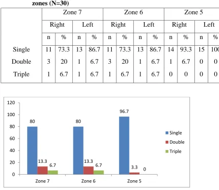
Table 1. EIP tendon frequency (n) and percentage (%) in three zones (N=30)
Zone 7 Zone 6 Zone 5
Right Left Right Left Right Left
n % n % n % n % n % n %
Absent 1 6.7 1 6.7 1 6.7 1 6.7 1 6.7 1 6.7
31
Figure 3. Percentage of EIP tendon in three zones
Extensor digitorum communis – index
Extensor digitorum communis tendon – index (EDC-index) was single in
29 hands (96.7%) and double in one hand (3.3%), as shown in Table 2 and Fig.5.
On the left side it was single in all hands (100%) with no difference between the
zones. On the right side it was single in 14 hands (93.3%) in zones 7 and 6. It
was double in one male right hand in zones 7 and 6. This double tendon fused
together before insertion making a single tendon in zone 5 (Fig. 4). In 93.3% of
hands it was bilaterally symmetrical.
Table 2. EDC-index tendon frequency (n) and percentage (%) in three zones (N=30)
6.7 6.7 6.7
93.3 93.3 93.3
0 10 20 30 40 50 60 70 80 90 100
Zone 7 Zone 6 Zone 5
Absent
Single
Zone 7 Zone 6 Zone 5
Right Left Right Left Right Left
n % n % n % n % n % n %
Single 14 93.3 15 100 14 93.3 15 100 15 100 15 100
33
Figure 5. Percentage of EDC-index tendon in three zones
Extensor digitorum communis – middle
Extensor digitorum communis – middle (EDC-middle) was single in 24
hands (80%), double in 4 hands (13.3%) and triple in 2 hands (6.7%). This
frequency was similar in zones 7 and 6. But in zone 5, single tendons were
observed in 96.7% of hands, double in 3.3% and triple tendons in none (Table 3,
Fig. 6). This showed that when multiple tendons are present, they usually fuse
together before insertion. The frequency of single tendons were slightly more on
the left side (86.7%) than on the right (73.3%) and double tendons were seen
more on the right side (20%) than on the left (6.7%) (Fig. 7). Triple tendons were
found in both hands of one male cadaver (Fig. 4) (Table 9). In zone 5 only
single tendons were seen on the left side (100%), whereas on the right side only
93.3% were seen because of fusion of multiple tendons into one before insertion.
One right hand showed double insertion in zone 5. 66.6% of hands showed
bilateral similarity with regard to single tendons.
96.7 96.7 100
3.3 3.3 0
0 20 40 60 80 100 120
Zone 7 Zone 6 Zone 5
Single
34
Table 3. EDC-middle tendon frequency (n) and percentage (%) in three zones (N=30)
Zone 7 Zone 6 Zone 5
Right Left Right Left Right Left
n % n % n % n % n % n %
Single 11 73.3 13 86.7 11 73.3 13 86.7 14 93.3 15 100
Double 3 20 1 6.7 3 20 1 6.7 1 6.7 0 0
Triple 1 6.7 1 6.7 1 6.7 1 6.7 0 0 0 0
Figure 6. Percentage of EDC-middle tendons in three zones
Extensor digitorum communis – ring
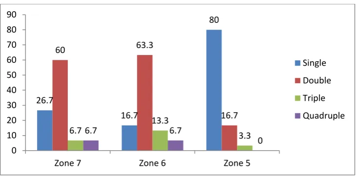
Extensor digitorum communis – ring (EDC-ring) tendon was single in 8
hands (26.7%), double in 18 hands (60%), triple in 2 hands (6.7%) and quadruple
in 2 hands (13.3%) in zone 7. In zone 6, single tendon was seen in 5 hands
(16.7%), double tendons in 19 hands (63.3%), triple in 4 hands (13.3%), and
quadruple in 2 hands (6.7%). In zone 5 single tendons were seen in 24 hands
(80%), double in 5 hands (16.7%), triple in one hand (6.7%) and quadruple in
80 80
96.7
13.3 13.3
3.3
6.7 6.7
0 0 20 40 60 80 100 120
Zone 7 Zone 6 Zone 5
Single
Double
[image:47.595.92.520.91.461.2]36
none (Table 4, Fig.10). This showed that even when majority of the tendons were
multiple they joined together as one before insertion. Comparison between sides
showed single tendons (Fig.8) were more on the left side in zones 6 and 5 and
same on both sides in zone 7. Double tendons had almost similar frequency in
right hands and left hands in zone 6.They were higher in right hand in zone 5 and
left hand in zone 7. Triple tendons (Fig. 9) were almost similar in all three zones
of right and left hands. The quadruple tendons (Fig. 4) were seen in two right
hands (13.3%) in zones 7 and 6 of two male hands (Table 9). None of the left
hands showed quadruple tendons.
In one female right hand extensor digitorum communis gave three tendons
to ring finger, of which two tendons passed to the dorsum of hand through the 4th
compartment of the extensor retinaculum and the third tendon passed through a
separate compartment between the fourth and fifth compartments of the extensor
retinaculum. In two hands, one female right and one male left hand, there were
double EDC-ring tendons, the ulnar of which gave a small slip to small finger.
Table 4. EDC-ring tendon frequency (n) and percentage (%) in three zones (N=30)
Zone 7 Zone 6 Zone 5
Right Left Right Left Right Left
n % n % n % n % n % n %
Single 4 26.7 4 26.7 2 13.3 3 20 10 66.7 14 93.3
Double 8 53.3 10 66.7 9 60 10 66.7 4 26.7 1 6.7
Triple 1 6.7 1 6.7 2 13.3 2 13.3 1 6.7 0 0
39
Figure10. Percentage of EDC-ring in three zones
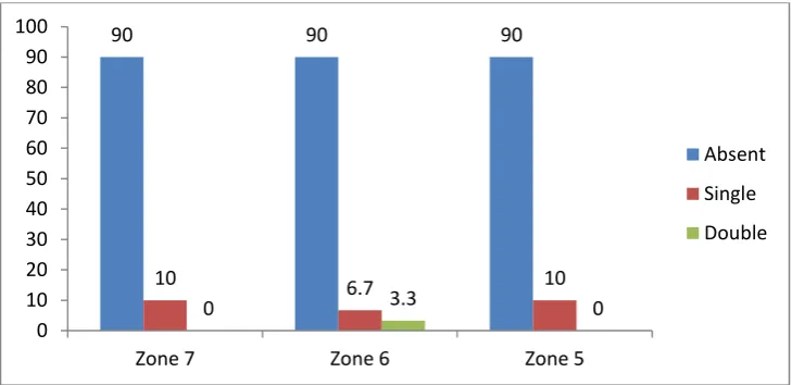
Extensor digitorum communis – small
Extensor digitorum communis – small tendon (EDC-small) was absent in
27 hands (90%) (Fig. 2) and single in 3 hands (10%) in zones 7 and 5 (Table 5,
Fig. 11). In zone 6, two hands were observed with single tendon (Fig.8).
Comparison between the sides showed that right and left sides had almost similar
frequency. On the left side, one hand had a single tendon in zone 7 which split
into two tendons in zone 6 but joined as single tendon before insertion into zone
5. In one male right hand EDC-small showed a small split at the head of 5th
[image:52.595.99.465.68.250.2]metacarpal bone which reunited before insertion into the small finger.
Table 5. EDC-small tendon frequency (n) and percentage (%) in three zones (N=30)
26.7
16.7
80
60 63.3
16.7 6.7
13.3
3.3
6.7 6.7
0 0 10 20 30 40 50 60 70 80 90
Zone 7 Zone 6 Zone 5
Single
Double
Triple
Quadruple
Zone 7 Zone 6 Zone 5
Right Left Right Left Right Left
n % n % n % n % n % n %
Absent 14 93.3 13 86.7 14 93.3 13 86.7 14 93.3 13 86.7
Single 1 6.7 2 13.3 1 6.7 1 6.7 1 6.7 2 13.3
40
Figure 11. Percentage of EDC-small in three zones
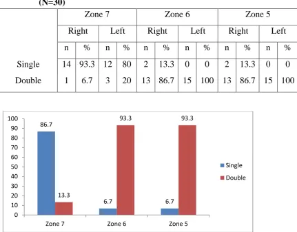
Extensor digiti minimi
Extensor digiti minimi (EDM) was seen as a single tendon in 26 hands
(86.7%) and double in 4 hands (13.3%) in zone 7. In zone 6, single tendons were
seen in 2 hands (6.7%) (Fig.4) and double tendons in 28 hands (93.3%) (Fig.2).
Zone 5 was similar to zone 6, with the majority of hands showing double tendons
(Table 6, Fig. 12). In zone 7, single tendons were slightly more on the right side
(93.3%) and double tendons on the left (20%). These double tendons were seen
only in male hands (Table 10). In zones 6 and 5, single tendons were observed
only in two right hands but none in left hands and the number of double tendons
were almost similar on both sides. One male left hand had two EDM tendons
from which the radial tendon gave a small slip which inserted into the 3y type of
juncturae tendinum. 86.6% of cadavers were bilaterally symmetrical.
90 90 90
10 6.7 10
0 3.3 0
0 10 20 30 40 50 60 70 80 90 100
Zone 7 Zone 6 Zone 5
Absent
Single
41
Table 6. EDM tendon frequency (n) and percentage (%) in three zones (N=30)
Zone 7 Zone 6 Zone 5
Right Left Right Left Right Left
n % n % n % n % n % n %
Single 14 93.3 12 80 2 13.3 0 0 2 13.3 0 0
Double 1 6.7 3 20 13 86.7 15 100 13 86.7 15 100
Figure12. Percentage of EDM in three zones
Extensor pollicis longus
Extensor pollicis longus (EPL) was a single tendon in all hands (100%) in
zone T5 (Fig.15) and 96.7% in zones T4 and T3 (Table 7, Fig. 13). In zones T4
and T3 one male left hand showed a double tendon (Fig. 14). When compared
between the sides, right and left frequencies were almost similar in all three
zones. 93.3% of cadavers were bilaterally symmetrical. 86.7
6.7 6.7
13.3
93.3 93.3
0 10 20 30 40 50 60 70 80 90 100
Zone 7 Zone 6 Zone 5
Single
[image:54.595.95.527.94.431.2]42
Table 7. EPL tendon frequency (n) and percentage (%) in three zones (N=30)
Zone T5 Zone T4 Zone T3
Right Left Right Left Right Left
n % n % n % n % n % n %
Single 15 100 15 100 15 100 14 93.3 15 100 14 93.3
Double 0 0 0 0 0 0 1 6.7 0 0 1 6.7
Figure 13. Percentage of EPL in three zones
Extensor pollicis brevis
Extensor pollicis brevis (EPB) was frequently a single tendon in 100% of
hands (Fig. 14) in zones T4 and T3 and 96.7% in zone T5 (Table 8, Fig.16). One
male hand had a double tendon in zone T5 (Table 10). Comparison between
sides showed frequencies were similar in zones T4 and T3. In zone T5, one left
hand had a double tendon (Fig 15). This accessory tendon inserted into base of
first metacarpal along with abductor pollicis longus. 93.3% of cadavers were
bilaterally symmetrical.
100 96.7 96.7
0 3.3 3.3
0 20 40 60 80 100 120
Zone T5 Zone T4 Zone T3
Single
[image:55.595.95.528.90.423.2]44
Table 8. EPB tendon frequency (n) and percentage (%) in three zones (N=30)
Zone T5 Zone T4 Zone T3
Right Left Right Left Right Left
n % n % n % n % n % n %
Single 15 100 14 93.3 15 100 15 100 15 100 15 100
[image:57.595.93.499.143.576.2]Double 0 0 1 6.7 0 0 0 0 0 0 0 0
Figure 16. Percentage of EPB in three zones
96.7 100 100
3.3 0 0
0 20 40 60 80 100 120
Zone T5 Zone T4 Zone T3
Single
45
Table 9. Tendon frequency (n) and percentage (%) in three zones in males and females (N=30)
Zone 7 Zone 6 Zone 5
Male Female Male Female Male Female
n % n % n % n % n % n %
EIP - absent 2 14.3 0 0 2 14.3 0 0 2 14.3 0 0
single 12 85.7 16 100 12 85.7 16 100 12 85.7 16 100
EDC-index
single 13 92.9 16 100 13 92.9 16 100 14 100 16 100
double 1 7.1 0 0 1 7.1 0 0 0 0 0 0
EDC-middle
single 8 57.1 16 100 10 71.4 14 87.5 13 92.9 16 100
double 4 28.6 0 0 2 14.3 2 12.5 1 7.1 0 0
triple 2 14.3 0 0 2 14.3 0 0 0 0 0 0
EDC-ring
single 2 14.3 6 37.5 1 7.1 4 25 12 85.7 12 75
double 9 64.3 9 56.2 10 71.4 9 56.2 1 7.1 4 25
triple 1 7.1 1 6.2 1 7.1 3 18.8 1 7.1 0 0
quadruple 2 14.3 0 0 2 14.3 0 0 0 0 0 0
46
Table 10. Tendon frequency (n) and percentage (%) in three zones in males and females (N=30)
Zone 7 Zone 6 Zone 5
Male Female Male Female Male Female
n % n % n % n % n % n %
EDC-small
absent 13 92.9 14 87.5 13 92.9 14 87.5 13 92.9 14 87.5
single 1 7.1 2 12.5 1 7.1 1 6.2 1 7.1 2 12.5
double 0 0 0 0 0 0 1 6.2 0 0 0 0
EDM –single 10 71.4 16 100 1 7.1 1 6.2 1 7.1 1 6.2
double 4 28.6 0 0 13 92.9 15 93.8 13 92.9 15 93.8
EPL – single 14 100 16 100 13 92.9 16 100 13 92.9 16 100
double 0 0 0 0 1 7.1 0 0 1 7.1 0 0
EPB – single 13 92.9 16 100 14 100 16 100 14 100 16 100
double 1 7.1 0 0 0 0 0 0 0 0 0 0
EDC – Extensor indicis communis; EDM – Extensor digiti minimi; EPL – Extensor pollicis longus; EPB – Extensor pollicis brevis
Juncturae tendinum
Juncturae tendinum comprising of three types were located in the
intermetacarpal spaces on the dorsum of hand. No juncturae tendinum was seen
47 Second intermetacarpal space
Type 1 juncturae tendinum was predominantly seen in the second
intermetacarpal space. It was observed in 28 hands (93.3%) and was found
equally in both right and left hands (Table 11). It was absent in one left female
hand. Type 2 juncturae tendinum was present in one male right hand (Table 14).
Type 3 juncturae tendinum was not found in the second intermetacarpal space.
86.6% of cadavers were bilaterally symmetrical without any gender difference.
Table 11. Distribution of juncturae tendinum in the second intermetacarpal space (N=30)
Right Left Total
n % n % n %
Type 1 14 93.3 14 93.3 28 93.3
Type 2 1 6.7 0 0 1 3.3
Type 3 0 0 0 0 0 0
Absent 0 0 1 6.7 1 3.3
n -number; %- percentage
Third intermetacarpal space
Type 2 juncturae tendinum was seen in 20 hands (66.7%) in the third
intermetacarpal space (Table 12). It was observed more in right hands (80%) than
in left hands (53.3%). Type 3 juncturae tendinum was found in 10 hands
48
was not seen in the third intermetacarpal space of any hand. Type 3 juncturae
tendinum was evenly distributed in males and females whereas Type 2
predominated in female hands (Table 14). 73.3% of cadavers were bilaterally
symmetrical without any gender difference.
Table 12. Distribution of juncturae tendinum in the third intermetacarpal space (N=30)
n -number; %- percentage
Fourth intermetacarpal space
Type 3 juncturae tendinum was observed in 20 hands (66.7%) (Table 13).
This was observed more on the left side (73.3%) than on the right (6%). Type 2
juncturae tendinum was found in 10 hands (33.3%) and this was observed more
on the right side (40%) than on the left (26.7%). Type 1 juncturae tendinum was
not observed in this space. Type 2 juncturae tendinum was common in males and
Type 3 was common in females. 73.3% of cadavers were bilaterally symmetrical
without any gender difference.
Right Left Total
n % n % n %
Type 1 0 0 0 0 0 0
Type 2 12 80 8 53.3 20 66.7
Type 3 3 20 7 46.7 10 33.3
49
Table 13. Distribution of juncturae tendinum in the fourth intermetacarpal space (N=30)
n -number; %- percentage
Type 1 juncturae tendinum
Type 1 juncturae tendinum is a thin filamentous one of fascial type
observed only in the second intermetacarpal space (Fig. 17). It was attached
between the extensor digitorum communis tendon for middle and index finger
but never to extensor indicis proprius. When extensor indicis proprius was
present ulnar to extensor digitorum communis tendon for index, this juncturae
passed superficial to this tendon. This juncturae ran in an oblique direction in
60.7% of hands and in a transverse direction in 39.3% of hands. When the
direction was oblique, 39.3% ran from an ulnar to radial direction and 21.4% ran
from radial to ulnar direction. Shape of this juncturae was rhomboid in 85.7% of
hands and trapezoid in 14.3% of hands. In relation to juncturae in other
metacarpal spaces, juncturae present in this space was proximal in 89.3% of
hands and almost at same level as other juncturae tendinum in 10.7 % of hands.
Right Left Total
n % n % n %
Type 1 0 0 0 0 0 0
Type 2 6 40 4 26.7 10 33.3
Type 3 9 60 11 73.3 20 66.7
50
These morphological features were present with almost similar frequency
between right and left hands. When compared between male and female hands,
Type 1 juncturae was more transversely placed in 53.8% male hands and
obliquely placed in female hands (the direction being ulnar to radial in 53.3% of
hands).
Type 2 juncturae tendinum
Type 2 juncturae tendinum is a ligamentous type attaching adjacent
tendons but not in continuity with the tendons (Fig. 17). They are thicker than
Type 1 but thinner than Type 3. They were predominantly seen in the third
intermetacarpal space. Of the juncturae present in the third intermetacarpal space,
66.7% of them were Type 2. They were all directed obliquely in an ulnar to
radial direction and attached between the tendons of extensor digitorum for
middle and ring fingers. 90% of them were rhomboid in shape and the rest were
trapezoid in shape. In 65% of hands, the juncturae in the third intermetacarpal
space were present just distal to the juncturae of second intermetacarpal space but
just proximal to juncturae of fourth intermetacarpal space. In 5% of hands it was
distal to juncturae of second space but at the same level to juncturae of fourth
space. In one hand juncturae of all the intermetacarpal spaces were at the same
52
Of the juncturae present in the fourth intermetacarpal space, 33.3% of
them were Type 2. Most of them were attached to the adjacent tendons of
extensor digitorum communis tendon to ring finger and small finger or to
extensor digitorum communis tendon to ring finger and extensor digiti minimi. It
was predominantly rhomboid in shape but trapezoid in one left hand. In male
hands, the direction was predominantly ulnar to radial and in female hands it was
predominantly radial to ulnar. One male right hand had Type 2 juncturae in
second intermetacarpal space attached to extensor digitorum tendon for index and
middle fingers.
Type 3 juncturae tendinum
Type 3 juncturae tendinum is a tendinous type in which the juncturae were
in continuity with the tendons. This type was present predominantly in the fourth
intermetacarpal space (66.7%) (Table 13). It was seen in the third intermetacarpal
space (33.3%) but never in the second intermetacarpal space (Table 12). It was
the thickest of all the types. All of them were attached from tendons of extensor
digitorum ring finger to extensor digitorum middle finger in an oblique direction
from ulnar to radial. They were intermediate in location in relation to juncturae of
the second and fourth intermetacarpal spaces. It was classified into two subtypes
‘r’ and ‘y’ based on its shape. In the ‘r’ subtype the juncturae branches out from
the base tendon like an ‘r’ (Fig. 18) whereas in the ‘y’ subtype it bifurcated from
53
seen in all of the hands. There was not much difference between male and female
hands. Type 3 made up 66.7% of juncturae of the fourth intermetacarpal space. In
90% of hands it was attached between the tendons of extensor digitorum to ring
finger and small finger tendon or to extensor digiti minimi. In the fourth space ‘r’
subtype was seen in 70% of the hands with no side difference. The ‘r’ subtype
was more common in female hands (83.3%) (Table 14). The ‘y’ subtype was
[image:66.612.94.531.319.529.2]seen in 30% of hands predominantly on the left side and more in males (50%).
Table 14: Distribution of juncturae tendinum in males and females (N=30)
IMC 2 IMC 3 IMC 4
Male Female Male Female Male Female
n % n % n % n % n % n %
Type 1 13 92.9 15 93.8 0 0 0 0 0 0 0 0
Type 2 1 7.1 0 0 9 64.3 11 68.8 6 42.9 4 25
Type 3 r 0 0 0 0 0 0 0 0 4 28.5 10 62.5
Type 3 y 0 0 0 0 5 35.7 5 31.2 4 28.5 2 12.5
IMC – intermetacarpal space
Patterns of juncturae combinations
The juncturae present in each intermetacarpal space may be fascial,
ligamentous or tendinous in type. Depending upon the type present the following
combinations in second, third and fourth intermetacarpal spaces were observed
54
Table15. Combination of Juncturae tendinum in Second-Third-Fourth Intermetacarpal Spaces (N=30)
Combination in Second-Third-Fourth Intermetacarpal Spaces
Frequency Percentage
Fascial – ligament – tendon 10 33.3
Fascial – ligament – ligament 9 30
Fascial – tendon – tendon 8 26.6
Fascial – tendon – ligament 1 3.3
Ligament – tendon – tendon 1 3.3
Absent – ligament – tendon 1 3.3
The first pattern was seen more in the right hand in females and in left
hand in males. The second pattern was seen more in the right hand in both males
and females. The third pattern was seen more in the left hand but there was no
gender difference. The fourth and sixth patterns were seen each in one left hand
of a female. The fifth pattern was