Histopathological Analysis of Salivary Gland Lesions and Role of Immunohistochemistry in Differential Diagnosis
Full text
Figure


Related documents
This tumor is usually located in the parotid gland and the minor salivary glands of the soft palate and represents less than 1% of all salivary gland tumors [2].. Several authors
The Effects of Manual Tooth Brushing on Parotid and Submandibular/Sublingual Gland Salivary Flow Rates in Healthy Young and Older Adults. Participant
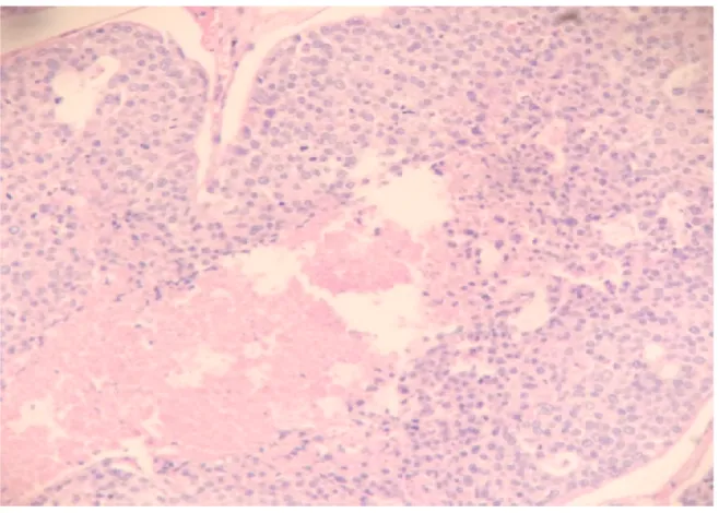
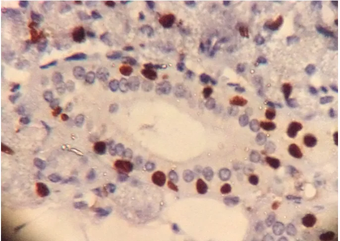
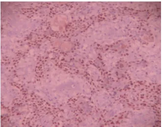
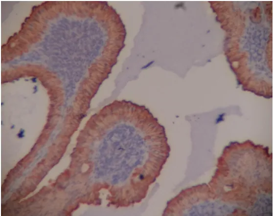
Conclusions: Histopathological examination is mandatory in the diagnosis of salivary gland lesions because of their wide spectrum of histomorphology.4. and sometimes within an
The most common aberrations in patients with salivary gland tumors were in the TP53 gene, followed by alterations in the cyclin pathway and the PI3K pathway (PIK3CA, PIK3R1, PTEN
The clinical presentation of minor salivary gland tumors is more diverse due their wide spread distribution throughout the upper aerodigestive track (Table 2). Most
Complete surgical excision is the primary treatment strategy for patients with salivary gland tumors, followed by radiotherapy or chemoradiotherapy in patients with
Male and parotid gland were the most affected and mucoepidermoid carcinoma was the most frequent lesion, followed by squamous cell carcinoma and adenoid cystic carcinoma..
Cysts of oral mucosa have similar historical and clinical features as benign mesenchymal and salivary gland tumors and are often included in the differential diagnosis of these
