PROSPECTIVE STUDY OF EFFECTS OF TURP ON OUTCOME,MORBIDITY AND MORTALITY IN PATIENTS WITH NON DIALYSIS REQUIRING RENAL INSUFFICIENCY
Dissertation Submitted to
THE TAMILNADU
DR.M.G.R. MEDICAL UNIVERSITY
CHENNAI
In Partial fulfillment of the regulations for the award of the degree of
M.CH DEGREE EXAMINATION BRANCH IV - UROLOGY
GOVERNMENT STANLEY MEDICAL COLLEGE
AND HOSPITAL
CHENNAI – 600001
CERTIFICATE
This is to certify that the dissertation entitled “PROSPECTIVE STUDY OF EFFECTS OF TURP ON OUTCOME, MORBIDITY,
MORTALITY IN PATIENTS WITH NON DIALYSIS
REQUIRING RENAL INSUFFICIENCY” is the bonafide original
work of Dr.AMAR NEEDHI GANESAN.B in partial fulfillment of
the requirements for MCH UROLOGY BRANCH – IV Examination
of the Tamilnadu Dr. M.G.R. Medical University to be held in August
2014. The period of postgraduate study and training was from August
2011 to July 2014.
GUIDE :
PROF. V.SELVARAJ , M.S.,MCH URO, PROFESSOR & HEAD
DEPARTMENT OF UROLOGY
GOVT. STANLEY MEDICAL COLLEGE CHENNAI-600 001.
PROF. V.SELVARAJ , M.S.,MCH URO, PROFESSOR & HEAD
DEPARTMENT OF UROLOGY
GOVT. STANLEY MEDICAL COLLEGE CHENNAI-600 001.
Dr. A.L.MEENAKSHI SUNDARAM, MD., DA, DEAN,
DECLARATION
I, Dr. AMAR NEEDHI GANESAN.B , solemnly declare that the dissertation titled, “Prospective Study of effects of TURP on
outcome,morbidity and mortality in patients with non dialysis requiring
renal insufficiency” is a bonafide work done by me at Govt. Stanley
Medical College & Hospital during 2011-2014 under the guidance of
and supervision of PROF.DR.V.SELVARAJ, M.S, M.CH URO,
Professor and Head, Department of Urology, Government Stanley
Medical College, Chennai-600 001.
The dissertation is submitted to The Tamilnadu Dr. M.G.R.
Medical University, towards partial fulfillment of requirement for the
award of M.CH UROLOGY (BRANCH – IV) in UROLOGY.
Place: Chennai.
Date:
ACKNOWLEDGEMENT
I express my profound gratitude to Dr. MEENAKSHI
SUNDARAM., M.D., Dean of Government Stanley Medical College and Hospital, Chennai–600001 for permitting me to use all the needed
resources for this dissertation work.
I sincerely express my grateful thanks to Prof. V.SELVARAJ.,
M.S, M.CH UROLOGY, Professor and Head, Department of Urology, Government Stanley Medical College for his unstinted support and
advice rendered throughout my study. I thank him for being a constant
source of encouragement, inspiration, not only in this study but in all my
professional endeavours. I thank Prof.P.Govindarajan, M.S,M.Ch
UROLOGY for guiding me throughout this study period.
I express my sincere thanks to all the Assistant Professors,
Dr.Deepak, Dr.Periasamy, Dr.Thiruvarul, Dr.Ayesha in Department of Urology, Stanley Medical College & Hospital, Chennai.
I also sincerely thank Ethical Committee, Govt. Stanley Medical
College, Chennai for approving my study.
I extend my sincere thanks to my subjects but for them the project
TABLE OF CONTENTS
Sl. No Topic Page No
1. INTRODUCTION 1
2. AIM OF THE STUDY 4
3. REVIEW OF LITERATURE 5
4. MATERIALS AND METHODS 42
5. OBSERVATION AND RESULTS 48
6. DISCUSSION 63
7. CONCLUSION 72
8. BIBLIOGRAPHY
9. ANNEXURES
Institutional ethical committee clearance
Anti Plagiarism Certificate
Proforma
Patient consent form
ABBREVIATIONS
BPH - Benign prostatic hyperplasia
TURP – Transurethral resection of prostate
IPSS - International Prostate symptoms score
1
INTRODUCTION
Benign Prostate Hyperplasia (BPH) is a common disease in adult
men and its incidence is age related. Prevalence of BPH is
approximately 25% in men aged 40 to 49 years, 50% in men aged 50 to
59 years and 80% in men aged 70 to 79 years.1
Renal failure and symptomatic benign prostatic hyperplasia (BPH)
are two common health problems, they usually co-exist in 5.9–13.6% of
the male population over 50 years of age. Actually going by the natural
history of the disease and its progression with relation to Benign prostatic
hyperplasia and its complications, it is noted that 13.6% of patients who
presented to undergo Transurethral resection were in renal failure. It is
usually not clear in this group of patients whether the reason for renal
insufficiency is or is not Benign prostatic hyperplasia. However, it has
been reported in some studies that the incidence of diabetes mellitus and
hypertension is higher in patients with renal failure (RF) and lower
urinary tract symptoms (LUTS) due to Benign prostatic hyperplasia. On
the other hand, it is known that due to chronic urinary obstruction, BPH
can lead to renal failure and even death occasionally.
The main constant indication for BPH surgery has been medical
2
but definite surgical indications usually includes upper urinary tract
deterioration. Renal failure increases the risk in transurethral prostatic
surgery, so there is a tendency for avoiding the surgery till there is a
detection of an absolute indication occurs. But these studies are based on
data from two or three decades ago not representing current practice.
As we understand that these patients with BPH whether
symptomatic or asymptomatic, if left untreated may present with renal
failure which could be chronic or acute. Despite the many possible causes
of renal failure in elderly patients, the common causes were BPH (38%),
neurogenic bladder (19%), obstructive pyelonephritis (15%).2
While the underlying mechanism for developing renal failure
associated with benign prostatic hyperplasia is likely multifactorial and
co-morbid factors in elderly men may contribute to renal impairment, we
wanted to evaluate the incidence of BPH with renal failure at our
institute.
TURP remains the gold standard surgical procedure for treatment
of these cases. However, patients in renal failure have an increased risk
for complications after TURP compared with patients with normal renal
function, so we wanted to study the treatment outcome and complications
3
Attending to high prevalence of BPH in older men with CKD it is
invaluable to take into consideration the relationship between these two
clinical entities. However, despite the high prevalence of renal failure and
BPH in elderly men, there is limited knowledge on the association
between these two conditions, there is very little information in the
literature regarding the role of only BPH as a causative factor in causing
renal failure and its treatment outcome.
The purpose of this study was to determine the incidence of renal
failure associated with BPH , effect of TURP in the morbilidity and
4
AIMS AND OBJECTIVES
To study the prevalence of co-morbid factors in patients with
Benign prostatic hyperplasia and non dialysis requiring renal
insufficiency.
Study of treatment outcome following the surgical management of
BPH with non dialysis requiring renal failure.
Study of complications associated with operative management in
5
REVIEW OF LITERATURE
Epidemiology of Benign prostatic hyperplasia :
Benign hyperplasia of Prostate occurs with increased growth of
non malignant tissue of prostate which surrounds urethra, it narrows the
lumen of urethra and subsequently gives rise to symptoms.63
Diagnosis of benign prostatic hyperplasia done conclusively on
histological evaluation of prostate. Histological evaluation is by taking
prostate by transurethral resection or trans rectal ultrasound guided or by
doing autopsy. But other measures, namely symptomatology, obstruction
of bladder with associated enlarged prostate is used to mark benign
prostatic hyperplasia. Because of this, the end point of benign prostatic
hyperplasia becomes difficult to assess.
These facts gives us sufficient details about the newly occurring
cases and disease progression.The prevalence of benign prostatic
enlargement is calculated by taking histological part (assessed by
autopsy) or clinically. (3) No men 30yrs and younger had evidence of
benign prostatic hyperplasia and the peak of prevalence increased along
with each age group, ultimately reaching 88% in men with eighties.4
Three things which are assessed separately which comprise of
6
three or two of them or only one. Hence prostatism has been now
changed to lower urinary tract symptoms (LUTS).5,6
Representative diagram showing relationship of various factors involved
Histologically, hyperplasia of prostate is seen in 8% men of age
31-40. Whereas it increases to 90% of men in ninth decade.4,7. In the setting
of Benign prostatic enlargement, untreated chronic kidney disease can
result in ESRD requiring dialysis or kidney transplantation.
Relationship between benign prostatic hyperplasia and Chronic KidneyDisease :
Etiology of benign hyperplasia of prostate is largely unknown, but
from clinical practice and studies, natural history of hyperplasia of
7
function with time. Both benign hyperplasia of prostate and chronic
kidney disease are commonly prevalent in ageing male.
Following are the studies conducted to find relationship between
Benign hyperplasia of prostate and chronic disease of kidney:
a. Study conducted by Epidemiology Project of Rochester,
concluded that association was significant between signs &
symptoms of benign prostatic hyperplasia and Chronic kidney
disease in their population-based sample which comprised of
white men numbering 476.9
b. But, another study conducted in Austria concluded that Lower
urinary tract symptoms in men was not an independent risk
factor for impaired kidney function.10
c. Another study that was conducted on 30,466 men study from
the HUNT II (the Nord-Trondeleg Health Study), they also
failed to show a connection between Lower urinary tract
symptoms and Chronic kidney disease.11
d. But recent evidence from two different studies have found out
an association of bening hyperplasia of prostate and chronic
disease of kidney.12,13
Eventhough the prevalence of chronic kidney disease is considered
8
consider it in patients presenting with obstructive lower urinary tract
symptoms mainly or with low peak flow rate or patients having
hypertension or diabetes. 13
Clinical guidelines for benign hyperplasia of prostate, which was
created in 1994 by Health Care Policy and Research Agency in 1994,
recommended routine serum creatinine measurement in men with lower
urinary tract symptoms, but in year 2003, a update discontinued the
routine serum creatinine measurement in these persons.9 So these
different approaches to benign prostatic hyperplasia patients, leads to a
significant amount of patients with Chronic kidney disease going
undetected.
By taking this data into account, we should always bear in our
minds that benign prostatic hyperplasia is almost an ubiquitous condition
in the setting of old men. Low occurrence of chronic kidney disease in
benign prostatic hyperplasia usually in clinical trials, should not be used
to infer a weak association between the two disease processes.
Signs and symptoms of Bladder outlet obstruction namely low
QMax, high residual urine (post void), Obstructive symptoms of lower
urinary tract are significant predictors of chronic kidney disease.9,12
Bladder outlet obstruction, probably, is the bridge connecting between
9
But we should never forget that chronic disease of kidney in the
setting of benign hyperplasia of prostate is a multifactorial process, and
thus it becomes difficult to separate the contribution of this condition
from all the other renal insults. This also affects and it takes its toll on the
design of studies as many men with concomitant disease are excluded,
and thus makes it harder for investigators, to take into account the true
influence of both these conditions.
Pathophysiology and Progression of Benign prostatic hyperplasia :
Although the etiology of benign hyperplasia of prostate is not
known, there are similarities found between benign hyperplasia and
morphogenesis of prostate during development. This prompted to suggest
hypothesis that benign hyperplasia of prosate results due to reawakening
of embryogenesis in adulthood.14
Most common renal pathology finding in men with obstructive
nephropathy with benign prostatic hyperplasia is, Chronic interstitial
nephritis15, 9 and 30% of cases have been attributed to obstructive
uropathy. End stage disease of kidney secondary to bladder outlet
obstruction can be prevented if recognised early, however it is still
difficult to recognise which men with benign prostatic hyperplasia can
10
So we have to recognize factors which are measurable during
evaluation of benign hyperplasia of prosate. This helps to prevent patients
from developing renal failure.
Anatomical
Benign hyperplasia of prostate initially in periurethral transition zone, then progressively enlarging
Benign hyperplasia of prosate develops first in periurethral
transition zone of the prostate. This transition zone has two separate glands located external to the preprostatic sphincter. Benign prostatic
hyperplasia also involves an increase in the number of glands, mostly the
periuretheral glands increase in number, simultaneously there is a
increase in smooth muscle and connective tissue in the periuretheral
region of prostate.16,3
Physiological
According to the physiological point, when the prostate enlarges,
urethral compression occurs, which prevents the outflow of urine and
contributing to the common lower urinary tract symptoms. Smooth
11
myosin heavy chain is upregulated along with down regulation of smooth
muscle myosin heavy chain. These things suggest that there is loss of
normal modulation pathways.16
We need to elucidate the factors determining passive tone in
prostate.(3)Adrenergic system regulates the active muscle tone in
prostate.(18) Adrenergic neurotransmitters have been involved in prostate
smooth muscle regulation as well as contraction, and α-adrenergic
blockade actually leads to a significant normal protein gene expression
downregulation, specifically smooth muscle myosin heavy chain.19,3
Clinical
Studies conducted recently, found there was a significant
correlation between urgency symptoms and prostate size.20 This also
proves a link between Chronic disease of kidney and benign hyperplasia
of prostate. Prostate size assessed by rectal examination is considered to
have poor reliability, so trans rectal ultrasound of prostate is used in
studies.3
So, prostate and its benign enlargement can contribute for outflow
obstruction, by two factors, one is the static component (periurethral
compression caused by stromal component), the second one is also by
dynamic component (smooth muscle cells and supplying adrenergic
12
BENIGN PROSTATIC HYPERPLASIA
PATHOPHYSIOLOGY AND PROGRESSION : Disease progression :
Three things contribute, they are Symtoms of lower urinary tract, Chronic retention of urine, Acute retention of urine.
Lower Urinary Tract Symptoms (LUTS)
Lower urinary tract symptoms (LUTS) are described as clinical
criteria to define a man with urinary problems. Most men with benign
hyperplasia of prostate have following symptoms namely voiding
dysfunction, nocturia, urgency, thin urinary stream, increased urinary
frequency and a sense of incomplete bladder emptying.
Many studies were done to achieve a scientific relation between
Lower urinary tract symptoms and Chronic kidney disease. But, until
recent years there was no palpable evidence mainly connecting these two
entities. A retrospective study which was done to find this, did not find
any relation between duration of symptoms and serum creatinine levels
measured in these patients.21
Our clinical practice shows us that many men with Lower urinary
tract symptoms routinely do not value their symptoms, and they avoid
seeking medical care. Those older men often tolerate their symptoms and
13
However we must take into account that the absence of these
symptoms in older men does not necessarily exclude benign prostatic
hyperplasia with urinary outlet obstruction. Silent prostatism or Silent
obstruction, a term which describes asymptomatic patients developing
chronic disease of kidney resulting from benign hyperplasia of prostate.
So, International prosate symptom score will not be useful in these
patients.23
Post-voiding residual urine volume – Chronic urinary retention
Chronic urinary retention is the mechanism, by which benign
prostatic hyperplasia can result in renal failure.9 Chronic retention of
urine is taken as post-void residual urine (PVR), higher than 100 mL. It
is significantly associated in Chronic disease of kidney.24,9,12
Acute urinary retention
Acute urinary retention is described as an acute complication of
Benign prostatic hyperplasia. These patients suffer from an acute, sudden
and painful inability to void. It needs emergency intervention. Acute
urinary retention was the main indication of surgery in 25% and 30% of
men undergoing trans urethral resection.3 Other causes that can trigger
acute urinary retention are surgery, anaesthesia, trauma, medications,
14
Acute urinary retention is uncommon in men under sixty years,
and may be responsible for the majority of acute renal failure cases due
to obstructive uropathy.25 Men in whom acute urinary retention is
promptly relieved by catheterizing the bladder, acute renal failure
usually does not develop but long-term tubular dysfunction can still
occur.9
So, dysfunction of renal tubules can persist after the acute
urinary retention episode and probably this might result in progressive
renal disease affecting these patients.
Pathophysiology of Progression to Renal failure
Remodelling of Bladder in response to urinary obstruction
Enlarged lateral lobes and median lobe of prostate producing bladder outlet obstruction with bladder
15
The bladder plays an important role in pathophysiology of benign
hyperplasia of prosate and its complications. So, the bladder responds by
adaptation, though it is a partial adaptation. Obstruction induces changes
in bladder is closely related to symptoms rather than the obstruction by
itself.
Two types of bladder changes are observed. First is detrusor
instability presenting with frequency and urgency and second one is
decreased contraction of detrusor leading to poor stream, hesitancy,
intermittency, increased residual urine. 3
Thickened bladder is measured by ultrasound. Trabeculations due
to hypertrophy of smooth muscle and permeation of connective tissue
accounts for elevated bladder pressure in patients with high pressure
chronic retention.26
The major detrusor changes and trabeculation were due to an
increase in detrusor collagen. Severe trabeculation is directly related to
significant post void residual urine27 suggesting that increases in collagen
in the bladder wall is mostly responsible for incomplete bladder emptying
rather than impaired detrusor muscle function.3
Detrusor hypertrophy is one of the first and foremost modification
in the bladder and, as studied in animal models, the response initially is
16
Cellular and physiological changes that occur in bladder muscle
and collagen, contributes to high pressure bladder that perpetuates itself
with worsening ability to empty and causing kidney lesions. These
mechanisms of bladder remodelling develop in a hypofunctional bladder,
with low compliance.28
Ureterovesical junction and upper tract dilation
Intravesical enlargement of prostate producing bilateral ureteric dilatation with ureterovesical junction
obstruction and bladder trabeculations
Generally, ureterovesical junction obstruction caused by
remodelling of bladder in chronic urinary retention is a contributing
mechanism for renal failure in benign prostatic enlargement.9 Upper tract
dilation occurs as a consequence of a continuing bladder outlet
obstruction and remodelling (detrusor hypertrophy and scarring) which
17
Upper urinary tract dilation or hydronephrosis is consistent with
chronic renal failure from obstructive uropathy. In men with benign
prostatic hyperplasia and increased serum creatinine, hydronephrosis is
common in about one third of patients, but it is found in 90% of men with
benign prostatic hyperplasia who are hospitalized with uremic
symptomatology. In ultrasound evaluation, it is common among patients
with bilateral hydroureteronephrosis, leading on to compression and renal
cortical thinning, with obvious impact in renal function.23
Other causes
- Recurrent urinary tract infections with chronic retention due to
benign hyperplasia of prostate may be a contributing factor leading
on to chronic renal failure.9
- Other cause being, Secondary hypertension occurring due to
chronic urinary retention is a described complication of benign
prostatic hyperplasia, leading to hypertensive kidney disease.30
- Nephrogenic diabetes insipidus can be caused by partial or chronic
urinary obstruction, this can result in renal failure.31
Other clinical entities like diabetes and hypertension are
independent factors that can lead to chronic kidney disease.(32)
Patients with benign prostatic hyperplasia are usually carriers of
18
renal function and must be taken into account as sombre conditioners of
renal disease.
Clinical symptomatology :
Benign prostatic hyperplasia is a chronic and progressive
condition,(33) where in patients generally have a history of lower urinary
tract symptoms and indolent obstructive uropathy. The clinical
presentation of benign hyperplasia of prostate and associated obstructive
uropathy varies and this reflects the source and duration of obstruction. In
benign prostatic hyperplasia, symptoms directly results from bladder
outlet obstruction (BOO) from prostate enlargement which is static
component dynamic component by enhanced smooth muscle tone and
increased resistance within the gland.
These physiologic issues reflect in voiding dysfunctions, that
significantly affects the health and quality of life of older men. Most of
the patients have characteristic symptomatology.
Patients complaints are usually thin urinary stream, nocturia,
urgency, with decreased flow rate, low values in Qmax and Qaverage, a
sense of incomplete bladder emptying, straining during micturition,
increased urinary frequency and dribbling during or after urination.9
Physical examination consists of digital rectal examination
19
lower abdominal percussion and palpation and focused neurological
examination to rule out neurogenic component involved in the
presentation.
Recurrent or persistent urinary tract infections (UTI) which are
associated with prolonged urinary stasis of lower urinary tract
obstruction, urgency, frequency, dysuria, hematuria are common
complaints among men with Urinary tract infection.
Chronic urinary retention due to benign hyperplasia of prostate is
defined as a palpable bladder, corresponding to high post void residual
urine34 and most of these patients with chronic urinary retention have an
indolent and progressive disease, with worsening of urinary symptoms
and the majority of these patients just seek for medical care in worsening
health conditions with acute renal insufficiency.
It becomes necessary to investigate the symptoms and signs
suggesting chronic kidney disease like vomiting, nausea, edema,
lethargy,hypertension.23
In some rare cases of patients who present to the emergency room
because of anuria, require interventional procedures like indwelling
catheter, nephrostomy tube insertion either unilaterally or bilaterally and
sometimes depending on the level of renal function, hemodialysis may be
20
Even though the signs and symptoms of benign hyperplasia of
prostate are normally present in a group of patients, there are a significant
number of patients who are relatively asymptomatic35 (without significant
voiding dysfunction), but can present primarily as a clinical sequel of
renal insufficiency namely uremic symptoms; with vomiting, nausea,
mental status changes and electrolyte disturbances namely hypercaliemia
and nonanion gap acidosis.
Older patients with voiding dysfunctions caused by chronic urinary
obstruction might present with hypertension due to hypervolemia in the
case of bilateral obstruction or increased renin release.35 Hypertension, on
other hand can be itself the sole cause of renal failure.
If left untreated, benign prostatic hyperplasia can cause serious
complications including hematuria, urinary tract infections, renal failure,
bladder stones, incontinence and mortality related with benign prostatic
hyperplasia.
Complications in Patients with Benign hyperplasia of prostate : Mortality :
Patients with renal failure undergoing transurethral resection have a
higher risk (25%), in comparison to normal renal parameters (17%),
21
Bladder stones
Bladder stones occur in line with retention of urine leading on to
stasis and urinary infection. These factors favour ion aggregation and
stone nucleation. This leads on to formation of vesical calculi.
Urinary tract infections
Urinary tract infections are usually due to chronic urinary
obstruction, due to increased post void residual urine, predisposing to
Urinary tract infections.39 This leads to multiple hospital admissions for
treating these urinary tract infections.
Incontinence of Urine
Incontinence can develop from transurethral surgical intervention
for benign prostatic enlargement 40. Other causes are overflow incontinece
due to overdistention of the bladder or due to instabilityof detrusor
resulting in urge incontinence.41,42,3
Hematuria
Gross hematuria with clots can be seen in patients with benign
hyperplasia of prostate. Microvessel density in prostate is higher in
hematuria patients than in controls3 suggesting that vascular lesions in the
22
Tests for diagnosing the condition :
Nowadays it is increasingly rare to find a patient with chronic renal
failure from chronic urinary retention due to benign prostatic hyperplasia,
about 13.6% (range from 0.3 to 30%) of men with benign prostatic
hyperplasia may present with chronic kidney disease defined by a
baseline serum creatinine of more than 133 mmol/L (1.5 mg/dL). This is
particularly true in older patients with cognitive deterioration and
autonomy impairment.
In order to diagnose and monitor the impact of a bladder outlet
obstruction due to benign prostatic hyperplasia in the upper urinary tract,
some laboratory and imaging tests:
Standardized questionnaires
Creatinine levels in serum or measuring estimated glomerular
filtration rate (eGFR)
Urine analysis
Serum prostatic specific antigen (PSA) levels
Uroflowmetry with peak flow rate determination (Q max)
Renal and bladder ultrasonography with detrusor thickness
evaluation
Transrectal prostate ultrasonography with measurement of prevoid
23
Cystometry, other urodynamic studies as needed
Urethrocystoscopy.
Symptom assessment by standardized questionnaires
Benign prostatic hyperplasia Impact Index (BII), a questionnaire
assessing symptoms effect in everyday, daily activities interference, so
informing us the impact of benign hyperplasia of prostate. This can be
used with International prostate symptom score.(American Urology
Association 2010).
Symptom quantification is useful for diagnosis, determination of
disease severity and monitoring of benign prostatic hyperplasia.
International Prostate Symptom Score is the standard got from the
American Urological Association Symptom Index published in 1992.43
A recent multivariate analysis, found relationship between
individual symptoms in theis questionnaire and chronic kidney disease
status. They found that obstructive symptoms like poor stream and
hesitancy were associated significantly with chronic kidney disease in
age.44 Where as Irritative symptoms had no positive correlation with
chronic kidney disease. Moderate to severe Lower urinary tract
Symptoms (IPSS > 7) were positively correlated with chronic kidney
24
Lower urinary tract Symptoms compared with men with no or mild
Lower urinary tract Symptoms.11
Kidney function decreases with age and age significantly correlates
with Symptoms of lower urinary tract.10
Although symptom score assessment do not directly correlates with
chronic kidney disease or can’t be used to establish the diagnosis of
benign prostatic hyperplasia, it may serve as a basis for symptom severity
and management approach to patients with Symptoms of lower urinary
25
Serum creatinine
American Urological Association 2010 Guidelines do not suggest
routine serum creatinine measurement in management of benign
hyperplasia of prostate. This recommendation is based on the conclusion
26
hyperplasia than in men of the same age group belonging to general
population.
But, the European Association of Urology Guidelines on benign
prostatic hyperplasia (2004) and the nephrology-focused NICE (National
Institute for Health and Clinical Excellence) guidelines advocated
measuring serum creatinine levels in all patients. This is relied on the
fact, that bladder outlet obstruction due to benign prostatic enlargement
can cause hydronephrosis and renal failure.23
Patients with benign prostatic enlargement and renal insufficiency
have relatively higher postoperative complications (25% complication
rate compared with 17% for patients without renal failure) and mortality
goes up to sixfold than those with normal renal function.37,38, 45 Estimated
glomerular filtration rate (eGFR) is a much more reliable measure to
define chronic kidney disease and is better than simple serum creatinine
measurement.46
Urinalysis
Urinalysis is an inexpensive and simple test that is recommended
as a primary evaluation of a patient with suspected benign prostatic
enlargement. It is used to rule out urinary tract infection and hematuria.
On the other hand, the finding of proteinuria and/or micro- albuminuria
27
Measuring Total PSA (Prostate specific antigen):
Total PSA is to be offered to patients with more than ten years of
life expectancy and in whom measuring Prostate specific antigen will
change the management of the lower urinary tract symptoms (AUA 2010
Guidelines). Combining digital rectal examination (DRE) and total PSA
measurement becomes the cornerstone of basic screening of prostate.
The risks of requiring surgery and developing acute retention is based on
measuring PSA and prostatic volume.
Usage of Uroflowmetry ( Peak urinary flow rate )
Uroflowmetry is a noninvasive, simple urodynamic test that
allows us to have a objective evaluation of the patient micturiating
pattern. Although uroflowmetry is an non specific evaluation tool, the
micturition graph may suggest some recognizable patterns (e.g. meatal
stenosis, urethral stricture, benign prostatic hyperplasia) and it is a tool
which is reproduced to quantify the urinary stream strength. It is a useful
preoperatively. Peak urinary flow rate (PFR), or Qmax, predicts surgical
outcome – by suggesting that, in patients with a preoperative Peak flow
rate more than 15 mL/s have poorer outcomes than patients with lesser
peak flow rate. It is also an independent predictor for chronic kidney
disease rather than reported Lower Urinary Tract Symptoms by
28
Uroflowmetry. A) Normal patient; B) BENIGN PROSTATIC HYPERPLASIA patient
Ultrasound of kidney,ureter, bladder :
Patients with an elevated serum creatinine level, increase post-void
residual urine volume are candidates requiring ultrasound of kidney.47
Renal ultrasonography has many advantages over intravenous urography
(IVU) for upper urinary tract. It can measure and evaluate bladder,
post-void residual urine volume and prostate, better characterization of renal
masses if found, no harm of radiation, no side-effects and lower cost.
Renal Ultrasound. Two ultrasound scans in benign prostatic hyperplasia patient showing bilateral
hydroureteronephrosis (right and left kidney respectively)
Bladder ultrasonography
Measuring bladder wall thickness by transabdominal ultrasound,
it is a method to assess bladder outlet obstruction.49 Measurement of
29
can detect bladder outlet obstruction, if it is more than 2 mm.(50) But it is
not recommended by guidelines as it lacks reproducibility.
Bladder Ultrasound. Two ultrasound scans in benign prostatic hyperplasia patient. It is possible to observe the
trabecullation, bladder wall thickening and diverticulum
Evaluation of Post-void residual urine:
Post-void residual urine volume can be measured non invasively
by transabdominal ultrasonography.(41) It can also be measured by
invasive methods like catheterization. Residual urine volumes more than
300 mL affect renal function in cases of benign hyperplasia of
prostate.51,9 Post Void Residual urine more than 100 mL is chronic
retention of urine which is associated with chronic kidney disease.9
Even residual urine volumes less than 100 mL can affect renal function as
the presence of residual urine in post void period is related with renal
function regardless of the quantity. Thus ultrasonographic evaluation of
post-void residual urine is a useful test in the prevention of chronic
30
Transrectal ultrasonography of Prostate :
Transrectal ultrasonography of prostate (TRUS) is performed for
asssessing prostate size and shape, tissue characterization and carcinoma
prostate. There is no relationship between prostatic enlargement measures
and chronic kidney disease.9
Ultrasonographical Tranabdominal grading of prostatomegaly:
Grade I - 3.0 to 3.8 cms, 30 Gms. Grade II - 3.8 to 4.5 cms, 30- 50 Gms.
Grade III- 4.5 to 5.5 cms, 50- 80 Gms. Grade IV - 5.5. cms, 80 Gms.3
Prostate Ultrasound. Prostate transrectal ultrasonography (sagital view)
Cystometry
It is usually not a routine examination for benign prostatic
enlargement evaluation. However, it can help to identify high bladder
pressure, low bladder compliance and detrusor instability that can
31
Use of Urodynamics :
Urodynamics is mainly useful for distinguishing between bladder
outlet obstruction and impaired contraction of detrusor. It is performed if
it is going to affect the therapeutic decision. Patients with history of
neurologic diseases known to affect bladder or sphincter and patients
with bothersome symptoms with peak flow rate more than 15 ml/s may
benefit.
Urethrocystoscopy
Urethrocystoscopy is not to be done routinely but is optional if
invasive treatment is contemplated.41,43 This also can confirm, other
causes of outflow obstruction, sametime eliminating intravesical
pathologies.
Cystoscopic view showing enlarged lateral lobes of prostate and verumontanum
Aspect of Treatment :
Patients with mild symptoms are managed by watchful waiting,
patients with moderate symptoms should receive pharmacotherapy and
32
So, a man with preoperative IPSS more than17, has 87% chance of
having symptom reduction.52
We have to identify a group of patients who are at increased risk
of progression (e.g. age, symptoms, Prostate specific antigen level,
Qmax, volume of prostate and post-void residual urine). Here we have to
give early preventive treatment.53,50 Because, a higher frequency of
kidney failure in patients presenting for prostate surgery than for non
prostate surgery has been shown, and several studies have shown
improvement in kidney function after prostatectomy.21
Emergency situations :
Patients who present to the casualty, with bladder outlet
obstruction and high serum creatinine, they should first be put in a
urethral catheter and subsequently they need to be evaluated to
distinguish between, whether it is an acute and chronic renal failure.
Hospitalization is required in these cases. If hydroureteronephrosis and
azotaemia persists despite decompressing the bladder, we should suspect
an ureterovesical junction obstruction and the next step would be bilateral
percutaneous nephrostomy or bilateral double J stents if possible, these
are done for temporarily drainage. Patients further may need urgent
hemodialysis. Ureteroneocystostomy after a prostate ablation may be
33
Benign prostatic hyperplasia - Medical treatment :
Medical approaches are not used to treat if complications are
associated with benign prostatic hyperplasia (one of them is chronic
kidney disease). They are used for Lower urinary tract symptoms relief
and for preventing the progression of benign prostatic hyperplasia
(especially 5 alpha reductase inhibitors - 5-ARI).It is also useful in
preventing benign prostatic hyperplasia complications such as chronic
kidney failure. However, they can’t revert chronic kidney disease
secondary to benign prostatic hyperplasia.
Surgical treatment
Surgical treatment is mainly offered to men developing
complications from benign hyperplasia of prostate. Health Care Policy
and Research agency and International Consensus Guidelines,
recommend surgery if patient has
1. refractory urinary retention (patient failing atleast one catheter
removal attempt)
2. Following conditions secondary to benign prostatic
hyperplasia- recurrent Urinary tract infection, bladder stones,
recurrent gross hematuria, renal insufficiency, large bladder
34
Some studies suggest that dialysis dependent patients may recover
renal function up to a year after prostatic surgery. Here efforts should be
made to identify and treat benign prostatic hyperplasia in patients under
dialysis.
Erectile dysfunction in about 4% to 10% and urinary incontinence
in 0.5% to 1.5% can develop in postoperative period.54,42 Recurrence of
benign hyperplasia of prostate following surgery at five years is about
2% to 10%.
Standard surgical procedures
Transurethral resection of the prostate is the standard and gold
standard in the sugery for Benign hyperplasia of prostate. Other
procedures are compared against this.55
Complications of Transurethral resection of Prostate :56
Patients with renal failure undergoing procedure need attention as the
complication rates increase in this group including mortality. The
35
Intra-operative and peri-operative complications of TURP56
36
Complications Bleeding
Arterial bleeding is more noticed in cases with preoperative
infection, retention of urine due to gland getting congested. Antiandrogen
preoperatively can help in decreasing bleeding. Venous bleeding due to
perforation of capsule and opening of venous sinusoid during surgery.
The amount of intraoperative bleeding usually depends on size of
prostate gland and amout resected.
Extravasation
If bladder neck division occurs or capsule perforated,
extravasation occurs. It is usually extraperitoneal, but if bladder injured
or diffusion occurs in large volume, it can become intraperitoneal.
Injury to Ureteric orifices :
It can occur during large median lobe resection where it becomes
difficult to identify ureteric orifice. Treatment depends on severity.
External sphincter injury :
Injury occurs mostly at ventral area ( at 12’0 clock postion)
because we cannot visualize verumontanum. But if verumontanum is
37
Postoperative complications57 Bladder tamponade
Evacuation of clots due to recurrent or persistent bleeding or
reintervention occurs in 1.3–5% patients. Changing of colour from clear
to red intermittently in irrigation suggests arterial bleeding, whereas
venous bleeding usually results in a irrigation fluid continuously showing
dark red colour.
Infection
The infection rate is usually low .Risk factors for infection :
- Preoperative bacteruria
- Duration of procedure exceeding 70 min
- Tamponade evacuation
- Preoperative stay longer than two days
Retention of Urine :
It occurs in 3–9% of patients. Mostly due to detrusor failure rather
than, incomplete resection. It is advised not to go for resurgery till
prostatic fossa is healed, exception is if transrectal ultrasound showing
significant tissue like ventile effects.
Incontinence postoperatively
Early incontinence occurs in 30–40% of patients, but late
38
Urethral stricture58
Literature suggests 2.2% to 9.8% occurrence and no relationship to
time present. The locations and reasons are
-Meatal strictures occurs due to inappropriate size of the instrument and
the diameter of meatus.
-Bulbar strictures are due to inadequate isolation by the lubricant, so
monopolar current leaks. We need to apply gel in urethra and along
resectoscope shaft. Reapply the gel if procedure is long. Avoid high
cutting current. Perform internal urethrotomy if meatus narrowed or
stricture present.
Bladder neck stenosis
It is around 0.3% to 9.2% in incidence, more with lesser than
30g gland resection. Prophylactic bladder neck incision while concluding
procedure may decrease incidence. Once it develops, treatment is by
incision by laser or electrical current.
Retrograde ejaculation
Avoiding of tissue around bladder neck leads to reduced incidence.
Especially in younger patients, try medical treatment. Transurethral
39
Erectile dysfunction
Around 3.4% to 32% develop erectile dysfunction. High frequency
current applied close to the capsule damages neurovascular bundle.
Recurrent BENIGN PROSTATIC HYPERPLASIA
Usually due to insufficient resection or natural course of disease,
but it is lesser with TURP than Transurethral microwave therapy and
Trans urethral needle ablation.
TUR syndrome59
It is around 2% risk that TUR-syndrome develops. It is due to
fluid intoxication, serum sodium level less than 130nmol/L. Large glands,
venous sinus opening, prolonged resection time, smoking history increase
risk.
Post-obstructive diuresis
Marked natriuresis along with water excretion characterize this disorder. In addition other serious electrolyte disorders such as
hypokalemia, hyponatremia and hypomagnesemia can occur.
The etiology is multifactorial, related to fluid and urea
accumulation in obstruction and tubular resistance to aldosterone and
40
Treatment by fluid replacement with 0.45% saline, at a rate
slightly less than urine output and replacement of electrolytes as they are
needed.
Renal recovery61,62
Complete or prolonged partial urinary tract obstruction leads to
tubular atrophy and eventually irreversible renal injury. Prognosis after
relief of obstruction is depends on duration, severity of obstruction.
Complete recovery of glomerular filtration rate occurs if relieved within
one week, but little or no recovery, occurs after 12 weeks.
However, measurement of the Glomerular Filtration Rate probably
overestimates the true degree of recovery. In a rat model in which
complete unilateral ureteral obstruction was induced for 24 hours,
approximately 15 percent of nephrons were nonfunctional as late as 60
days after release, a presumed reflection of irreversible injury .Despite
this nephron loss, the Glomerular filtration rate can return to normal
because of hypertrophy and hyper filtration in the remaining functional
nephrons. It is likely that a similar process of compensatory hypertrophy
occurs in human obstruction, as it has been demonstrated in other
diseases such as lupus nephropathy. The course of partial obstruction is
41
obstruction, as well as other potential complicating factors, such as
hypertension, infection, or preexisting renal disease.
Functional recovery61,62
Radionuclide scanning and renal ultrasonography have been used in an attempt to predict the likelihood of functional recovery. Adverse
prognostic findings thought to be indicative of severe and usually
irreversible disease include total nonvisualization on renal scan and
marked cortical thinning on ultrasonography.
But, these findings may not be useful in the individual patient,
since their presence does not preclude substantial return or even near
normalization of the GFR following release of the obstruction
Most of the functional recovery will usually be seen in the first 7 to
10 days after relief of the obstruction. However, some patients with
severe renal failure may, after the obstruction is relieved, require dialysis
for a period of weeks until sufficient improvement occurs to allow
dialysis to be discontinued.
Only partial recovery is seen in this setting with the plasma
creatinine concentration generally stabilizing at a value above 3 mg/dL
42
MATERIALS AND METHODS
Study Design : Prospective study
Duration : August 2012 to Dec 2013
Setting : Govt. Stanley Medical college and Hospital, Chennai,
This is a clinical study of 40 cases of Benign prostatic hyperplasia in
normal and in patients with non dialysis requiring renal failure who
underwent surgical therapy-TURP.
PATIENT SELECTION
INCLUSION CRITERIA
All patients with Non dialysis requiring Renal Dysfunction
associated with Benign Enlargement of Prostate in the Department of
Urology in our institute.
EXCLUSION CRITERIA
Histologically proven malignant prostatomegaly
Patients undergoing open prostatectomy
43
DATA COLLECTION
This is a clinical study of 40 cases of BPH who underwent surgical
therapy – Transurethral resection of prostate in our institute, out of which
20 patients presented with elevated renal parameters.
The screening was done by selecting all the patients presenting with BPH (350 patients) at our institute during the study period, out of
which patients who had associated renal failure on the basis of serum
creatinine value were selected. Serum creatinine level of greater than
1.4mg/dl was taken as criteria to determine the presence of renal failure.
Among the patients who had BPH with renal failure cases which satisfied
the inclusion criteria (20 patients) were selected and rest of the cases i.e.
cases which required dialysis, prostatic malignancy, causes of obstructive
uropathy other than BPH were filtered out.
All the patients in the study group presented to our institute with
severe obstructive voiding symptoms including retention of urine who
underwent urethral catheterization and residual urine were measured. All
the patients underwent ultrasound abdomen, some of the patients before
and some after catheterisation.
Serum Prostate Specific Antigen (PSA) levels were done only in
44
Urodynamic study was done in selected patients to rule out neurogenic
bladder. After the stabilization of renal function (RFT) all the patients
underwent diagnostic Cystoscopy followed by TURP. Indwelling 20 Fr
three way Foley’s catheter was inserted which was removed on 4th post
operative day. Patients who went for retention after catheter removal
were re catheterised and check cystoscopy done later to rule out the
possible obstructive causes of retention. Patients who voided successfully
were discharged. Histology of the resected prostate confirmed benign
prostatic hyperplasia in all cases.
In the post operative period Serum creatinine estimation was done
on 2nd,7th day and at 6weeks. Ultrasound was done 6 weeks post
operatively.Patients with non dialysis requiring renal insufficiency and
normal patients were grouped as groups 1 and 2, respectively.Patient age,
comorbid diseases, IPSS, residual urine volume, prostate volume, urea,
creatinine ( at presentation, post cathetrisation), Sodium, potassium,
hemoglobin levels recorded preoperatively. Bleeding time and clotting
time done preoperatively in all patients to rule out any coagulation
abnormality.If any patient in group 1 shows a drop of s.creatinine below
1.4 in the post cathetrisation setting, they were shifted to group 2 along
45
Transurethral resection of prostate was performed using a 24
French Storz resectoscope and 1.5% glycine solution or with normal
saline (bipolar). Regional anaesthesia was employed. Early postoperative
values of hemoglobin, Na, K, and creatinine levels which were measured
24 hours after the operation were recorded. The need for blood
transfusion and presence of a TUR syndrome were also evaluated. The
catheters of the patients were removed in 4th Postoperative day after the
urine became clear.
DATA ANALYSIS:
STATISTICAL METHODS APPLIED
Frequencies
The Frequencies procedure provides statistics and graphical displays that
are useful for describing many types of variables. The Frequencies
procedure is a good place to start looking at your data.
Descriptives
The Descriptives procedure displays univariate summary statistics for
several variables in a single table and calculates standardized values (z
46
or descending order), alphabetically, or by the order in which you select
the variables (the default).
Chi-Square Test
The Chi-Square Test procedure tabulates a variable into categories and
computes a chi-square statistic. This goodness-of-fit test compares the
observed and expected frequencies in each category to test either that all
categories contain the same proportion of values or that each category
contains a user-specified proportion of values.
Crosstabs (Contingency coefficient test)
The Crosstabs procedure forms two-way and multiway tables and
provides a variety of tests and measures of association for two-way
tables. The structure of the table and whether categories are ordered
determine what test or measure to use.
Paired samples t test
The Paired-Samples T Test procedure compares the means of two
variables for a single group. It computes the differences between values
of the two variables for each case and tests whether the average differs
47
Independent-Samples T Test
The Independent-Samples T Test procedure compares means for two
groups of cases. Ideally, for this test, the subjects should be randomly
assigned to two groups, so that any difference in response is due to the
treatment (or lack of treatment) and not to other factors.
All the statistical calculations were done through SPSS 16.0 (2007) for
windows.
Multinomial Logistic Regression
Multinomial Logistic Regression is useful for situations in which you
want to be able to classify subjects based on values of a set of predictor
variables.
Pearson’s correlations coefficient
The Bivariate Correlations procedures computes Pearson’s correlations
coefficient. Correlations measure how variables or the Rank orders are
related.
The statistical operations were done through SPSS 16.0 (2007) for
48
OBSERVATION AND RESULTS:
TotTotal No. of patients screened for BPH
attending our institute- Government Stanley Medical college and Hospital, during the study period between August 2012-Dec 2013
350
• Total BPH=678
Among patients with BPH, patients with renal failure were selected
61
Among these patients, 20 patients who had renal failure (S.Creatinine 1.4 to 3)
associated with BPH and those who underwent TURP and 20 patients randomnly selected from normal pool
who underwent TURP were taken.
Patients without renal failure i.e. Sr.Creatinine
< 1.4 were excluded
Exclusions:
Prostatic Malignancy
Cases selected for Open prostatectomy
49
In our study total of 350 patients at our institute were screened during
the study period and 20 of those patients who satisfied the inclusion
criteria were included in the study group. The observation and results of
the study were as follows.
[image:55.595.157.470.341.719.2]1.Descriptive Study
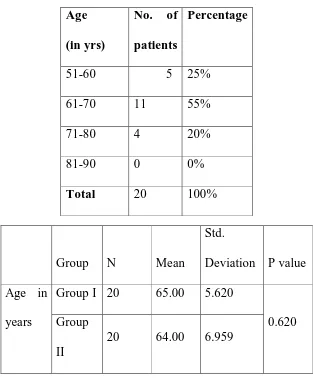
Table 1.0. Age Distribution of study population : Group 1
Age distribution pattern in Group 1 patients Age
(in yrs)
No. of patients
Percentage
51-60 5 25%
61-70 11 55%
71-80 4 20%
81-90 0 0%
Total 20 100%
Group N Mean
Std.
Deviation P value
Age in
years
Group I 20 65.00 5.620
0.620 Group
II
50
In a total of 20 patients in Group 1, the youngest was 55 years and oldest
was 75 years with a mean age of 64 years and standard deviation of 5.62
with the predominant age group 61-70 years.
In Group 2,
Age distribution pattern in Group 2 patients\
In Group 2, the youngest patient was 52 years and oldest patient was
76 years with a mean of 64 years with a standard deviation of 6.95.
Age (in yrs)
No. of patients
Percentage
51-60 8 40%
61-70 7 35%
71-80 5 25%
81-90 0 0%
51
Group 1
Figure 1. Frequency distribution of population in Group 1
In Group 2
Figure 1. Frequency distribution of population in Group 2
0 2 4 6 8 10 12
51-60 61-70 71-80 81-90
0 1 2 3 4 5 6 7 8
52
Symptom score and Quality of Life
N Minimum Maximum Mean Std.
Deviation
IPSS
Group 1 20 20
24.95 1.986 .444
Group 2 20 20 24.65 1.899 .425
Table 1.3. Symptom score
All the patients had severe IPSS (majority had obstructive symptoms)
with a mean score of 24.95 in Group 1 and 24.65 in Group 2,minimum
and maximum score of 20 and 25 respectively with a standard deviation
of 1.98. On analysis of IPSS score it was found that mean obstructive
symptoms score was 14.3 as compared to the mean irritative symptoms
score of 10.5.
Figure 3. IPSS score in this study population
0 5 10 15 20 25 30
53
Figure 4. IPSS score (obstructive vs. irritative score)
Comorbities associated with groups
In Group 1, out of 20 patient who presented, nine people had comorbidities, where as in goup two eight people had comorbidities.
The difference between these two groups was not significant.
Correlation between comorbidities like hypertension, diabetes in both groups
14.3 10.5
IPSS score
Mean obstructive score Mean irritative score
Comorbities
Both DM
HT Nil
C
o
u
n
t
14
12
10
8
6
4
2
0
Group
Group I
54
Group Total
Group I Group II
Comorbities Nil Count 11 12 23
% within
Comorbities
47.8% 52.2% 100.0%
% within Group 55.0% 60.0% 57.5%
HT Count 3 3 6
% within
Comorbities
50.0% 50.0% 100.0%
% within Group 15.0% 15.0% 15.0%
DM Count 5 4 9
% within
Comorbities
55.6% 44.4% 100.0%
% within Group 25.0% 20.0% 22.5%
Both Count 1 1 2
% within
Comorbities
50.0% 50.0% 100.0%
% within Group 5.0% 5.0% 5.0%
Total Count 20 20 40
% within
Comorbities
50.0% 50.0% 100.0%
% within Group 100.0% 100.0% 100.0%
55
Ultrasound prostate size :
Comparing the prostate size by ultrasound in both groups, the
mean prostate volume was 46cc in group 1 with a standard deviation of
12.47 and 42 cc in group 2 with a standard deviation of 14.19.
Residual urine measurement :
When we compare the residual urine in both groups, residual urine in patients in group1 was in the range of 165 ml and in group 2 it was in
the range of 136ml.
S.creatinine at presentation :
In group 1, patients who presented with elevated s.creatinine values, the following values were recorded at presentation
Picture representing levels of S.creatinine in patients during presentation
0 0.5 1 1.5 2 2.5 3 3.5 4
56
It ranged from 3.4 to 1.7, with a mean value of 2.170 and a standard
deviation of 0.5. All these patients underwent catherization accordingly,
then their s.creatinine was recorded once it got stabilized. Whereas in
group 2, the mean serum creatinine was around 0.925 with a standard
deviation of 0.1.
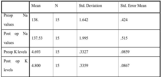
Preoperative Serum creatinine :
Once the serum creatinine values stabilized, the readings were recorded. Out of 20 patients who presented with elevated renal
parameters, (patient with s.creatinine more than 1.4 ) five patients
subsequently showed fall in serum creatinine below 1.4, hence were
considered along with normal patients in group 1. Because of this, the
subsequent serum creatinine values of rest of 15 patients stabilized at a
mean of 1.7 with a standard deviation of 0.3. Interestingly patients whose
serum creatinine which stabilized at a value of more than 1.4, some of
them had coexistent diabetes, hypertension. This might be an explanation
that these patients have developed preexisting renal disease which was
worsened by their developing benign prostatic hyperplasia.
Group N Mean
Std.
Deviation
Std. Error
Mean
Preop
S.creatinine
Group I 15 1.747 .3021 .0780
Group II 25 .960 .1258 .0252
57
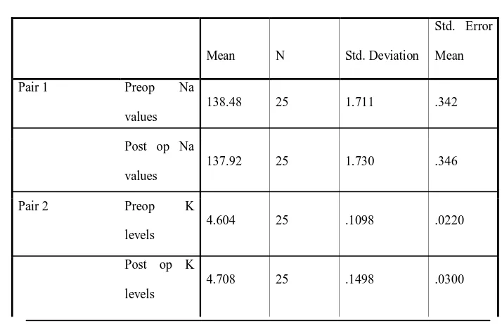
Comparison of preoperative sodium, potassium values in both groups:
Preop Na values Group I 15 138.13 1.642 .424
Group II 25 138.48 1.711 .342
Preop K levels Group I 15 4.693 .3327 .0859
[image:63.595.205.457.501.654.2]Group II 25 4.604 .1098 .0220
Table showing mean preoperative sodium and potassium levels in both groups
As depicted above, in group 1, the mean sodium value was 138,
whereas in group 2, the mean sodium value was 138.48. There was no
significant difference in these two groups. Similarly, the mean
preoperative potassium levels in both groups were not significantly
different.
Cystourethroscopy findings :
Cystourethroscopy findings of prostate and bladder in both groups.
On cystourethroscopy, out of 40 patients 16 had bilobar and 24 had
trilobar enlargement.
Bladder cystoscopy
Tri Bi
C
o
u
n
t
14
13
12
11
10
9
8
7
6
Group
58
CYSTOSCOPY – BLADDER TRABECULATIONS:
Group 1: In group 1, out of 15 , 10 patietns had grade 2 trabeculations, rest with grade 3 trabeculations.
Figure 10. Cystoscopy-Bladder Trabeculations in Group 1
Group 2 : In group 2, out of 20, 17 patients had grade 2 trabeculations, rest with grade 3 trabeculations.
Figure 10. Cystoscopy-Bladder Trabeculations in Group 2
Resection time during surgery :
The mean resection time in group 1 was 53 minutes , with a standard deviation of 8.8 minutes. In group 2, the resection time was 56
minutes mean value with a standard deviation of 9.7 minutes.
0 5 10 15
T2 T3
0 5 10 15