A CLINICAL STUDY OF INCIDENCE,
RISK FACTORS, MATERNAL AND PERINATAL
OUTCOME IN PREGNANCIES WITH
ABRUPTIO PLACENTAE
Dissertation submitted in partial fulfillment of requirements for
M.S. DEGREE BRANCH II
OBSTETRICS AND GYNAECOLOGY
MADRAS MEDICAL COLLEGE
CHENNAI
THE TAMIL NADU Dr. M.G.R. MEDICAL UNIVERSITY
CHENNAI
CERTIFICATE
This is to certify that the dissertation titled
"
A CLINICAL STUDY OF INCIDENCE, RISK FACTORS,MATERNAL AND PERINATAL OUTCOME IN
PREGNANCIES WITH ABRUPTIO PLACENTAE
"
is a
bonafide work done
by Dr.S.SHEBA MATHAVIin the Institute
of Obstetrics and Gynaecology (Madras Medical College)
Egmore, Chennai in partial fulfillment of the university rules
and regulations for award of MS degree in Obstetrics and
Gynaecology under my guidanceand supervision during the
academic year 2012-2014.
Prof. DR.V.KANAGASABAI
M.D
Dean
MadrasMedicalCollege &
Rajiv Gandhi Govt.General Hospital Chennai – 3
Prof.DR.MEENAUMACHANDER
M.D.,DGO. Director and superintendent
Institute of Obstetrics and Gynaecology
Madras Medical College, Chennai – 3.
Prof.DR.T.KRISHNAVENI M.D.,DGO.
Guide
Institute of Obstetrics and Gynaecology
DECLARATION
I solemnly declare that this dissertation titled "
"
A CLINICAL STUDY OF INCIDENCE, RISK FACTORS,MATERNAL AND PERINATAL OUTCOME IN
PREGNANCIES WITH ABRUPTIO PLACENTAE
"
was done
by me at Institute of Obstetrics and Gynaecology, Madras
Medical College during the year 2012 - 2014 under the
guidance
and
supervision
of
Prof.DR.T.KRISHNAVENIM.D.,DGO. This dissertation is
submitted to The Tamil Nadu Dr.M.G.R. Medical University
towards the partial fulfillment of requirements for the award of
M.S. Degree in Obstetrics and Gynaecology (Branch -II)
Place : Signature of the candidate
Date : Dr.S.SHEBA MATHAVI
MS Post Graduate Student
Institute of Obstetrics and Gynaecology Madras Medical College, Chennai -3
Prof.DR.T.KRISHNAVENIM.D.,DGO
Guide
ACKNOWLEDGEMENT
I gratefully acknowledge and sincerely thank the
Prof.Dr.V.KANAGASABAI,MD, DEAN
Madras Medical
College and Rajiv Gandhi Govt. General
Hospital,Chennai-600003 for permitting me to conduct the study and use the
facilities of the institution for my study.
It
is
with
immense
honour
and
gratitude
I
thank
our
Director
and
Superintendent,
Prof.Dr.MEENAUMACHANDER MD.,DGO,
Institute of
Obstetrics and Gynaecology, Egmore, Chennai a very
dynamic person, for helping me all through the study.
I am extremely thankful to
Prof.Dr.T.KRISHNAVENI
MD., DGO,
for her valuable guidance, and constructive
comments she provided with great affection.
My sincere thanks to
Dr.PadmanabhanPh.D.,
for his
help in statistical analysis of the data and results.
I wish to express my sincere thanks to all other Unit
Chiefs and Assistant Professors of our department for their
support during the study.
I wish to thank my postgraduate colleagues who helped
me carry out this study.
I would be failing in my duty if I don’t place my sincere
thanks to those patients who were the subjects of my study.
ABSTRACT
AIMS AND OBJECTIVES:To study the incidence , risk factors,maternal
,perinatal morbidity and mortality of patients with
placentalabruption.MATERIALS AND METHODS:A Prospective
observational study;Studyperiod-oneyear,Studygroup:Patientsadmitted with
clinical diagnoses of abruption with gestational age more than 28 weeks.,cases
diagnosed retrospectively with retroplacentalclot.The maternal outcome in
terms of anaemia,shock,DIC,renalfailure,postpartumhaemorrhageand death
were evaluated.Perinatal outcome in terms of birth weight,Apgarscore,perinatal
mortality were noted.RESULTS:Among 191 patients of antepartum
haemorrhage the incidence of abruption was 0.76%(1 in132
deliveries.Maximum incidence was between 33-36 weeks of gestation and in
multiparae.Hypertension and Anaemia are significant risk factors with ODDS
ratio of 10.64 and 5.941 respectively.Coagulation
failure:23.7%;Hypovolemic shock:37.62%;Renal failure:3.96%;Postpartum
haemorrhage:34.65% and Couvelaire uterus:5.94% of patients.There were 2
maternal deaths in our study and the perinatal death rate
was48%.CONCLUSION:Correction of anaemia and hypertension would help to
reduce the incidence and early referrals to the teritiary care centres will decrease
CONTENTS
S.No. Particulars Page No.
1. Introduction 1
2. Aims and Objectives 3
3. Review of Literature 4
4. Materials and Methods 38
5. Results of the Study 40
6. Discussion 63
7. Summary 73
8. Conclusion 76
9. Bibliography
10. Annexure
11. Abbreviations
12. Proforma
LIST OF TABLES
S.No. Particulars Page No.
1. Distribution of Antepartum Haemorrhage 40
2. Age distribution among cases of placental
abruption 42
3. Distribution based on pairty 43
4. Distribution based on Antenatal booking status 44
5. Distribution based on gestational age 45
6. Distribution based on presenting signs and
symptoms 46
7. Distribution based on Risk factors 47
8. Distribution based on Grades of abruption 49
9. Distribution based on types of abruption 50
10. Distribution based on Fetal presentation 51
11. Distribution based on Membrane status 52
12. Distribution based on Mode of delivery 53
13. Break up of the course of vaginal deliveries 54
14. Breakup of Caesarean excluding Grade 0 cases 55
S.No. Particulars Page No.
16. Distribution based on weight of RP clots 57
17. Distribution based on Maternal Complications of
abruption 58
18. Perinatal mortality in vaginal and Caesarean
deliveries 59
19. Perinatal mortality in vaginal delivery based on
birth weight 59
20. Perinatal mortality in Caesarean delivery based on
birth weight 60
21. Comparison of incidence in various institutions 63
22. Comparison of age group distribution in various
institutions 64
23. Comparison of cases based on parity in various
institutions 65
24. Comparison of cases based on booking status in
various institutions 66
25. Comparison of cases based on gestational age in
various institutions 67
26. Comparison of presenting signs and symptoms in
various institutions 68
27. Comparison of Risk Factors in various institutions 69
28. Comparison based on types of abruption 70
1
INTRODUCTION
The term abruptio placentae refers to a condition when
haemorrhage occurs as a result of premature separation of normally
situated placenta. It is one of the obstetric emergencies posing a threat
tomaternal and fetal life.
The present study is done to know the incidence, risk factors of
this disorder in our institution as well as the impact of this disorder on
maternal and fetal outcome.
Though abruption is an obstetric emergency and a leading cause
of perinatal morbidity and mortality the availability of diagnostic
modalities like ultrasonagram has proved helpful in excluding other
causes of ante partum haemorrhage like placenta previa and for timely
management of the condition. Moreover the increased use of
caesarean section and the better availability of haematological
facilities has made a good impact in the recent studies.
All patients with diagnoses of abruption confirmed either
clinically or by ultrasonagram or retrospectively following delivery
were evaluated in our study and the required intervention was made to
2
clinical assessment of the patient and simultaneous treatment of
haemodynamic instability or coagulation abnormality was carried out.
The data generated from this study would help to improve maternal
and fetal morbidity and mortality by planning prompt management of
3
AIMS AND OBJECTIVES
To study the incidence and risk factors of patients presenting
with placental abruption.
To study the outcome of patients with abruptio placentae in
4
REVIEW OF LITERATURE
GENERAL ASPECTS:
GENERAL ASPECTS: Third trimester bleeding is one of the most
omnious complications of pregnancy and it complicates 3% of
pregnancies. Third trimester obstetric haemorrhage seems to be one of
the three leading causes of both maternal and perinatal morbidity and
mortality. The obstetric causes of bleeding in third trimester being
very hazardous has to be differentiated from the non obstetric causes.
Causes of third trimester bleeding
OBSTETRIC NON OBSTETRIC
1.Bloody show 1.Cervicitis
2.placentaprevia 2.Cervical polyp
3.abruptio placenta 3.Cervical erosion
4.vasaprevia 4.Vaginitis
5.Disseminated intravascular
Coagulation
5.Genital tract trauma
6.Uterine rupture 6.Vulval,vaginalvaricosities
5
DEVELOPMENT OFTHE PLACENTA:
The placenta is a remarkable organ performing many diverse
functions including transport of gases and metabolites, immunological
protection and production of steroid and protein harmones.
The zygote after repeated mitotic divisions gets converted in to
a blastocyst. The outer layer of the blastocyst is called trophoblast and
the inner cell mass is called embryoblast. The blastocyst after
embedding in to the endometrium, the trophoblast rapidly proliferates
and differentiates in to outer syncitiotrophoblast and an inner
cytotrophoblast. The primary, secondary and tertiary villi are formed
by the syncitiotrophoblasts and the intervillous space is formed by
cytotrophoblasts. Until the end of sixteenth week the placenta grows
in thickness and circumference due to the growth of the chorionic villi
and expansion of the intervillousspace. After that there is little
increase in thickness but it increases circumferentially until term. The
implanted placenta by nature separates during the third stage of labor
by a multiphasic process.
Abruptio placentae is the premature separation of a normally
implanted placenta with haemorrhage in to the decidua basalis (Konje
6
pregnancies. Placenta previa and Abruptio placenta remain the two
importantcauses of antepartum haemorrhage.
HISTORICAL ASPECTS:
The term abruption in Latin means “breaking away” which
describes the process by which the placental attachment to the uterus
is disrupted by haemorrhage.
In 1609 Louis Bourgeois recognised the premature separation of
placenta. DeLee and Coole in1848 coined the term ABRUPTIO
PLACENTAE todenote sudden forcible separation of placenta from its
normal site. In 1775, Edward Rigby of NORWICH made the first
clinical differentiation between abruptio placentae and placenta previa.
He also called abruptio placentae as accidental haemorrhage
EPIDEMIOLOGY
The overall incidence of abruptio placentae varies from 0.5 to
1% (Ananth et al 1996; Baumannetal2000)3. Diagnoses is always
clinical.
A pathologist often quotes a higher incidence of them have an
7
The incidence varies due to variable diagnostic criteria as well
as the increased recognition of milder forms of this disorder. Although
various studies report a higher incidence of abruption in older women
and those with increased parity (Konje andTaylor2001)1. There is
conflicting evidence about the relationship of age and parity and
abruption (Kramer etal 1997)5.
ETIOLOGY:
The etiology is obscure but impaired placentation, placental
insufficiency, uteroplacental underperfusion seem to be the underlying
mechanisms
IMMUNOLOGICAL:
Immunological defects also may lead to an extensive
inflammatory response with release of cytokines and result in shallow
trophoblastic invasion, defective spiral artery remodeling, placental
infarctions and thrombosis (Matthiesen et al1995)6
In placental abruption there is no suppression of cell mediated
immunity and upregulation of humoral immunity which occurs in a
normal pregnancy. This leads to exaggerated immune rejection of the
8
agents. HLA-G levels which avoid fetal rejection are decreased in
abruption. If the signalling between trophoblastic and NK cells is poor
it causes defective trophoblastic invasion and spiral artery remodeling
which ultimately leads to dysfunctional placenta, thromboses,
infarction and a generalised inflammation in which systemic
endothelial dysfunction is a component. Hence placental abruption
may be due to dysfunctional placenta caused by defective maternal
immune response to paternal antigens. (Baumann et al 2000)
INFLAMMATION:
There is an increase of neutrophils and macrophages in
placentae of women with abruption. There is increased production of
TNF ALPHA and IL-BETA1 that increase the production of Matrix
metalloproteinases by trophoblasts and other cell types. Increased
premature production of Matrix metlloproteinases may destroy the
extracellular matrix and cell to cell interactions causing premature
placental detachment. (Ananth et al 2006a)
VASCULAR DISEASE:
A placental growth factor deficiency and sFlt-1 excess results
9
arteries. This defective remodeling leads to high resistance flow and
finally rupture of decidual arteries.
PATHOGENESIS AND PATHOPHYSIOLOGY OF ABRUPTIO
PLACENTA:
Abruption is due to rupture of the decidual artery. The decidua
is separated from the placenta by the accumulating blood. The
collected blood when small in quantity may hardly produce any
morbid pathological changes in the uterine wall or on the placenta.
When the blood continues to dissect through the placental decidual
interface it may lead to complete or near complete placental
separation. After this the blood may escape through the potential space
between chorion and decidua until it reaches the cervix. Blood may
seep in to myometrium producing the so called COUVELAIRE
UTERUS or reach the amniotic cavity producing blood stained
amniotic fluid.
COUVELAIRE
UTERUS
(UTEROPLACENTAL
10
It is a pathological condition first described by Couvelaire and
is met with severe forms of abruption. There is massive intravasation
of blood in to the myometrium up to the serous coat. The condition is
diagnosed only on laparatomy. The uterus is of dark port wine colour
.It occurs initially at the cornu before spreading to other areas .Free
blood may be present in the peritoneum or there may be a broad
ligament haematoma. This myometrial haematoma rarely interferes
with uterine contractions and hence it is not an indication per se for
hysterectomy.
HEPATIC AND RENAL CHANGES:
In severe types of abruption placentae a fibrin knot which is a
thrombotic lesion in the hepatic sinusoids has been described to be
specific of abruptio placentae.
In renal changes oliguria and anuria may develop due to acute
tubular necrosis in milder forms of abruption and renal cortical
necrosis in severe forms of abruption.
RETROPLACENTAL HAEMOTOMA17:
It is a universal accompaniment of separation of placenta but is
usually of little clinical significance. A fresh clot loosely adherent to
11
is retained in utero some time after delivery but it is not the genuine
retroplacental haematoma. If the placenta is not routinely seen many
true retroplacental haematomas may be missed. Although many small
lesions go unnoticed the large excavating retroplacental clot is readily
apparent. Lesser degrees of indentations are produced by smaller
lesions but the clot is usually firmly adherent to the maternal surface
.
LARGE
RETROPLACENTAL
CLOT
ON
THE
12
The colour of the haematoma is a function of the age. They lie
between the basal plate and underlying endometrium and they are
considered to be dissecting haemorrhages of the decidua. They have
features similar to that of an intervillous thrombus histologically.
Recurrent small haemorrhages may give a laminated appearance the
oldest clot being near the placental surface.
Incidence of retroplacental clots is 5%.In known preeclamptics
the associated abnormalities in decidual arterioles may be implicated.
In otherwise normal pregnancies there is no clear indication of
precipitating factors.
The clinical importance of retroplacental haematoma is two
fold. Firstly the extent to which placenta remains deprived of its
maternal blood supply clearly determines the survival of the fetus.
However in otherwise normal pregnancies majority of these lesions
have a little impact on fetal outcome. Secondly large retroplacental
13
RISK FACTORS:
AGE:
The incidence of abruption increases with maternal age. In the
First and Second Trimester Evaluation of Risk trial(FASTER TRIAL)
women older than 40 years were 2.3 times at more risk of abruption
than those at35 years or younger. In a study by Bryan M Hibbard there
is an increased incidence with maternal age and patients over 35 years
are twice as prone to abruption as patients under 25 years. The
unadjusted risk of abruptio placentae for women over the age of 35
years was 2.2 times the risk for women aged 19-34 years.
(Williams et al)
PARITY:
Although Pritchard and colleagues (1991) reported the
incidence of abruption to be higher in women of great parity, Toohey
and associates (1995) did not find this. HibbardBM and JEFFCOATE
TNA30 in their studies found an association between high parity and
14
NUTRITION:
Folic acid deficiency seems to be an important factor in etiology
of abruptio placentae. The possible association between folic acid
deficiency and abruption is proved by bone marrow biopsy studies
showing megaloblastic erythropoiesis. (Hourihana et al)
HYPERTENSIVE DISORDERS OF PREGNANCY:
The most common condition associated with abruption seems to
be some form of hypertension (gestational hypertension, chronic
hypertension, preeclampsia). Sibai and coworkers (1998) reported that
1.5% of pregnant women with chronic hypertension suffered placental
abruption. Ananth and associates (2007) reported a2.4fold increased
incidence of placental abruption with chronic hypertension and this
was increased further if there was superimposed preeclampsia or
IUGR. The severity of hypertension does not correlate with the
incidence of abruption. (Witlin and colleagues Zetterstrom and
coworkers 2005).
PREMATURE RUPTURE OF MEMBRANES:
There is an increased frequency of placental abruption in
patients with early rupture of membranes. The incidence was 13%
15
29-32 weeks. (Holmgren Paetal1997 Histological chorioamnionitis is
associated with increased incidence of placental abruption and this
association is dependent on its severity. (Nath et al 2007)25
SMOKING AND COCAINE ABUSE:
Smoking increases the overall risk of abruptioplacentae.
According to a prospective cohort study the increase is by40% for
each year of smoking prior to pregnancy. The rate of abruption has
been reported to be13-35% in patients who abuse cocaine and seems
to be dose dependent.
TRAUMA:
Blunt abdominal trauma or rapid decompression of uterus may
cause shearing of placenta due to sudden stretching or contraction of
uterine wall. Severe maternal trauma has been associated with a six
fold risk of abruption.
EXTERNAL CEPHALIC VERSION:
When external cephalic version was a common practice
abruption was a well known complication of it, especially when
version was performed under anaesthesia. The incidence of abruption
is said to be 2 to9% according to Savona Ventura (1986) who quotes 4
16
the addition of to colytic drugs the complication rates are reported to
be low (Lancet Leader 1984)
THROMBOPHILIAS:
The association between placental abruption and the maternal
thrombophilias supports the fact that abruption is a final acute clinical
presentation of a chronic placental disease. (WENDY LKINZER etal)
MULTIPLE PREGNANCY:
The risk of placental abruption is increased 2-3 fold in multiple
gestations (Salihu et al 200520, Ananth et al2001). In patients with
twin gestation associated with placental abruption the risk of preterm
birth or SGA is higher.
PLACENTA PREVIA:
About 10% women with placenta previa may have placenta
previacoexistently. (Konje and taylor2001)1
OTHERS:
First trimester ultrasound examination showing a subchorionic
or retroplacental haematomaincreases the subsequent placental
17
When the membranes rupture as in the case of poly hydramnios
there is asudden decompression of the uterus .This leads to the
reduction in uterine volume and a corresponding loss of surface area
and as a result the placenta sheers off.
Abruption has been reported as a result of snakebite (Zugaib et
al 1985) but has not been reported as a result of anticoagulant therapy.
(Howell et al 19838)
Uterine malformations may lead to poor decidualisation and
placentation. The contractility of a malformed uterus may lead to
uncoordinated uterine action resulting in increased risk of placental
abruption (Dabrashrafi et al1999).The risk of abruption is increased in
patients with lower segment uterine scar due to impaired placental
attachment.(Rasmussen et al199916; Lydon Rochelle et al 2001)
RECCURRENT PLACENTAL ABRUPTION:
Ina women with a previous history of abruption there is a7-20
fold risk in a subsequent pregnancy (Karegard and Genser 198611,
18
PREDICTORS:
BIOCHEMICAL MARKERS:
Unexplained second trimester elevation in MSAFP may be
associated with subsequent adverse obstetric outcome including
placental abruption. (Dugoff et al200512, Smith et al2006).This is due
to chronic villitis and vascular thromboses or infarction (Salafia et
al198813).AFP levels >2.0MoM were detected in 17% of pregnancies
with subsequent placental abruption (Finish study2002). Though
MSAFP levels is a good marker for placental abruption it has not been
used for this purpose widely in clinical practise. High levels of
maternal serum beta hcg have been linked to placental abruption.
(Liu et al1999).Decreased placental perfusion may lead to increased
beta hcg.
Low levels of PAPP-A detected in the first trimester (29% at
lowest 5th percentile and 43%at lowest 10th percentile) have been
linked with placental abruption (Pilalis et al200714). Proangiogenic
placental growth factor (PIGF) and antiangiogenic soluble - fms like
tyrosine kinase-1(sflt-1) are the regulators of angiogenesis in
pregnancy. Increased levels of sflt1/PIG F ratios at21-32 weeks of
19
has developed preeclampsia or PIH (Signnora et al 200629)
Fibronectin produced in the endothelial cell seems to be
increased with placental abruption (Kanayama and Terao1992).
Levels of thrombomodulin which is a marker of endothelial
cell damage may be increased with placental abruption (Magriples et
al 1999).
D - dimer a by product of clot analysis may be used in early
diagnoses of abruption (Nolan et al1993)
Uterine artery flow measurement:
High uterine artery pulsatility index at11-14 weeks or notching
of the uterine artery waveform at 20-24 gestational weeks also may
predict subsequent placental abruption (Pilalis et al200714) but these
methods have not been accepted universally in the prediction of
abruption.
FAMILY HISTORY:
5% of women with abruption may have first degree relatives
20
CLINICAL PRESENTATION:
The clinical presentation varies from one patient to another.
Vaginal bleeding, painabdomen, uterine tenderness, loss of fetal
movements seem to be the classic symptoms. All these symptoms
need not be present and asymptomatic presentation does not exclude
placental abruption.
Placental abruption may be revealed or concealed. In the
revealed type blood tracks between the membranes and the decidua
escaping through the vagina. The less common concealed type occurs
when blood collects behind the placenta without any external bleeding
(Oyelese and Ananth20062)
21
Four grades of placental abruption based on
PAGE'S CLASSIFICATION:
Grade0 : An asymptomatic retroplacental clot seen after
placental delivery.
Grade1 : Present with vaginal bleeding and mild uterine
tenderness.
Grade2 : Vaginal bleeding may or may not be present but
placental separation is significant enough to cause
fetal compromise.
Grade3 : Vaginal bleeding may be moderate or severe but
there may be significant maternal complications
along with late stage of fetal compromise or fetal
death.
Geofrrey Sher (1978) proposed a clinical grading system
Grade1 : Diagnoses is made retrospectively by seeing a
retroplacental clot.
Grade2 : Cases in which the fetus is alive. The retroplacental
clot volume is usually 150 to 500gms.
Grade3 : Features of Grade2 but fetal demise is confirmed.
Grade 3 is further subdivided based on A) presence
22
Vaginal bleeding is present in 70-80%of placental abruption
though its amount poorly correlates with the degree of abruption.
Uterine tendernss or pain is present in 66%. Hypertonic uterine
contractions may be seen in 34% cases. Abdominal pain is less
common in posterior placentations.
DIFFERENTIAL DIAGNOSES IN CASES OF CONCEALED
OR MIXED TYPES:
1. Rupture uterus
2. Acutehydramnios
3. Rectus sheath haematoma
4. Appendicular or intestinal perforation
5. volvulus
6. Twisted ovarian tumor
7. Tonic uterine contraction
8. Red degeneration of fibroid.
Points in favour of concealed abruption:
1. Shock out of proportion to external bleeding
2. Unexplainedanaemia with tense uterus
23
4. uterus may be tense tender
5. absent fetal heart
6. oliguria
7. coagulopathies
ULTASONAGRAM IN ABRUPTION:
Ultrasound abdomen is done
1. to rule out placenta previa
2. to reveal the state of fetus and degree of placental separation
In acute phase of abruption the haematoma appears hyperechoic
to isoechoic when compared to placenta. On resolution the haematoma
becomes hypoechoic within 1 week and sonoluscent within 2 weeks
24
A. Normal Placenta B, C Large Retro Placental Abruption
D. Thickened Placenta with Heterogenous Appearance, Arrow Heads
Show Areas of Haemorrhage.
Although there is marked improvement in, the diagnoses of
abruption was correct only in 25%. (Glantz and Purnell200231). When
a clot was visualised by USG, the positive predictive value of
25 Jello Sign:
The placenta may jiggle when sudden pressure is applied with
transducer. However the ultrasound though inaccurate in diagnoses of
abruption may be very helpful in excluding a placenta previa.
CARDIOTOCOGRAPHIC CHANGES:
Placental abruption may be associated with a variety of CTG
patterns and this includes repetitive, late and variable decelerations,
decreased beat to beat variability, bradycardia or sinusoidal fetal heart
rate pattern (Oyelese andAnanth2006)2
CTG OF A DEAD FETUS WITH UTERUS SHOWING
26
MATERNAL MORBIDITY:
The maternal complications primarily depend on the severity of
abruption. They include haemorhage and need for blood transfusions,
coagulopathy, renal failure and hypovolemic shock, hysterectomy and
less commonly maternal death. In cases of abruption severe enough to
cause death of the fetus DIC sets in. Hypovolemiaor DIC may be a
forerunner of acute renal failure.
HAEMORRHAGICSHOCK:
There is significant loss of blood volume, either revealed
vaginally or concealed within the uterus.
In pregnancy the increased blood volume and increased levels
of coagulation factors such as fibrinogen and factor 7, 8 and 10
provide protection against haemorrhage. But when there is more than
25% loss of total blood volume, rapid haemodynamic deteroriation
occurs.
When there is continuous bleeding and development of
anaerobic metabolism with acidosis, there may be development of left
27
DISSEMINATEDINTRAVASCULAR COAGULATION:
This is another common complication met with severe grades
of abruptions associated with fetal death. In abruption placentae the
release of procoagulant substances lead to release of tissue
thromboplastins (from placental injury) in to the maternal circulation.
Due to the hypovolemia and hypoxia induced endothelial
response there is white cell activation and production of
proinflammatory cytokines and oxygen free radicals. These increase
the oxidative stress and promote lipid peroxidation once antioxidant
mechanisms are saturated. So there is loss of vascular integrity and
hence increased vascular permeability.
The thromboplstins and the endothelial damage lead to
widespread activation of coagulation cascade. If this goes unchecked,
there is rapid consumption of coagulation factors and platelets with
deposition of fibrin in microcirculation and thrombus formation in the
placental surface. This leads to defibrination, thrombocytopenia and
hemostatic failure. Disseminated intravascular coagulation also
stimulates fibrinolysis and the resultant fibrin degradation products
interferes with clot formation producing haemorrhage and also has a
28
intravascular coagulation is based on a combination of clinical and
laboratory diagnoses. Bleeding can occur at venepuncture sites, gums,
nose, rectum etc. Platelet counts and coagulation tests are abnormal
.As more than 50% clotting factors must be consumed before the
coagulation tests become abnormal, these tests become insensitive. As
fibrinogen levels are increased in normal pregnancy, even low normal
levels should be of concern. Fibrin degradation products including
D-dimer are measured in the circulation and their abnormal levels help
to confirm the presence of coagulopathy.
RHESUS ISOIMMUNISATION:
There may be a significant amount of fetomaternal
haemorrhage. So it is advisable for all rhesus negative women with
abruptio placentae to undergo a kleihauverbetke testto determine the
correct dose of ANTI-D Immunoglobulin.
Maternal mortality however has decreased in the past few years
due to increased maternal health care facilities. Also women with a
history of placental abruption are less likely to become pregnant again
29
A study of long term effect of placental abruption on maternal
health (Ray et al2005)32 shows a risk of development of premature
cardiovascular disease in 70% of these women. But the cause remains
unclear.
PERINATAL MORBIDITY AND MORTALITY:
Perinatal morbidity includes low birth weight, preterm
deliveries, birthasphyxia, stillbirth, intrauterinedeath, neonataldeaths.
Here also the fetal survival depends on the grade of abruption as well
as the gestational age.
If more than 50% of placental surface is involved fetal death
invariably occurs. The perinatal mortality also depends on the
prematurity of the fetus and the neonatal facilities available. Both
spontaneous and iatrogenic preterm deliveries are common in
placental abruption.
Fetal growth restriction is so well associated with abruption,
that it could be taken as a single marker for risk of abruption (Ananth
and Wilcox 2001)24.Even congenital fetal malformations are
approximately two times higher than in general population (Konje and
30
scores and cord blood pH values are due to maternal hypoxia (Spinillo
et al 1993)9.The risk of birth asphyxia is 3.7 fold. Long term risk of
neonates born to mothers with placental abruption are development of
cystic periventricular leukomalacia or intra ventricular haemorrhage.
These neural defects increase with prematurity and low birth weight
(Spinillo etal1993)9
GENERAL MANAGEMENT:
A rapid clinical assessment should bedone in women presenting
with antepartum haemorrhage whether urgent intervention is required
to manage maternal or fetal compromise. In women presenting with
massive haemorrhage resuscitation should be started immediately
since mother is the priority and should be stabilised prior to
establishing the fetal condition. Meanwhile blood should be collected
for investigations to identify complications.
The investigations may include:
The investigations may include
Blood Hb%
Platelet count
Leucocyte count
31 Blood for grouping and typing
Bleeding and clotting time
Clot observation and retraction time
Prothrombin time
Activated partial thromboplastin time
Serum fibrinogen level
Fibrin degradation product levels
D-dimer levels
Liver function tests
Renal function tests
Serum electrolytes
Arterial blood gas analysis
Kleivauher Betketest (if available)
OBSTETRIC MANAGEMENT:
The clinical presentation of placental abruption being variable,
the obstetrical management is individualised on a case by case basis.
The presentation, gestaional age and the degree of maternal and fetal
compromise decides the management. In cases of abruption at or near
term with a live fetus, prompt delivery is indicated. If there is fetal
32
be performed. When the maternal and fetal conditions are reassuring
vaginal delivery may be reasonable.
RUPTURING THE MEMBRANES AND HASTENING
DELIVERY:
The main purose of doing an amniotomy is to hasten the onset
of labour and by encouraging uterine contractions, to reduce uterine
bleeding. It is effective and useful in most cases but seems to be
dangerous in a few. If the uterus has become atonic as happens rarely,
reduction of intrauterine pressure encourages further bleeding which
fills the space. An intravenous infusion of oxytocin should always be
started.
If patient is in established labor it may be allowed to progress in
these conditions. On the other hand induction of labor may be planned
if the labor has not been established. There should be close monitoring
of both mother and fetus and when fetal heart rate becomes
nonreassuring, caesarean section is indicated.
In patients with severe placental abruption resulting in
intrauterine fetal death, if the mother is stable, it is reasonable to allow
for a vaginal delivery in the absence of any obstetric indications.
33
contractions and an amniotomy may speed up delivery. When labor
does not progress rapidly or in case of obstetrical indications like
cephalopelvic disproportion, a scarred uterus or a fetal
malpresentation.
Caesarean delivery may be performed to avoid worsening of
coagulopathy. But stabilisation of the patient and correction of any
coagulation defects is very much essential during surgery. The patient
should be closely monitored paying attention to vital signs, amount of
blood loss, urine output. Uterus may remain hypotonic necessitating
emergency hysterectomy. There should be a high index of suspicions
of preeclampsia in patients with abruption because this may be
34
MANAGEMENT OF MATERNAL COMPLICATIONS:
Haemorrhagic shock is a major complication of abruption.
Hypovolemia should be corrected early. Since there may be a greater
degree of vasospasm blood pressure may not be a good guide to assess
shock. So regardless of the patient’s general condition atleast one litre
of blood should be transfused. Central venous pressure is the best
guide to monitor the patient .Haematocrit should be atleast 30% and
urine output more than 30 ml per hour.
Disseminated intravascular coagulation is due to release of
tissue thromboplastin in placental abruption leading to consumptive
coagulopathy. Whole blood transfusion replenishes not only the
fibrinogen but also other procoagulants. Fresh frozen plasma helps in
the administration of fibrinogen, a necessary procoagulant. Prompt
replacement of blood volume and coagulation factors helps a lot in the
management of these coagulation disorders. There is no role of
heparin or anticoagulants in disseminated intravascular coagulation
caused by abruption.
Steps for immediate delivery of the fetus should be taken that
would help to improve the haemostatic competence in vast majority of
35
there is no clinically evident coagulopathy 12 after the delivery.
Renal failure in its early phase can be corrected in most cases of
abruption by volume replacement .But however in its late phases the
help of a nephrologist is needed.
EXPECTANT MANAGEMENT:
In case of preterm gestational ages when there is minimal
abruption evidenced by ultrasound, there is a role of conservative
management. But this should be individualised on a case to case basis.
These patients usually present with a small vaginal bleed and a
localised area of uterine tenderness or an ultrasound evidence of
minimal separation of placenta. In such circumstances there is
something gained from allowing the pregnancy to continue but the
decision to terminate will depend on the length of gestation, whether
there was a previous episode the state of fetus and the extent of
placental separation.
Abruption in a posteriorly situated placenta is dangerous
because the only symptom being backache and also the placenta is out
36
there is some damage to the integrity and function of placenta. And
these patients need an extremely close monitoring because of a
significant risk of fetal death at any period of time. Initial
hospitalisation for further evaluation and fetal wellbeing is necessary
and serial ultrasonagrams may be needed to evaluate progression or
regression of abruption.
PREVENTION OF ABRUPTIO PLACENTAE:
It aims at prevention of risk factors likely to cause abruption, early
detection of anaemia in the antenatal period and its correction and
prompt identification and commencement of therapy to minimise
grave complications namely shock, coagulopathy, renalfailure.
Prevention of factors likely to cause placental separation are:
1. Early diagnoses and treatment of preeclampsia and other
hypertensive disorders of pregnancy.
2. Avoidance of sudden rupture of membranes.
3. Avoidance of trauma especially forceful external cephalic
version.
37
MANAGEMENT OF A SUBSEQUEUENT PREGNANCY:
These women are at a ten fold risk of having an abruption in the next
pregnancy. They are also at increased risk of other pregnancy
outcomes like preterm birth and preeclampsia. Some
recommendations are possible in this women. Women who smoke
tobacco or use cocaine should be encouraged to quit before next
pregnancy. Hypertension should be controlled well before the next
pregnancy. It is reasonable to treat women with inherited
thrombophilias with thrombophylaxis although no clear benefit has
been demonstrated. Because patients with abruption have an increased
risk of impaired uteroplacental perfusion it is reasonable to do serial
38
MATERIALS AND METHODS
A prospective observational clinical study was carried out in our
institution from Oct’12 to Sept’13 to determine the incidence, risk
factors, maternal and perinatal outcome in patients presenting with
abruption placentae. Study group consists of patients admitted to our
labour room with clinical diagnosis of abruption and gestational age
more than 28weeks. Cases diagnosed retrospectively with
retroplacental clots were included in this study.
INCLUSION CRITERIA:
Pregnant women with gestational age more than 28 weeks with
complaints of bleeding per vaginum.
Patients with retroplacental clots diagnosed retrospectively.
EXCLUSION CRITERIA:
Patients with atypical signs and symptoms were excluded from
the study after their delivery if there was no clinical evidence of
abruption.
A detailed history of the patient including the obstetric history
39
were done for the patients to arrive at a diagnoses and the patients
diagnosed to have placental abruption were followed up for maternal
and fetal outcome.
Risk factors for abruption were analysed by the following
statistical methods in the patients presenting with abruption.
1. Chi square test,
2. Fisher exact test
Investigations including coagulation profile and the renal
function tests were done to identify complications.
The grade of abruption presenting in the patient was identified
and the severity was correlated with the maternal morbidity and
mortality as well as fetal outcome in terms of intrauterine fetal death,
stillbirths, neonatal admissions and neonatal deaths.
The fetal outcome was also measured on the basis of route of
40
[image:49.612.120.506.128.383.2]RESULTS OF THE STUDY
TABLE I
Total No. of Deliveries in study period : 13239
Incidence %
Antepartum
Haemorrhage
191 1.44%
Abruptio Placentae 101 0.76%
Placenta Previa 78 0.5%
Others 12 0.09%
The total number of deliveries in the study period was 13239.
.Among these patients presenting with antepartum haemorrhage
accounted for 191caseswith an incidence rate of 1.44%.The two main
causes of antepartum haemorrhage are Placenta previa and placental
abruption. In our study placental abruption accounted for 101 cases
The number of patients presenting with Placenta previa
with an incidence of 0.5%.The remaining patients
haemorrhage were12 and make up an incidence of0.09 %.
40.84%
6.28%
DISTRIBUTION OF ANTEPARTUM
41
The number of patients presenting with Placenta previa
with an incidence of 0.5%.The remaining patients of antepartum
haemorrhage were12 and make up an incidence of0.09 %.
52.88% 6.28%
DISTRIBUTION OF ANTEPARTUM
HAEMORRHAGE
Abruptio Placentae
Placenta Previa
Others
The number of patients presenting with Placenta previa were78
of antepartum
DISTRIBUTION OF ANTEPARTUM
Abruptio Placentae
0% 10% 20% 30% 40% 50% 60%
< 20 years 21-25 years
DISTRIBUTION BASED ON AGE IN PLACENTAL ABRUPTION AGE DISTRIBUTION AMONG CASES OF
Age Group
< 20 years
21-25 years
25-30 years
31-35 years
> 35 years
When the age was analysed the highest incidence was among
21-25 years accounting
abruption occurred was 18 years and the highest age was 38 years.
42 25 years 25-30 years 31-35 years
> 35 years
DISTRIBUTION BASED ON AGE IN PLACENTAL ABRUPTION
[image:51.612.127.472.469.675.2]< 20 years 21-25 years 25-30 years 31-35 years > 35 years
TABLE II
AGE DISTRIBUTION AMONG CASES OF PLACENTAL
ABRUPTION
Incidence %
7 6.93%
52 51.48%
35 34.65%
6 5.94%
1 0.99%
When the age was analysed the highest incidence was among
accounting for 52%.The youngest age at which
abruption occurred was 18 years and the highest age was 38 years.
25 years 30 years 35 years PLACENTAL 6.93% 51.48% 34.65% 5.94% 0.99%
When the age was analysed the highest incidence was among
for 52%.The youngest age at which
DISTRIBUTION BASED ON PARITY (n
Parity
Primi
Multi
The incidence of abruption was seen to be
61.38%
DISTRIBUTION BASED ON PARITY
[image:52.612.126.510.341.612.2]43
TABLE III
DISTRIBUTION BASED ON PARITY (n=101 CASES)
Incidence %
39 38.61%
62 61.38%
The incidence of abruption was seen to be highest among multipara.
38.61%
DISTRIBUTION BASED ON PARITY
=101 CASES)
38.61%
61.38%
highest among multipara.
Primi
BASED ON ANTENATAL BOOKING STATUS
Booked
Unbooked
In our study the nu
DISTRIBUTION BASED ON
ANTENATAL BOOKING STATUS
[image:53.612.128.501.287.553.2]44
TABLE IV
BASED ON ANTENATAL BOOKING STATUS
No. of Women %
95 94.05%
6 5.94%
In our study the number of booked women were 94.05%
94.05% 5.94%
DISTRIBUTION BASED ON
ANTENATAL BOOKING STATUS
Booked
Unbooked 94.05%
5.94%
mber of booked women were 94.05%
Booked
BASED ON GESTATIONAL AGE
Gestational Age In Weeks
28-32 weeks
33-36 weeks
37 Weeks & Above
In our study there was a higher incidence of
in women belonging to gestational age of 33 to36 weeks.
0.00% 25.00% 50.00%
28-32 weeks 26.73%
Based on Gestational Age
[image:54.612.128.469.394.632.2]45
TABLE V
BASED ON GESTATIONAL AGE
Gestational Age In No. of Patients
(n=101) %
27 26.73%
47 46.53%
27 26.73%
In our study there was a higher incidence of placental abruption
belonging to gestational age of 33 to36 weeks.
32 weeks
33-36 weeks
37 Weeks & Above 26.73%
46.53%
26.73%
Based on Gestational Age
26.73%
46.53%
26.73%
46
TABLE VI
DISTRIBUTION BASED ON PRESENTING SIGNS AND
SYMPTOMS
Signs and Symptoms No. of Patients %
Vaginal Bleeding 82 81.18%
Pain Abdomen 88 87.12%
Tense, Tender Uterus 73 72.27%
Absent Fetal Heart 37 36.63%
Shock 38 37.62%
Hypertension 62 61.30%
The clinical presentation was either single or a combination of above
symptoms and signs. The most common presenting sign was vaginal
47
TABLE VII
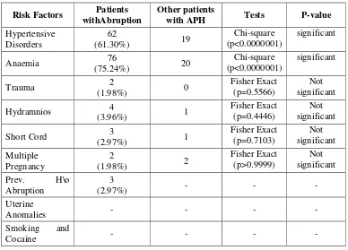
DISTRIBUTION BASED ON RISK FACTORS
Hypertensive disorders of pregnancy in the form of pregnancy
induced hypertension accounted for 56 cases while 6 cases were due
to chronic hypertension
.From the above table we infer that anaemia and hypertension
are the two significant associated factors.
1. The patients with anaemia are having ten times risk for
developing abruption,(ODDS RATIO =10.64)
Risk Factors Patients
withAbruption
Other patients
with APH Tests P-value
Hypertensive Disorders
62
(61.30%) 19
Chi-square (p<0.0000001)
significant
Anaemia 76
(75.24%) 20
Chi-square (p<0.0000001)
significant
Trauma 2
(1.98%) 0
Fisher Exact (p=0.5566)
Not significant
Hydramnios 4
(3.96%) 1
Fisher Exact (p=0.4446)
Not significant
Short Cord 3
(2.97%) 1
Fisher Exact (p=0.7103) Not significant Multiple Pregnancy 2
(1.98%) 2
Fisher Exact (p>0.9999)
Not significant
Prev. H\o
Abruption
3
(2.97%) - - -
Uterine
Anomalies - - - -
Smoking and
48
2. The patients with hypertension have five times more risk of
developing abruption.(ODDS RATIO=5.941)
There were 2 cases of trauma abdomen which was due to a fall
from a height. There were 4 cases of hydramnios. Short umblical cord
was recognised postnatally in 3 cases of abruption. There were 2 cases
of twin gestation in the study group. No significant anomalies could be
made out in the study group. Three of the women gave a previous
history of abruption of which had a short interpregnancy interval of
one year. Drug abuse history was not obtained from any of the women
DISTRIBUTION BASED ON GRADES OF ABRUPTION Grades Grade 0 Grade 1 Grade 2 Grade 3
Grades of abruption were based on Page’s classification.
3 abruption which is a severe grade is seen in 37
patients had invariably fetal demise.
maternal complications also.
36.63%
DISTRIBUTION BASED ON GRADES
OF ABRUPTION
49
TABLE VIII
DISTRIBUTION BASED ON GRADES OF ABRUPTION
No. of Patients %
21 20.79%
14 13.86%
29 28.71%
37 36.63%
Grades of abruption were based on Page’s classification.
is a severe grade is seen in 37patients.These
patients had invariably fetal demise. Some of them developed
maternal complications also.
20.79%
13.86%
28.71%
DISTRIBUTION BASED ON GRADES
OF ABRUPTION
Grade 0
Grade 1
Grade 2
Grade 3
DISTRIBUTION BASED ON GRADES OF ABRUPTION
20.79%
13.86%
28.71%
36.63%
Grades of abruption were based on Page’s classification. Grade
patients.These
ome of them developed
Grade 0
Grade 1
Grade 2
DISTRIBUTION BASED ON TYPES OF ABRUPTION
Distribution Based on Types of Abruption
Types
Concealed
Revealed
Mixed
The maximum
with onely revealed type of abruption was only 2.97%.
0.00% 50.00% 100.00%
Concealed 20.79%
Distribution Based on Types of Abruption
[image:59.612.128.469.380.585.2]50
TABLE IX
DISTRIBUTION BASED ON TYPES OF ABRUPTION
Distribution Based on Types of Abruption
No. of Patients %
21 20.79%
3 2.97%
77 76.23%
number of cases were in the mixed type.
with onely revealed type of abruption was only 2.97%.
Concealed Revealed Mixed 20.79% 2.97% 76.23%
Distribution Based on Types of Abruption
DISTRIBUTION BASED ON TYPES OF ABRUPTION
20.79%
2.97%
76.23%
DISTRIBUTION BASED ON FETAL PRESENTATION
Presentation
Cephalic
Breech
Transverse Lie
Majority of cases were in cephalic presentation.
cephalic presentations were due to prematurity.
13.86%
DISTRIBUTION BASED ON FETAL
[image:60.612.162.505.360.654.2]51
TABLE X
DISTRIBUTION BASED ON FETAL PRESENTATION
No. of Patients %
85 84.15%
14 13.86%
2 1.98%
Majority of cases were in cephalic presentation. The non
phalic presentations were due to prematurity.
84.15% 13.86% 1.98%
DISTRIBUTION BASED ON FETAL PRESENTATION
Cephalic Breech Transverse Lie
DISTRIBUTION BASED ON FETAL PRESENTATION
84.15%
13.86%
1.98%
DISTRIBUTION BASED ON MEMBRANE STATUS
Status of Membrane on Admission
Absent Membranes
Intact Membranes
Most of them had intact membranes on admission
DISTRIBUTION BASED ON STATUS OF
Absent Membranes
[image:61.612.129.471.306.657.2]52
TABLE XI
DISTRIBUTION BASED ON MEMBRANE STATUS
Status of Membrane
No. of Patients %
12 11.88%
89 88.11%
Most of them had intact membranes on admission
11.88%
88.11%
DISTRIBUTION BASED ON STATUS OF MEMBRANES
Absent Membranes Intact Membranes
DISTRIBUTION BASED ON MEMBRANE STATUS
11.88%
59%
DISTRIBUTION BASED ON MODE OF
DISTRIBUTION BASED ON MODE OF DELIVERY
Mode of Delivery
Vaginal Delivery
Caesarean
There were 100 deliveries
death occurred antenatally with fetus in utero.
deliveries there were 41 vaginal deliveries
Out of the 41 vaginal deliveries Grade 0 abruption diagnosed
retrospectively constituted 18
53
41%
DISTRIBUTION BASED ON MODE OF DELIVERY
[image:62.612.128.471.414.637.2]Vaginal Delivery Caesarean
TABLE XII
DISTRIBUTION BASED ON MODE OF DELIVERY
No. of Patients
(n=100) %
41 41%
59 59%
There were 100 deliveries among 101 cases. One maternal
death occurred antenatally with fetus in utero. Among the 100
deliveries there were 41 vaginal deliveries and 59 caesarean deliveries.
Out of the 41 vaginal deliveries Grade 0 abruption diagnosed
retrospectively constituted 18 cases.
Vaginal Delivery
DISTRIBUTION BASED ON MODE OF DELIVERY
41%
59%
among 101 cases. One maternal
Among the 100
and 59 caesarean deliveries.
54
TABLE XIII
BREAK UP OF THE COURSE OF VAGINAL
DELIVERIES IS SHOWN IN THE TABLE BELOW
Nature of Progress of Vaginal Delivery
Vaginal Deliveries
(n=23) %
Spontaneous Nil Nil
ARM 3 13.04%
ARM + Oxytocin 15 65.21%
Prostaglandin
Induction 3 13.04%
Among 41 vaginal deliveries excluding the18 Grade 0 cases the
progress of labour in remaining 23 were studied. Three of them were
delivered by amniotomy alone, fifteen of them needed an oxytocin
55
TABLE XIV
BREAKUP OF CAESAREAN SECTION EXCLUDING GRADE
‘0’ CASES (3 CASES)
No. of Patients
(n=56) %
ARM + Oxytocin
followed by LSCS 20 35.71%
Direct LSCS 36 64.28%
Excluding the 3Grade 0 cases, out of the remining 56 cases 36
patients had direct caesarean section without attempting for a vaginal
delivery. Most of them were with a history of previous LSCS or grade
2 abruptions with fetal distress.
The remaining 20 patients had a Caesarean done after an
DISTRIBUTION BASED ON ABRUPTION DELIVERY
Abruption Delivery Interval
< 2 hrs
2-4 hrs
5-8 hrs
> 8 hrs
Most of the patients were delivered with in 2
abruption.
17.82%
DISTRIBUTION BASED ON ABRUPTION
56
TABLE XV
DISTRIBUTION BASED ON ABRUPTION DELIVERY
INTERVAL
Abruption Delivery No. of Women
(n=80) %
5 6.25%
54 57.42%
17 21.25%
-
Most of the patients were delivered with in 2-4 hours of
5%
57.42% 17.82%
0%
DISTRIBUTION BASED ON ABRUPTION DELIVERY INTERVAL
DISTRIBUTION BASED ON ABRUPTION DELIVERY
6.25%
57.42%
21.25%
-
hours of
DISTRIBUTION BASED ON ABRUPTION
< 2 hrs
2-4 hrs
5-8 hrs
0% 25% 50% 75%
< 150 gms 21%
Distribution Based on Weight of Retroplacental Clots
DISTRIBUTION BASED ON WEIGHT OF
RETROPLACENTAL CLOTS
Weight of RP Clots
< 150 gms
150-500 gms
> 500 gms
Majority of patients had retroplacental clots weighing 500gms.
57 150-500 gms
> 500 gms 63%
16%
[image:66.612.128.471.374.674.2]Distribution Based on Weight of Retroplacental Clots
TABLE XVI
DISTRIBUTION BASED ON WEIGHT OF
RETROPLACENTAL CLOTS
Total No. of Cases
(n=100) %
21 21%
63 63%
16 16%
Majority of patients had retroplacental clots weighing %
%
%
150-58
TABLE XVII
DISTRIBUTION BASED ON MATERNAL COMPLICATIONS
OF ABRUPTION
Maternal Complications
Total No. of Cases
(n=101) %
Hypovolemic Shock 38 37.62%
Coagulation Failure 24 23.76%
Renal Failure 4 3.96%
Post Partum
Haemorrhage 35 34.65
Couvelaire Uterus 6 5.94%
Hypovolemic shock was the major complication noted. It was
corrected with crystalloids, whole blood, transfusion. Coagulation
failure detected by our coagulation tests was corrected by fresh frozen
plasma as well as whole blood transfusions. Renal failure was
corrected by correction of depleted intravascular volume and in none
of them a haemodialysis was necessary.
Postpartum haemorrhage was medically managed in most of
cases. Only 1 patient had a caesarean hysterectomy. In patients who
59
TABLE XVIII
PERINATAL MORTALITY IN VAGINAL & CAESAREAN
DELIVERIES
Mode of Delivery Alive Dead Total Perinatal
Mortality
Vaginal Delivery (41) 24 17
50
Caesarean (59) 28(1T) 33(1T)
Tables 19 and 20 give a break up for fetal fate based on fetal
[image:68.612.123.525.476.665.2]weight and route of delivery.
TABLE XIX
PERINATAL MORTALITY IN VAGINAL DELIVERY BASED
ON BIRTH WEIGHT
Birth
Weight <1 kg
1000 – 1499 gms 1500 – 2499 gms > 2500gm s Total Perinatal Mortality Infants alive
on admission - 1 7 17 25
15
IUFD 2 1 7 3 13
Still Birth - 1 - - 1
Neonatal
Death - 1 - - 1
Infants
60
TABLE XX
PERINATAL MORTALITY IN CESAREANDELIVERY
BASED ON BIRTH WEIGHT
Birth
Weight <1 kg
1000 – 1499 gms 1500 – 2499 gms > 2500gms Total Perinatal Mortality
Infants alive on
admission
- 2 20(1T) 14 36
33
IUFD 5 (1T) 3 9 7 -
Still Birth - 1 - - -
Neonatal
Death - 1 4 3 -
Infants
Survived - 1 16 11 28
Among fetuses weighing 1-1.499 kg, 3 were alive on admission
and4 were dead in utero. Among the live foetuses of this group one
was delivered vaginally and the same died on second day of life. The
rest 2 were delivered by caesarean section and one of them died after 6
hours of life. There were 4 intrauterine fetal deaths in this group.
Among them for 3 cases Caesarean delivery was done for maternal
indications. There were 2 stillbirths in this group and one was
61
In the group weighing 1500-2499 gms 27 fetuses were alive on
admission .Among them 20 fetuses were delivered by Caesarean
section including a twin delivery. The 7 babies delivered vaginally
survived. Out of the 20 delivered by Caesarean section16 of them
survived and there were 4 neonatal deaths. There were 16 IUFDs in
this group due to severe abruption. Out of them7 could be delivered
vaginally.9 of them were delivered by caesarean because most of them
had a previous history of caesarean.
In the group with birth weight more than 2500 grams there were
44 babies .Among them 7 delivered vaginally survived. There were 3
IUDs delivered vaginally and for 7 IUDs Caesarean section has to be
done for maternal reasons. Among 14 babies delivered by Caesarean
section only 11 survived.
MATERNAL MORTALITY:
Among the 101 cases of placental abruption, there were 2
maternal deaths. One patient was a 22 year old G5P1A3LOwith 31
weeks of gestation referred from another medical college with severe
preeclampsia, bleeding p/v 2 hours duration ,placental abruption with
absent fetal heart sounds with coagulation, liver, renal failure. She
62
The other patient was a 30 year female, G2P1L1with a history
of previous Caesarean section presenting at 29 weeks of gestation with
bleeding P/V. She was delivered by Caesarean section a dead born
female fetus of 1.6 kg after initial stabilisation of the condition. The
admission delivery interval was1 hour. There was Couvelaire uterus at
laparotomy but it contracted with uterotonics. There was 650 grams of
retro placental clots. After surgery, there was bleeding from the
surgical site and reduced renal output .Whole blood and fresh frozen
plasma were transfused. Even after intensive care and expert
63
DISCUSSION OF THE RESULTS:
TABLE XXI
COMPARISON OF INCIDENCE IN VARIOUS INSTITUTION
Study Total No. of
Cases
No. of Cases of
Abruption Incidence
B.N. Purandare,33 NowrosjeeWadia
Maternity Hospital Mumbai
80,419 518 0.63%
VVH, Bangalore34 10,467 202 1.93%
Present Series, IOG,
Chennai 13,239 101 0.76%
Our study has an incidence comparable to that of the study by
B.N. Purandare’s study at Mumbai. Also the variation in incidence is
64
TABLE XXII
COMPARISON OF AGE GROUP DISTRIBUTION IN
VARIOUS INSTITUTION
Age Group in Years
G.S. Mondal,
Eden Hospital, Culcutta35
NowrosjeeWadia Maternity
Hospital, Mumbai33
VVH, Bangalore34
Present Series,
IOG Chennai
< 20 years 8.33% 10.52% 23% 6.93%
21-25 years 44.4% 24.73% 36% 51.48%
25-30 years 20.7% 37.36% 30% 34.65%
31-35 years 15.7% 23.15% 9% 5.94%
> 35years 3.72% 4.2% 2% 0.99%
When age was compared the highest incidence was in the 21-25
years age group. The reason is mor