DETECTION AND CHARACTERISATION OF METALLO BETA LACTAMASE PRODUCTION IN PSEUDOMONAS AERUGINOSA
BY PHENOTYPIC AND MOLECULAR METHODS FROM CLINICAL ISOLATES IN A TERTIARY CARE HOSPITAL
DISSERTATION SUBMITTED TO
In partial fulfillment of the requirement for the degree of DOCTOR OF MEDICINE IN MICROBIOLOGY
(Branch IV) M. D. (MICROBIOLOGY) of
THE TAMIL NADU DR. M. G. R MEDICAL UNIVERSITY CHENNAI- 600032
DEPARTMENT OF MICROBIOLOGY TIRUNELVELI MEDICAL COLLEGE
TIRUNELVELI- 11
CERTIFICATE
This is to certify that the Dissertation “DETECTION AND
CHARACTERISATION OF METALLO BETA LACTAMASE
PRODUCTION IN PSEUDOMONAS AERUGINOSA BY
PHENOTYPIC AND MOLECULAR METHODS FROM
CLINICAL ISOLATES IN A TERTIARY CARE HOSPITAL” presented herein by Dr. V.G. SRI DEVI is an original work done in the Department of Microbiology,Tirunelveli Medical College Hospital, Tirunelveli for the award of Degree of M.D.( Branch IV ) Microbiology under my guidance and supervision during the academic period of 2012-2015.
The DEAN
Tirunelveli Medical College, Tirunelveli-627011
CERTIFICATE
This is to certify that the dissertation entitled “DETECTION AND
CHARACTERISATION OF METALLO BETA LACTAMASE
PRODUCTION IN PSEUDOMONAS AERUGINOSA BY
PHENOTYPIC AND MOLECULAR METHODS FROM
CLINICAL ISOLATES IN A TERTIARY CARE HOSPITAL” submitted by Dr. V.G. SRI DEVI to the Tamilnadu Dr. M.G.R Medical University, Chennai, in partial fulfillment of the requirement for the award of M.D. Degree Branch – IV (Microbiology) is a bonafide research work carried out by her under direct supervision & guidance.
Dr. C . Revathy M.D.,
Guide, Professor and Head,
Department of Microbiology, Department of Microbiology, Tirunelveli Medical College, Tirunelveli Medical College,
DECLARATION
I, Dr. V.G. SRI DEVI declare that, I carried out this work on “DETECTION AND CHARACTERISATION OF METALLO
BETA LACTAMASE PRODUCTION IN PSEUDOMONAS
AERUGINOSA BY PHENOTYPIC AND MOLECULAR METHODS FROM CLINICAL ISOLATES IN A TERTIARY CARE HOSPITAL” at the Department of Microbiology, Tirunelveli Medical College, I also declare that this bonafide work or a part of this work was not submitted by me or any others for any award, degree, or diploma to any other University, Board, either in India or abroad.
This is submitted to the Tamilnadu Dr. M.G.R. Medical University, Chennai in partial fulfillment of the rules and regulations for the M.D Degree examination in Microbiology.
Place: Tirunelveli Dr. V.G. Sri Devi
ACKNOWLEDGEMENT
I am grateful to The Dean, Dr.L.D. Thulasi Ram M.S., Tirunelveli Medical College and, Tirunelveli Medical College hospital Tirunelveli for permitting me to carry out this study.
I extend my sincere thanks to Prof.Dr.C.Revathy M.D., for her valuable guidance and support during the study period.
I extend my sincere thanks to Prof.Dr.R. Napolean M.D., for his great guidance and encouragement given to me throughout this study.
I would like to express my sincere thanks to Prof. Dr. S. Poongodi @ Lakshmi M.D., for her constant guidance and encouragement given to me throughout this study.
I am highly indebted to Prof. Dr.Ramesh Babu M.D., whose sincere guidance and encouragement were a source of strength for the successful completion of this study.
I would like to express my sincere thanks to all my Assistant Professors Dr.B.Cinthuja M.D., Dr. G.Velvizhi M.D., Dr. G.Sucila Thangam, M.D., Dr V.P Amudha M.D., Dr I.M Regitha M.D., Dr.S.Gowri M.D., Dr.M.Kanagapriya M.D. for their valuable suggestions, guidance and support throughout the study.
Dr.R.Shiny, Dr.R.Poornakala, Dr.V.Indhumathi and Dr.S.Prarthana, Dr.P.Anbumathi for their support and cooperation rendered during the work.
I extend my thanks to all staff members, Department of Microbiology for giving full cooperation and timely help in carrying out the laboratory studies.
Special thanks are due to my Messer V.Parthasarathy, V.Chandran, S.Pannerselvam, S.Santhi, S.Venkateshwari. M.Mali, S.Arifal Beevi, A.S.Abul Kalam, A.Kavitha, K.Vadakasi, T.Jeya, K.Sindhu, K.Umayavel, Sreelakshmi and are other supporting staffs for their services rendered.
I am greatly indebted to Mr. Heber for his expert guidance in statistics. I thank the Almighty for without Him nothing would have been possible. I extend my thanks to all the patients for their participation and kind co-operation throughout the study period.
CONTENTS
Abbreviations List of tables
Sl.
No. Title Page No.
1 INTRODUCTION 1
2 AIMS & OBJECTIVES 5
3 REVIEW OF LITERATURE 6
4 MATERIALS AND METHODS 47
5 RESULTS 65
6 DISCUSSION 89
7 SUMMARY 103
8 CONCLUSION 106
9 BIBLIOGRAPHY
10 ANNEXURE
LIST OF ABBREVATIONS
P.aeruginosa Pseudomonas aeruginosa
P.putida Pseudomonas putida
MBL Metallo betalactamase Opr Outer membrane porin
CLSI Clinical Laboratory Standards Institute CDT Combined Disk Synergy Test
DDST Double Disk Synergy Test
EDTA Ethylene Diamine Tetra Acetic Acid E test Epsilometer test
MIC Minimum Inhibitory Concentration
MEM Meropenem
MP Meropenem
MPI Meropenem with EDTA
CFU Colony Forming Unit
MHA Muller Hinton Agar
ATCC American Type Culture Collection
MRPA Meropenem Resistant Pseudomonas Aeruginosa
MSPA Meropenem Sensitive Pseudomonas Aeruginosa
IMP Imipenemases
VIM Verona integrin encoded metallo β lactamase SPM Sao Paulo metallo β lactamase
GIM German imipenemase SIM Seoul imipenemase
bla Betalactamase CSF Cerebrospinal Fluid
μg microgram
PCR Polymerase chain reaction DNA Deoxy Ribonucleic Acid IC Internal Control
LIST OF TABLES
S.No Contents Page
1. Interpretation of Antibiotic susceptibility testing 51
2. bla IMP reaction mix for samples 61
3. bla VIM reaction mix for samples 61
4. Amplification profile for bla IMP and blaVIM gene 63
5. Interpretation of results 64
6. Age – Sex distribution of study group 66 7. Distribution of Pseudomonas aeruginosa in clinical isolates 68
8. Resistance patterns of Pseudomonas aeruginosa to
antibiotics 70
9. Meropenem Resistance in Pseudomonas aeruginosa isolates 72 10. Sex-wise distribution of MRPA isolates 73 11. Distribution of MRPA isolates from wards 74 12. Categorization of MRPA isolates according to Infections 76
13. Prevalence of blaVIM and blaIMP gene in the Meropenem
resistant isolates 78
14. Analysis of various methods for the detection of the MBL
production 80
15. Comparison of CDT and PCR for detection of MBL 81 16. Comparison of DDST and PCR for detection of MBL 82 17. Comparison of MBL E test and PCR for detection of MBL 83
18. Duration of hospital stay 84
ABSTRACT
DETECTION AND CHARACTERISATION OF METALLO BETA LACTAMASE PRODUCTION IN PSEUDOMONAS AERUGINOSA BY
PHENOTYPIC AND MOLECULAR METHODS FROM CLINICAL ISOLATES IN A TERTIARY CARE HOSPITAL.
INTRODUCTION:
P.aeruginosa is an opportunistic pathogen associated with a range of nosocomial infections. It flourishes as a saprophyte, with innate resistance to many antibiotics. In addition to its innate resistance, acquired resistance is particularly associated with indiscriminate use of antimicrobials. Carbapenams is the last resort drug used against this organism isolated from patients. Resistance to carbapenams has emerged by various mechanisms; one growing factor leading to the resistance is the production of metallo beta lactamases. With worldwide increase in the occurrence and dissemination of MBLs, early detection is crucial, the benefits of which include timely implementation of strict infection control practices and treatment with alternative antimicrobials. OBJECTIVE:
To detect the prevalence of MBL production in Pseudomonas
aeruginosa isolates in Tirunelveli Medical College.
To detect the MBL production in Pseudomonas aeruginosa isolates by
phenotypic and genotypic methods.
METHODS AND MATERIALS:
This study will be conducted in the Department of Microbiology in Tirunelveli medical college hospital after approval from the institutional ethical committee. A total of 100 consecutive non repetitive clinical isolates of
P.aeruginosa are subjected to three different phenotypic methods such as combined disc test (CDT), double disc synergy test (DDST), MBL E test using Meropenem and confirmed genotypically by RT-PCR for the presence of bla
IMP and bla VIM gene. RESULTS:
Out of 100 P.aeruginosa isolates, 21 were resistant to Meropenem. Out of 21 Meropenem resistant P.aeruginosa isolates 14 were detected as MBL producer by CDT, 12 and 10 were detected as MBL producer by DDST and MBL E test respectively. The PCR detected 10 isolates as MBL producer and all of them were positive for bla VIM gene and no bla IMP gene was detected in any of the isolates.
CONCLUSION:
This study shows that DDST is the simple and cost effective method in the detection of MBL and it should be confirmed with the MBL E test if PCR is not available in the clinical laboratory settings.
1
1.
INTRODUCTION
Pseudomonas aeruginosa are aerobic non-spore-forming, gram negative rods motile with the help of one or more polar flagella can grow normally using minimal nutritional components. Many of them are saprophytes and others are opportunistic pathogens of humans.
Pseudomonas aeruginosa has been found in many things used in laboratories and medical practice.
P.aeruginosa can survive strict environmental conditions and shows intrinsic resistance towards many antimicrobials which facilitate the organism to survive in the hospital setting. Within the hospital, the colonization of P. aeruginosa is seen mostly in the moist surfaces of patients on the axilla, ear, and perineum. It is also isolated from other moist environments such as in water sinks and drains, toilets and showers. The equipment used in the hospital that comes in contact with water, such as mops, respiratory ventilators, cleaning solutions can also form the sources of P. aeruginosa1.
2
wounds, usage of contaminated needles and trauma to the eyes with contaminated contact lenses which results in infections of the skin and bone, septicemia, wound and eye infections2.
For immunocompromised patients the infections due to P.aeruginosa
are severe and life threatening. In cystic fibrosis patients these organisms have predilection to the respiratory tract and cause severe pneumonia. It may cause invasive malignant otitis externa in diabetic patients and in any condition which results in leukopenia, P.aeruginosa assumes an opportunistic pathogenic role3. There is also a problem of community acquired pseudomonas infection due to the exposure to the moist surfaces like swimming pools and other types of tubs4.
3
multiple mechanisms, one major factor leading to resistance is the production of metallo beta lactamases. Metallo beta Lactamases (MBL) hydrolyses all beta lactams including carbapenems except Aztreonam.
The prevalence of MBL producing P. aeruginosa ranges from 8 – 14% in various parts of India8. The resistance to the last resort drug Carbapenem due to the production of MBL is the emerging problem to the clinicians in treating the P. aeruginosa infections. Hence there should be urgency in detecting the resistance in P. aeruginosa in clinical laboratory.
There are different methods to detect phenotypically the production of the MBL production in the clinical laboratory. But there are no formulated guidelines from the CLSI for the detection of the metallo beta lactamase producing P. aeruginosa. The various phenotypic methods that are available to detect the MBL include, combined disc test (CDT), double disc synergy test (DDST), MBL E test, Modified hodge test. The genotypic method to detect and confirm MBL is the PCR (Polymerase Chain Reaction) which identifies the common genes like IMP and VIM encoding for MBL production. The molecular detection of the metallo beta lactamase using PCR is available only in the reference center is the limitation regarding the gene detection.
4
antibiotic strategy to prevent the spread of these MBL producing strains, and treatment with alternative antimicrobials.
Hence the present study is intended to detect the presence of MBL producing P. aeruginosa and the susceptibility pattern of these strains to other antibiotics in clinical isolates in our hospital using both phenotypic and genotypic tests. All the Pseudomonas aeruginosa isolates are subjected to three different phenotypic methods such as combined disc test (CDT), double disc synergy test (DDST) and MBL E test using Meropenem disc and EDTA as chelator to find out their effectiveness in the detection of the production of MBL. The Meropenem disc is used to perform the phenotypic tests, as stated in the CLSI guidelines (2013) that the Imipenem disc performs poorly as a screen for carbapenemases. The PCR is also performed to check the presence of blaIMP and blaVIM gene responsible for the production of MBL.
5
2. AIMS AND OBJECTIVES
To detect the prevalence of MBL production in Pseudomonas
aeruginosa isolates in Tirunelveli Medical College.
To detect the MBL production in Pseudomonas aeruginosa isolates
by phenotypic and genotypic methods.
To evaluate various phenotypic methods in the detection of MBL. To assess antibiotic susceptibility pattern of Pseudomonas
6
3. REVIEW OF LITERATURE
3.1 HISTORY
Pseudomonas aeruginosa first documented as the source of „blue pus‟ in wounds and hence named Bacillus pyocyaneus by Gessard. The name was altered by S edillot in to Pseudomonas pyocyanea in 1850. Luke noted the rod shaped bacteria in the blue green pus from the surgical dressings and later Schroeter named it Pseudomonas aeruginosa in 1872. Osler in 1925 suggested that this organism is said to invade the damaged tissues rather the healthy tissue1. P. aeruginosa produces the pigment pyocyanin that gives rise to a typical green colony on solid media. The name aeruginosa was derived from the green hue seen in colonies of clinical isolates.
In 1960s P. aeruginosa emerged as a major human pathogen due to its ability to cause infections in immunocompromised and burn patients and in those who were in modern medical equipment. Since then, P. aeruginosa
has become one of the most severe causes of nosocomial infections, particularly in the lung, wound infections, blood and urinary tract4.
3.2 TAXONOMY:
7
diminuta. The name Pseudomonas was reserved for rRNA group I, which included Pseudomonas aeruginosa. The species of Pseudomonas can be subdivided into two groups, the fluorescent and the non-fluorescent species. The new edition of Bergey‟s Manual of systematic bacteriology includes
about 65 species in Pseudomonas1. RNA Group I:
Flouresent Group
P. aeruginosa
P. fluorescence
P. putida
Stutzeri Group
P. stutzeri
P. mendocina
CDC group Vb-3 Alcaligenes Group
P. alcaligenes
P. pseudoalcaligenes
Pseudomonas species group-1
Fluorescent Group:
8
species, Pseudomonas aeruginosa, produces the distinctive blue pigment pyocyanin which is water soluble. Pseudomonas aeruginosa is the pseudomonad most frequently recovered from clinical specimens9.
3.3 MORPHOLOGY:
Pseudomonas aeruginosa are aerobic gram negative rods. They are non-spore-forming, straight or slightly curved rods measuring 0.5 to 1.0 by 1.5 to 5.0 µm. They are motile, with single polar flagella.
3.4 CULTURAL CHARACTERISTICS:
Pseudomonas aeruginosa is nutritionally adaptable and grows readily on most common diagnostic media. They have a strictly aerobic respiratory metabolism and uses oxygen as the terminal electron acceptor. The nitrate can also be used as an alternative electron acceptor and grows anerobically. They are oxidase positive, catalase positive. The growth can occur in the temperature between 5 and 420 C and the optimum temperature is 370C. The optimum pH ranges from 7.4-7.6. The cultures produce a characteristics grape or earthy like smell of acetaminophenone.
On nutrient agar, six distinct colonial types of Pseudomonas aeruginosa are seen10.
Type I: Large rough colonies surrounded by serrated skirts of growth. Type II: Coliform like, Small, smooth domed colonies.
9
Type V: Mucoid Alginate producing colonies Type VI: Dwarf colonies.
The type 2 colonies are most commonly isolated from the environmental sources and the type 5 is frequently isolated from respiratory secretions from cystic fibrosis patients.
On blood agar, a flat spreading colony with serrated edges often shows metallic sheen. It produces beta hemolytic colonies.
On Mac-conkey agar, colourless colonies which do not ferment lactose are produced.
It forms a dense turbidity with a surface pellicle in the broth. 3.5 PRODUCTION OF PIGMENTS:
The characteristic feature of the P. aeruginosa is the production of pigments. These pigments diffuse into the culture media as they are water-soluble.
Types of pigments produced by P. aeruginosa:
o Pyocyanin
o Pyorubin
o Pyomelanin
o Pyoverdin
Pyocyanin is the blue phenazine pigment which is characteristic of
10
depends on the pH. The presence of this pigment and a characteristic fruity odor are the valuable characters for the preliminary identification.
Pyorubrin is a red pigment and the addition of glutamate enhances its production. Pyomelanin is a brown pigment which is produced from aromatic amino acids such as tyrosine or phenylalanine. The pyoverdin is of three types and it enhances the virulence of the organism. This pigment is used for the purpose of typing (siderotyping).
Some of the strains produce pigments like phenazine-1-carboxylate, dihydroxyphenazine-1-carboxylic acid, chlororaphin and oxychlororaphin. 3.6 BIOCHEMICAL REACTIONS:
Oxidase positive, does not ferment carbohydrates, glucose is utilized oxidatively forming acid only. It appears inert. Indole and H2S are not
produced.
Voges – Proskauer and Methyl red reactions are negative. Nitrates are reduced to nitrites and to gaseous nitrogen.
Catalase, oxidase and Arginine dihydrolase tests are positive. Lactose, sucrose and mannitol are not fermented but xylose is fermented.
On Triple sugar iron agar alkaline slant and alkaline butt are produced with no gas and H2S production.
11
The selective medium for the isolation of Pseudomonas aeruginosa is the cetrimide agar in which the cetrimide acts as detergent which inhibits most bacteria and enhances the pigment production.
3.7 Minimum requirements for the definitive diagnosis of P.aeruginosa9:
Gram negative rod
Oxidase positive
Typical smell i.e fruity grape like odour
Recognizable colony morphology
Large green pigmented colonies with metallic sheen and
beta hemolysis on blood agar.
On Mac conkey agar, colourless, non-lactose fermenting,
colonies 3.8 TYPING METHODS:
The typing methods are used mainly for epidemiological purposes. There are three methods of typing namely, bacteriocin typing, serotyping and DNA restriction analysis.
3.8.1 Bacteriocin typing:
12
produced by P.aeruginosa. About 90% of the P.aeruginosa strains produce pyocins10.
3.8.2 Serotyping:
The serotyping is done based on the reaction of heat stable O-antigen with a set of antisera. The antisera are prepared against the Pseudomonas
cell suspensions.
3.8.3 DNA restriction analysis:
The endonucleases are used in the restriction analysis. Pseudomonas
is rich in G+C pairs, and after digestion they will give more fragments. The separation is usually done by electrophoresis.
3.9 EPIDEMIOLOGY:
13
flowers, brought by visitors or health personnel that colonize the hospital environment like respiratory instruments, ventilators and in many equipment used in clinical laboratories and medical practice4.
P. aeruginosa skin infections (Jacuzzi) are related to use of hot tubs and swimming pools are the common clinical presentations of community-acquired infection. The community- community-acquired P. aeruginosa infection is considered a minor problem when compared to the nosocomial infection.
Gastrointestinal (GI) colonization occurs secondary to the use of antibiotics that disrupt the normal microbial flora of the GI tract which sometimes can lead to aspiration results in the respiratory tract colonization. The extended-wear of contact lenses has increased risk of P. aeruginosa ulcerative keratitis due to the presence of the organism in the lens solution.
Patients with burn wounds are at high risk for P. aeruginosa infection due to the loss of physical barrier of the intact dermis. Patients, who receive mechanical ventilation, become colonized with P. aeruginosa results in pneumonia known as ventilator associated pneumonia.
O.Oncul, et al from Iraq in his studies reported that the rate of infection with the P.aeruginosa is increased in burn patients11.
14
of nosocomial urinary tract infection that represents 7 % of all nosocomial infections. Community acquired urinary tract infections are not commonly caused by P. aeruginosa12.
In the United States, the health care associated pneumonias accounted for about 18.1%, 9.5% of surgical site infections and 3.4% of septicemia in intensive care units are due to P. aeruginosa in the year 20034.
Invasive P. aeruginosa infection usually precedes a period of colonization in 50% of infected patients with an underlying risk factor for infection.
In hospitals the most common opportunistic pathogen is P. aeruginosa. This infection is a leading cause of morbidity and mortality of the patients. This increases the problem in the treatment of this infection.
Elizabeth B Hirsch et al reported that in a study with 136 patients, the mortality rate increases with the inappropriate and delay in the treatment of MDR P. aeruginosa6.
3.10 PATHOGENESIS:
P. aeruginosa commonly requires a break in defenses to initiate infection. In burns and wound infections, P. aeruginosa grows better in dead or poorly perfused tissue, to allow it to seed the blood and devastate the immunity of the host4.
15
aeruginosa turn out to be a major pathogen. The fundamental mechanism related to increased host susceptibility to P. aeruginosa infection is loss of tissue integrity.
Adherence of the organism to the host tissue is the first step in infection and it is mediated by adhesins like pili, flagella, and the extracellular polysaccharide slime which bind to the receptors. The receptors include sialic acid and N acetyl glucosamine and the attachment is enhanced by loss of surface fibronectin, which explains in part the susceptibility for debilitated persons. Entry of P. aeruginosa in the host occurs by the oral or respiratory route.
Host tissues provide a stressed environment for the organism with reduced availability of nutrients, oxygen, and other growth factors. The response of the microbe to the in vivo environment and the antimicrobial response of the host regulate the progression from colonization to infection and the disease. The virulence factors such as exotoxin A, exotoxin S, and elastase are directly injected into host cells by a special contact secretion system3.
The dissemination from burn wounds is associated with Exoenzyme S which destructs the cytoskeleton of the cell. The elastase in the
16
The ability to form biofilms and the mechanism of quorum sensing plays an important role in the pathogenesis of the disease. The P. aeruginosa can grow anaerobically if nitrate is present and it is the significant pathogen of implanted medical devices such as intravenous catheters, orthopedic implants on which the organism can colonize and form a biofilm which disseminate systemically. The major components of a biofilm are a matrix composed of polysaccharide called a glycocalyx.
3.11 VIRULENCE FACTORS9:
P. aeruginosa produces numerous virulence factors in the form of enzymes and toxins that is responsible for its complex pathogenesis.
Exotoxin A (ETA) is the most toxic extracellular enzymes produced by P. aeruginosa. It inhibits the protein synthesis which is similar to that of the diphtheria toxin.
Alkaline proteases and elastase cause vascular lesions, necrosis and shedding of respiratory epithelial cells. These two metallo protease enzymes are inhibited by chelating agents.
Alginate is a capsular polysaccharide which is helpful in the adherence of the bacteria to the lung epithelium and forms biofilm. It resists phagocytosis and opsonisation killing by the host cells.
17
which enhances the microbial growth by providing iron as a source. It also has efflux pumps to remove antibiotics. The pyoverdine and pyochelin are the two siderophores produced by the Pseudomonas aeruginosa. The heat-labile phospholipase C and heat-stable rhamnolipid are the two hemolysins produced by P. aeruginosa.
There are three types of proteases secreted by the organism namely LasA protease, LasB elastase and alkaline protease which degrades host immune effectors like antibody and complement. Ferripyochelin and Pyocyanin are the secreted oxidative factors which produce reactive oxygen radicals that disrupt epithelial cell function and cause oxidative damage to tissues. It also damages the respiratory ciliary activity.
Lipopolysaccharide is endotoxic and antiphagocytic which causes sepsis syndrome, DIC and metabolic abnormalities.
Neuraminidase is the virulent enzyme that facilitates the binding of pili by removing the sialic acid residues from GM-1 ganglioside receptors. The Phospholipase C destructs the cytoplasmic membrane and pulmonary surfactant and results in the respiratory infection. The other factors like Leucocidin, inhibits the function of lymphocyte and neutrophils. The enterotoxin causes diarrhea.
18
Exotoxin and elastases are responsible for burns infections and
septicemia.
The Quorum sensing molecules and Alginate are related to the
biofilm formation in chronic pulmonary colonization.
Pyochelin and pyoverdin are the bacterial siderophores that
enhances the virulence.
In Cystic Fibrosis, the mucoid colonies of P. aeruginosa strains are due to the increased production of a polysaccharide polymer known as Alginate. The pulmonary infection can also be caused by the virulence of flagella and fimbriae. The non pilus adhesins of P. aeruginosa adhere to the mucous surfaces of the lungs and increase the pathogenicity. The Pyocyanin pigment induces the release of interleukin-8 in cystic fibrosis that increases the intracellular oxidant stress.
3.11.1 Quorum Sensing1:
The communication system between the cells population in which the regulation of transcription of genes occurs only when a critical mass of the cell is achieved. The system of quorum sensing (QS) allows the organism to communicate with each other after the critical mass is reached.
19
There are three major interrelated quorum sensing systems are known to exist in P. aeruginosa. These include the las, rhl, and Pseudomonas
quinolone system (PQS).
The expression of several virulence factors, including elastase and rhamnolid are regulated by the quorum-sensing system of P. aeruginosa. 3.11.2 Biofilm as a mechanism of resistance4:
The other mechanism that involves in the virulence is the biofilm formation that protects the organism from many antibiotics. Biofilm plays a major role in the pathogenesis of chronic infections such as osteomyelitis and cystic fibrosis.
The mechanism of resistance behind the biofilm formation include quorum sensing , decreased diffusion of antibiotics through the matrix polysaccharide alginate , synthesis of glucans that specifically bind antibiotics , and anaerobic growth of biofilm bacteria.
3.12 CLINICAL MANIFESTATIONS:
P. aeruginosa can produce any of the opportunistic infections. It causes infections in burns and wound, urinary tract, skin, eye, ear and respiratory infections which may progress to septicemia.
20
al in a similar study concluded that P. aeruginosa was isolated in 32% out of 940 clinical isolates from wound infections14.
It is the common pathogen isolated in burn patients. Kalantar E, et al in his studies reported that swab from 176 burn patients, 100 swabs were turned to be P. aeruginosa15.
Rajput A, et al, 2007 in his study with 154 isolates from burn patients, the most common organisms isolated were Pseudomonas aeruginosa
(55%)16.
In 1959-1963 the burn wound sepsis is the major cause of death in 60 % of the burns patients in US army5. In a study conducted in Iran by Leila Azmi et al said that the burn patients are more vulnerable to the colonization of P. aeruginosa in the environment. Among 717 samples taken from the burns, P. aeruginosa was isolated from 324 specimens. The mortality rate was 12% and all the patients who died had one culture positive with P. aeruginosa 17.
21
Pseudomonas aeruginosa is responsible for nosocomial pneumonia especially among patients who have been hospitalized in intensive care units and under mechanical ventilation. Ventilator associated pneumonia (VAP) caused by Pseudomonas aeruginosa remains a severe and dreaded complication 19.
In a study by Mayank Dwivedi et al showed that VAP was the most common infection among the hospital acquired infections due to
Pseudomonas aeruginosa and it was about 63 (31.5%) patients out of 200 patients admitted in the ICU20.
Crouch Brewer et al revealed that about 38% of the deaths are directly associated with the P. aeruginosa in the ventilated patients. Septic shock and multiple organ dysfunction syndromes are the main causes of death among these VAP patients21.
It also causes osteomyelitis in environmentally contaminated wounds of compound fractures or nail puncture wounds of the foot and also in prosthesis. The etiologic agent in 4 to 6% of infected orthopedic devices was Pseudomonas aeruginosa22.
P. aeruginosa causes pneumonia particularly in patients with neutropenia. Pulmonary infection in cystic fibrosis is a chronic infection with alveolar necrosis, vascular invasion, infarcts and bacteremia.
22
lenses for an extended period are at increased risk of P. aeruginosa
ulcerative keratitis. Cheng KH et al in his study reviewed about 96 cases of microbial keratitis that wear contact lenses for a long period and stated that
P. aeruginosa was the commonest organism isolated23.
In P. aeruginosa bacteremia, cutaneous papules develop into necrotic ulcers called ecthyma gangrenosum due to the destruction of blood vessel walls by the organism. CNS infections are rare and are always secondary to head injury or surgical procedure4.
3.13. TREATMENT OF INFECTIONS DUE TO PSEUDOMONAS AERUGINOSA:
P. aeruginosa is usually resistant to penicillin, ampicillin, tetracycline, chloramphenicol, sulfonamides, cephalothin, and the earlier aminoglycosides like streptomycin, kanamycin. The antibiotics with antipseudomonal activity include the newer aminoglycosides like gentamicin, tobramycin, and amikacin, ticarcillin, ureidopenicillins, third generation cephalosporins such as ceftazidime, cefepime and cefoperazone, quinolones, aztreonam and the carbapenems.
Carbapenems have a wider spectrum of activity against multiresistant
23
3.14. MECHANISM OF ANTIBIOTIC RESISTANCE:
This microorganism very often possesses various mechanisms to express the resistance towards almost all the available antibiotics.
3.14.1. Penicillins and Cephalosporins24:
The carbenicillin is semi-synthetic penicillin and was introduced in 1967 against P. aeruginosa. It has a carboxyl group in their side chains in order to alter the susceptibility to inactivating enzymes like β-lactamases and to enhance the effectiveness of antibacterial activity and the pharmacological properties of the drug.
The first cephalosporins obtained from Cephalosporium acremonium
and the active nucleus is 7-aminocephalosporanic acid, which consists of a β-lactam ring fused to a dihydrothiazine ring. The side chains are added to
produce semisynthetic compounds with wider range of antibacterial activity. Cephalosporins are classified by generations based on general features of their antibacterial activity. The first and second generation cephalosporins are not active against the Pseudomonas aeruginosa. The ceftazidime and cefoperazone which belongs to the third generation cephalosporins have potent action against Pseudomonas aeruginosa.
3.14.1.1. Mechanism of Action:
24
which is a heteropolymeric component of the cell wall that gives stability to the organism.
3.14.1.2. Mechanism of Resistance7:
P. aeruginosa possesses an inducible chromosomal AmpC β-lactamase which causes resistant to penicillins, first generation and second-generation cephalosporins. It also confers resistance to cefotaxime and ceftriaxone.
The other mechanism of resistance to the β-lactam antibiotics is the presence of multiple efflux pumps in P. aeruginosa that can result in expulsion of β -lactams. The acquired resistance is by the β-lactamases that are encoded in plasmids, on transposons or integrons which confer resistance to antipseudomonal penicillins, ceftazidime and cefipime.
3.14.2 β-LACTAMASE INHIBITORS:
The molecules that inactivate the enzyme β-lactamase, and thereby prevent the destruction of β lactam antibiotics are called β-lactamase
inhibitors. They are mostly active against plasmid-encoded β-lactamases that hydrolyze ceftazidime and cefotaxime. Clavulanic acid, sulbactam, tazobactam are the common β-lactamase inhibitors used against P. aeruginosa infections now a day.
3.14.3 MONOBACTAMS7:
25
cell wall synthesis. Like carbapenems it is also stable against commonly occurring plasmid and chromosomally mediated β-lactamases. Aztreonam is
the only monobactam available for clinical use.
3.14.4 Resistance Mechanism of Aminoglycosides, Quinolones and other antibiotics7:
There are various mechanisms responsible for the intrinsic resistance of the organism towards many antibiotics. The impermeability mutation due to the loss of the OprD porin that results in the resistance of aminoglycosides, quinolones and colistin. The aminoglycoside-modifying enzymes often encoded on transposons and integrons result in various combinations of resistance to gentamicin, tobramycin or amikacin.
These enzymes also carry resistance determinants for other classes of antibiotics such as sulfonamides, β-lactams and chloramphenicol. Along with the impermeability mutations these Aminoglycoside-modifying enzymes result in the broad-spectrum aminoglycoside resistance. It has been described that the broad-spectrum aminoglycoside resistance is due to a gene rmtA.
The intrinsic mechanism which includes efflux pump systems mediated by some regulatory genes can cause expulsion of β -lactams,
26
The overproduction of the efflux pump is due to the upregulation of the mexR gene which can cause resistance to antibiotics such as quinolones, penicillins, cephalosporins and aztreonam.
3.14.5 CARBAPENEMS:
Carbapenems are distinctive of all classes of β-lactam antibiotics. They have a hydroxyl ethyl side chain at position 6 and lack sulphur or oxygen atom in the nucleus.
3.14.5.1 MECHANISM OF ACTION OF CARBAPENAMS:
Carbapenems bind to penicillin binding protein (PBP 1 and PBP 2) of gram-negative and gram-positive bacteria. They cause elongation of cell and lysis. They are not hydrolyzed by most plasmid or chromosomally mediated β-lactamases. The peculiar stereochemistry of the hydroxyethyl side chain gives stability against β-lactamases. The currently available carbapenems
for clinical use are doripenem, ertapenem, imipenem, and meropenem. 3.14.5.2 MECHANISM OF RESISTANCE:
The resistance towards carbapenem is classified into enzymatic and non-enzymatic. The non-enzymatic reasons for the resistance towards carbapenem are,
3.14.5.2.1 Over- expression of efflux pumps:
27
The overproduction of the efflux pump is due to the upregulation or mutation in the mexR gene which results in the resistance to Meropenem.
When the over-expression efflux pump coexists with the reduced outer membrane permeability then there develops resistance to other carbapenems also 25.
3.14.5.2.2 Diminished outer membrane permeability:
Carbapenems enter the P.aeruginosa through the outer membrane porin OprD. Due to the mutation in the OprD gene there will be loss of porins which results in the Imipenem resistance. The down regulation in the OprD outer membrane porin leads to reduced susceptibility to Meropenem but the β lactams are not affected. The loss of OprD porin or diminished
expression is the frequent causes of the failure in the treatment with Imipenem7.
In a study done by J.K. Lee et al in Korea concludes that mutation in OprD gene from P. aeruginosa isolates was linked with the down-regulation of OprD transcription and Imipenem resistance26.
Carbapenem resistant but beta-lactam susceptible strains are mainly due to the diminished expression of OprD. But these are not seen in the multidrug resistant strains.
3.14.5.2.3 Enzymatic – Beta-lactamases:
28
reaction. The positively charged residue of the enzyme attracts the negatively charged carboxylate group of the β-lactam antibiotic and binds with the help of hydrogen bonding. The β-lactam is acylated. A strategically positioned water molecule deacylates the β-lactam antibiotic and regenerates the enzyme β- lactamase7
. 3.14.5.2.3.1 Classification of β-lactamases:
β-lactamases are classified according to two properties, the functional and molecular level.
3.14.5.2.3.1.1 Functional classification:
Functional classification proposed by Bush-Jacoby in 1988 is widely accepted and it classifies the β-lactamases into four major functional groups.
Group 1 cephalosporinases,
Group 2 penicillinases,
Group 3 metallo-β-lactamase,
Group 4 is unclassified.
In the functional classification, carbapenemases are found principally in groups 2f and 3.
3.14.5.2.3.1.2 Molecular classification:
The structural classification by Ambler includes four molecular classes:
29
B) The Metallo-Beta-Lactamases. C) The Cephalosporinases.
D) The Oxacillinases.
The enzymes in Classes A, C and D have serine in their active site. The MBLs bear zinc in their active site. The amide bond of the beta-lactam ring is cleaved by the serine enzymes to inactivate the antibiotic, while MBLs use one or two divalent Zn2+ cations to catalyze the same chemical reaction.
The carbapenemases in P.aeruginosa belong to Ambler class B, commonly referred to as metalloenzymes.
3.14.5.2.3.1.3 Bush Jacoby Classification:
They classified the beta lactamases into 4 groups (1-4) and 5 subgroups (a-f)
Group 1: involves cephalosporinases that resist to clavulanic acid, comparable with the class C in Ambler scheme.
Group 2: consists of penicillinases and cephalosporinases that are inhibited by clavulanic acid, comparable with the class A and D in Ambler classification. The TEM and SHV were originally classified in this group. Due to the increase in the number of TEM and SHV derived beta lactamases, this group as divided into 2 subclasses, 2a and 2b.
30
Subgroup 2b: consists of broad spectrum beta lactamases which can inactivate penicillins and cephalosporins at the same rate.
There are sub-subgroups, 2be and 2br classified under the subgroup 2b.
Sub-subgroup 2be: is consisted of Extended spectrum beta lactamases that can hydrolyse the third generation cephalosporins like cefotaxime, ceftazidime and cefpodoxime. It also hydrolyzes the monobactams (aztreonam).
Sub-subgroup 2br: this group contains the enzymes that are resistant to clavulanic acid and sulbactam. This is named „r‟ due to its reduced ability to bind to clavulanic acid and sulbactam. This group is called inhibitor-resistant TEM derivative enzymes but is still sensitive to tazobactam.
Subgroup c: this group was later separated from subgroup 2b which have the property to inactivate carbenicillin more efficiently than benzyl penicillin.
Subgroup 2d: this group of enzymes is able to hydrolyze cloxacillin and carbenicillin more than the benzyl penicillin and also inhibited by clavulanic acid.
Subgroup 2f: this group consists of serine based carbapenemases but is different from zinc based carbapenemases in group 3.
31
which requires the metal ion zinc for their catalytic activity. These enzymes hydrolyze penicillins, cephalosporins and carbapenems.
Group 4: is the group composed of penicillinases that resist to clavulanic acid and not comparable with any of the group in the Ambler class.
3.15 METALLO-β-LACTAMASE: (MBL)
The first metallo-β-lactamase isolated were chromosomal enzymes that were present in environmental and opportunistic bacteria. These chromosomal enzymes express serine β lactamase, inducible only after the exposure to β lactam antibiotics. The genes responsible for the chromosomal
enzymes are not easily transferred.
There are some acquired or transferable families of these metallo enzymes which spread through plasmids or transposons between bacteria. The most common metallo β lactamase families include the VIM, IMP
genes located in integrons. Transferable carbapenem resistance in a P.
aeruginosa isolate was first discovered in Japan in 199027.
The important features of metallo β lactamase are listed below,
Class B metallo β lactamases use a Zn2+
cation for hydrolysis of the β-lactam ring.
They hydrolyse all β-lactam antibiotics like penicillins,
cephalosporins and even carbapenems except monobactam.
They are susceptible to ion chelators such as ethylene diamene
32
They are not inhibited by the β-lactamase inhibitor such as
clavulanic acid.
3.15.1 CHEMICAL NATURE OF MBLs7:
MBLs carry a set of amino acids with two zinc ions at their active site. The zinc ions have two water molecules which is essential for hydrolysis. The major zinc-binding site is histidine-X- histidine-X-aspartic acid (HXHXD). These two zinc binding sites function separately with the primary zinc binding site assisted by the secondary site. This is common to all types of MBLs except the class B2 enzymes. The class B2 enzymes have only one zinc ion at their active site.
These MBL enzymes contain a αββα site with a central β and two
helices on each side. The zinc ions are held in place by three histidines molecule and a water molecule. The water molecule plays a critical role in catalysis of the antibiotics.
The zinc in the active site positions the β-lactam bond to enable
nucleophilic attack by zinc-bound water and hydroxides. These enzymes are resistant to the inhibitors such as clavulanic acid and sulbactum they will not hydrolyze aztreonam particularly well.
3.15.2 CLASSIFICATION OF METALLO-β-LACTAMASES: 3.15.2.1 Depending on the zinc requirement:
Metallo β lactamases can be clustered into three different
33
B1 enzymes: They are fully active with one or two zinc ions. These include IMP-1, VIM-2, and CcrA.
B2 enzymes: These enzymes are inhibited by the addition of second zinc ion. The example is CphA.
B3 enzymes: These enzymes require two zinc ions. Eg: L1 3.15.2.2 Depending on the carbapenem hydrolyzing capacity:
On the basis of carbapenem hydrolyzing capacity the class B enzymes are divided into three groups
3a: It has a broad spectrum activity. The examples include BCII, IMP-I, Ccr A, VIM, GIM, SPM-1. It has property of binding of two zinc atoms for optimal hydrolysis. Three molecules of histidine are present in the one binding site of the zinc and asparagine, cysteine and histidine are present in the second binding site of zinc in the enzyme.
3b: It has preferential activity towards carbapenem. The examples include Cph A, Sfh-1. It has two molecules of histidine and one molecule of asparagine in the first binding site of the zinc atom. The addition of second zinc atom is inhibitory to the catalytic activity of the enzyme.
34
3.15.2.3 Depending on the location of the MBL gene:
According to the location of MBL gene they are classified into two types,
Transferable MBLs
Chromosomally mediated MBLs
3.15.2.3.1 Chromosomally Mediated MBLs:
Most of the metallo β lactamases are chromosomally encoded. Their
expression can be constitutive or inducible. In B. cereus, Stenotrophomonas maltophilia, Aeromonas hydrophila, and Aeromonas jandaei the metallo β
lactamases are inducible.
Fortunately, these bacteria are generally opportunistic pathogens and are not frequently associated with serious nosocomial infections. But these bacteria show high level resistant to β lactams. As these MBL genes are chromosomally encoded, they are not easily transferred.
3.15.2.3.2 TRANSFERABLE MBLs28:
The Transferable MBLs are classified into five types according to the gene constituent,
i. IMP ( “Imipenemases”)
ii. VIM (“Verona integron-encoded metallo β lactamase”) iii. SPM (“Sao Paulo metallo β lactamase”)
35
The Transferable MBLs are located in the integrons. Integrons are the genetic elements of variable length that consists of three regions namely, 5‟conserved region, 3‟ conserved region, and a variable region and has 59
conserved integrase gene (int), an integration site for the gene cassette, attI
(attachment) and mobile antibiotic resistance genes called cassettes respectively.
By using a site-specific recombination system, integrons capture antibiotic resistance gene cassettes. There are about five distinct integron classes are associated with antibiotic resistance gene cassettes. Three major classes of integrons class1, class2 and class 3 have been described for gram-negative bacteria.
Integrons which have β lactamases are commonly present in A.
baumannii, P. aeruginosa, and other species of gram negative bacteria included in Ambler class A, B, and D β lactamases.
The integrons which carry multiple resistance gene cassettes can be readily mobilized. Hence they are significant in the spread of antibiotic resistance.
Integrons are found to be an important source for the spread of bla
36
3.15.2.3.2.1 IMP type MBLs:
IMP (Imipenemase) was the first transferable MBL isolated from
P.aeruginosa strain GN17203 in Japan during 1988. It has a high MIC for imipenem and also exhibited resistance to cephalosporins such as ceftazidime. These resistance alleles were set up on a mobile conjugative plasmid which can be easily transferred to other P.aeruginosa strains. These alleles were found in the class 1 integron.
The same identical gene isolated in Serratia marcescens Tn9106 from an UTI patient, three years later was found in class 3 integron.
To date there are up to 18 members in the IMP family listed on the β lactamases nomenclature.
Johann D. D. Pitout et al 2004, in his studies obtained about 2% of the IMP gene from 241 clinical strains of imipenem resistant P. aeruginosa
from the centralized laboratory in the Calgary Health Region29.
In a study Fallah F, et al out of 83 imipenem resistant strains 48 were MBL producers. PCR and Sequencing methods proved that these 48 isolates were positive for blaIMP-1 genes, none of them were positive for blaVIM genes30.
37
A study by Dong et al. reported that among 146 strains of multi drug resistant Pseudomonas aeruginosa, 46 (31.5%) were positive for the MBL production and carried the blaIMP and blaVIM gene.32
Nam Hee Ryoo et al in his study stated that 31 Pseudomonas aeruginosa isolates were producing the MBL enzyme and 21 among them were positive for IMP gene and 10 were positive for VIM gene33.
3.15.2.3.2.2 VIM type MBLs: (“Verona integron-encoded metallo β lactamase”)
The VIM type of enzymes is the second main group of acquired MBLs. VIM-1 was first described in Verona in 1997, from a P. aeruginosa
isolate. It was found to be resistant to most of the β lactams and also the aztreonem. The resistance to the aztreonam may be due to efflux mechanism and increased production of cephalosporinases.
In 1996, VIM-2 was isolated in France from a P. aeruginosa isolate. This isolate was resistant to most β lactams but they are susceptible to aztreonam. VIM-2 is identical with 90% amino acid of VIM-1. The VIM family at present consists of 14 members and VIM-2 is the most-reported metallo β lactamase worldwide34
.
38
In a study on molecular characterization of carbapenem resistant strains by Masoumeh Doosti, et al with 70 nosocomial isolates, forty four were resistant to Imipenem and DNA sequencing revealed the VIM-2 determinant36.
Fereshteh Shahcheraghi et al in their study stated that out of 610
Pseudomonas aeruginosa isolates, 68 were found to be imipenem resistant with MIC ≥4 μg/ml. The PCR results for these 68 isolates showed only 16
isolates harbored blaVIM gene. No other genes like IMP, SPM were detected in this study37.
Mariana Castanheira et al (2009) in their study isolated 301
P.aeruginosa collected from 10 Indian hospitals. The isolates producing VIM gene were detected in most of the participating hospitals and 5 bla
VIM variants were detected38.
Horieh Saderi et al in a study reported that out of 100 Pseudomonas aeruginosa isolates in burn patients, 65% isolates were found to be imipenem resistant and 13 among these isolates when subjected to PCR results in detection of VIM 2 gene. MBL genes like IMP were not detectable39.
39
About 51.4% isolates were positive for the presence of blaVIM and blaIMP and concluded that the VIM gene was more prevalent40.
3.15.2.3.2.3 SPM (Sao Paulo MBL):
This was isolated from a P. aeruginosa isolate in 1997 in Sao Paulo in Brazil. The strains were found to be resistant to all antibiotics against gram-negative bacteria except Polymixin E. This is more often related to IMP gene.
Alexandre R. Marra et al 2005, from Sau Paulo Brazil, reported that out of the 76 P. aeruginosa isolates, four(5.3%) of them showed MBL gene
blaSPM-1 recovered from the blood stream infections and the other genes obtained were blaIMP [3.9%] and no blaVIM MBL gene was detected41.
Maria Renata Gomes Franco et al in their study reported that about 30.4% of the carbapenem resistant P. aeruginosa isolates were MBL positive, with 81% positive for SPM-1 and 19% positive for VIM-2. They also concluded that SPM-1 (Sao Paulo) metallo-b-lactamase is the most prevalent MBL in Brazil42.
3.15.2.3.2.4 GIM-1 (German imipenemase) 26:
In Germany, a newer β-lactamase named GIM-1 was isolated from
five P. aeruginosa isolates in 2002. These isolates were susceptible only to polymyxin B.
40
3.15.2.3.2.5 SIM-1 (“Seoul imipenemase”) 25:
The newer family of acquired metallo-β-lactamases was discovered from Korea. This SIM-1 enzyme has about 64 to 69% amino acid identity to the IMP family. SIM-1 metallo -β-lactamase is located within a class 1 integron.
The SPM, GIM, and SIM metallo-β-lactamases have not spread beyond their countries of origin. But, VIM and IMP genes are detected worldwide. These two particular genes are currently spreading into the
Enterobacteriaceae beyond the P. aeruginosa.
In two different studies Bashir et al 8., 2011, Pitout et al 29., 2007 concluded that MBL genes have spread from P.aeruginosa to Enterobacteriaceae in recent years.
3.15.2.3.2.6 NDM-1(New Delhi Metallo β lactamase)
41
Refath Farzana et al in Bangaladesh in their study reported that out of 31 MBL positive isolates by PCR, 8 isolates were positive for NDM gene44. 3.16 MULTIDRUG RESISTANT PSEUDOMONAS AERUGINOSA: (MDRPA)
The P. aeruginosa resistant to three or more antimicrobial classes are said to be multidrug resistant P. aeruginosa 45, 46. The projected annual incidence of MDRPA infected patients have been increased from 2007 to 201047. The rates of multidrug resistance increased from 4 to 14% from the year 1993 to 2002, with decreasing susceptibility to most of the aminoglycosides, beta lactams and ciprofloxacin45.
Elizabeth et al in their studies reported that the multidrug resistant P. aeruginosa isolates in the blood stream are about 10-17% from 2005 to 20076.
3.17 PHENOTYPIC METHODS FOR MBL DETECTION:
Metallo beta lactamases (MBLs) produced by Pseudomnonas aeruginosa poses a serious problem due to their ability of transmission between the same as well as different bacterial species. Though there are no CLSI guidelines in the detection of MBL there are many different methods to detect MBLs which may be performed in the laboratory.
3.17.1 Meropenem – EDTA combined disc test (CDT):
42
Meropenem discs are used as per the CLSI guidelines. Two 10 μg Meropenem discs (Himedia) are placed on the plate. To one of the discs, 10 μL of 0.5 M EDTA solution is added. The plate is incubated aerobically at
35°C for 16 to 18 hours. The zone of inhibition of the Meropenem and Meropenem-EDTA discs are compared. In the CDT, if the zone of inhibition of the Meropenem with the EDTA disc is ≥ 7 mm than the Meropenem disc alone, then it is considered as MBL positive.
Valenza G, et al in 2010, with 489 clinical isolates when investigated for MBL production from Germany university hospital suggested that the CDT is a valid alternative to the molecular investigation of MBL detection48.
Deeba Bashir et al from Kashmir in 2008 in their study regarding detection of MBL in clinical isolates reported that out of 283 P. aeruginosa
isolates 38 were resistant to imipenem and 33 were found to be MBL producers by CDT and all of them showed reduction in MIC with E test and agar dilution method. This study suggested that due to the cost constraints of the E test and laborious procedure of the agar dilution method, a simple screening test like the CDT can be used8.
43
A study by P Pandya et al 2011, on evaluation of phenotypic methods on MBL detection in gram negative bacilli reported that out of 450 P. aeruginosa isolates about 27 were positive for MBL production. The production of MBL was highest in Pseudomonas (9.92%) than other gram negative bacilli. The CDT detected 26 isolates (96.30%) and DDST detected 22 isolates (81.48%) as MBL producer and the author concluded that the CDT is the most sensitive method for detection of MBL production in gram negative bacilli and there is subjective variation in interpreting the result of DDST. The results were more obviously interpreted in CDT than DDST50.
3.17.2 Meropenam-EDTA double disk synergy test (DDST):
The test strain is inoculated on to the Mueller Hinton agar plate. A Meropenem (10 μg) disc was placed 10 mm centre to centre from a blank disc containing 10 μL of 0.5 M EDTA (750 μg)51
. The zone of inhibition in the area between Meropenem and the EDTA disc is enhanced in comparison with the zone of inhibition on the far side of the drug is interpreted as a positive.
44
In a study by Kalantar E, et al the DDST for the detection of MBL showed that out of the 100 P. aeruginosa isolates, 22 (22%) were positive for MBL 15.
Yousefi S, et al from Iran in his study stated that among 104 clinical isolates of Pseudomonas aeruginosa,39 (37.50%) were MBL positive by DDST53.
Bogiel T, et al in his study reported that from 67 carbapenam resistant strains from intensive care units, about 72.9% of the isolates are positive for MBL by DDST.54
Johann D.D Pitout et al., 2005 stated that the DDST has 100% sensitivity and 97% specificity in detecting MBL production in
P.aeruginosa and suggested that it is better than the MBL E test29. 3.17.3 MBL E-test:
The MBL E-Test strip contains a dual sided dilution range of Meropenam (MP) (0.125 to 8μg/mL) and Meropenem with EDTA (MPI) (0.032 to 2μg/mL) has been reported to be the most sensitive format for MBL detection. The E-test was done according to manufacturer's instructions. MIC ratio of MP (Meropenem)/MPI (Meropenem-EDTA) of >8 or deformation of ellipse or phantom zone is considered to be positive for MBL production.
45
and the specificity for the E test was 100% and 95% respectively in the detection of MBL56.
Saha R et al in their study reported that out of 87 isolates of
P.aeruginosa 31 isolates were resistant to carbapenem and 61% of these 31 isolates, were MBL producers by CDT, while 48% were detected by E-test method57.
3.18 INDIAN SCENARIO:
Various studies from India quoted the prevalence of MBL varying from 8-14% (Bashir et al8., 2011; Varaiya et al58., 2008; Hirakata et al59., 1998). Ram Gopalakrishnan et al in his study on a retrospective review of surveillance over the period 2001-2008 reported the antimicrobial resistance patterns in Indian hospitals have a high prevalence that up to 40% of P. aeruginosa was resistant to carbapenems60.
Prashant Durwas Peshattiwar et al from Bijapur 2010, in his study reported that about 7.8% of P.aeruginosa was MBL producers61. Deeba Bashir et al from Kashmir in his study stated that the prevalence of MBL producing P.aeruginosa were about 11.66%8. Agrawal et al from Nagpur concluded that about 8.05 % of the P.aeruginosa
46
There are some studies that showed higher prevalence rates in India. Vikas kumar et al revealed in his study from North India that 87.17% were metallo beta -lactamase producers 64. Noyal et al from Pondicherry reported 50% of the resistant isolates were MBL producers65. The prevalence rate of MBL in P.aeruginosa isolates was 51.4% reported by Shanthi amudhan et al40 from Chennai.
3.19 GLOBAL SCENARIO:
Various studies across the world have shown different rates of MBL production in P.aeruginosa isolates. The prevalence rate ranges from 43% - 82% in countries like Bangaladesh(Nasrin et al66), Brazil(Pellegrino et al67., Magalhaes et al68.), Iran(H.Saderi et al39.), Italy(Lagatolla et al69) and Korea(K.Lee et al70).
47
4. MATERIALS AND METHODS
In the present study 100 Pseudomonas aeruginosa isolates obtained from various clinical specimens like pus, urine, burn, wound, sputum, pleural fluid and CSF from inpatients admitted to Tirunelveli medical college, Tirunelveli were included. The study period was from April 2013 to May 2014.These isolates were studied for detection of MBL production in
Pseudomonas aeruginosa including their antibiogram. 4.1 INCLUSION CRITERIA
1. Specimens from patients admitted in wards were only included.
2. All isolates of Pseudomonas aeruginosa confirmed by biochemical reactions
3. Isolates showing resistance to Meropenem were only tested for production of metallo beta lactamase enzyme.
4.2 EXCLUSION CRITERIA
1. Specimens from outpatient department. 4.3 METHODOLOGY
4.3.1 Collection and Processing Of Various Samples:
48
culture. The broth which showed turbidity was sub-cultured onto the MacConkey Agar and blood Agar media using sterile technique.
The urine specimens were centrifuged and inoculated on to the culture media. The wound swab and the other specimens such as pus, sputum were inoculated in the culture media and was also used for direct gram stain. The incubation was done at 37˚C for a period of 18-24 hours
aerobically.
4.3.2 Ethical clearance and Informed consent :
The ethical committee clearance was obtained from our institution and informed consent was obtained from all patients included in the study. 4.3.3 Proforma:
The proforma was filled with the details like name, age, sex, ward, clinical diagnosis, risk factors, undergone any surgery, duration of hospital stay and other parameters significant to the present study.
4.3.4 Sample storage:
The Pseudomonas aeruginosa isolates were sub-cultured on to nutrient agar slope and stored at 2 to 8˚C. The isolates were sub-cultured in
every two weeks.
4.3.5 CULTURE IDENTIFICATION OF PSEUDOMONAS
AERUGINOSA:
49
media like Nutrient agar, MacConkey agar and Blood agar, the characteristic odour in culture plates, oxidase test, motility, biochemical reactions and growth at 42˚C.
4.3.5.1 GRAM STAINING:
Pink colored bacilli arranged in discrete pattern were identified as Gram negative bacilli.
4.3.5.2 COLONY MORPHOLOGY:
Nutrient agar: smooth, large, green pigmented, translucent colonies with earthy odour.
MacConkey agar: Colourless, non-Lactose fermenting colonies with spreading edges.
Blood agar: Beta hemolytic, greyish white colonies with metallic sheen. 4.3.5.3 MOTILITY (HANGING DROP METHOD):
The active motility, characteristic of P.aeruginosa was seen. 4.3.5.4 BIOCHEMICAL REACTIONS:
The P.aeruginosa gave the following biochemical reactions,
Positive catalase test
Positive oxidase test
Oxidative pattern in Hugh – Leifson Oxidation – Fermentation test
Nitrate reduction to nitrite
50
Citrate utilized
Urea not hydrolyzed
Alkaline / Alkaline with no gas in Triple sugar iron agar
Methyl red negative
Voges – Proskaeur negative
4.3.6 ANTIBIOTIC SUSCEPTIBILITY TESTING:
According to the CLSI guidelines the antibiotic susceptibility testing was done in the entire Pseudomonas aeruginosa isolates by Kirby Bauer disc diffusion method.
4.3.6.1 Kirby-Bauer’s disc diffusion method:
About 3-5 colonies of the test organism were inoculated in 2 ml of peptone water and incubated for 2-4 hrs at 37˚C. Using Wickham‟s chart the turbidity of the inoculum was adjusted to 0.5 McFarland standards (1.5x10⁸ CFU/ ml). A sterile cotton swab was soaked in the inoculum and a lawn culture was made on to the Muller-Hinton agar (MHA). By rotating the swab against the inner side of the test tube, excess broth was expressed. The panel of antibiotic discs was applied and incubated at 37˚C for 18-24 hours.
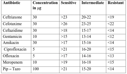
The zone size was recorded and interpreted as per the CLSI guidelines 2013. The three interpretive categories are described as follows.
Susceptible: