They say, when you have the Attitude of Gratitude, Express it.
A heartfelt thanks to all who made this project possible.
I bow down to the Omnipresent Almighty God, for showering His
blessings upon me and being the Alpha and Omega of my life; His Amazing
Grace gives me the strength for each day.
It is with great pride and honor that I express my gratitude to my
Mentor, my Inspiration and Head of the Department Professor
Dr M Veerabahu, for his guidance, concern, overwhelming knowledge and
constant attention that has been a paddle throughout my post-graduation
period. I am indebted to him for bringing out all the hidden talents in me and
molding me to a person that I am today, without whose constant
encouragement and assurance it would have not been possible for me to
accomplish the tasks that I have done so far. Sir the time I spent with you was
an enriching experience for me.
I sincerely thank our beloved Principal, Professor Dr Azhagarasan,
for allowing us to use the scientific literature and research facilities of the
1
I am extremely grateful to my Guide and inspiration Professor
Dr J A. Nathan for his relentless guidance, precious timely advice, words of
encouragement, valuable corrections, throughout my study. His way of
explaining complex things with simple illustrations, discipline, and
imparting courage has made me come a long way. His guidance towards this
project is immense and was constant at each step of the way, without whose
support it would have not been possible for me to complete my task. I thank
him wholeheartedly for his guidance and support in helping me finish the
project on time. The wealth of knowledge and experience that I gained from
you Sir, I will cherish for a life time.
I extent my heartfelt gratitude to Professor Dr B Vikraman for being a
great teacher and philosopher, whose insights and teachings are second to
none. I have been an admirer of his surgical skills, creative thinking,
meticulous planning and passion to understand the latest technology. Sir, I
am grateful to you for imparting some of these qualities in me. I learnt from
you to appreciate all the aspects of life, keep things simple and be cheeky &
humorous whenever possible. Sir, your motivation and guidance stirred up a
lot of enthusiasm in me.
I take this opportunity to thank our respected Professor Dr Malini
Jayaraj, for lending her help and guidance all throughout my
post-graduation period. Her zeal to make the department better was displayed by
2
encouragement, reassurance and her loving, and kind nature were a great
support to me.
I am greatly indebted to Dr Radhika Krishnan, Anesthesiologist for
sharing and imparting her vast experience in medicine. I am thankful to her
for being a person to look up to for suggestions and help whenever there was
a need.
Words aren’t sufficient to thank Professor Dr Shankar, for his
generous and courteous support in the completion of the dissertation and
throughout my education. His belief on my ability was very re-assuring and
a constant source of inspiration.
I sincerely thank Dr Sathyabama, for helping me push boundaries in
the strive for excellence leaving behind no stone unturned and helping me to
understand my own abilities.
I would fail in my duties if I don’t thank my former teacher Dr Seema
Alice Matthew for all encouragement and support.
I am truly grateful to Dr Satheesh, Reader for his little bits of advice,
every now and then which was of great importance upon looking back. His
extended effort to push my limits to get the best out of me was great indeed.
I wholeheartedly thank him for helping me to assist in the various cases
3
extending his knowledge and guidance at each step of the way. His
reassurance was a constant source of encouragement.
A Big ‘thank you’ to Dr James Bhagat for being my teacher beyond
measure. He was always giving a spark to my motivation right from my
undergraduation days and Dr Harish for always building up and
maintaining a cohesive environment and learning atmosphere for me in the
department.
I also thank all my teachers both past and present for their valuable
contribution at each step of the way. It is by their blessings that I stand where
I am today.
I generously thank my batchmates Dr Ajit.C, Dr Arun Vignesh, Dr
Kishok, Dr Deepan, Dr Manoj for their united support and, constructive
criticism at every step and self-less cooperation during my post-graduation
years in the institution. I wish them a successful carrier ahead.
I thank my juniors, Dr Veeraragavan, Dr Alka, Dr Diana, Dr Arvind
for all their constant support and help throughout the college days. I thank
my sub-juniors, Dr Badruddin, Dr Abinaya, Dr Priyanka, Dr Hemavathy,
Dr Priyadardhini, Dr Moni Vikshini for all their help.
I thank my seniors Dr Nirmal Tony, Dr Vivek, Dr Sriraman,
4
Narasimman, Dr Nambi, Dr Siva, & Dr Nirmal for their guidance and
corrections throughout my college days. I also sincerely thank Dr Pavithra
for her timely help in completing the statistics for the dissertation.
I thank all my friends and well-wishers, with special mention to
Dr Priyanka N.P, who has been a pillar of support to me and Dr Rahul Bhandu, Dr Aswin Ku, who always help me with their encouragement.
I also thank my friends, Dr Akila, Dr Gayathri, Dr Sura Dinesh,
Dr Chinu, Dr Darlene, Dr Dhanu whose friendship I have extended from my
undergraduation days.
I thank Dr Asish, Dr Manimala, Dr Maniamudhu from the department
of prosthodontics for helping me with the prosthetic rehabilitation of my
implant patients. I thank Dr Fiaz & Ankhor Dental Lab members for all their
help.
I take this opportunity to thank the Sisters of our Department Mrs.
Deepa, Mrs. Laila, Mrs. Mala, Mr. Venugopal, Mrs. Malathi and other
non-teaching staff.
Saved the best for the last. It is very difficult to put pen to paper in
thanking my parents MR. JOSHUA MUTHUCHAND and Dr
ELIZABETH JOSHUA for their sacrificial love and care they have shown
5
person everyday was there to be seen in me always. It is nothing but their
guidance that has led me thus far. I thank my brother DR
SILAS DANIELRAJ for being an anchor of support in my life. I am also
thankful to my late grandpa Dr Rajendran whose footsteps I wish to follow,
and also my late grandma Mrs. Sakunthala Rajendran whose blessings are
6
ABSTRACT
PURPOSE: The aim of the study is to assess and evaluate the outcome of Alveolar sockets by placing either Bio-Oss or Bio-Oss with Platelet rich fibrin
immediately following extraction; followed by Endossous Dental Implant
placement as a delayed procedure.
MATERIALS AND METHOD: This is a prospective study including 16 preservation sites in 14 patients undergoing extraction immediately followed by
placement of Bio-Oss alone in 8 sites & Bio-Oss mixed with PRF in 8 sites and
the patients were between the age group of 18-50 years (adults). The first group
of patients (Group A) received Bio-Oss which is a bone graft and the second
group (Group B) received Bio-Oss mixed with Platelet Rich Fibrin (5ml patients
own blood, centrifuged at 3000 rpm for 10 min) as the bone preservation agent.
The buccolingual width, the clinical height, the radiological vertical height
(long cone paralleling angle technique which is a measurement from the
adjacent tooth’s root tip/CEJ to the visible outer cortical lining) and soft tissue
status were assessed immediately following extraction (Stage 1), 10 days after
during suture removal & prior to implant placement after approx. 3 months
(Stage 2). The implants were placed between 3-10 months and the stability and
survival rate of the implants were measured. Finally, after evidence of
7
RESULTS: Both the groups A & B were evenly distributed with respect to age, gender, site, periodontal status. An overall reduction of the clinical
bucco-palatal width, clinical height and radiographic height were found in all
3 stages of evaluation but was not significant with respect to both the groups.
Both Group A and Group B show similar socket dimensional changes.
CONCLUSION:
Within the limitations of the study, it can be inferred that the use of PRF along with bone grafts for socket preservation improves handling
properties & soft tissue healing but with respect to maintaining the socket
dimensions there is no clear statistical evidence to justify its use along with
bone grafts.
Key words: Bio-Oss, Platelet-Rich fibrin, Socket preservation,
8
CONTENTS
S. No. TITLE PAGE No.
1. INTRODUCTION 1
2. AIMS AND OBJECTIVES 6
3. REVIEW OF LITERATURE 7
4. MATERIALS AND METHODS 24
5. RESULTS 39
6. DISCUSSION 43
7. SUMMARY AND CONCLUSION 51
8. BIBILOGRAPHY 53
9
LIST OF TABLES
TABLE No.
TITLE
1. GENDER DISTRIBUTION
2. SMOKING STATUS DISTRIBUTION
3. ARCH DISTRIBUTION
4. SITE DISTRIBUTION
5. GINGIVAL STATUS DISTRIBUTION
6. PERIODONTAL STATUS DISTRIBUTION
7. STAGE 1 VARIABLES
8. STAGE 2 VARIABLES
9. STAGE 3 VARIABLES
10. TEST OF SIGNIFICANCE FOR BOTH GROUPS
11. DISTRIBUTION BASED ON ADDITIONAL BONE GRAFTING PROCEDURE
10
LIST OF GRAPHS
GRAPH No. TITLE
1 GENDER DISTRIBUTION
2 SMOKING STATUS DISTRIBUTION
3 ARCH DISTRIBUTION
4 SITE DISTRIBUTION
5 GINGIVAL STATUS DISTRIBUTION
6 PERIODONTAL STATUS DISTRIBUTION
7 DISTRIBUTION BASED ON USE OF ADDITIONAL BONE GRAFT
8 DISTRIBUTION BASED ON USE OF OSTEOTOME
9 STAGE 1 VALUES
10 STAGE 2 VALUES
11 STAGE 3 VALUES
1
INTRODUCTION
Since time immortal there has been events recorded in history of lost
tooth being replaced by artificial means. This led to the field of dentistry
and further advancements followed [1][2]. The thought of having an artificial tooth fixed in the alveolus performing the normal function has eluded the
minds of people for some time but not for long. The field of Oral
Implantology has grown in leaps and bounds and is currently the go to
option when it comes to fixed replacement and prosthesis [3].
As a good foundation is for a successful structure to be in place, so is
a good alveolar ridge a prerequisite for successful dental implant
placement. The major issue with it is that, there is definite loss of alveolar
structure when a tooth is lost, in all three dimensions. The actual bone that
is present before the tooth was lost is the same amount required for similar
and strong implant placement. Socket preservation technique is as old as the
field of Implantology and is by far the most reliable technique when it
comes to maintaining the residual alveolar bone and maintaining the width
and height [4][5].
The resorption pattern seen after tooth extraction is characterized by
2
thereafter [6].Horizontal buccal bone resorption is as much as 56%, while
lingual bone resorption has been reported to be upto 30% [7].Major bone resorption of the vestibular wall of the extraction socket can be attributed to
the higher proportion of bundle bone, a tooth-dependent tissue through
which the periodontal ligament fibers are anchored to the jaws, which
undergoes resorption due to the loss of its function.
The volume reduction of the alveolar process may prevent or render
difficult implant installation in a prosthetically driven position,
simultaneously jeopardizing the functional and aesthetic outcomes. Thus,
ridge preservation treatment protocols have been developed in order to
maintain the alveolar bone volume existing at the time of tooth extraction,
and to ensure the support of an adequate ridge profile
Alveolar socket preservation (ASP) is a procedure in which a graft
material is placed in the socket of the extracted tooth at the time of
extraction, with or without the application of barrier membranes or soft
tissue coverage, to preserve or improve the original ridge dimensions and to
allow an ideal implant location [8].
A successful esthetic and functional restoration of the implant
depends on its optimal placement, which is influenced by its height and
buccolingual position as well as by the alveolar ridge dimensions [9]. Prerequisites for successful implant therapy are integration of the implant,
3
require sufficient alveolar bone volume and favorable ridge architecture
coupled with an appropriate surgical technique [10]. Socket preservation most of the time eliminates or often at least minimizes the necessity for
future augmentation procedure. Success of dental implants are not only
weighed by osseointegration but also the long-term stability of the
prosthesis including form, function and aesthetics [11].
Healing of extraction socket involves retention of clot followed by a
sequence of events that lead to changes in the alveolar process in all three
dimensions. The key element is to act at the time of tooth extraction and to
prevent the collapse of the ridge [12]. Since the surgical placement of implants have become a routine part of treatment planning, clinicians need
to consider socket preservation at the time of extraction, as an increasingly
predictable method of ridge preservation [13]. Several techniques have been
employed as ridge preservation procedures involving the use of bone grafts,
barrier membranes, and biologics to provide a better restorative outcome
[14].
Site preservation through socket grafting will help optimize bony fill
within the extraction socket, thereby maintaining vertical bone height and
helping to stabilize the marginal soft tissue at the site [15]. Maintenance of the hard and soft tissue envelope and stable ridge volume are important
aims to allow simplifying subsequent treatments and optimizing their
4
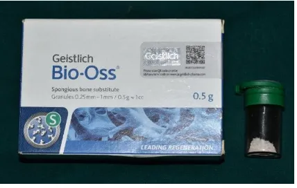
It is well documented and researched in the literature that Bio-Oss
(Geistlich Pharma AG, Wolhusen, Switzerland) has various beneficial
properties when used as a bone preserving agent. Bio-Oss is a Deproteinized
Bovine Bone Mineral (DBBM) and is a defatted and deproteinized xenograft
reduced to porous grains of different dimensions (0.25–2 mm) and deprived
of all its organic components through high-temperature processes in order to
minimize the immune response.
The particles in the Bio-Oss graft have properties to encourage bone
fill and act as a stable grafting substance. It is noted that socket dimensions
are reduced by 21-43% by spontaneous healing after tooth extraction
without the use of bone grafts but the use of the Bio-Oss bone graft
substance has significantly held the socket in place and the socket
dimensions reduce only by 8-17%. The use of Bio-Oss bone graft is
considered here [17].
Recently the use of platelet concentrates has been proposed as an aid
for enhancing regeneration of osseous and epithelial tissue in oral surgery
[18]. Platelets play a significant role in wound healing as they have the
various growth factors necessary for the sequel of wound healing.
Platelet rich plasma (PRP), an autologous concentrate of human
platelets in a small volume of plasma have demonstrated to induce healing.
5
since its introduction but the use of it, has declined ever since. PRF has
taken over because of the ease of preparation with better results [19].
Platelet Rich Fibrin (PRF) is a 2nd generation platelet concentrate with reduced preparation time and better handling properties developed by
Choukrounet al 2001. Unlike PRP, it is derived by natural and progressive
polymerization occurring during centrifugation without addition of any
anticoagulant [20]. PRF has also been studied about its bone preservation properties in the past [21].
Therefore, in this study either Bio-Oss separately or Bio-Oss mixed
with PRF was used as a socket preservation agent followed by delayed
implant placement and loading protocol.
6
AIMS AND OBJECTIVES
This prospective clinical study is designed to evaluate the outcome of
either Bio-Oss separately or Bio-Oss mixed with PRF as socket
preservation agent
1. Evaluate and tabulate the socket preservation site’s dimensional
changes followed by delayed Endossous Dental Implant placement
and delayed loading protocol
2. Evaluate the overall stability and survival rate of the Implants
7
REVIEW OF LITERATURE
Pietrokovski, Jaime et al (1967) [22] studied about the alveolar ridge
resorption in 149 plaster casts with single teeth missing through extraction and
concluded that
1. The buccal plate in the both maxilla and mandible was resorbed to a
greater extent than the palatal plate. The centre of the edentulous ridge
therefore, shifts to a position closer to the palatal plate of bone.
2. The amount of tissue resorption was significantly greater in the
edentulous molar region than in the incisor and premolar regions of
both jaws.
Bragger, U., Pasquali, L., & Kornman, K. S. (1988) [23]. This was
one of the early articles where the term ‘Alveolar Socket Preservation’ was
used, where the author analysed the socket dimensions along with Interdental
bone changes after various flap procedures. The study was assessed by
computer-assisted densitometric image analysis (CADIA). Standardized
radiographs were digitized and quantitative information regarding density
changes was obtained. The study showed a path towards socket preservation
which can be an effective procedure in maintaining the dimensions and
8
Mecall & Rosenfeld (1992) [24] discussed the influence of residual
ridge resorption patterns on Implant placement and tooth position. The
incorporation of prosthetic information into a presurgical implant treatment
plan is essential to provide accurate guidelines for fixture placement by the
surgeon. The first part of the article gives a modified surgical technique to
construct a prosthesis compatible with the final tooth position. The second part
describes a technique in which a computer profile image is used to identify the
determinants of the optimal final tooth position and discusses the impact of
these determinants on the prosthetic design.
Smukler et al, (1999) [25]determined the fate of demineralized
freeze-dried bone allograft (DFDBA) used in conjunction with a barrier membrane in
the management of 5 extraction sockets and compared it with the amount of
bone formed in 5 untreated sites. After an adequate healing time (8-23
months) just prior to implant placement, bone biopsy was taken and
Histomorphometric analysis done. They concluded that treatment with
DFDBA in conjunction with cell occlusive membranes will result in new bone
formation, predominantly by the process of conduction, which appears to be
similar in amount and nature to that found in cores harvested from healed
non-functional edentulous areas.
Berglundh & lindhe (1997) [26] did an animal study to check the
Healing around implants placed in bone defects treated with Bio-Oss and
9
newly formed bone. 5 beagle dogs, about 1-year-old, were used. Bio-Oss was
seen to fulfil the criteria of an osteoconductive material. It was also observed
that 4 months after implant installation, the titanium/hard tissue interface at
test and control sites exhibited, from both a quantitative and qualitative aspect,
a similar degree of "osseointegration".
Artzi et al (2000)[27] histomorphologically analysed the healing of
porous bovine bone graft (PBBM) in 15 human extraction sockets and
concluded that newly formed bone was characterized by abundance of cellular
woven-type bone in the coronal area, while lamellar arrangements could be
identified only in the more apical region. New osseous tissue adhered to the
PBBM. Therefore, it is seen that PBBM particles are an appropriate
biocompatible bone derivative in fresh extraction sockets for ridge
preservation. The resorbability of this xenograft could not be recognized in a
9-month period as all the samples had residual bone graft materials.
Tassos Irinakis (2006) [28]concluded in his study that post-extraction
maintenance of the alveolar ridge minimizes residual ridge resorption and
thus, allows placement of an implant that satisfies aesthetic and functional
criteria. Recent advances in bone grafting materials and techniques allow the
dentist to place implants in sites that were considered compromised in the
past.
I Darby, S Chen, R De Poi, (2008) [29] Implant treatment can be
10
maintaining sufficient bone for optimal implant placement and consequently
the appearance. Consideration has been given to healing of extraction sockets
and previously published studies that have attempted to preserve the alveolar
ridge. The majority of teeth are extracted by general dental practitioners and it
is hoped that this article will stimulate some thought on the topic of ridge
preservation. Not all extraction sockets need to be preserved, but the authors
feel that ridge preservation ought to be considered at the time of tooth
removal.
Michael Danesh-Meyer (2008) [15] Site preservation through socket
grafting will help to optimize bony fill within the extraction socket, thereby
maintaining vertical bone height and helping to stabilize the marginal soft
tissues at the site which plays an integral part in surgical placement of dental
implants. Guided bone regeneration with the use of particulate bone graft and
resorbable membranes remains the most widely used technique at present.
Fickl S, Zuhr O, Wachtel H, Stappert CFJ, Stein JM, Hu¨rzeler MB (2008) [30] concluded that Socket preservation techniques, used in the
present experiment, were not able to entirely compensate for the alterations
after tooth extraction. Yet, incorporation of Bio-Oss Collagen seems to have
the potential to limit but not avoid the postoperative contour shrinkage. The
study clearly stated that complete ridge preservation is not possible with the
11
Fickl S, Zuhr O, Wachtel H, Bolz W, Huerzeler MB (2008) [31]
concluded that the findings from the present study disclose that incorporation
of Bio-Oss Collagens into the extraction socket has only limited impact on the
subsequent biologic process with particular respect to the buccal bone plate.
The horizontal measurement of the alveolar ridge depicted that the loss of the
buccal bone plate was replaced to a certain amount by newly generated bone
guided by the Bio-Oss collagens scaffold. It seems that the mechanical
stability provided by Bio-Oss collagens and furthermore by a free gingival
graft could act as a placeholder preventing the soft tissue from collapsing.
Fickl S, Schneider D, Zuhr O, Hinze M, Ender A, Jung RE, Hurzeler MB (2009) [32] Did an animal study and studied the dimensional
changes of the ridge contour after socket preservation and buccal
overbuilding. It was concluded and statistically proved from their study that
overbuilding the buccal aspect in combination with socket preservation is not
a suitable technique to compensate for the alterations after tooth extraction.
Ivan Darby, Stephen T. Chen, Daniel Buser (2009) [10]Authors did a
review of literature of the articles relating to ridge preservation and concluded
that Ridge preservation procedures are effective in limiting horizontal and
vertical ridge alterations in post-extraction sites. The authors stressed the
importance of knowing the internal and external changes that happen in the
sequence of events that follow in the extraction of a tooth. There is no
12
also no conclusive evidence that ridge preservation procedures improve the
ability to place implants.
Mardas N, Chadha V, Donos N (2010) [33] compared the potential of
a synthetic bone substitute, Straumann Bone Ceramics (SBC) or a
bovine-derived xenograft, Bio-Oss deproteinized bovine bone mineral (DBBM)
combined with a collagen membrane to preserve the alveolar ridge dimensions
following tooth extraction in 26 patients. It was seen that both biomaterials
partially preserved the width and the interproximal bone. The histologic
analysis showed new bone formation in the apical part of the biopsies, which
was in direct contact with the bone grafting particles. The coronal part of the
biopsies were filled with dense fibrous connective tissue surrounding the bone
grafting particles.
Luis André Mezzomo, Rosemary Sadami Shinkai et al (2010) [9]
Several techniques and materials have been suggested for alveolar ridge
preservation (ARP) after dental extraction and before implant placement.
Ridge preservation procedures are efficient in limiting the post-extraction
dimensional loss of the ridge and are accompanied by a different degree of
bone regeneration, with varied amounts of residual graft material particles.
Fawad Javed a, George E. Romanos (2010) [34]reviewed and stated
that, it is evident that the core issue to observe during immediate loading is the
establishment of a good implant primary stability. There is sufficient evidence
13
dependent on several factors including bone density and quality, implant
shape, design and surface characteristics and surgical technique including
implants placed in a preserved site. But if the stability of the implant cannot be
achieved then site preservation protocols could be followed for a delayed
implant placement.
Gholami GA, Najafi B, Mashhadiabbas F, Goetz W, Najafi S (2011) [35] compared a nanocrystalline hydroxyapatite (NCHA), NanoBone and a deproteinized bovine bone mineral (DBBM), Bio-Oss with a collagen
membrane on the horizontal ridge width. Alterations following tooth
extraction in 28 symmetrical, non-molar extraction sockets using a split-mouth
design in the adult population, was found and socket preservation with both
the materials resulted in similar and limited horizontal ridge width alterations
following tooth extraction. In the DBBM group, the width decreased from
7.75 +/- 1.55 to 6.68 +/- 1.85 mm whereas the width of the NCHA group
decreased from 7.36 +/- 1.94 to 6.43 +/- 2.08 mm. In both the groups the
readings were not statistically significant.
CHF Hammerle, Arau´ jo MG, Simion M (2011) [16] demonstrated
that the alveolar ridge undergoes a mean horizontal reduction in width of 3.8
mm and a mean vertical reduction in height of 1.24 mm within 6 months after
tooth extraction. The techniques aimed at ridge preservation encompassed two
different approaches: i) maintaining the ridge profile, ii) enlarging the ridge
14
immediate implant placement leads to high implant survival rates. This
procedure is primarily recommended in premolar sites with low aesthetic
importance and favourable anatomy. In the aesthetic zone, however, a high
risk for mucosal recession was reported. Hence, it should only be used in
stringently selected situations with lower risks and only by experienced
clinicians. In molar sites a high need for soft and hard tissue augmentation was
identified.
Hassan, Marei, and Alagl et al (2011) [36] studied the osseous defects
after surgical removal of impacted 3rd molars and covered the extracted site with combined autogenous graft with Bio-Oss bone graft materials. From their
study they concluded that grafting of osseous defects and extraction site with
autogenous bone graft combined with Bio-Oss materials will predictably result
in a decreased risk of developing a periodontal defect.
K. Patel, N. Mardas, N. Donos (2012) [37] stated that there was no
significant difference in radiographic bone levels and success and survival of
implants placed in sites previously preserved with SBC (Straumann bone
ceramic) or DBBM (Bio-Oss) at 1-year follow-up (post-loading). The study
was done in 27 patients who were randomly assigned into the groups. SBC
could therefore be a suitable alternative to DBBM for ridge preservation prior
to implant placement. In literature, DBBM forms a standard upon which other
15
Ahmad Kutkut, Sebastiano Andreana, Hyeong-ll Kim, and Edward Monaco Jr (2012) [38] stated that major changes in an extraction site
happened during the 1st year after tooth extraction, with two thirds of the bone loss occurring within the first 3 months. The study was done on 16 patients
assigned into two groups. Medical-grade calcium sulfate hemihydrate
(MGCSH) mixed with platelet-rich plasma (PRP) showed greater vital bone volume at 3 months with rapid enhancement of bone healing compared to
PRP-free collagen resorbable graft based on Histomorphometric analysis.
There was no clear statistically significant difference clinically in both vertical
and horizontal bone resorption between the groups.
XU lixin, Ding yun, Lei chaofeng, Jiang weipeng (2012) [39] showed
the use of Advance Platelet-rich fibrin (platelet-rich fibrin PRF) in 6 patients
with immediate implants in the aesthetic zone. It was seen that even in the
presence of chronic periapical lesions which were debrided, the implant sites
healed well and the pink esthetic score reached 10 or more. The various
growth factors in the PRF synergies and promote tissue regeneration/repair.
The earlier clinical success rate of bone graft & anti-infective ability were the
substantial benefits.
Robert Horowitz, Danny Holtzclaw, Paul S. Rosen (2012) [5] There
appears to be consensus from the reviewed literature supporting ridge
preservation techniques as a whole. Multiple studies demonstrated less ridge
16
versus the placement of no graft material in fresh alveolar sockets. The
analysis did not show any grafting materials demonstrating a clear benefit over
any others or that a barrier membrane is necessary. The evidence is also too
premature about whether socket preservation efforts require primary closure.
In the emerging area of growth factors, there is no high-quality evidence to
either support or refute the use of socket grafting measures.
Marco Tatullo, Massimo Marrelli, et al (2012) [40]The present study
is to investigated, clinically and histologically, the potential use of PRF,
associated with deproteinized bovine bone (Bio-Oss), as grafting materials in
pre-implantology sinus grafting of severe maxillary atrophy, in comparison
with a control group, in which onlydeproteinized bovine bone (Bio-Oss) was
used as reconstructive material. With the adding of PRF the lamellar bone
tissue with an interposed stroma appeared relaxed and richly vascularized. The
author concludes that the use of PRF and piezo-surgery reduced the healing
time, compared to the 150 days described in literature, favouring optimal bone
regeneration. At 106 days, it is already possible to achieve good primary
stability of endosseous implants, though lacking functional loading.
Giorgio Pagni, Gaia Pellegrini, William V. Giannobile and Giulio Rasperini (2012) [41]reviewed the literature and stated that Socket grafting is
a commonly adopted therapy for the preservation of alveolar bone structures
17
recommend that a less invasive grafting technique to be adopted when
indicated especially in the esthetic zones. Invasive procedures like guided
bone regeneration and sinus lift are less frequently needed if a proper socket
preservation is adopted.
Jung RE, Philipp A, Annen BM, Signorelli L, Thoma DS, Ha¨mmerle CHF, Attin T, Schmidlin P (2013)[17]evaluated the radiographic
changes of the alveolar ridge following application of different ridge
preservation techniques 6 months after tooth extraction. Four treatment
modalities were randomly assigned in 40 patients:
b-tricalcium-phosphate-particles with polylactid coating (b-TCP), demineralized bovine bone mineral
with 10% collagen covered with a collagen matrix (DBBM-C/CM), DBBM-C
covered with an autogenous soft-tissue graft (DBBM-C/PG) and spontaneous
healing (control). Cone-beam computed tomography scans were performed
after treatment and 6 months later. Application of DBBM-C, covered with CM
or PG, resulted in less vertical and horizontal changes of the alveolar ridge as
compared with controls 6 months after extraction.
Qi Li, Shuang Pan, Smit J. Dangaria, Gokul Gopinathan, Antonia Kolokythas, Shunli Chu, Yajun Geng (2013) [42] study suggest that PRF
enhances osteogenic lineage differentiation of alveolar bone progenitors more
than of periodontal progenitors by augmenting osteoblast differentiation,
RUNX2 expression, and mineralized nodule formation via its principal
18
regenerative scaffold promoting both tissue-specific alveolar bone
augmentation and surrounding periodontal soft tissue regeneration via
progenitor-specific mechanisms.
B Kassim, S Ivanovski, N Mattheos (2014) [43] Ridge preservation
techniques are effective in minimizing post-extraction alveolar ridge
contraction. However, there is insufficient evidence to suggest that the use of
these techniques in conjunction with dental implant treatment improves
implant treatment outcomes. Furthermore, ridge preservation does not
necessarily eliminate the need for further simultaneous augmentation at the
time of implant placement. The delayed healing associated with ridge
preservation using socket grafting necessitates a commitment to a delayed
placement protocol. The extended treatment time, compromised healing and
expense related to ridge preservation suggests a more cautious approach with
regards to the indication of such techniques.
Elizabeth M. Tomlin, Shelby J. Nelson and Jeffrey A. Rossmann (2014)[12] Healing of the extraction socket after tooth removal involves
retention of the blood clot followed by a sequence of events that lead to
changes in the alveolar process in a three-dimensional fashion. This normal
healing event results in a minimal loss of vertical height (around 1 mm), but a
substantial loss of width in the buccal-lingual plane (4-6 mm). During the first
three months following extraction that loss has been shown to be significant
19
ability to restore the site with acceptable aesthetics. Procedures that reduce the
resorptive process have been shown to be predictable and potentially capable
of eliminating secondary surgery for site preparation when implant therapy is
planned. The key element is prior planning by the dental therapist to act at the
time of extraction to prevent the collapse of the ridge due to the loss of the
alveolus.
Several techniques have been employed as ridge preservation
procedures involving the use of bone grafts, barrier membranes and biologics
to provide a better restorative outcome. This review explored the evidence
behind each technique and their efficacy in accomplishing site preparation.
The literature does not identify a single technique as superior to others;
however, all accepted therapeutic procedures for ridge preservation have been
shown to be more effective than blood clot alone in randomized controlled
studies.
Dhurat R, Sukesh MS (2014) [44] The utility of platelet-rich plasma
(PRP) has spanned various fields of dermatology from chronic ulcer
management to trichology and aesthetics, due to its role in wound healing.
There is also a wide variation in the reported protocols for standardization and
preparation of PRP, in addition to lack of accurate characterization of the
tested products in most articles on the topic. Additionally, the high cost of
20
Jie Liu and David G. Kerns (2014)[45] Guided Bone Regeneration
(GBR) is a surgical procedure that uses barrier membranes with or without
particulate bone grafts or/and bone substitutes. Angiogenesis and ample blood
supply play a critical role in promoting bone regeneration. Guided bone
regeneration can be achieved with using particulate autogenous bone grafts,
allografts, xenografts, or alloplastic grafting materials and resorbable or
non-resorbable barrier membranes techniques.
Dias DR, Leles CR, Lindh C, Ribeiro-Rotta RF (2014) [46]
Evaluated the changes in marginal bone level (MBL) around implants, in sites
with different bone types, affecting the over-time implant stability measured
by resonance frequency analysis (RFA). Implant bone sites were grouped
according to the Lekholm and Zarb bone classification. The implant stability
quotient (ISQ) was measured by RFA at four time points: implant placement,
uncovering, rehabilitation, and at 1-year follow-up after loading. Percent
change in bone level was calculated based on the difference between the
implant length and height from the crestal bone level to the implant apex. It
was concluded that there was increased implant stability after implant
placement, but it was not affected by changes in marginal bone level during
the first year of loading.
Markus Glocker, Thomas Attin and Patrick R. Schmidlin (2014)
[47]It has also been suggested that resorption of the buccal bundle bone can be
21
because the biological integrity of the buccal periodontium (bundle bone)
remains untouched.
Eduardo Borie, Daniel García Oliví, Iara Augusta Orsi, Katia Garlet, Benjamín Weber, Víctor Beltrán, Ramón Fuentes (2015) [18] in
their article meticulously jotted the various applications and the versatility of
the use of PRF in dentistry. Platelet rich fibrin (PRF) appears as a natural and
satisfactory alternative with respect to bioactive surgical additives to regulate
inflammation to increase the speed of healing process, with favourable results
and low risks. PRF alone or in combination with other biomaterials seems to
have several advantages and indications both for medicine and dentistry, as it
is a minimally invasive technique with low risks and satisfactory clinical
results. It is said to increase the healing rate of the grafted bone when used in
combination with the same.
Chihiro Masaki, Tetsuji Nakamoto, Taro Mukaibo (2015) [11] The
anterior maxilla is in the esthetic zone, and the thickness of the bone on the
labial side around the natural tooth is less than 1 mm in many cases.
Therefore, it is impossible to prevent bone resorption completely, even if ridge
preservation and immediate or early implant placement are performed after
tooth extraction. It is necessary to obtain stable and long-term esthetics by
combining connective tissue and free gingival grafts, in addition to hard tissue
22
Zeeshan Sheikh, Nader Hamdan, Yuichi Ikeda, Marc Grynpas, Bernhard Ganss and Michael Glogauer (2017) [48] in their article reviewed
the importance on the placement of a barrier membrane to limit the epithelial
down growth. In an attempt to overcome complications related to the epithelial
down-growth and/or collapse of the non-rigid barrier membrane and to
maintain space, clinicians are recommended to commonly use a combination
of membranes with hard tissue grafts.
Pâmela Letícia Santos, Edilson José Ferreira, Marcos M. Kuabara et al (2017) [14] Two and a half months after the dental extraction and the
filling with Bio-Oss, it was possible to observe through tomograpic image that
the alveoli of the teeth 11 and 21 were completely filled with mineralized
tissue. Authors concluded that the biomaterial Bio-Oss is efficient for the
preservation of the alveolar bone after dental extraction, acting as a framework
for the bone neoformation for a later installation of osseo-integrated implants.
Carlo Maiorana, Pier Paolo Poli et al (2017) [8] Noted a trend
towards reduction of horizontal and vertical socket dimensions from baseline
to the final examination. The mean width and height of resorption were 1.21
mm (P=0.005) and 0.46 mm (P=0.004), respectively. Socket preservation
using demineralised bovine bone mineral (Bio-Oss) provided stable
dimensional changes of the alveolar ridge associated with good
23
Madhan G, Singh M (2017) [21] The 22 patients in the study received
either PRF or Collaplug (Zimmer) as the preservation agent. Both the groups
were analysed for vertical buccal crestal. Bone heights were measured
immediately after extraction; at 4 months and implants were placed. It was
concluded that both the two materials tested seem to be effective in the
treatment of extraction sockets. The study lacked a negative control and could
not completely attribute the extent of the clinical improvement of PRF per se,
but it was stated that preparation of PRF is not very cumbersome and
inexpensive, which makes it a better socket preservation material.
Tomasi C, Donati M, Cecchinato D (2018) [64] analysed in their study
the characteristics of fresh extraction sockets subsequent dimensional changes.
Three dimensional virtual models were analysed using Geomagic software and
the dimensional changes were calculated. It was concluded that the width of
the marginal portion (2mm-4mm apically of the crest) of the ridge shrunk by
20%-30%. Eventually, about 6mm of the horizontal dimension was lost at the
extracted site. It was also seen that the ridge reduction was twice as large at
the buccal/facial when compared to the lingual/palatal aspect thus after first 3
months, the horizontal dimension was reduced by 30% and at 12 months, it
was from 30-40%. It was stated that the hard tissue change following tooth
24
MATERIALS AND METHODS
For this prospective study, the study subjects were the patients who
had tooth/teeth that are indicated for extraction and consented for a socket
preservation procedure with a future implant placement in mind. These were
patients who reported to the Department of Oral and Maxillofacial Surgery,
Ragas Dental College and Hospital, Chennai. For the study the patients were
randomly assigned into two groups. The first group received only Bio-Oss
bone grafting material as the bone preservation agent whereas the second
group received Bio-Oss mixed with PRF (5ml patients own blood, centrifuged
at 3000 rpm for 10 min) as the bone preserving agent. In both the groups a
standard resorbable Guided Tissue Regeneration membrane was used to cover
and hold the bone graft in place.
There was a total of 8 socket preservation sites in which implants were
placed in each group, accounting for a total of 16 sites in 14 patients. All
patients had a single tooth to be replaced except 2 patients who had two sites
(one in each group). The implant placement was planned approx. 4-6 months
after the socket preservation procedure.
Before the commencement of the study an Ethical committee approval
was obtained from the Institutional Review Board in Ragas Dental College
25
procedure and they agreed to be a part of the study protocol by submitting a
written Informed consent (Annexure II). All patients were recalled and
reviewed postoperatively at definite time intervals.
INCLUSION CRITERIA: -
1. Tooth indicated for extraction (caries, endodontic complications,
crown fractures)
2. All patients who give a written consent
3. Age group between 16-50 years.
4. Both sexes were included
5. Desire for Implant placement following extraction of non - restorable
tooth and socket preservation.
6. Good general health
7. Be available for follow-up examinations
8. Patients with fair periodontal status
EXCLUSION CRITERIA:
1. History of systemic diseases that would contraindicate surgical
treatment.
26
3. Patients who do not accept the use of xenograft (religious reason)
4. Acute infection in the surgical site
5. Periodontal disease with bone loss
6. Known allergy to any of the materials used in the study
7. Failure to sign a informed consent
8. Ankylosed tooth.
9. Pre-existing bone loss >50%
ARMAMENTARIUM USED: -
1. 2% Lignocaine with 1:80,000 Epinephrine
2. Extraction forceps & Periotomes
3. Bio Oss
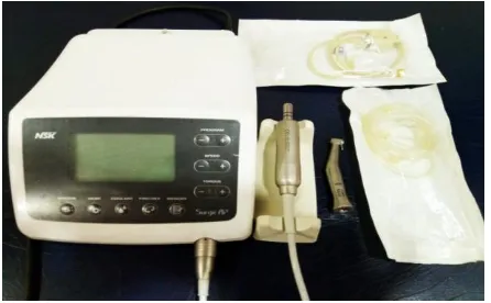
4. (Universal Centrifuge) Platelet rich fibrin
5. Ethilon sutures
6. Caliper/marked probe.
7. Implants (Adin universal system)
27
Pre-operative a-silicon impressions were taken and diagnostic
casts poured, the Intra Oral Periapical Radiographs were taken (long
cone paralleling angle technique for reproducibility) clinically the site
to be preserved was measured with a caliper/probe with markings. No
pre-op medications were prescribed for both groups. The study
precedes with a formal written consent from the patient.
SURGICAL TECHNIQUE for socket preservation: -
Patient was draped and asked to wash with 0.2% betadine solution.
Extraction of the concerned non-restorable tooth was performed with great
care to preserve the buccal bone plate and the surrounding soft and hard
tissues. After administration of local anaesthetic with adrenaline (2% Lignox
1:80,000), crevicular incisions were made around the tooth structure with a
scalpel, no 15. Minimal flap elevation was done just to visualize the
surrounding bone level. Initially the tooth was gently luxated with the use of a
sharp Luxator (GDC Luxatip set) at the junction between the tooth-bone
interface. No attempt was made to elevate but just to make the tooth structure
inside the socket mobile, any inadvertent expansion of the socket during
luxation was beneficial for the socket dimensions as long as the damage to the
socket walls was minimal. The mobile tooth was grasped by root forceps and
rotated & extracted along the axis of the tooth without any buccal traction.
After tooth extraction, careful removal of the granulation tissue (if
28
width of the socket was measured mesio-distally and bucco-lingually to the
base of the crestal bone, the height of the buccal and lingual bone plate was
clinically measured to the nearest 0.5 mm at the mid-buccal and mid-lingual
aspect using a periodontal probe. The extracted tooth’s dimensions were also
measured
Group A:
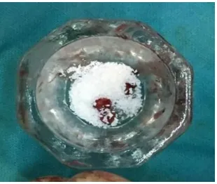
Extraction was followed by gently packing of the alveolar sockets with
Bio-Oss bone graft alone, which is wetted with saline in a sterile Dappen dish.
Excessive pressure was avoided as the spaces in between the bone graft
particles were allowed to be filled by blood. The socket is completely packed
till the crestal bone level anticipating a slight crestal bone resorption. Any
bone particles which overflowed were carefully removed with a sharp
instrument. A resorbable GTR membrane (Healiguide, APT Pharma), with
dimensions relating to the socket was adapted over the bone cover and tucked
into the flaps. No sutures were placed over the resorbable membrane, but flaps
were approximated by either 3-0 or 4-0 silk sutures to get maximum cover. A
slight expose of the membrane to the oral environment was inevitable in most
situations.
Group B:
Just before the extraction procedure, 10ml of patient’s own blood is
29
anticoagulant. The blood containing test tubes were immediately transferred to
a centrifuge approx. 400g (Remi R&C Lab centifuge) for 3000 rpm for 10
mins and allowed to rest for a further 15 mins for the clot formed to mature
and form the actual PRF, this method is slightly different to original protocol
formulated by Dolan & Choukroun, 2006[49,50,51]. It was observed that the resting of the test tubes for 15 mins allowed for a stable and consolidated
formation of the PRF jelly. The PRF was taken from the test tube by a sharp
tweezer, and the PRF- residual RBC junction visualized. The separation of the
PRF was done more towards the residual RBC side as it is seen that the many
vital growth factors reside here; care was taken not to eliminate the vital
growth factors. The PRF jelly was mixed with Bio-Oss bone graft particles
and gently packed into the socket, similar to Group A. A part of the PRF jelly
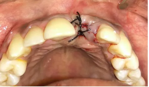
was flattened and kept over the admixture. A resorbable membrane was placed
over the socket and sutured with either 3-0 or 4-0 silk sutures to get maximum
cover.
Post-op instructions:
Strict oral hygiene instructions were given.
Patients were instructed to rinse twice a day with 0.2% Chlorhexidine
and avoid any alcohol containing mouthwashes.
Patients were also instructed to avoid chewing at the treated sites,
30
Patients received pain killer medication (Imol plus) and antibiotics
(Amoxicillin) (Metronidazole) for 5 days.
Postoperative follow-up:
All patients were recalled at 7–14 days for check-up and suture
removal. They were also inspected for any swelling or release of graft
material. The condition of the membrane was documented.
Interim prosthesis was fabricated either as Maryland winged Bridge
which is resin bonded on the lingual aspect or as Partial denture with adequate
relief over the preserved site in the anterior esthetic zone.
Patients were advised not to inadvertently give any pressure or trauma
over the site which is preserved.
The preserved site is allowed to heal, post-operative evaluation is done
after 3 days, at suture removal approx..2 weeks, 1 month and finally around
3-4 months before an implant procedure was planned.
After a healing phase of approx. 3 months, the augmented alveolar
segment was again measured clinically & radiographically.
Depending on the measurements obtained the implant size was
determined. After which an implant procedure was planned.
Prior to the implant placement procedure, a-silicon impressions were
31
Surgical technique for Implant placement: -
Patient was draped and site preparation done by scrubbing betadine
solution. Local anaesthesia was given and a full thickness flap was raised. The
ridge visualized and dimensions measured with the help of a marked probe.
The width of the socket was measured mesio-distally and bucco-lingually to
the base of the crestal bone, the height of the buccal and lingual bone plate
was clinically measured to the nearest 0.5 mm at the buccal and
mid-lingual aspect. These are the same measurements done at the time of socket
preservation both in Group A and Group B. With the help of the marking drill
(Lancet drill) the point of placement of the Implant is marked, with an initial
depth of approx. 3mm. after which sequential drilling done with a proper axis
and the implant is gently screwed in place.
Based on the density of bone (ZARB classification) D2, D3 bone were
expanded with lateral condensing osteotomes as shown by Lee H Silverstein et
al, 1999 [52] to maintain the bone structure which otherwise could be lost due
to drilling. The Endossous implant (Adin Universal system) implants were
screwed in place and in all cases except one the primary stability was above 40
N. The implants were slightly submerged to 1mm into the bone to compensate
for the crestal bone loss. In cases where there was thread exposure, a
32
This was followed by approximation of the mucoperiosteal flap and
closure was done with 3-0 or 4-0 ethilon suture. Final irrigation of the site was
done with saline and the regular oral surgical case post op instructions were
given to all patients.
Suture removal was done at about 2 weeks and the patient was
reviewed for any signs of pain or discomfort. The patients were advised to
wait for approx. 3-4 months for the integration of the implants depending on
the site in which they were placed.
Second stage surgery:
The implants were left for 3-4 months approx. after which IOPA
radiograph was taken to check for any evidence of radiolucency surrounding
the implant. Radiolucency extending till the apex of the implant was
considered as failure. The implants were also clinically checked for stability.
After confirming osseointegration the implants proceeded for prosthetic rehab.
In all patients, the cover screw was exposed under local anaesthesia
infiltration and healing abutment placed with minimal flap elevation. The
healing abutment was left in place till a sufficient amount of soft tissue cuff
was formed around it. Patients were advised strict oral hygiene measures with
peri-mucositis in mind.
After 10-14 days the healing abutment was removed and open tray
33
was fabricated and the crown was placed in proper functional and esthetic
position.
EVALUATION CRITERIA: -
1. Following extraction, the desired socket preservation method is employed
and the socket measured clinically by caliper/marked probe (in mm) and
radiographically (in mm).
2. Clinically the Bucco-lingual width & vertical height with the
mucogingival junction as reference
3. Radiographically the vertical height is measured with the Cemento-enamel
junction off the adjacent tooth as reference
4. Implant success is calculated by the ICOI criteria [53].
The clinical buccolingual width and mesiodistal width was measured
with a caliper or a marked periodontal probe, the radiological vertical height
(long cone paralleling angle technique which is a measurement from the
adjacent tooth’s root tip/CEJ to the visible outer cortical lining) are measured
immediately following extraction, prior to implant placement after approx. 3
months and at the time of loading at approx. 6-8 months. The point of highest
prominence of mesial and distal was connected, form the median to the socket
tip (apical foramen) is done to calculate the radiograph height. The other
variables like age, gender, smoking status, gingival and periodontal status etc.,
were calculated and seen that both the groups were evenly distributed. The
34
RAGAS DENTAL COLLEGE
CASE HISTORY
Name: Age/Gender:
Address:
Nativity:
Contact no:
Occupation:
Religion:
Chief complaint:
History of presenting illness: pain/sensitivity
Past Medical history:
Past Dental history:
35 Personal history:
Pernicious habits: nail biting/tongue thrusting/thumb sucking/mouth breathing/bruxism
Deleterious habits:
Oral hygiene practice:
Brushing Type:
Duration:
Frequency:
GENERAL EXAMINATION:
Vitals:
Pulse: Respiratory rate:
Blood pressure: Temperature:
EXTRAORAL EXAMINATION:
TMJ: CLICKING/DEVIATION
Max. Mouth opening:
Lymph node examination:
INTRA ORAL EXAMINATION: Number of teeth present:
Decayed:
36 Filled:
Trauma from occlusion:
Type of occlusion:
Concerned tooth to be replaced with an implant:
Reason for extraction of the non-restorable tooth: 20 caries/periodontal problem/RCT failure/periapical pathology/fracture/
History of RCT in the associated tooth:
History of trauma associated with the tooth:
Periodontal status of the tooth: healthy/unhealthy
Clinical Alveolar width:
Clinical Alveolar height:
Gingiva: Flat & thick/ Scalloped & thin
Height of the opposite occluding tooth:
If present in the esthetic zone:
Smile line:
Dental midline/Facial midline:
Lip length:
Lip line:
37 RADIOGRAPHIC ASSESSMENT:
Interproximal bone height:
Density of bone: Class 1/Class 2/Class 3/Class 4
Gingival contour:
Radiographic Length of the non-restorable tooth:
Radiographic mesio-distal width of the non-restorable tooth at crown:
Radiographic mesio-distal width of the non-restorable tooth at root:
Interdental bone thickness:
Root orientation:
Distance from the canal/sinus:
AFTER EXTRACTION:
Buccal bone thickness:
Lingual bone thickness:
Socket dimensions:
Extracted tooth dimensions:
PATIENT ASSESSMENT:
38 Awareness about implants: yes/no
Character: hysterical/cooperative
Overall satisfaction:
Addition findings/comments:
S.no After socket
preservation
Before implant placement
At the time of loading of the implant
Clinical bone height
Clinical mesiodistal width clinical buccolingual width Radiographic height mesial Radiographic height distal Interdental bone
INSTRUMENTATION
[image:59.595.176.420.344.485.2]Fig. 1 Surgical Instruments
Fig.2 Adin Dental Implant kit
[image:59.595.187.410.545.683.2]Fig. 4 Armamentarium for Socket measurements
Fig. 5 Bio-Oss Bone Graft
CASE 1 – GROUP A (BIO-OSS)
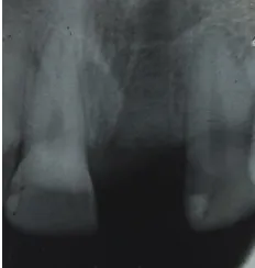
[image:61.595.243.353.387.503.2]Fig. 7 Non-restorable tooth in relation to 21
Fig. 8 Pre-Op IOPA
[image:61.595.221.376.562.693.2]
Fig. 10 Socket filled with Bio-Oss bone graft
Fig. 11 Resorbable membrane placed over bone graft
Fig. 13 Preserved socket ready to receive the Dental Implant
Fig. 14 IOPA of the healed socket
Fig. 16 IOPA of the placed Implant
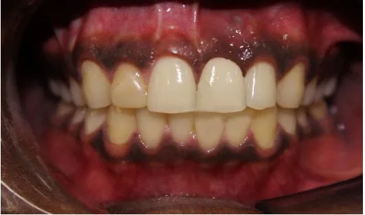
CASE 2 – GROUP B (BIO-OSS WITH PRF)
Fig. 18 Preoperative Intraoral photograph – tooth concerned 21
Fig. 19 Preoperative IOPA of 21 region
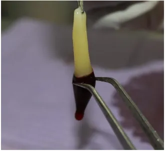
Fig. 21 PRF prepared prior to socket preservation
Fig. 22 Extraction of PRF
Fig. 24 Packing the Site with the Mixture
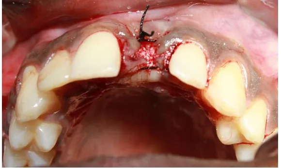
Fig. 25 Final suturing
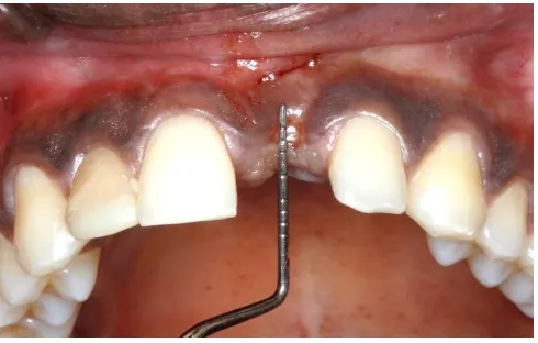
Fig. 27 Preserved socket Mesio-distal measurement
Fig. 28 Preserved socket Bucco-lingual measurement
Fig. 30 Suturing with 4-0 ethilon
Fig. 31 Implant placed in the Preserved site
39
RESULTS
Our study was done to evaluate the dimensional changes in the socket
preservation site with either Bio-Oss bone graft alone (Group A) or Bio-Oss
mixed with PRF (Group B) and to finally check the success of implants placed
in the site loaded after successful evidence of osseo-intergation. Based on the
inclusion criteria the patients requiring socket preservation with a future
implant placement in mind were selected. They were randomly assigned into
the two groups. This study evaluated 16 sites in 14 patients who reported to
the outpatient department, Department of Oral and Maxillofacial Surgery,
Ragas Dental College, Uthandi, Chennai.
Data obtained was entered in excel sheet and analyzed using SPSS
version 20. Normality of data was tested using Shapiro Wilks test and
Unpaired ‘t’ test was used for data that followed normal distribution and Mann
Whitney ‘u’ test was used for non-parametric data. Mean and standard
deviation was calculated for all the quantitative variables. Frequency
distribution was calculated for qualitative data and chi square test was used to
test for any difference between the two groups.
Both the groups were evenly distributed with respect to Gender, Site,
Smoking status, Gingival and Periodontal Status, root orientation and ease of