PAPER • OPEN ACCESS
Electrospun gelatin-based scaffolds as a novel 3D
platform to study the function of contractile smooth
muscle cells in vitro
To cite this article: J C Bridge et al 2018 Biomed. Phys. Eng. Express 4 045039
View the article online for updates and enhancements.
Related content
Comparative study of gelatin methacrylate hydrogels from different sources for biofabrication applications
Zongjie Wang, Zhenlin Tian, Fredric Menard et al.
-Contact guidance for cardiac tissue engineering using 3D bioprinted gelatin patterned hydrogel
Ajay Tijore, Scott Alexander Irvine, Udi Sarig et al.
-3D porous collagen scaffolds reinforced by glycation with ribose for tissue engineering application
Natalia Gostynska, Gopal Shankar Krishnakumar, Elisabetta Campodoni et al.
PAPER
Electrospun gelatin-based scaffolds as a novel 3D platform to study
the function of contractile smooth muscle cells
in vitro
J C Bridge1,2,6
, M Amer1,6
, G E Morris1
, N R W Martin2
, D J Player2,5
, A J Knox4
, J W Aylott1
, M P Lewis2,3
and F R A J Rose1
1 Division of Regenerative Medicine and Cellular Therapies, Centre for Biomolecular Sciences, School of Pharmacy, University of
Nottingham, United Kingdom
2 School of Sport, Exercise and Health Sciences, Loughborough University, United Kingdom
3 National Centre for Sport and Exercise Medicine, Loughborough University, United Kingdom
4 Division of Respiratory Medicine, School of Clinical Sciences, University of Nottingham, United Kingdom
5 Institute of Orthopaedics and Musculoskeletal Science, Division of Surgery and Interventional Science, University College London,
United Kingdom 6 Jointfirst author.
E-mail:felicity.rose@nottingham.ac.uk
Keywords:electrospinning, 3D cell culture, GelMA, smooth muscle,in vitromodel, tissue engineering, contractile Supplementary material for this article is availableonline
Abstract
Contractile dysfunction of smooth muscle
(
SM
)
is a feature of chronic cardiovascular, respiratory and
gastro-intestinal diseases. Owing to the low availability of human
ex vivo
tissue for the assessment of SM
contractile function, the aim of this study was to develop a novel
in vitro
SM model that possesses the
ability to contract, and a method to measure its contractility. A range of electrospun scaffolds were
produced from crosslinked gelatin and methacrylated gelatin
(
GelMA
)
, generating highly aligned scaffolds
with average
fi
bre diameters ranging from 200 nm to several micrometres. Young
’
s moduli of the scaffolds
ranged from 1
×
10
5to 1
×
10
7Pa. Primary aortic smooth muscle cells
(
AoSMCs; rat
)
cells readily
adhered to and proliferated on the
fi
brous scaffolds for up to 10 days. They formed highly aligned
populations following the topographical cues of the aligned scaffolds and stained positive for SM markers,
indicating a contractile phenotype. Cell-seeded GelMA scaffolds were able, upon stimulation with uridine
5
′
-triphosphate
(
UTP
)
, to contract and their attachment to a force transducer allowed the force of
contraction to be measured. Hence, these electrospun GelMA
fi
bres can be used as biomimetic scaffolds
for SM cell culture and
in vitro
model development, and enables the contractile forces generated by the
aligned three-dimensional sheet of cells to be directly measured. This will supplement
in vitro
drug
screening tools and facilitate discovery of disease mechanisms.
1. Introduction
Smooth muscle(SM)is a key component of respiratory, cardiovascular, and gastrointestinal systems. Certain disease states arise due to dysfunction in the smooth muscle component and, as yet, such diseases not fully understood; an example is asthma[1]. Technical and ethical difficulties associated with in vivo human research and the maintenance of human primary cells
in vitro have significantly limited studies aimed at elucidating the interrelationship between the main cell types and the extracellular matrix involved in such pathologies. In addition, the low availability of human tissue suitable for ex vivo assessment of contractile
function has restricted current methods for studying smooth muscle(such as airway smooth muscle; ASM) to those that requireex vivotissues, animal models or 2Din vitrosystems[2]. Conventional 2D culture models represent a non-physiological mechanical environment where contraction is assessed at a single cell level.Ex vivomodels replicate thein vivosituationin vitrobut these techniques(such as the precision cut lung slice model)are technically challenging, the cell and matrix components cannot be easily manipulated, and the construct is essentially dying during the experiment. Currently available animal models represent poor relevance to human disease and only mimic aspects of the human phenotype[3].
OPEN ACCESS
RECEIVED 24 March 2018
REVISED 11 June 2018
ACCEPTED FOR PUBLICATION 22 June 2018
PUBLISHED 10 July 2018
Original content from this work may be used under the terms of theCreative Commons Attribution 3.0 licence.
Any further distribution of this work must maintain attribution to the author(s)and the title of the work, journal citation and DOI.
Tissue engineering principles have been applied to the generation of 3D culture models created using cells
in vitrocultured on a 3D scaffold to provide a more physiologically relevant environment than 2D cultures [4]. Natural polymers, such as collagen[5]and gelatin, exhibit high cellular biocompatibility and are more elastic in nature than synthetic polymers(e.g.: poly
(ethylene terephthalate) PET) [6]. Electrospinning producesfibrous, porous, 3D scaffolds that resemble the structure of the natural extracellular matrix[7]. Fluorinated alcohols used as electrospinning solvents have been reported to cause denaturation of collagen [8,9]. As a result, there is little difference between the resultant electrospun collagenfibres when compared to those fabricated from gelatin[10]. Therefore, as an alternative to collagen, electrospinning gelatin is a more cost-effective way of producing scaffolds that are chemically similar to collagen whilst still being bio-compatible and biodegradable[11].
Crosslinking gelatin provides stability against enzymatic degradation, lower water solubility and an opportunity to modulate mechanical properties[12]. Gelatin methacrylate(GelMA), synthesised via a reac-tion between gelatin and methacrylic anhydride, con-sists of multiple methacrylamide groups[13]. These can form chemical crosslinks between gelatin mole-cules in the presence of free radicals from a photo-initiator following light exposure allowing the tuning of the mechanical and degradation properties of the resultant scaffold. GelMA has been utilised in hydrogel cultures [14–16], micro-patterning [17], and 3D-printing [18, 19]. Three dimensional (3D) GelMA hydrogels closely resemble the native extracellular matrix(ECM)due to the presence of cell-attachment and matrix metalloproteinase responsive peptide motifs[20,21]. GelMA has been used to coat electro-spun polycaprolactone fibres [22] and has recently been electrospun into nanofibre scaffolds to investi-gate wound healing and cutaneous regenera-tion[23,24].
Despite the clear clinical need for more effective therapeutics to treat disease, such as asthma, that involve the SM component, very few new classes of drugs have made it to the clinic over the past 40 years and it is clear that one of the reasons for this is the lack of relevantin vitroandin vivomodels[25]. A tissue engineered approach allows the generation of‘living’ tissue constructs from both animal(validation), and human cells(relevance)which can be used to accu-rately measure contraction. One approach to study SMC function is to decellularise native vessels and/or cross-link them for arterial grafting[26,27]. Although this does provide a native ECM with some preserva-tion of structure and biochemical composipreserva-tion, the acquisition of native vessels for decellularisation is limited and suffers from issues with sample-to-sample variability. In addition, this approach is extremely lim-ited in the study of paediatric disease(due to the lack of
tissue donated for research purposes). These aspects limit the application of this approach.
We have previously reported that electrospun PET scaffolds allow control overfibre alignment, allowing the generation of aligned sheets of smooth muscle[7]. Given that the Young’s moduli ofin vivoSM, such as human arteries, range from 0.1 to 1.0 MPa[28], which is 100–1000 times lower than values reported for the Young’s moduli of synthetic fibrous scaffolds (for example those electrospun from PET exhibiting a Young’s modulus of 200–300 MPa[7]), we set out to develop a GelMA-based model that provides a suitable matrix for culture of SMCs. This presents a standar-dised culture platform that bypasses the need for native tissue and allows tailoring of the scaffold’s mechanical properties. The aim and novelty of this study therefore was to develop anin vitromodel of SM that possesses the ability to contract and importantly, to develop a method to directly and quantitatively measure this contractility.
2. Materials and methods
2.1. Synthesis of gelatin methacrylate
Gelatin methacrylate(GelMA)was synthesised follow-ing a previously published method[14]. Briefly, 10 g of gelatin(type A 300 bloom)was dissolved in 95 ml PBS at 50°C and stirred for 1 h. Methacrylic anhydride
(8.0 ml)was added to the gelatin solution and stirred for a further 3 h at 50°C. PBS(400 ml)was added to the mixture and stirred for an additional 30 min The solution was then transferred into three dialysis membranes (12–14 kDa molecular weight cut-off). The membranes were placed in 3.0 L of distilled water and stirred at 50°C. The dialysis water was changed twice daily for 7 days before the membranes were removed and the solution frozen overnight at–80°C. The frozen GelMA solution was lyophilised and stored at room temperature. The percentage of methacryla-tion within the synthesised GelMA was calculated using the following equation:
= ´
´ ´
DM% I5.6 ppm 0.3836 0.84 ppmI 0.0385 100
2.2. Production of electrospun gelatin and GelMA scaffolds
Solutions of gelatin at various concentrations(6, 8 and 10% w/v) were made by dissolving gelatin powder
humidity in a ventilated fume cabinet. For each scaffold, 4 ml of solution was electrospun at aflow rate of 1.2 ml h−1. The needle collector distance was 15 cm and the voltage across the apparatus was 15 kV. The collector mandrel was set to spin at a speed of 2000 rpm. Upon completion of electrospinning, scaffolds were cut from the mandrel using a scalpel blade and stored in aluminium foil at room temperature.
2.3. Crosslinking of electrospun gelatin scaffolds
Prior to crosslinking the scaffolds, they were first secured in an acetate frame(5star™, Cambridge, UK) using aquarium sealant (Sinclair animal and house-hold care Ltd, Gainsborough, UK)on either side of the scaffold. The acetate frame size was either 23×42 mm with an internal window of 13×32 mm or 25×25 mm with an internal window of 15×15 mm. Once the acetate frames were adhered, individual scaffolds were cut out and left to dry overnight. The gelatin scaffolds were crosslinked with EDC and NHS[29], whereby a solution of 250 mM 1-Ethyl-3-(3-dimethylaminopropyl) carbodiimide
(EDC) (Applichem, Darmstadt, Germany) and 100 mM N-Hydroxysuccinimide (NHS) (TCI Europe, Zwijndrecht, Belgium)was prepared in distilled water. Ethanol(Sigma Aldrich) was added to the solution until the total volume was 10 times the original volume. Scaffolds were then placed in the EDC/NHS solution for 24 h at 4°C and washed in distilled water
(x3)before being lyophilised overnight.
2.4. Crosslinking of GelMA scaffolds
Prior to crosslinking, GelMA scaffolds were first secured to an acetate frame (section 2.3). Scaffolds were then submerged in a 1% w/v solution of 2-Hydroxy-4′-(2-hydroxyethoxy) -2-methylpropio-phenone(photoinitiator)in a solvent mix of ethanol and water. Solvent ratios used ranged from 1:0 to 9:1 ethanol:water. Scaffolds were then exposed to UV radiation(0.5 Wm2)for 10 min and washed in PBS three times. The proposed mechanism of the cross-linking process is displayed in; no further crosscross-linking of the sample occurs when UV sterilised for cell culture due to the wash step removing excess photoinitiator. Samples used for SEM imaging were washed indH2O and freeze-dried prior to analysis. Samples used for cell culture were incubated in antibiotic/antimycotic solution (50 000 units mL−1 penicillin G, 500 mgmL−1 streptomycin sulphate and 125μgmL−1 amphotericin B) (Fisher Scientific)in PBS at 4°C prior to use.
2.5. NMR analysis
Samples of gelatin and GelMA(as synthesised)were dissolved in D2O in glass NMR tubes and the NMR spectrum of each sample was determined using a Bruker Avance 600 MHz spectrometer(Bruker, Cov-entry, UK). Spectra were analysed using the
MestReNova LITE software package (Mestrelab research, Hereford, UK).
2.6. Scaffold morphology
Samples of electrospun scaffolds were cut using an 8.0 mm diameter biopsy pen and mounted on a holder using graphite adhesive SEM pads (Agar Scientific, Essex, UK). Samples were gold coated for 5 min
(Balzers Union SCD 030, Balzers Union Ltd, Liechten-stein) and were then imaged at 20–30 kV using a scanning electron microscope (JEOL JMS-6060 LV, JEOL Ltd, Hertfordshire, UK)at different magnifi ca-tions (as identified on the images). Scaffold fibre diameter and fibre alignment were determined by analysis of SEM micrographs using the image analysis software packages MeasureIT(Olympus Soft Imaging solutions GmbH, Münster, Germany) and ImageJ respectively. Alignment was calculated by expressing individualfibre angles as deviations from the sample meanfibre angle. All measurements were achieved by measuring 20 fibres from images of 3 individual scaffolds(60fibres in total).
2.7. Tensile measurements of GelMA scaffolds
Samples of electrospun GelMA scaffolds were cross-linked in acetate frames(25×25 mm with an internal window of 15×15 mm; the frames were used to prevent shrinkage of the scaffold)as described above. Crosslinked scaffolds were cut away from the edges of the scaffold along the two sides parallel to thefibre direction using a scalpel. Scaffolds were then placed in a TA HDPlus Texture Analyser(Stable Micro Systems Ltd, Surrey, UK)with a 5 kg load cell with thefibre direction parallel to the testing direction. Samples were tested at an extension rate of 6 mm min−1. Young’s moduli of the samples were calculated from the resultant stress/strain curves.
2.8. Isolation and culture of primary rat aortic smooth muscle(AoSM)cells
Male Wistar rats(200–225 g)were killed by stunning and exsanguination, using an approved Schedule 1 method of euthanasia. All procedures were approved by the animal welfare and ethical review body
in low Ca2+ dissection buffer before incubation at 37°C for 45 min. The partially digested aortic tissue was extracted from the papain solution and washed in BSA solution(Sigma Aldrich)3 times before transfer-ring into 3 ml AoSMC media(high glucose DMEM supplemented with 10% (v/v) FBS and 1% (v/v) antibiotic/antimycotic solution (10 000 units ml−1 penicillin G, 100 mg ml−1streptomycin sulphate and 25μg ml−1 amphotericin B). Tissue was firmly agi-tated by pipetting for 30 s to release cells from the partially digested tissue before the cell suspension was transferred into two collagen-coated (0.03 mg mL−1 type I bovine collagen(PureCol®, Advanced Bioma-trix, San Diego, CA)in PBS)T-25flasks containing 5 ml AoSMCs media. Flasks were incubated(at 37°C, 5% CO2)for 48 h before the media was changed to remove decellularised tissue and unattached cells; the cells were cultured for a further 5 days before thefirst passage. Subsequent passages were carried out every 7 days up to passage 3 in collagen-coated flasks; cells were used at passage 2–3.
2.9. PrestoBlue®cell viability assay
AoSMCs cell viability was measured post-seeding on aligned scaffolds using the PrestoBlue® assay at various time points across a 10-day period. Samples were washed with PBS prior to incubation with 1 ml PrestoBlue working solution(10%(v/v)PrestoBlue in HASM culture media)for 10 min at 37°C. PrestoBlue was then collected(100μl aliquots)and replaced with media and the constructs returned to the incubator. Fluorescence was measured in duplicate on a Tecan Infinite M200 plate reader(Tecan, Reading, UK)using excitation and emission wavelengths of 560/590 nm. Resultant readings were expressed as a percentage of thefluorescence reading at day 0.
2.10. Immunocytochemistry
All samples were washed with PBS prior tofixation with 3.8%(w/v)p-formaldehyde for 10 min at room temperature. Samples were permeablised using 0.5%
(v/v)Triton X-100 in PBS(5 min, 4°C)then blocked sequentially with 3% (v/v) BSA, 1% glycine (w/v)
solution for one hour, followed by 10% (v/v) goat serum solution in PBS for 1 h at room temperature. The following primary antibodies were used at 1:200 dilution in 10%(v/v)goat serum in PBS:α-smooth muscle actin(Alexa Fluor 488-conjugated phalloidin; Life Technologies, Paisley, UK), SM22α (ab14106; Abcam, Cambridge, MA), desmin (D1033; Sigma-Aldrich, UK), connexin(C6219; Sigma-Aldrich, UK), vinculin (V4505; Sigma-Aldrich, UK) and calponin
(ab46794; Abcam, Cambridge, MA). Samples were incubated with the relevant primary antibody at 4°C overnight. Nuclei were stained with Hoechst 33342 at a 1:833 dilution for 10 min(Fisher, UK). Samples were imaged using a Leica TCS SP2 laser scanning confocal inverted microscope(Leica Microsystems Ltd, Milton
Keynes, UK) or a Leica DM2500 M fluorescent microscope. Images were then analysed using ImageJ. Nuclei alignment was calculated using particle analysis on binary images of Hoechst-stained cell nuclei. Surface coverage was determined by calculating the fraction of confocal z-stack images that were not stained for SM22α. SM marker positivity was calcu-lated by counting the number of nuclei co-expressing the marker of interest and subtracting from the total nuclei present. Cell density was determined by divid-ing the number of nuclei visible per micrograph by micrograph area.
2.11. Scaffold contraction assays
Samples of crosslinked 10% w/v gelatin or GelMA scaffolds were placed in 6-well plates and sterilised by UV exposure (20 min), followed by incubation in media(60 min, 37°C)prior to cell seeding). Gelatin/ GelMA scaffolds were seeded with AoSMCs at a density of 2×10⁵
cells cm−2 and incubated for 10 days at 37°C until confluency. Following incubation, culture media was aspirated and 1 ml serum-free DMEM added to each sample; scaffolds were cut out from their acetate frames and allowing them to be free-floating in the well-plates. Serum free DMEM
(1 ml)±smooth muscle contraction agonist, uridine 5′-triphosphate(200μM UTP)was then added to each sample with minimum disturbance, allowed to equili-brate and imaged using a flatbed scanner at time intervals up to 30 min. Scanned images were analysed using ImageJ which was used to measure the surface area of the construct.
2.12. Direct force measurement of AoSMC-seeded GelMA scaffold contraction using muscle
physiology apparatus
A force monitoring apparatus design has been pre-viously reported by Dennis and Kosnik[30], and later adapted to measure the contractile force of cultured skeletal muscle constructs[31,32]. This was further adapted here to measure uniaxial contractile force. Rectangular GelMA scaffolds (13×32 mm) were prepared, sterilised and seeded with AoSMCs at 2×10⁵
free-floating end, which was then attached to a model 403 A force transducer (Aurora Scientific, Dublin, Ireland)using canning wax. The force transducer was connected to a Powerlab 4/25 T unit with associated software(AD Instruments, Oxford, UK). Force was measured at a frequency of 1 kHz. Once stable, the baseline force of the resting scaffolds was measured for 3 min before the addition of 500μl of 1 mM UTP in serum-free DMEM (500μl serum free DMEM in control experiments)using a pipette(disturbance to the scaffolds was reduced to a minimum by careful pipetting). Contraction was then measured continu-ously for 60 min.
2.13. Statistical analysis
All statistical tests were carried out using the GraphPad Prism 6 (GraphPad Software Inc. San Diego, CA). Each statistical test carried out is stated in the relevant section and figure legends. Statistically significant results are represented with asterisk(s) (*,**,***,****) and representpvalues0.05, 0.01, 0.001 and 0.0001 respectively.
3. Results
The aim of this study was to develop a novelin vitro
model of contractile smooth muscle tissue. We have previously shown that smooth muscle cells follow topographical cues from their environment; when cultured upon aligned electrospun PET scaffolds, the cells quickly formed an aligned population of cells [7, 33]. However, one noticeable feature of these scaffolds was that they were very stiff, with Young’s moduli much higher than seen inin vivomuscle tissue [28]. To achieve a contractile SMC construct, we electrospun and crosslinked gelatin-based scaffolds that better mimic the mechanical properties of the extracellular matrix(ECM). These scaffolds were then seeded and cultured with rat aortic smooth muscle cells (AoSMCs), which were used as an exemplar smooth muscle cell type. The ability of these cells to align according to the alignedfibrous nature of the scaffolds, to express proteins associated with SMC phenotype and the ability to measure contraction in response to chemical stimulus is presented here.
3.1. Electrospinning aligned gelatinfibre scaffolds
Gelatin was chosen due to its more elastic properties and its chemical similarity to collagen, which has been shown to allow the contraction of smooth muscle cells [34]. Gelatin was electrospun from solutions ranging 6% to 10%(w/v) (figures1(A),(C)and(E))to provide the topographical cues we had previously observed important for cell alignment[7, 33]. Scaffolds were crosslinked using EDC and NHS in an ethanol/water mixture with shrinkage prevented by securing the scaffolds in place during crosslinking using acetate
frames. SEM images of the electrospun and cross-linked gelatin scaffolds are shown infigures1(B),(D) and(F).
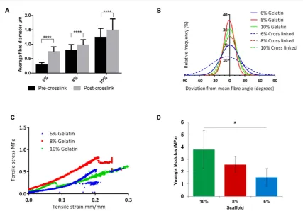
The average fibre diameter and degree of fibre alignment were analysed for each scaffold. Measure-ments were taken before and after the scaffolds were crosslinked to assess the effect of crosslinking on the morphology of the gelatin scaffolds. Fibre diameter of gelatin scaffolds increased with increasing concentra-tion of the gelatin soluconcentra-tion; 10% gelatin soluconcentra-tions pro-duced fibres with an average diameter of 1.2μm whereas 8% and 6% produced scaffolds with average fibre diameters of 788 and 286 nm respectively. Cross-linking of these scaffolds significantly affected the average fibre diameter with merging of individual fibres apparent(p<0.0001). In all cases, the average fibre diameter of the 10%, 8% and 6% gelatin scaffolds increased to 1.49μm, 978 nm and 746 nm, an increase of 20%, 24% and 161% respectively(figure2(A)). All gelatin scaffolds were highly aligned, with 58%, 65% and 43% offibres within 10°of the meanfibre angle in 10%, 8% and 6% scaffolds respectively which decreased in all cases(to 51%, 48% and 23% respec-tively)after the crosslinking process(figure2(B)).
Tensile testing was performed on crosslinked 6%, 8% and 10%(w/v)aligned gelatin scaffolds. Representa-tive stress/strain curves for each scaffold are displayed in figure2(C). The gradients of these curves were used to calculate the Young’s modulus of each scaffold. The stiff-est scaffold was that fabricated from 10% gelatin, with a mean Young’s modulus value of 3.8±1.7 MPa. The mean Young’s moduli of the 8% and 6% gelatin scaf-folds were 2.6±0.7 and 1.5±0.8 MPa respectively
(mean±SD). There was a statistical difference between the stiffness of the 10% and 6% gelatin scaffolds as shown infigure2(D) (p<0.05).
3.2. Culture of AoSMCs on gelatin alignedfibre scaffolds
The differences seen in crosslinked gelatin scaffold alignment (10%>8%>6%) is reflected in the nuclear alignment of cells cultured upon the scaffolds. AoSMCs were more aligned when cultured on the 10% crosslinked gelatin scaffolds(38% within 10°of the mean) than on the 8% (27%) and 6% (21%) scaffolds (figure 3(A); a high magnification image illustrating nuclear alignment on thefibre scaffolds as identified by Hoescht staining is presented in figure3(F)). Due to the increased alignment of cells on 10% gelatin scaffolds, and demonstrating only a slight difference in stiffness compared to the other gelatin scaffolds, the 10% cross-linked gelatin scaffolds were chosen to be used for further cell culture studies.
was achieved after 9 days when scaffolds were seeded at a density of 2×10⁵cells cm−2. To confirm SM pheno-type, cells cultured on the gelatinfibre scaffolds were immunostained and found to express the SM markers SM22α (figure3(C))and calponin(figure3(E)). Cells also stained positive for the gap junction protein con-nexin, and negatively for the intermediatefilament pro-tein desmin (figure 3(D)). Individual focal adhesions were observed in cells stained for vinculin(figure3(F)). Cells exhibited a spindle-like morphology, with most focal adhesions occurring at the spindle poles.
3.3. Contraction of AoSMCs seeded on gelatin(10%) fibre scaffolds
Crosslinked 10% gelatin electrospun scaffolds were seeded with AoSMCs at 2×10⁵
cells cm−2and incu-bated for 10 days before scaffolds were cut away from the
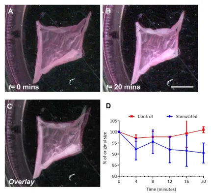
supporting acetate frames. The cell-fibre constructs were submerged in media(free-floating)and the cells stimu-lated with 100μM UTP and imaged to assess the degree of scaffold contraction. Figures 4(A) and (B) displays images of a cell-seeded gelatin scaffold at timest=0 and t=20 min respectively. By comparing the surface area of
the construct across this period, constructs reduced to 90±4.5%(mean±SD)of their original size, and no further contraction was seen after 20 min(figures4(C) and(D)). Unstimulated controls remained approximately 100% their original size, with insignificant spontaneous contraction observed, throughout the study.
3.4. Production of electrospun GelMAfibre scaffolds
[image:7.595.123.553.60.524.2]tenability to modulate the stiffness of the scaffold. The degree of gelatin methacrylation can be controlled by the ratio of gelatin:methacrylic anhydride in the initial reaction mixture in addition to length of exposure to UV light and concentration of photoinitiator. GelMA was synthesised in-house and the degree of methacry-lation calculated using proton NMR spectroscopy. Comparing the 1HNMR spectrum of synthesised GelMA to that of gelatin, there are two clear additional peaks in the GelMA spectrum at approximately 5.3 and 5.6 ppm due to the two protons found on the methacrylate vinyl group. The degree of methacryla-tion was calculated using a previously published method[18]. The peak at 0.84 ppm can be used as a reference peak ascribed to the hydrophobic side chains of valine, leucine and isoleucine; the integration of this peak(I0.84)corresponds to 0.3836 mol/100 g(sum of known composition of these amino acids in gelatin). The total amount of available amine groups in gelatin is equal to 0.0385 mol/100 g. Therefore, the percent-age of methacrylation within the synthesised GelMA can be expressed using the following equation:
= ´
´ ´
DM% I5.6 ppm 0.3836 0.84 ppmI 0.0385 100
Using this equation, the percentage of methacryla-tion was calculated to be 81.0%.
The synthesised GelMA was electrospun using a 10% (w/v) solution as for the gelatin scaffolds
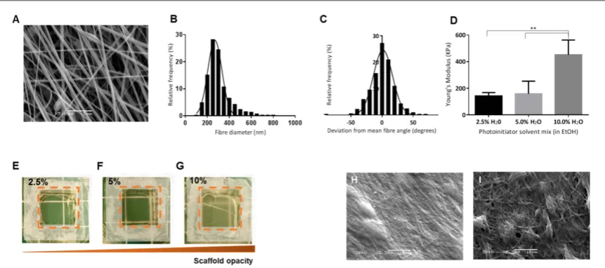
(figure5(A)). SEM image analysis was used to deter-mine the average fibre diameter(297.4±101.1 nm
(mean±SD)and degree offibre alignment(50% of fibres within 10°of the meanfibre angle)of the elec-trospun scaffolds. Distribution curves of thefibre dia-meter and angle are displayed infigures5(B)and(C) respectively. GelMA scaffolds were cut and adhered to acetate frames before being crosslinked using UV light in the presence of a photoinitiator(1%w/vsolution of 2-Hydroxy-4′-(2-hydroxyethoxy) -2-methylpropio-phenone). To prevent the GelMA scaffolds from dis-solving during the crosslinking process, taking into account a single intensity UV lamp was used, a num-ber of solvent mixtures were investigated. These ran-ged from pure ethanol to a 9:1 ethanol:water mixture. Scaffolds would not crosslink in pure ethanol and dis-solved in solutions containing greater than 10% H2O. Three different ethanol:water solutions were explored further including 39:1(2.5% H2O), 19:1 (5% H2O) and 9:1(10% H2O). Tensile tests of the different cross-linked GelMA scaffolds showed that the Young’s mod-ulus increased with increasing water content in the crosslinking solution: average Young’s moduli were 142.2±25.4, 158.7±95.3 and 451.5±111.3 kPa
[image:8.595.123.551.61.359.2]and 10% water in ethanol solutions respectively
(figure 5(D)). Scaffolds crosslinked in 10% H2O in ethanol were significantly stiffer than those cross-linked in 5% and 2.5% H2O in ethanol solutions
(p<0.01). Scaffold opacity increased with increasing crosslinking solution water content(figures5(E)–(G)). In addition, the failure rate of the scaffolds during post-crosslinking washing was much higher when crosslinking in 2.5% H2O in ethanol. Individualfibre analysis post-crosslinking was not possible due to a lack offibre resolution;fibres appeared to merge toge-ther during crosslinking although the aligned fibre morphology was evident when crosslinked in 5% and 10% H2O in ethanol solutions(figures5(H),(I)). Due to the high failure rates of 2.5% H2O in ethanol-cross-linked scaffolds and the significantly higher Young’s modulus of the 10% H2O crosslinked scaffolds, scaf-folds crosslinked in 5% H2O in ethanol were chosen for the cell culture studies.
3.5. Culture of AoSMCs on GelMA scaffolds
AoSMCs were cultured on crosslinked aligned GelMA scaffolds for a period of 10 days, during which cellular metabolic activity was monitored using the Presto-Blue® assay. Cell metabolism increased steadily throughout the 10-day period(figure6(A)), indicating cell proliferation over this time. Samples werefixed after 10 days and stained for the SM markers SM22α and calponin, and for the gap junction protein
connexin (figures 6(B)–(D) respectively). Samples stained positive for all markers and stained negatively for desmin (figure 6(D)). Unlike AoSMCs cultured upon gelatin fibre scaffolds, those cultured upon GelMA fibre scaffolds did not stain positively for vinculin, with no clear focal adhesions present. Cell alignment on GelMA scaffolds was similar to the degree of alignment achieved on 8% and 6% gelatin scaffolds, with 23% of nuclei oriented within 10°of the mean(figure6(E)). A comparison of the morpho-logical and mechanical properties of the scaffolds produced and smooth muscle ECM is shown in table1.
3.6. Contraction of AoSMC-seeded GelMAfibre scaffolds
Crosslinked GelMA scaffolds were seeded with AoSMCs at a density of 2×10⁵
cells cm−2 and incubated for 10 days before scaffolds were stimulated with 100μM UTP and imaged to assess the degree of contraction. Figures7(A)and(B)display images of a cell-seeded gelatin scaffold at times t=0 and t=30 min respectively. By comparing the construct’s surface area across this period, AoSMCs contraction resulted in the reduction to 78±2.5%(mean±SD) of their original size(22% reduction in size), and no further contraction was observed after 30 min
[image:9.595.123.553.62.357.2](figures 7(C) and (D)). Unstimulated controls remained close to 100% their original size throughout Figure 3.Culture of AoSMCs on crosslinked gelatin scaffolds.(A)Distribution curves illustrating the alignment of AoSMCs cultured on the cross-linked scaffolds.(B)Cell metabolism was monitored using the PrestoBlue®assay for 10 days. Error bars represent SEM;
n=6.(C)–(F)AoSMCs were stained with SM22α(C), connexin(D)and calponin(E, F) (all green). Cells were also stained for desmin
the study. This level of contraction was higher than witnessed in the AoSMC-seeded gelatin scaffolds
(reducing to 90±4.5% of their original size). Follow-ing these results, the direct force measurement(using a force transducer)of AoSMC-seeded GelMA construct contraction was measured. AoSMCs were cultured upon crosslinked GelMA scaffolds for 10 days. After this culture period, constructs were cut from their acetate frames and attached to the force transducer
(figure7(E)). Once attached, the force was allowed to stabilise before recording started. The stable force reading was measured for 3 min before the constructs were stimulated with 100μM UTP. Control studies were stimulated with serum-free DMEM. An increase in force was detected within seconds of adding the agonist solution and continued to rise quickly for 20 min before slowing. Force measured continued to increase for a further 30 min before beginning to plateau(figure7(F)). The maximal force generated by the AoSMCs constructs ranged from 755.2μN to 1356.4μN, with the average max force being
1008.1±251.6μN (mean±SD). The majority of contraction occurred within thefirst 5 min following stimulation, with 42±3% of the maximal force measured occurring within this time; 60±4% mea-sured 10 min following stimulation and 95±2% after 40 min stimulation.
4. Discussion
The aim of this study was to develop anin vitromodel of SM that possesses the ability to contract. This study explores the production of a range of electrospun crosslinked gelatin scaffolds which possess elastic properties and an alignedfibrous morphology to serve as a topographical cue to the cells. Primary rat aortic smooth muscle cells (AoSMCs) were explored as a model smooth muscle cell type, and the contraction of the cell-scaffold constructs in response to agonists were measured.
[image:10.595.121.552.63.455.2]moduli ranging from a few hundred kPa[35]to several hundred MPa [36]. This wide range in stiffness can greatly impact attachment[37], proliferation[38], and differentiation[39]of mammalian cells cultured upon them. It was therefore important to choose a material with mechanical properties that closely match the mechanical properties that cells experiencein vivoto produce tissue constructs with functional outputs
(such as contraction). In this study, the contractile behaviour of AoSMCs cultured upon electrospun scaf-folds was explored. In addition, using the scaffold that supported SM contraction, the physical force gener-ated during agonist-induced SM contraction was actively measured using muscle physiology apparatus.
In a previous study, HASM cells were cultured upon electrospun PET scaffolds for up to 14 days and expressed markers of a contractile SM phenotype[7]. In addition, we showed that HASM cells rapidly aligned on these scaffolds following the topographical cues from thefibres. However, the Young’s modulus ofin vivoSM is much lower than the Young’s moduli of the PET scaffolds(200–300 MPa). For example, the Young’s moduli of human arteries and porcine bronchi range from 0.1 to 1.0 MPa and 0.35 to 1.35 MPa respectively[28,40]. These values are 100 to 1000 times lower than the values obtained for the PET scaffolds although some caution must be taken when comparing such values and native and synthetic mate-rials are likely to have different porosities. As a result, a much less stiff material was required to provide sui-table mechanical properties that would facilitate SM contraction. Gelatin was investigated, which is bio-compatible, chemically similar to collagen, inexpen-sive, and possesses favourable mechanical properties.
Gelatin fibrous scaffolds were successfully fabri-cated using electrospinning. The fibre diameter increased with increasing gelatin concentration and ranged from 286 nm to 1.24μm, a range similar to previously published work when similar concentra-tions of gelatin influorinated alcohols were electro-spun [10, 38]. When gelatin is electrospun using aqueous solutions, much greater concentrations
(30%–40%w/v)need to be used to achieve similar sized fibres[12,41]. This once again illustrates the effect that different electrospinning parameters(e.g.: solvent used) can have on the resultant fibres [42]. Scaffoldfibre alignment was high for all scaffolds fab-ricated. Image analysis of the scaffolds after cross-linking showed that scaffolds appear slightly less uniform and the degree of alignment had decreased slightly. This is due to severalfibres merging together during crosslinking as crosslinks formed between fibres, also leading to a reduction in visible pores between fibres; however, the aligned fibrous topo-graphy of the scaffolds remained. Crosslinking also resulted in an increase infibre diameter in all of the gelatin scaffolds; this has also been reported by Zhang
[image:11.595.121.552.62.253.2]et al[12], who crosslinked gelatin scaffolds with EDC and NHS at 25 mM EDC. Fibre swelling, due to absorption of water, has also been reported when crosslinking gelatin scaffolds with genipin[41]. In this case, the crosslinked scaffolds appear to behave like a hydrogel, swelling in size with reduced stiffness, whilst retaining their fibrous structure. Zhang et al also found that when wet, the Young’s modulus of cross-linked gelatin scaffolds dramatically decreases[12]. As the scaffolds produced in this study were to be used for cell culture, only the wet state Young’s modulus was measured, which ranged from 3.80 to 1.54 MPa— Figure 5.Properties of electrospun GelMA scaffolds.(A)SEM image of aligned electrospun GelMA spun from a 10% w/v solution before cross-linking.(B),(C)Histograms and Gaussian distribution curves offibre diameter(B)andfibre alignment(C)are presented
(n=60, 20 measurements taken from 3 images of independent scaffolds).(D)Average Young’s moduli of GelMA scaffolds crosslinked in different H2O:EtOH mixtures. Error bars represent standard deviation(n=4). One way ANOVA with Tukey’s
multiple comparisons test was carried out between the Young’s modulus of all scaffolds(p<0.01) (E),(F),(G). Representative photographs of scaffolds crosslinked in 2.5%, 5.0% and 10.0%(v/v)H2O in ethanol respectively, showing increasing scaffold opacity
values similar in magnitude to those measured by Zhang for similar sized scaffolds. Future work will determine the mechanical properties of individual fibres using atomic force microscopy to identify the forces experienced by individual cells[43].
Scaffolds spun from 10% w/v gelatin solutions were chosen to assess cell attachment, proliferation and phenotype. AoSMCs displayed high alignment when cultured upon the scaffolds. Cells achieved con-fluency over 9 days with increasing metabolic activity over the same period, signifying that cells were pro-liferating on the scaffolds. Immunostaining for SM22αand calponin showed that both proteins were present, in addition to illustrating the aligned, spindle-shaped morphology of the cells, all of which are indi-cative of a contractile SM phenotype. In addition, cells stained positive for the gap junction protein connexin, with gap junctions visible between cells. Focal adhe-sions were also seen on scaffolds, with cells staining positive for vinculin. Once again, cells stained negative for desmin. Vascular smooth muscle cells possess little
to no desmin, with vimentin being the key constituent of intermediatefilaments[44].
Few studies have investigated the contractile force generated by seeding SM cells onto materials [2,45,46]. Very few have directly measured this force, and those that did were carried out on cell-seeded col-lagen hydrogels[47]. In this study, cell-seeded con-structs were stimulated with 100μM UTP and imaged periodically to measure scaffold contraction. Scaffolds reduced in size by an average of 9.5% over 20 min fol-lowing stimulation relative to unstimulated controls. Although the level of contraction was small, it was deemed possible to create AoSMC-seeded electrospun aligned scaffold constructs that contract by reducing scaffold stiffness further.
[image:12.595.113.552.61.339.2]Due to the multiple ways in which the mechanical properties of GelMA hydrogels can be manipulated, GelMA was considered as an attractive material that could potentially produce electrospun scaffolds with Young’s moduli lower than those seen in the gelatin scaffolds by varying the degree of cross-linking. Figure 6.Culture of AoSMCs on crosslinked GelMA scaffolds.(A)AoSMCs were cultured on crosslinked GelMA scaffold for 10 days, cell metabolism was monitored periodically using the PrestoBlue®assay; error bars represent standard error of the mean(n=6).(B),
(C),(D)Immunostaining of AoSMCs on electrospun GelMA scaffolds for SM22α(B), calponin(C), and connexin(D) (all green). Cells were also stained for desmin(D)and vinculin(C) (both red). All samples were additionally stained with Hoechst 33342(blue). Scale bar=100μm.(E)Samples werefixed after 10 days and cell nuclei stained. Image analysis of stained samples was carried out in order to determine cell alignment(n=6).
Table 1.Comparison of morphological and mechanical properties of the scaffolds produced in this study and smooth muscle ECM.
Material Gelatinfibres GelMAfibres Native SM ECM
Averagefibre diameter(μm) 1.24 0.38 —
Fibre alignment(% within 5°mean) 48 37 —
Nuclear Alignment(% within 10°mean) 38 23 65[7]
[image:12.595.118.555.444.513.2]GelMA was successfully synthesised following a pre-viously published method[14,17]. The level of GelMA crosslinking can be controlled by the initial level of methacrylation, the amount of photoinitiator used, the length of UV exposure and the solvents used in the crosslinking process. Following synthesis, the degree of methacrylation of gelatin was calculated using NMR spectra and found to be approximately 80%. This level of methacrylation matches previously published cal-culated values for the same gelatin: methacrylic anhy-dride ratio (degree of methacrylation of ∼70%– 80%)[14,17].
Synthesised GelMA was electrospun at a con-centration of 10% w/v generating aligned fibres around 300 nm in diameter, which were much thinner compared to gelatin fibres electrospun from the equivalent concentration (average diameter of 1.2μm). This is most probably due to a change in the overall net charge of the molecule during GelMA synthesis, whereby consumption of the majority of free amines by methacrylation(and no change to the number of free carboxylate groups)led to an increased negative charge at neutral pH due to the presence of deprotonated carboxylic acid groups[48]. This in turn affected the conductivity of the electrospinning solu-tion, leading to thinnerfibres. A crosslinking method had to be developed where the scaffolds retained their fibrous architecture whilst submerged in a photo-initiator solution. Previous studies have crosslinked GelMA hydrogels in PBS[14,17,18], but this was not possible with the electrospun scaffolds due to the high solubility of these GelMA fibres upon contact with aqueous solutions. In order to overcome this issue, the EDC and NHS gelatin crosslinking method was
adapted[12], which utilises a 9:1 ethanol:water mix for crosslinking. A range of ethanol:water mixtures were investigated for use as the solvent for the photo-initiator solution. Mixtures with a water content higher than 10%(v/v)caused the scaffolds to dissolve, and those with a water content of less than 2.5% failed to sufficiently crosslink scaffolds under UV light, lead-ing to dissolution of the scaffolds upon washlead-ing with PBS. By changing the ethanol to water ratio it was pos-sible to partially control the level of GelMA cross-linking and, as a result, the mechanical properties of the scaffolds. Young’s moduli of the scaffolds ranged from 142 kPa(2.5% water)to 451 kPa(10% water) which was in line with values published for human arteries(0.1 to 1.0 MPa[28]). SEM images of cross-linked scaffolds suggested that scaffolds appeared to lose their porous structure due tofibre swelling upon crosslinking, but the aligned fibrous topography of scaffolds was still visible.
AoSMCs were cultured upon GelMA scaffolds crosslinked in a 5% water in ethanol solution. These scaffolds were chosen for further cell culture experi-ments due to their low Young’s modulus and high crosslinking success rate relative to 10% and 2.5%
[image:13.595.120.553.62.274.2]alignment seen when crosslinking the GelMA scaffolds. AoSMCs once again stained positive for SM22α, calpo-nin and connexion. Unlike when cultured on gelatin scaffolds, AoSMCs on GelMA scaffolds showed no clear positive staining for vinculin, signifying that no focal adhesion points could be identified. It has been pre-viously documented that the level of vinculin bound to the cytoskeleton, and the amount of vinculin localizing at focal adhesions is larger on stiffer surfaces than on more elastic ones[49,50]. The difference in stiffness between the GelMA and gelatin scaffolds could there-fore be the reason why no vinculin localisation was seen on the GelMA scaffolds. As previously attempted with the gelatin scaffolds, AoSMCs were cultured upon square crosslinked electrospun GelMA scaffolds for 10 days prior to contraction studies. When stimulated to contract with 100μM UTP, the AoSMC-seeded GelMA constructs reduced in surface area by an average of 22% due to AoSMCs contraction. Since this level of contrac-tion was higher than that seen in AoSMC-seeded gelatin constructs, GelMA scaffolds were chosen to be used for the measurement of the physical force exerted by SM cells during contraction.
AoSMCs were seeded on crosslinked GelMA scaf-folds and cultured for 10 days to achieve a confluent layer of cells. These constructs were then attached to the force transducer and stimulated with 100μM UTP; these constructs contracted instantly and gener-ated average maximal forces in the region of 1000μN. Although electrospun gelatin scaffolds have been used to produce contractile tissues such as cardiac[35]and skeletal [43]muscle previously, this is thefirst time that SM contraction has been assessed using electro-spun scaffolds. In addition, this is also thefirst time that the physical force of contraction from any cell type has been directly measured on electrospun scaf-folds. In addition, the ability to modulate the stiffness of the scaffold allows the impact of matrix stiffness, relevant in the study of inflammatory diseases such as asthma where tissue remodelling occurs, upon cell phenotype and function to be studied.
5. Conclusions
This work represents thefirst time that the contractile forces generated by a confluent, aligned sheet of SM cells cultured upon gelatin based electrospun scaffolds have been directly measured. We also describe novel methods for the crosslinking of electrospun GelMA scaffolds. Using these methods, the mechanical prop-erties of the GelMA scaffolds were manipulated by controlling the amount of water in the ethanol based photoinitiator solution. SM cells readily attached to, proliferated and aligned upon both gelatin and GelMA fibrous scaffolds, expressing contractile SM markers in all cases. SM cells were able to contract both gelatin and GelMA scaffolds, with greater contraction seen on
the less stiff GelMA scaffolds. Thisin vitromodel of contractile smooth muscle holds great potential for the study of disease arising from matrix remodelling and in the discovery of new therapeutic entities to treat such diseases.
Acknowledgments
This work was supported by the Engineering and Physical Sciences Research Council [grant number EP/F500491/1], Doctoral Training Centre awarded to J. C. Bridge, U.K. GE Morris was employed on a related project funded by the National Centre for the Replace-ment, Refinement, and Reduction of Animals in Research (NC3Rs; G1001804/1). We also wish to acknowledge Dr William Dunn at the University of Nottingham for the procurement of rat aortas for the isolation of aortic smooth muscle cells.
ORCID iDs
M P Lewis https://orcid.org/0000-0002-8430-4479 F R A J Rose https://orcid.org/
0000-0001-6640-8840
References
[1]Holgate S T, Arshad H S, Roberts G C, Howarth P H, Thurner P and Davies D E 2010 A new look at the pathogenesis of asthmaClin Sci118439–50
[2]West A Ret al2013 Development and characterization of a 3D multicell microtissue culture model of airway smooth muscle Am J Physiol Lung Cell Mol Physiol304L4–16
[3]Bates J H, Rincon M and Irvin C G 2009 Animal models of asthmaAm J Physiol Lung Cell Mol Physiol297L401–10 [4]Griffith L G and Swartz M A 2006 Capturing complex 3D tissue
physiologyin vitro Nat. Rev. Mol. Cell Biol.7211–24 [5]Powell H M, Supp D M and Boyce S T 2008 Influence of
electrospun collagen on wound contraction of engineered skin substitutesBiomaterials29834–43
[6]Hinds S, Bian W, Dennis R G and Bursac N 2011 The role of extracellular matrix composition in structure and function of bioengineered skeletal muscleBiomaterials323575–83 [7]Morris G E, Bridge J C, Eltboli O M I, Knox A J, Aylott J W,
Brightling C E, Ghaemmaghami A M and Rose F R A J 2014 Human airway smooth muscle maintain in situ cell orientation and phenotype when cultured on aligned electrospun scaffolds Am J Physiol-LungC307L38–47
[8]Liu T, Teng W K, Chan B P and Chew S Y 2010 Photochemical crosslinked electrospun collagen nanofibers: synthesis, characterization and neural stem cell interactionsJ Biomed Mater ResA95a276–82
[9]Yang L, Fitie C F C, van der Werf K O, Bennink M L, Dijkstra P J and Feijen J 2008 Mechanical properties of single electrospun collagen type IfibersBiomaterials29955–62 [10]Tsai S W, Liou H M, Lin C J, Kuo K L, Hung S, Weng R C and
Hsu F Y 2012 MG63 osteoblast-like cells exhibit different behavior when grown on electrospun collagen matrix versus electrospun gelatin matrixPlos One7e31200
[11]Yeh M K, Liang Y M, Cheng K M, Dai N T, Liu C C and Young J J 2011 A novel cell support membrane for skin tissue engineering: gelatinfilm cross-linked with 2-chloro-1-methylpyridinium iodidePolymer52996–1003
electrospinning of aqueous gelatin solution for guided tissue regenerationJ Biomed Mater ResA90671–9
[13]Van den Bulcke A I, Bogdanov B, De Rooze N, Schacht E H, Cornelissen M and Berghmans H 2000 Structural and rheological properties of methacrylamide modified gelatin hydrogelsBiomacromolecules131–8
[14]Nichol J W, Koshy S T, Bae H, Hwang C M, Yamanlar S and Khademhosseini A 2010 Cell-laden microengineered gelatin methacrylate hydrogelsBiomaterials315536–44
[15]Zuo Y, Liu X, Wei D, Sun J, Xiao W, Zhao H, Guo L, Wei Q, Fan H and Zhang X 2015 Photo-cross-linkable methacrylated gelatin and hydroxyapatite hybrid hydrogel for modularly engineering biomimetic osteonACS Appl Mater Interfaces7 10386–94
[16]Zhao X, Lang Q, Yildirimer L, Lin Z Y, Cui W, Annabi N, Ng K W, Dokmeci M R, Ghaemmaghami A M and Khademhosseini A 2016 Photocrosslinkable gelatin hydrogel for epidermal tissue engineeringAdv Healthc Mater5108–18 [17]Nikkhah Met al2012 Directed endothelial cell morphogenesis
in micropatterned gelatin methacrylate hydrogelsBiomaterials 339009–18
[18]Ovsianikov A, Deiwick A, Van Vlierberghe S, Dubruel P, Moller L, Drager G and Chichkov B 2011 Laser fabrication of three-dimensional CAD scaffolds from photosensitive gelatin for applications in tissue engineeringBiomacromolecules12851–8 [19]Bertassoni L Eet al2014 Direct-write bioprinting of cell-laden
methacrylated gelatin hydrogelsBiofabrication6024105 [20]Yue K, Trujillo-de Santiago G, Alvarez M M, Tamayol A,
Annabi N and Khademhosseini A 2015 Synthesis, properties, and biomedical applications of gelatin methacryloyl(GelMA) hydrogelsBiomaterials73254–71
[21]Liu Y and Chan-Park M B 2010 A biomimetic hydrogel based on methacrylated dextran-graft-lysine and gelatin for 3D smooth muscle cell cultureBiomaterials311158–70 [22]Correia T R, Ferreira P, Vaz R, Alves P, Figueiredo M M,
Correia I J and Coimbra P 2016 Development of UV cross-linked gelatin coated electrospun poly(caprolactone)fibrous scaffolds for tissue engineeringInt J Biol Macromol931539–48 [23]Sun X Met al2017 Electrospun photocrosslinkable hydrogel
fibrous scaffolds for rapidin vivovascularized skinflap regenerationAdv. Funct. Mater.271604617
[24]Zhao X, Sun X, Yildirimer L, Lang Q, Lin Z Y, Zheng R, Zhang Y, Cui W, Annabi N and Khademhosseini A 2017 Cell infiltrative hydrogelfibrous scaffolds for accelerated wound healingActa Biomater4966–77
[25]Holmes A M, Solari R and Holgate S T 2011 Animal models of asthma: value, limitations and opportunities for alternative approachesDrug Discov Today16659–70
[26]Schmidt C E and Baier J M 2000 Acellular vascular tissues: natural biomaterials for tissue repair and tissue engineering Biomaterials212215–31
[27]Yazdani S K, Watts B, Machingal M, Jarajapu Y P,
Van Dyke M E and Christ G J 2009 Smooth muscle cell seeding of decellularized scaffolds: the importance of bioreactor preconditioning to development of a more native architecture for tissue-engineered blood vesselsTissue Eng PartA15827–40 [28]Riley W A, Barnes R W, Evans G W and Burke G L 1992
Ultrasonic measurement of the elastic modulus of the common carotid artery. The Atherosclerosis Risk in Communities(ARIC)studyStroke23952–6
[29]Kuijpers A J, Engbers G H, Krijgsveld J, Zaat S A, Dankert J and Feijen J 2000 Cross-linking and characterisation of gelatin matrices for biomedical applicationsJ Biomater Sci Polym Ed11225–43 [30]Dennis R G and Kosnik P E 2nd Excitability and isometric
contractile properties of mammalian skeletal muscle constructs engineeredin vitro In Vitro Cell Dev Biol Anim36 2000 327–35
[31]Huang Y C, Dennis R G, Larkin L and Baar K 2005 Rapid formation of functional musclein vitrousingfibrin gelsJ. Appl. Physiol.98706–13
[32]Khodabukus A and Baar K 2009 Regulatingfibrinolysis to engineer skeletal muscle from the C2C12 cell lineTissue Eng Part C-Me15501–11
[33]Bridge J C, Aylott J W, Brightling C E, Ghaemmaghami A M, Knox A J, Lewis M P, Rose F R and Morris G E 2015 Adapting the electrospinning process to provide three unique environments for a tri-layeredin vitromodel of the airway wall J. Vis. Exp.101e52986
[34]Matsumoto H, Moir L M, Oliver B G, Burgess J K, Roth M, Black J L and McParland B E 2007 Comparison of gel contraction mediated by airway smooth muscle cells from patients with and without asthmaThorax62848–54 [35]Kharaziha M, Nikkhah M, Shin S R, Annabi N, Masoumi N,
Gaharwar A K, Camci-Unal G and Khademhosseini A 2013 PGS: gelatin nanofibrous scaffolds with tunable mechanical and structural properties for engineering cardiac tissues Biomaterials346355–66
[36]Hadjizadeh A, Ajji A and Bureau M N 2011 Nano/micro electro-spun polyethylene terephthalatefibrous mat preparation and characterizationJ Mech Behav Biomed Mater4340–51 [37]Haugh M G, Murphy C M, McKiernan R C,
Altenbuchner C and O’Brien F J 2011 Crosslinking and mechanical properties significantly influence cell attachment, proliferation, and migration within collagen
glycosaminoglycan scaffoldsTissue Eng PartA171201–8 [38]Skotak M, Noriega S, Larsen G and Subramanian A 2010
Electrospun cross-linked gelatinfibers with controlled diameter: the effect of matrix stiffness on proliferative and biosynthetic activity of chondrocytes culturedin vitro J Biomed Mater ResA95828–36
[39]Park J S, Chu J S, Tsou A D, Diop R, Tang Z Y, Wang A J and Li S 2011 The effect of matrix stiffness on the differentiation of mesenchymal stem cells in response to TGF-betaBiomaterials 323921–30
[40]Wang J-Y, Mesquida P, Pallai P, Corrigan C J and Lee T H 2011 Dynamic properties of human bronchial airway tissues eprint arXiv:1111.5645
[41]Panzavolta S, Gioffre M, Focarete M L, Gualandi C, Foroni L and Bigi A 2011 Electrospun gelatin nanofibers: optimization of genipin cross-linking to preservefiber morphology after exposure to waterActa Biomaterialia71702–9
[42]Ruiter F A A, Alexander C, Rose F R A J and Segal J I 2017 A design of experiments approach to identify the influencing parameters that determine poly-D, L-lactic acid(PDLLA) electrospun scaffold morphologiesBiomed. Mater.12 [43]Ostrovidov Set al2014 Myotube formation on gelatin
nanofibers—multi-walled carbon nanotubes hybrid scaffolds Biomaterials356268–77
[44]Gabbiani G, Schmid E, Winter S, Chaponnier C,
de Ckhastonay C, Vandekerckhove J, Weber K and Franke W W 1981 Vascular smooth muscle cells differ from other smooth muscle cells: predominance of vimentinfilaments and a specific alpha-type actinProc. Natl Acad. Sci. USA78298–302 [45]Grosberg A, Nesmith A P, Goss J A, Brigham M D,
McCain M L and Parker K K 2012 Muscle on a chip:in vitro contractility assays for smooth and striated muscleJ Pharmacol Toxicol Methods65126–35
[46]Nesmith A P, Agarwal A, McCain M L and Parker K K 2014 Human airway musculature on a chip: anin vitromodel of allergic asthmatic bronchoconstriction and bronchodilation Lab Chip143925–36
[47]Oishi K, Itoh Y, Isshiki Y, Kai C, Takeda Y, Yamaura K, Takano-Ohmuro H and Uchida M K 2000 Agonist-induced isometric contraction of smooth muscle cell-populated collagen gelfiberAm J Physiol Cell Physiol279 C1432–42
[48]Zhou L, Tan G X, Tan Y, Wang H, Liao J W and Ning C Y 2014 Biomimetic mineralization of anionic gelatin hydrogels: effect of degree of methacrylationRsc Adv421997–2008
[49]Yamashita H, Ichikawa T, Matsuyama D, Kimura Y, Ueda K, Craig S W, Harada I and Kioka N 2014 The role of the interaction of the vinculin proline-rich linker region with vinexin alpha in sensing the stiffness of the extracellular matrix J Cell Sci1271875–86