STAPHYLOCOCCUS AUREUS
STRAIN TO
SENSITIVITY
IN VIVO
Sheree Alice de Malmanche
A thesis presented in partial fulfilment
of the requirements for the degree of
MASTER OF SCIENCE in MICROBIOLOGY
at Massey University
In 1986 during an outbreak of MRSA infection at Palmerston North Hospital an MRSA strain (PN MRSA) was recovered from a patient who was treated and subsequently discharged. In 1990 prior to readmission an isolate of S.aureus which produced small colonies typical of MRSA was recovered from the same patient. This isolate was resistant to several antibiotics but unexpectedly was sensitive to methicillin. This investigation examines the possibility that this atypical methicillin sensitive S.aureus (AMSSA) strain was derived in vivo from the resistant strain, possibly by a reversible mutation, and examines the possibility that exposure of this sensitive strain to analogues of methicillin may lead to reversion to resistance. The PN MRSA and the AMSSA strain were compared with various other methicillin resistant and sensitive staphylococci by phage typing, reverse phage typing, plasmid profiles, and total genomic digests using the restriction enzymes HindIII and Smal. In all instances results showed that the PN MRSA and the AMSSA strain were more similar to each other than they were to any of the other staphylococci examined. Probing of total genomic andSmaI-digested DNA with the methicillin resistance gene mec showed that the gene was present in all the 'high level multiply resistant' and 'low level singularly resistant' MRSA strains examined but absent from the AMSSA strain and the other methicillin sensitive isolates. The 143 kb fragment which contained the mec gene in the PN MRSA was absent from the SmaI restriction profile of the AMS SA strain. The loss of this fragment and another fragment ( 104 kb) followed by the gain of a 203 kb fragment in the profile of the AMSSA strain was consistant with a deletion ( 44 kilobases) which spans a SmaI site. The deletion corresponds to the estimated size of the mec gene complex.
ACKNOWLEDGEMENTS
I am particularly grateful to my supervisor, Associate Professor John Clarke, for the guidance he provided in the writing of this thesis and the advice he gave throughout the investigation.
I also wish to thank:
Dr Gordon Scrimgeour of the Palmerston North Hospital for instigating the project and providing the isolates for the study.
Dr George Ionas, and Dr Lawrence Ward for advice on methodology.
Dr Gordon Archer of the Medical College of Virginia for providing the mec gene probe
ABSTRACT . . . 11
ACKNOWLEDGEMENTS . . . 111
LIST OF TABLES . . . . . . . vn LIST OF FIGURES AND GRAPHS . . . vm CHAPTER 1 INTRODUCTION TO THESIS . . . . . . . . . . . . . . 2
HISTORICAL REVIEW . . . 3
1.1 HISTORICAL PERSPECTIVE . . . 3
1.2 MRSA IN NEW ZEALAND . . . . . . . . . . . 3
1.3 HETEROGENEOUS AND HOMOGENEOUS RESISTANCE . . . 4
1.4 MECHANISM OF METHICILLIN RESISTANCE . . . 5
PBPs . . . . . . . . . . . . . . . . . . . . . . 5
PBP2a . . . . . . 6
1.5 GENETICS OF METHICILLIN RESISTANCE . . . 6
Mee . . . 6
Mee encodes PBP2a . . . 7
Origin of mee . . . . . . . . . . . . . . . . . . . . . 7
Regulation of PBP2a production . . . . . . 8
Transposition of mee . . . . 8
Genetic basis for the accumulation of resistance determinants in the mee region . . . . . . . . . . . . 9
Other factors . . . 10
1.6 SUMMARY . . . 11
CHAPTER 2 COMPARISON OF THE COLONY MORPHOLOGY, GROWTH RA TE, AND MINIMUM INHIBITORY CONCENTRATION OF METHICILLIN FOR VARIOUS STAPHYLOCOCCUS AUREUS STRAINS . . . . . . . . . . . 12
2.1 INTRODUCTION . . . 12
2.2 MATERIALS . . . . . . . . . 13
2.3 METHODS . . . 15
2.3.1 S.aureus strains . . . 15
2.3.2 Isolation of S.aureus from healthy adults . . . 15
2.3.3 Screening of S.aureus strains for methicillin . . . 16
2.3.4 Colony Morphology . . . . . 17
2.3.5 Growth Rates . . . . . . . . . . . . . . . . 18
2.4 RESULTS . . . 19
2.4.1 Minimum inhibitory concentration . . . 19
2.4.2 Colony morphology . . . . . . . . . . . . . . 19
2.4.3 Growth rates . . . . . . . . . 20
PROBING OF S.A URE US DNA FOR THE MEC GENE . . . 27
3.1 INTRODUCTION . . . 27
3.2 MATERIALS . . . . . . . . . . . . 30
3.3 METHODS . . . 36
3.3.1 Extraction of S.aureus DNA . . . . . . . . 36
3.3.2 Preparation of slot blots of S.aureus DNA . . . 37
3.3.3 Isolation of the pUC18 plasmid from E.coli . . . . . . . . . . 37
3.3.4 Linearisation of Plasmid DNA with EcoRl . . . . . . . . . . 38
3.3.5 Labelling the PBP2a gene probe . . . 38
Labelling the probe using radioactive 32 P . . . 38
Labelling the probe using nonradioactive digoxigenin-dUTP . . . 40
3.4 RESULTS . . . . . . . . . 42
3.5 DISCUSSION . . . . . . . . . 43
CHAPTER4 COMPARISON OF THE AMSSA STRAIN WITH THE PN MRSA AND OTHER METHICILLIN RESISTANT AND SENSITIVE ISOLATES . . . 46
4.1 INTRODUCTION . . . . . . . . . . . . . 46
4.2 MATERIALS . . . 47
4.3 METHODS . . . 50
4.3.1 Comparison of S.aureus isolates using phage typing and reverse phage typing . . . 50
4.3.2 Preparation of DNA from S.aureus strains for plasmid analysis and restriction endonuclease digestion . . . 52
4.4 RESULTS . . . . . . . 55
4.4.1 Phage typing with and without heat shock . . . 55
4.4.2 Reverse phage typing . . . . . . 56
4.4.3 Plasmid profiles . . . . . . . 56
4.4.4 Digestion of total DNA with HindIII . . . . . . . . . . . . . . . 57
4.5 DISCUSSION . . . . . . . 57
CHAPTER 5 COMPARISON OF S.A UREUSISOLATES USING SMAI RESTRICTION ENZYME ANALYSIS 5.1 INTRODUCTION . . . 64
5.2 MATERIALS . . . . . . . . . . . . . . . . . . . 65
5.3 METHODS . . . 67
5.3.1 Preparation of DNA plugs . . . . . . . 67
5.3.2 Digestion of S.aureus DNA with SmaI and separation of bands using pulsed-field gel electrophoresis . . . . . . 67
5.3.3 Probing the SmaI digests for the mec gene . . . 69
5.4 RESULTS . . . . . . . . . 69
5.5 DISCUSSION . . . . . . . . . . . 69
Table 2.1.
S.aureus isolates studied in this thesis Table 2.2.
14
Scheme for Preparing Dilutions of Methicillin to be used in Agar Dilution Susceptibility Tests (NCCLS 1990) . . . . . . . . . . . . . . . . . . . . . . . . 18 Table 2.3.
Minimum Inhibitory Concentration of Methicillin for S.aureus isolates 21 Table 2.4.
Absorbance versus Time for S.aureus isolates Table 3.1.
Antimicrobial Susceptibility of S.aureus isolates Table 3.2.
Order of S.aureus isolates on the Nylon membrane Table 4.1.
International Basic Set of Phage for Typing Staphylococcus aureus Table 4.2.
Phage Patterns of S.aureus isolates Table 4.3.
Reverse Phage Patterns of S.aureus isolates Table 4.4.
25
29
44
51
59
60
Figure 2.1.
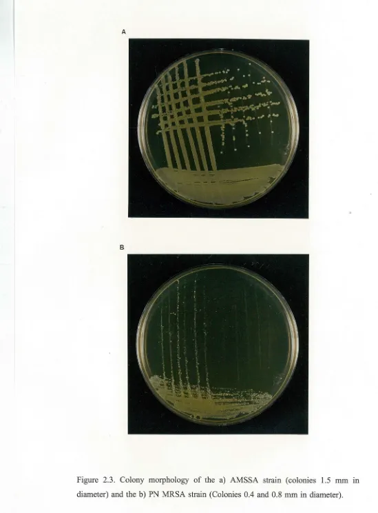
Colony morphology of methicillin sensitive S.aureus isolates a)SAl and b )SA4 22 Figure 2.2.
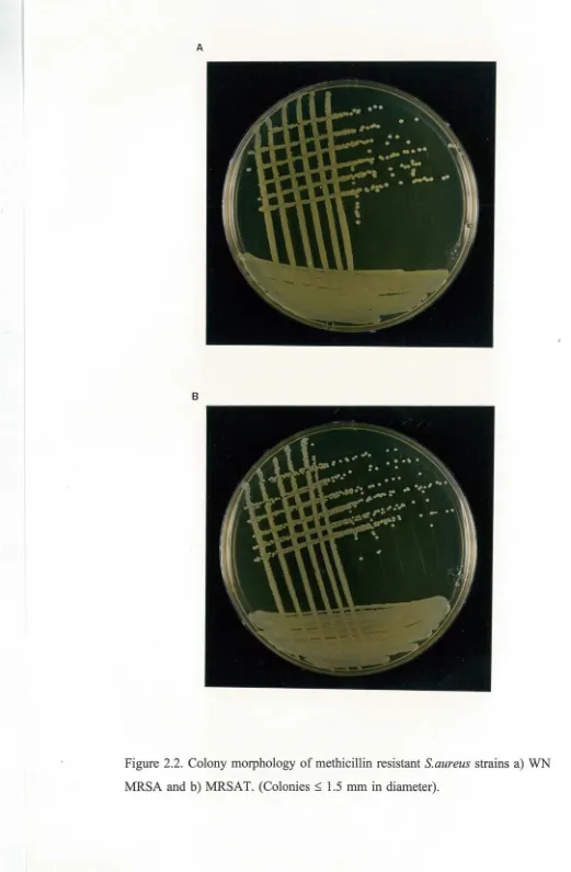
Colony morphology of methicillin resistant S.aureus strains a)WN MRSA and b)MRSAT . . . 23 Figure 2.3.
Colony morphology of the a) AMSSA strain and the b )PN MRSA . . . . . . . . 24 Graph 2.1.
Absorbance versus Time for S.aureus isolates . . . . . . . . . . . . . . . . . . . . 26 Figure 3.1.
Hybridisation of S.aureus DNA with a)32P-labelled orb) digoxigenin-dUTP labelled mec gene probe . . . . . . . . . . . . . . . . . . . . . . 45 Figure 4.1.
Plasmid Profiles of S.aureus isolates . . . . . . . . . . . . . . . . . . . 62 Figure 4.2.
HindIII restriction patterns of S. aureus isolates . . . . . . . . . . . . . . . . . . . 63 Figure 5.1.
Pulsed field gel electrophoresis of Smal digested DNA and hybridisation with a 32
P-labelled mec gene probe . . . . . . . . . . . . . . . . . . . . . . 70 Figure 6.1.
CHAPTER 1
INTRODUCTION TO THESIS
Methicillin resistant strains of Staphylococcus aureus (MRSA) are important nosocomial pathogens as they are usually resistant not only to methicillin but also to a wide range of unrelated antibiotics such as erythromycin, chloramphenicol, tetracycline, and aminoglycosides (Thornsberry 1988). This makes MRSA infections difficult to treat. The only antibiotic to which all strains of MRSA are susceptible is vancomycin (Hackbarth and Chambers 1989b) but this is expensive, may produce side effects, and if used frequently could lead to the development of vancomycin resistant strains.
Methicillin resistance in S.aureus is due to the production of a novel penicillin binding protein, PBP2a, which has a low affinity for methicillin (Chambers 1988). PBP2a is encoded by the mec gene which resides on the chromosome. The region of DNA surrounding the mec gene harbours many other resistance determinants, the number, nature and arrangement of which can vary significantly among different isolates (Inglis et al 1990, Dubin et al 1992). This region can be up to 50 kilobases in size and has no allelic equivalent in the chromosomes of methicillin sensitive strains of S. aureus.
The elucidation of the mec gene complex and further studies to investigate the genetic mechanisms leading to the acquisition of resistance determinants in the mec region have largely involved the conversion, in vitro, of methicillin resistant strains to sensitivity and the subsequent study (in the MRSA strain) of that segment of DNA lost in the sensitive derivative (Inglis et al 1990). The MRSA strains were converted in vitro to sensitivity using techniques such as acriflavin treatment (Matthews et al
the deletion endpoints occurred at or near either a transposon attachment site or the insertion sequence IS257 (Inglis et al 1990, Wada et al 1991).
Spontaneous in vivo loss of resistance to methicillin has not to our knowledge been reported. Although recent evidence by Inglis et al (1993) suggests that deletion of large amounts of DNA may occur in the mec region of the chromosome of MRSA in the clinical environment. However in that study the methicillin resistant and sensitive isolates examined were from different patients and the sequence in which they were recovered was not known in two of the three cases. Therefore the differences seen may have been due to insertion rather than deletion, of DNA. This thesis reports evidence for the loss of methicillin resistance in vivo.
In 1986, during an outbreak of MRSA infection at Palmerston North hospital, an MRSA strain (PN MRSA) was recovered from a patient who was treated and subsequently discharged. In 1990 prior to readmission an isolate of S.aureus, which produced small colonies typical of an MRSA strain, was recovered from the same patient. This isolate was resistant to several antibiotics but unexpectedly was sensitive to methicillin. The medical microbiologist at Palmerston North hospital was concerned that this atypical methicillin sensitive S.aureus (AMSSA) strain (which was assumed to be derived from the PN MRSA strain) could rapidly become resistant to methicillin if the patient was treated with that antibiotic.
This thesis reports an investigation into this possibility and addresses the question as to whether minor, easily reversible, changes (eg point mutations) led to sensitivity or, in contrast to the above, sensitivity is due to the loss of the mec gene. Consequently this investigation addresses four related questions:
1) Are the properties of the original (MRSA) isolate and the second (methicillin sensitive) isolate so similar as to imply that one was derived from the other?. 2)Was sensitivity in the AMSSA strain due to a reversible mutation such as a point mutation or a small deletion? or
HISTORICAL REVIEW
1.1 HISTORICAL PERSPECTIVE
Before the antibiotic era, the prognosis for patients with severe Staphylococcus
aureus infection was often very poor. The introduction of penicillin into clinical use in the early 1940's brought about a dramatic reversal in this situation (Lyon and Skurray 1987, Thornsberry 1988). For the first time, invasive S.aureus infections, such as those that develop from accidental or operative trauma, bums, and other serious skin lesions, could be treated effectively. However, this was to be short lived. Within a few years Kirby (1944) reported the isolation of an enzyme from a strain of S.aureus that could inactivate penicillin. This enzyme called penicillinase or B-lactamase, catalysed the hydrolysis of the B-lactam ring of penicillin, forming penicillonic acid, which had no antibactericidal properties (Thornsberry 1988). The gene which encoded resistance to penicillin was borne on a plasmid (Thornsberry 1988) and so spread rapidly. By 1946 it was estimated that 60% of hospital isolates in the United Kingdom were resistant to penicillin (Barber and Rozwadowska-Dowzenko 1948, Lyon and Skurray 1987). By the 1950's the high prevalence of staphylococci resistant to penicillin had seriously reduced the value of this antibiotic and alternative agents were needed.
The introduction of methicillin, the first of the penicillinase-resistant semisynthetic penicillins, into clinical practice in 1959 and 1960 solved this problem for a time (Chambers 1988, Thornsberry 1988). Strains of S.aureus resistant to methicillin were detected almost immediately (Jevons 1961). However, these strains accounted for only approximately 1 % of isolates from the United Kingdom (Lyon and Skurray 1987) and were not considered to pose a serious threat to the overall effectiveness of the antibiotic.
outbreaks and, in contrast to the strains isolated earlier, a higher proportion of these new strains were resistant to several antibiotics (Lyon and Skurray 1987, Thornsberry 1988). These multiply resistant MRSA strains have continued to cause major problems in hospitals worldwide to the present day.
1.2 MRSA IN NEW ZEALAND
Methicillin resistant strains of S.aureus (MRSA) were first isolated in New Zealand in 1975 (Humble 1976). From 1975 to 1984 relatively few cases were identified with a maximum of 13 infections in any one year (CDNZ suppl.1 1992). In 1985 however, the number of cases began to increase and from 1985 to 1987 two major outbreaks in Palmerston North and Wellington occurred, involving 258 persons at Palmerston North Hospital and 79 persons at Wellington Hospital (Jones 1987, Scrimgeour 1987, Martin 1987, Martin et al 1989). Although fewer cases have been reported in subsequent years, MRSA continues to affect a substantial number of persons in New Zealand (Heffernan et al 1993).
In contrast to MRSA overseas, the majority of MRSA in New Zealand in recent years are not resistant to multiple antibiotics (CDNZ suppl.1 1992, Heffernan et al 1993). Furthermore, the majority ofMRSA isolates display only low level methicillin resistance with minimum inhibitory concentrations (MICs) to methicillin of 8 to 32 µg/ml (CDNZ suppl.1 1992, Heffernan et al 1993).
1.3 HETEROGENEOUS AND HOMOGENEOUS RESISTANCE
Heterogeneous Resistance
express higher resistance levels is reproducible but strain dependent, and ranges from 10-2 to 10-3 (Tomasz et al 1991, Berger-Bachi et al 1992).
Expression of resistance is enhanced by growth in high concentrations of methicillin because the susceptible subpopulation is eliminated and the highly resistant subpopulation is selected. These antibiotic-selected cells are more uniformly resistant than the parent strain, however, this trait is unstable, and so with repeated subculturing in drug-free medium the culture reverts to its heterogeneous pattern of resistance (Tomasz et al 1991, Hacbarth and Chambers 1989b).
Homogeneous Resistance
A minority of strains are homogeneous in their expression of resistance ie cells are uniform in their expression of resistance and can grow in high concentrations of methicillin (Chambers 1988, Tomasz et al 1991). They maintain this trait even with repeated subculturing in the absence of antibiotics.
1.4 MECHANISM OF METHICILLIN RESISTANCE
PBPs
B-lactam antibiotics (such as penicillin and methicillin) act by inhibiting enzymes involved in assembling the bacterial cell wall (Lyon and Skurray 1987, Chambers 1988, Hackbarth and Chambers 1989a). These enzymes are found in the membrane and catalyze the linking reactions between peptidoglycan polymers. The cross-linking gives the wall additional ridigity, which is essential to maintain the viability of the cell. B-lactam antibiotics covalently bind to the active site of these enzymes and inhibit cross-linking (Waxman and Strominger 1983, Hackbarth and Chambers 1989). This leads to weakening of the cell wall and ultimately lysis. Because these enzymes bind penicillin at their active site they are called penicillin binding proteins (PBPs). The fundamental difference between susceptible staphylococci and methicillin resistant strains is their PBPs.
respectively (Wyke et al 1984, Chambers 1988). The specific physiologic function or functions of these staphylococcal PBPs as transpeptidases, endopeptidases, and carboxypeptidases (the three enzymatic activities which may be possessed by PBPs) have not been defined completely. However PBPs 1, 2, and 3 appear to be necessary for cell growth and survival (Georgopapadakou et al 1986, Reynolds 1988).
PBP2a
Methicillin resistant strains of S.aureus produce an additional 78 kilodalton PBP, termed PBP2a which has a low binding affinity for B-lactam antibiotics (Chambers 1988, Hackbarth and Chambers 1989a). Methicillin resistant strains of coagulase negative staphylococci also produce PBP2a (Chambers 1987).
In contrast to other staphylococcal PBPs, which generally bind B-lactam antibiotics at low concentrations, PBP2a binds B-lactam antibiotics only at a relatively high antibiotic concentration (Brown and Reynolds 1980, Chambers 1988). Since the presence of PBP2a confers methicillin resistance presumably it can substitute for the 'normal' PBPs when these have been saturated by drug, ie PBP2a can perform the functions necessary for cell wall assembly (Chambers 1988).
1.5 GENETICS OF METHICILLIN RESISTANCE
Mee
Stewart and Roseblmn (1980) have shown using cotransduction studies, that no allele equivalent to mec exists in susceptible strains of S.aureus. Beck and colleagues (1986) confirmed this by showing that mec probes hybridised with chromosomal DNA from unrelated resistant strains, but not with chromosomal DNA from susceptible strains.
Mee encodes PBP2a
The PBP2a gene has been shown to be part ofmec (Hackbarth and Chambers 1989a). Transformation of mec from S.epidermidis to a susceptible strain S.carnosus caused the recipient strain to produce PBP2a (Tesch et al 1988), and DNA from the transformants hybridised with mec specific DNA (Beck et al 1986), whereas DNA from the recipient strain did not. Furthermore, a 4 kilobase fragment of mec has the same restriction enzyme map as a cloned fragment which produces PBP2a (Matsuhashi et al 1986, Inglis et al 1988).
Origin of mec
Southern blot analysis suggests a unique origin of mec and indicates that the gene is highly conserved (Beck et al 1986, Song et al 1987, Hackbarth and Chambers 1989a). Likewise, the PBP2a gene product is also highly conserved structurally. After partial proteolytic digestion PBP2a's from several unrelated strains of S.aureus and coagulase negative staphylococci had identical fragments which were different from those of other staphylococcal PBPs (Hackbarth and Chambers 1989a).
(Staphylococcal special average 30 to 35% G+C (Novick 1990)). This observation is in opposition to the roughly 50% G+C content observed in E.coli DNA (Wu et al 1992). Furthermore, the high degree of identity at the DNA level (>99%) among mec genes from distant geographical origins suggests that relatively little time has passed since the mec gene entered staphylococci (Wu et al 1992). Thus it appears likely that the mec gene was transferred to staphylococci in a relatively recent horizontal transfer event from a species which perhaps shares the low G+C content characteristic of staphylococcal DNA. Candidate donor organisms which might fill the role include members of the genera Streptococcus, Leuconostoc, Pediococcus, Lactobacillus or Sarcina (Wu et al 1992).
Regulation of PBP2a production
PBP2a is inducible by B-lactam antibiotics although the protein can be produced constitutively (Chambers 1988, Hackbarth and Chambers 1989a). PBP2a is inducible in strains in which the penicillinase plasmid is present and constitutive in strains lacking the plasmid (Ubukata et al 1985). Thus the repressor of the penicillinase gene may play a role in the regulation of PBP2a.
A repressor may also be contained within mec (Hackbarth and Chambers 1989a). An upstream open reading frame is present on the strand of DNA opposite that which encodes PBP2a (Song et al 1987). An open reading frame of similar location is associated with penicillinase genes in other bacterial species, and its product is believed to be a repressor (Hackbarth and Chambers 1989a). This may explain why in some strains PBP2a is inducible even in the absence of the penicillinase plasmid.
Transposition of mec
1989, Dubin et al 1991). IS257 has also been found on the chromosome of methicillin sensitive strains of S.aureus (Barbaris-Maino et al 1987).
Evidence suggests that 1S257 may play a role in the transfer of mec between strains and the subsequent integration of mec into the chromosome (Hackbarth and Chambers 1989). When mec was transformed from S.epidermidis into a susceptible strain of S.carnosus (Tesch et al 1988), the cloned fragment that conferred resistance did not integrate into the chromosome but remained on the plasmid (Tesch et al 1988). Not surprisingly no 1S257-like sequences were detected on the S.carnosus chromosome. In addition mec cannot be transduced into a methicillin sensitive strain of S.epidermidis that is sensitive to mercury or cadmium (Blanchard et al 1986), presumably because the proper insertion sequences required for integration are missing.
Trees and Iandolo (1988) demonstrated that the penicillinase plasmid (which contains IS257) has an active role in the transduction of methicillin resistance into susceptible strains by providing a temporary insertion sequence for the mec-containing transposon. This requirement of penicillinase for the transduction of mec into S.aureus has been known for some time (Stewart and Rosenblum 1980). Perhaps mec insertion into the penicillinase plasmid is necessary for it to acquire the information needed to integrate into its chromosomal site.
Genetic Basis for the Accumulation of resistance determinants in the mec region
MRSA strains are usually resistant not only to methicillin but also to a wide range of unrelated antibiotics (Lyon and Skurray 1987) and in such multiply resistant strains the DNA surrounding the mec gene often contains these resistant determinants (Inglis et al 1990, Dubin et al 1992). This region varies significantly among different isolates and may be up to 50 kilobases in size (Inglis et al 1990, Dubin et al 1991, Dubin et al 1992). No allele equivalent to mec or its surrounding DNA has been found in the chromosome of methicillin sensitive strains of S.aureus (Hackbarth and Chambers 1989).
either:
1) flanked by direct repeats of the insertion sequence IS257 eg the mercury resistance operon (Inglis et al 1990),
2) part of a transposon, eg the macrolide-lincosamide-streptogramin B (MLS) resistance genes in Tn554, or the cadmium resistance gene in UTn554 (Inglis et al 1990, Chikramane et al 1991 Dubin et al 1992),or
3) part of plasmids which have integrated into the chromosome and are flanked by direct repeats of IS257, eg the tetracycline resistance plasmid, pT181 and the kanamycin resistance plasmid pUBl 10 (Dubin et al 1991)
The genetic basis for the accumulation of these resistant determinants in the mec region of the staphylococcal chromosome could be explained by either or both of the following:
1) Firstly, the region may have been created by the insertion of a plasmid which had previously assembled these resistance genes and insertion sequences (Inglis et al 1990). Large staphylococcal plasmids carrying multiple resistance genes and insertion sequences such as IS257 have been described (Lyon and Skurray 1987, Skurray et al 1988).
2) Secondly, features of this reg10n of the chromosome might have promoted recombination by providing target sites for homologous recombination or for transposon insertion ( eg Tn554 only integrates at specific chromosomal attachment sites (Chikramane 1991)). Insertion sequences (eg IS257) or transposon attachment sites might function as target sequences in sensitive strains, which could provide the recombinational sites for resistance genes coming into the cell on plasmids, phages, or conjugative transposons (Inglis et al 1990).
Other Factors
Berger-Bachi and Kohler 1983, Kornblum et al 1986). These genes termed fem factors (factors essential for the expression of methicillin resistance) are found in both the chromosome of methicillin resistant and sensitive strains (Berger-Bachi 1992). Two of these factors, femA and femB are involved in the peptidoglycan cross-bridge formation of S.aureus peptidoglycan (Berger-Bachi 1992, Henze et al 1993). Inactivation of these genes by Tn551 insertion lowers the level of methicillin resistance (Berger-Bachi 1989).
1.6 SUMMARY
Methicillin resistance is genetically and biochemically complex. PBP2a, the protein associated with methicillin resistance is encoded by the mec gene, a fusion product between a staphylococcal penicillinase gene and a PBP from a nonstaphylococcal source. Two different repressors may control expression of methicillin resistance. However within a single population various subpopulations may express different levels of resistance. What governs the switch from low level resistance to various higher levels is unknown. However the level of resistance does not correlate with the with the quantity of PBP2a present. Other factors not linked to mec are known to influence the level of methicillin resistance.
MRSA strains are usually resistant not only to methicillin but also to a wide range of unrelated antibiotics, and in such multiply resistant strains, the region of DNA surrounding the mec gene harbours many of these resistance determinants. This region can be up to 50 kilobases in size and has no allelic equivalent in the chromosomes of methicillin sensitive strains of S.aureus.
CHAPTER 2
COMPARISON OF THE COLONY MORPHOLOGY, GROWTH RATE, AND
MINIMUM INHIBITORY CONCENTRATION OF METHICILLIN FOR
VARIOUS STAPHYLOCOCCUS AUREUS STRAINS
2.1 INTRODUCTION
2.2 MATERIALS
S.aureus Isolation
DNase Test Agar DNA
Tryptose (Bacto) NaCl
Agar Water to
Agar Dilution Method
12800 µg/ml Methicillin. Stock Solution Methicillin
Distilled water to
Preparation of the Standard Inoculum
0.048 M BaC12
BaCl2.H20
Sterile distilled water to
0.36 M H2SO4 (1 % v/v)
Sterile Brain Heart Infusion broth concentrated H2SO4
0.5 McFarland Standard
2g 20 g 5 g
15 g
1000 ml
0.128 g 10.0 ml
1.173 g
100.0 ml
99.0 ml 1.0 ml
0.048 M BaC12 0.5 ml
0.36 M H2SO4 99.5 ml
Place 5.0 ml aliquots into sterile tubes, seal tightly and store in the dark. Equivalent to a bacterial culture with a density of 108 organisms/ml
0.15 M Saline (Physiological salt concentration) NaCl
Distilled water to
8.78 g
Table 2.1.
S.aureus isolates studied in this thesis
Description of isolates Source of isolates
Palmerston North outbreak MRSA Patient x: Palmerston North Hospital (PN MRSA)
Atypical methicillin sensitive strain Patient x: Palmerston North Hospital (AMSSA)
Wellington outbreak MRSA (WN Wellington Hospital MRSA)
MRSAT Palmerston North Hospital
MRSAB Palmerston North Hospital
MRSAG Palmerston North Hospital
Methicillin sensitive isolate (SA 1) Palmerston North Hospital
S.aureus isolates (SA2, SA4, SA201, Nasal swab from healthy individuals SA203, SA209, SA214, SA217,
SA301, SA303, SA304, SA312)
MRSA (ST90 410) Auckland Hospital
MRSA (ST90 371) Auckland Hospital
MRSA (ST90 236) Rotorua Hospital
MRSA (ST90 211) Auckland Hospital
MRSA (ST90 176) Auckland Hospital
MRSA (ST90 103) Auckland Hospital
MRSA (ST88 13 77) Auckland Hospital
MRSA (ST90 267) Palmerston North Hospital
MRSA (AS89 103) Dunedin Hospital
MRSA (AS89 105) Wellington Hospital MRSA (ST90 380) Palmerston North Hospital
MRSA (ST89 580) Auckland Hospital (Singapore isolate)
MRSA (ST89 1302) Auckland Hospital (Japan isolate)
MRSA (ST88 988) Auckland Hospital
2.3 METHODS
2.3.1 S.AUREUS STRAINS
All the strains which were studied in this thesis are listed in Table 2.1.
The PN MRSA, the AMSSA strain, three methicillin resistant strains MRSAT, MRSAB, MRSAG, and the methicillin sensitive isolate SAl were provided by Dr Gordon Scrimgeour of the Palmerston North Hospital.
The WN MRSA and all the other methicillin resistant strains (ST# and AS#) were kindly provided by Helen Heffernan of the New Zealand Communicable Disease Centre.
All the other S.aureus isolates (SA#) were recovered from nasal swabs of healthy individuals on the assumption that 30% of healthy adults are colonised with S.aureus. (Davis et al 1990).
2.3.2 ISOLATION OF S.AUREUS FROM HEALTHY ADULTS
A swab wetted in 0.15 M saline was rotated around the inside of the nostril. The swab was spread over the surface of a BHI agar plate and the plate incubated at 37°c overnight. White or golden colonies, 1 to 3 mm in size, were tested for catalase activity. Catalase positive organisms were Gram stained and tested for DNase and coagulase activity.
1) Catalase Test
A section of a colony was picked with a toothpick and added to a drop of hydrogen peroxide on a glass slide. The production of bubbles indicates catalase activity. 2) Gram stain
seconds. The slide was then examined under the microscope at 1 000x magnification. Staphylococcal species appear as Gram positive (purple) cocci in grape-like clusters.
3) DNase Test
A colony was picked with a straight wire and stabbed into a DNA plate. The plate was incubated at 37°c overnight. The plate was then flooded with 1 M HCL and left at room temperature for 15 minutes. A clearing around the inoculum indicates DNase activity.
4) Coagulase Test
A single colony was added to a tube containing 0.5 ml of rabbit plasma and incubated at 3 7°c. The mixture was examined for clotting at 30 minute intervals over 4 hours.
Isolates were identified as Staphylococcus aureus if they were catalase positive, DNase positive, coagulase positive, and produced Gram positive cocci in clumps.
2.3.3 SCREENING OF S.A URE US STRAINS FOR METHICILLIN
RESISTANCE
AGAR DILUTION METHOD
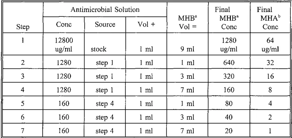
A stock solution of methicillin (12800 µg/ml) was diluted from 1280 µg/ml to 20 µg/ml using the scheme outlined in Table 2.2 (pg 18)(recommended by NCCLS 1990). 2 ml of each antibiotic solution was then added to 38 ml of Mueller Hinton agar at 50°c, mixed, and poured into two petri dishes on a level surface. 2 ml of sterile distilled water was added to 38 ml of agar for control plates.
Preparation of the Standard Inoculum
forming units per ml). The suspension was then diluted 10-fold by adding 1 ml of bacteria to 9 ml of sterile 0.15 M saline.
Inoculation of Plates
1 µl of the diluted bacterial suspension was spotted onto a Mueller Hinton agar plate using a calibrated loop to give a final inoculum of approximately 104 CFU on an area
5 to 8 mm in diameter. 32 strains were spotted onto each plate using a grid. A plate containing no methicillin was inoculated first. Then starting at the lowest concentration, the plates containing methicillin were inoculated. Finally another control plate (no methicillin) was inoculated to ensure that there was no contamination or antimicrobial carry over during inoculation. Once the inoculum spots were dry the plates were incubated at 30°c for 16 to 20 hours.
The MIC of a strain was taken as the lowest concentration of antibiotic that inhibited growth. A few small colonies or a flat haze were disregarded.
2.3.4 COLONY MORPHOLOGY
A 100 µl aliquot of S.aureus cultures stored at -70° were thawed and inoculated into 5 ml ofBHI broth. The cultures were incubated at 37°c overnight on a rotary shaker. A loopful was streaked to produce isolated colonies on a BHI plate. Plates were incubated at 37°c for 24 hours and photographed.
2.3.5 GROWTH RA TES
Table 2.2.
Scheme for Preparing Dilutions of Methicillin to be used in Agar Dilution Susceptibility Tests (NCCLS 1990)
Antimicrobial Solution Final
MHBa MHBa
Step Cone Source Vol+ Vol= Cone
1 12800 1280
ug/ml stock 1 ml 9 ml ug/ml
2 1280 step 1 1 ml 1 ml 640
3 1280 step 1 I ml 3 ml 320
4 1280 step 1 I ml 7 ml 160
5 160 step 4 I ml I ml 80
6 160 step 4 I ml 3 ml 40
7 160 step 4 I ml 7 ml 20
a. MHB = Mueller Hinton Broth b. MHA = Mueller Hinton Agar
Final MHAb
Cone
64 ug/ml
32
16
8
4
2
[image:27.599.88.560.149.373.2]2.4 RESULTS
2.4.1 MINIMUM INHIBITORY CONCENTRATION
The minimum concentrations of methicillin which inhibited the growth of various S.aureus strains are shown in Table 2.3 (pg 21).
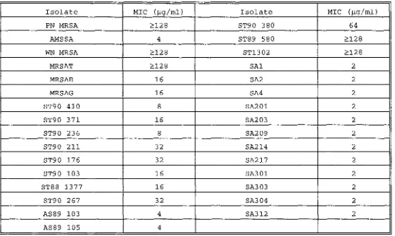
An isolate is considered resistant (NCCLS 1990) if its mm1mum inhibitory concentration (MIC) is ~8 µg/ml. The degree of antibiotic resistance shown varied tremendously from one isolate to another with MICs ranging from 8 µg/ml to ~128 µg/ml (the highest level of antibiotic used in the test). The PN MRSA strain is highly resistant to methicillin with an MIC ~128 µg/ml. The AMSSA strain had an MIC of 4 µg/ml whereas all the other methicillin sensitive strains tested had MICs to methicillin of 2 µg/ml.
2.4.2 COLONY MORPHOLOGY
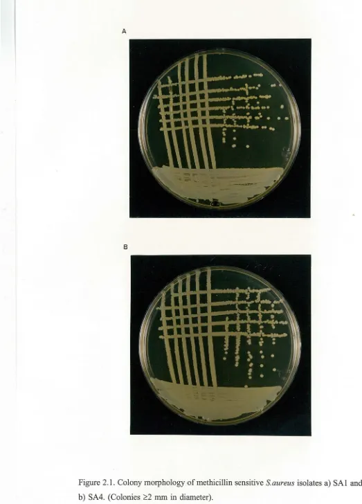
S.aureus isolates could be divided into three distinct groups based upon their colony morphology.
The first . group always produced relatively large colonies greater than 2 mm in diameter (Figure 2.1 ). All the methicillin sensitive strains (MIC
=
2 µg/ml) ( except the AMSSA strain) belonged to this group.The second group always produced smaller colonies no larger than 1.5mm in diameter (Figure 2.2). Most of the methicillin resistant strains tested belonged to this group. This group also included the AMSSA strain (Figure 2.3a) (the only methicillin sensitive strain to produce small colonies).
2.4.3 GROWTH RATES
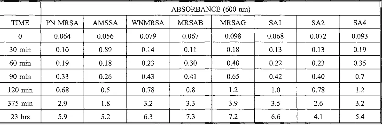
The growth rates (measured as aborsorbance (600 nm) over time) of the PN MRSA, the AMSSA strain and other methicillin resistant and sensitive strains are shown in Table 2.4 and Graph 2.1.
All of the isolates tested had similar growth rates in BHI broth.
2.5 DISCUSSION
In general, there seems to be a good correlation between an isolates colony morphology and its MIC of methicillin. Methicillin sensitive strains (MIC ~4 µg/ml) produce large colonies (>2 mm in diameter), while methicillin resistant strains (MIC ~8 µg/ml) produce smaller colonies (<1.5 mm in diameter).
Palmerston North Hospital supplied the AMSSA strain with the comment that it produced colonies typical of MRSA strains. The above results confirm this observation and show that this trait is atypical for a methicillin sensitive isolate.
Minimum Inhibitory Concentration of Methicillin for S.aureus isolates
Isolate MIC (µg/ml) Isolate MIC (µg/ml)
PN MRSA :?:128 ST90 380 64
AMSSA 4 ST89 580 :?:12 8
WN MRSA :?:128 ST1302 :?:128
MRSAT :?:128 SAl 2
MRSAB 16 SA2 2
MRSAG 16 SA4 2
ST90 410 8 SA201 2
ST90 371 16 SA203 2
ST90 236 8 SA209 2
ST90 211 32 SA214 2
ST90 176 32 SA217 2
ST90 103 16 SA301 2
ST88 1377 16 SA303 2
ST90 267 32 SA304 2
AS89 103 4 SA312 2
[image:30.846.70.631.131.466.2]B
Figure 2.1. Colony morphology of methicillin sensitive S.aureus isolates a) SAl and
B
[image:32.597.6.527.25.820.2],-,;;./!' • • •
...
. .
.. ,
...
-:.•...
.
.
...
'
,·.;:••... .
..
8
Figure 2.3. Colony morphology of the a) AMSSA strain (colonies 1.5 mm m
[image:33.596.7.556.27.771.2]Absorbance versus Time for S.aureus isolates
ABSORBANCE (600 nm)
TIME PN MRSA AMSSA WNMRSA MRSAB MRSAG SAl SA2 SA4
0 0.064 0.056 0.079 0.067 0.098 0.068 0.072 0.093
30 min 0.10 0.89 0.14 0.11 0.18 0.13 0.13 0.19
60 min 0.19 0.18 0.23 0.30 0.40 0.22 0.23 0.35
90 min 0.33 0.26 0.43 0.41 0.65 0.42 0.40 0.7
120 min 0.68 0.5 0.78 0.8 1.2 1.0 0.78 1.2
375 min 2.9 1.8 3.2 3.3 3.9 3.5 2.6 3.2
[image:34.846.75.704.135.341.2]A (600nm)
0.1
0.01
20 40 60 80 100 120
Time (Minutes)
KEY
a PN MRSA d MRSAG
b AMSSA e SAl
C WNMRSA f SA2
CHAPTER3
PROBING OF S.AUREUS DNA FOR THE MEC GENE
3.1 INTRODUCTION
Do all MRSA with a MIC 28 µg/ml possess the mec gene?
MRSA isolates recovered from a range of countries throughout the world are typically resistant to high levels of methicillin (ie c:64 µg/ml). These strains are typically multiply resistant (Lyon and Skurray 1987, Heffernan et al 1993). Such strains have been isolated in New Zealand (isolates 11 to 16 in Table 3.1, pg 29) and include the Palmerston North and Wellington outbreak MRSA strains (PN MRSA and WN MRSA). However the majority of MRSA strains isolated in New Zealand in recent years are resistant to only low levels of methicillin (ie 8 to 32 µg/ml), and, unlike the MRSA found overseas, are not generally multiply resistant (CDNZ suppl. I 1992) (isolates 2 to 8 Table 3.1).
In multiply resistant MRSA strains the mec gene and many of the other resistance determinants are found on the chromosome as part of a large 'mec gene complex' (up to 50 kilobases in size) (Dubin et al 1992). The 'low level singularly resistant' MRSA strains found in New Zealand must lack that part of the 'mec gene complex' that confers resistance to antibiotics other than methicillin and, as they are resistant to only low levels of methicillin it is possible that they also lack the mec gene itself. If so, some other mechanism besides the possession of a gene coding for PBP2a, may mediate the low level of methicillin resistance shown by these isolates.
Does the AMSSA strain possess the mec gene ?
Table 3.1.
Antimicrobial Susceptibility of S.aureus isolates (provided by M. Brett, NZCDC)
No. Isolate Methicillin MIC µg/ml
1 ST90 410 8
2 ST90 371 16
3 ST90 236 8
4 ST90 211 32
5 ST90 176 16
6 ST90 103 8
7 ST88 1377 16
8 ST90 267 16
9 AS89 103 4
10 AS89 105 4
11 ST90 380 64
12 WNMRSA ~128
Antimicrobial Resistance
Me, Pn, Em, Gm
Me, Pn
Me, Pn
Me, Pn
Me, Pn Me, Pn, Em
Me,PN,
Me, Pn
Ox,Pn
Ox,Pn
Me, Pn, Em, Co
Me, Pn, Cm, Em, Co
13 ST89 580 ~128 Me, Pn, Cm, Em, Gm, Co
14 ST89 1302 ~128 Me, Pn, Em, Gm
15 ST88 988 64 Me, Pn, Em, Gm
16 PN MRSA ~128 Mn, Em, Gm, Co
17 AMSSA 4 Pn, Em, Co
Antimicrobials Tested:
Methicillin (Mn)
Penicillin (Pn)
Chloramphenicol (Cm) Erythromycin (Em) Fusidic acid (Fa)
Gentamycin (Gm)
Vancomycin (Vm)
Cotrimoxazole (Co)
Rifampicin (Rf)
3.2 MATERIALS
Commonly Used Reagents
0.2 M EDTA
EDTA (disodium salt) pH to 8.0 with NaOH Distilled water to
1 M Tris-HCL (pH 8.0) Trizma Base (Sigma) pH to 8.0 with HCI Distilled water to
5 M NaCl NaCl
Distilled water to
Extraction of S.aureus DNA
Lysis of S.aureus cells
Lysostaphin 1 mg/ml Lysostaphin (Sigma) Sterile distilled water to
Lysis Buffer (pH 8.0) 1 M Glucose
0.2 M EDTA
1 M Tris-HCL (pH 8.0) 5 M NaCl
Distilled water to
Sodium dodecyl sulphate (SDS) 10% SDS (Sigma)
Distilled water to
Pronase Type XIV 10 mg/ml Pronase (Sigma)
Sterile distilled water to
7.44 g
100.0 ml
12.11 g
100.0 ml
29.22 g 100.0 ml
5 mg 5.0 ml
2.5 ml
2.5 ml 1.25 ml 1.5 ml 50.0 ml
10.0 g 100.0 ml
0.2 g 20.0 ml
RN ase 10 mg/ml
RNase - from bovine pancreas (Sigma) Sterile distilled water to
0.1 g 10.0 ml
This solution was preincubated at 90°c for 10 minutes to destroy DNase activity. It was then stored at -20°c.
5 M Sodium perchlorate Sodium perchlorate Distilled water to
DNA Extraction
TE Buffer (Tris-EDTA) 1 M Tris-HCL (pH 7.5) 0.2 M EDTA pH7.5 Distilled water to
Phenol/chloroform/isoamyl alcohol solution A 25:24:1 solution, respectively, was prepared.
61.22 g 100.0 ml
10.0 ml 5.0 ml 1000.0 ml
1/10 the volume of STE Buffer was then bubbled through the mixture.
Preparation of Slot Blots
20x Standard Saline Citrate (SSC) Stock Solution NaCl
Sodium citrate Distilled water to
0.4 M NaOH NaOH
Distilled water to
2 M Ammonium acetate (pH 7.0) Ammonium acetate
adjust pH to 7.0 Distilled water to Store at 4°c
E.coli Plasmid Preparation
Luria Broth NaCl
Tryptone (Bacto) Yeast extract (Difeo)
pH to 7-7.5 with 5 M NaOH Distilled water to
175.3 g 88.2 g 1000.0 ml
1.6 g 100.0 ml
15.4 g
100.0 ml
5 g
Luria Agar NaCl Tryptone Yeast extract
pH to 7-7.5 with 5 M NaOH Agar
Distilled water to
Ampicillin 5 mg/ml Ampicillin
Distilled water to
Luria Broth+ Ampicillin (100 µg/ml) Luria Broth
Ampicillin (5 mg/ml)
Luria Agar+ Ampicillin (110 µg/ml) Luria Agar
Ampicillin (5 mg/ml)
Melt agar and equilibrate to 50°c before adding ampicillin
Solution I
Glucose
1 M Tris-HCl (pH 8.0) 0.2 M EDTA (disodium salt) Distilled water to
Autoclave at 121 °c for 15 minutes
Solution II (prepare fresh) NaOH
SDS
Distilled water to
Solution III Potassium acetate Glacial acetic acid Distilled water to
Lysozyme 50 mg/ml (prepare fresh) Lysozyme
Distilled water to
Megaprime DNA Labelling Protocol
Megaprime DNA Labelling Kit (Amsersham) Primer solution
Random nonamer primers in an aqueous solution
5 g
10 g 5 g
0.5 ml 15 g 1000.0 ml
0.05 g 10.0 ml
4.9 ml 0.1 ml
196.0 ml 4.0 ml
4.95 g
12.5 ml 25.0 ml 500.0 ml
0.8 g 1.0 g
100.0 ml
29.44 g
11.5 ml 100.0 ml
Megaprime reaction Buffer
dA TP, dGTP, and dTTP in Reaction buffer.
Reaction Buffer
A concentrated reaction buffer containing: Tris-HCl (pH7.5)
MgCl
2-mercaptoethanol
Enzyme Solution
I unit/µl DNA polmerase I 'Klenow' fragment (cloned) in: 50 mM potassium phosphate (pH 6.5)
10 mM 2-mercaptoethanol 50% glycerol
Hybridisation Buffer
IM Hepes Buffer (pH 7.0) 20x SSC
Herringsperm DNA (3 mg/ml) 20% SDS
Ficoll (Sigma)
Bovine Serum Albumin (Sigma) Polyvinyl pyrrolidone (PVP-10 Sigma) Distilled water to
Store at 4°c
Warm to 37°c before use
25.0 ml 75.0 ml 3.0 ml 2.5 ml 1.0 g 1.0 g 1.0 g 500.0 ml
DIG Labeling and Detection Kit (Boeringer Mannheim Biochemica)
Hybridisation
Hexanucltotide Mixture (DIG kit) I Ox concetrated hexanucleotide mixture
dNTP Labeling Mixture (DIG kit)
1 Ox concentrated dNTP labeling mixture containing: 1 mmol/1 dATP
1 mmol/1 dCTP 1 mmol/1 dGTP 0.65 mmol/1 dTTP 0.35 mmol/1 Dig-dUTP pH 6.5 (20°c)
Klenow Enzyme, labeling grade, 2 U/µl (DIG kit)
4 M LiCl LiCl
Distilled water to
Hybridistaion Solution 20xSSC
10% N-lauroyl sarcosine 10% SDS
Blocking reagent (DIG kit) Sterile distilled water
Washing the Membrane Wash Solution I
20xSSC 10% SDS
Distilled water to
Wash Solution II 20x SSC
10% SDS
Distilled water to
Colour Detection Buffer 1
Trisma Base NaCl
Distilled water to
Buffer 2
Blocking reagent (DIG kit) Buffer 1
62.5 ml 2.5 ml 500 µl 1.25 g 184.5 ml
40 ml 4 ml 400 ml
2 ml 4 ml 400 ml
12.1 g 8.77 g 1000.0 ml
2.5 g 500.0 ml
Prepare 1 hour in advance and dissolve at 50-70°c (Buffer remains turbid).
Buffer 3
Trisma Base NaCl
MgC12
pH to 9.5 with NaOH Distilled water to 250.0 ml
Anti-digoxigenin alkaline phosphatase conjugate (DIG kit) 3.0 g 1.46 g 1.19 g
750 U/ml polyclonal sheep anti-digoxigenin Fab-fragments conjugated to alkaline phosphatase
antibody-conjugate (150 U/ml) anti-digoxigenin-AP conjugate Buffer 1
NBT-solution (DIG kit)
4 µl 20.0 ml
X-Phosphate-solution (DIG kit)
50 mg/ml 5-bromo-4-chloro-3-indolyl phosphate toluidinium salt m dimethylformamide
Colour Solution NET-solution
X-Phosphate-solution Buffer 3
Prepare fresh.
45 µl
35 µl
3.3 METHODS
3.3.1 EXTRACTION OF S.AUREUS DNA
Lysis of S.aureus cells
A 10 µl aliquot oflysostaphin (1 mg/ml) was added to 190 µl ofLysis buffer (Archer and Pennell 1990). A loopful of bacteria, obtained from heavy growth on a BHI agar plate was then added to the mixture and incubated for I hour at 37°C. 150 µ1 of
10% SDS was then added, and the bacterial suspension incubated at 50°C for 10 minutes. Following this 10 µl of pronase (10 mg/ml) was added and the lysate was incubated at 50°C overnight to ensure that the cells were lysed and that their proteins digested. 10 µl of RNase (10 mg/ml) was then added and incubated for 1 hour at 50°C. 90 µl of 5 M sodium perchlorate was added and the lysate was incubated at 50°C for a further 60 minutes.
DNA Extraction
3.3.2 PREPARATION OF SLOT BLOTS OF S.AUREUS DNA
Three sheets of Bio-Dot Format Filter paper were washed in 6xSSC and placed in a Slot-Blot apparatus (Biorad). A nylon membrane, cut slightly bigger, was then placed on top of the filter paper, and the apparatus was screwed together as tightly as possible. 15 µl of S.aureus DNA was denatured by incubation in 100 µl of 0.4 M NaOH for 5-10 minutes, neutralized by the addition of 100 µl of cold (4°C) ammonium acetate solution and added to the wells under gentle vacuum (tap open). Once all the liquid had been drawn through (1-2 hours) each slot was washed with 200 µl of 2x SSC, drawn through under full vacuum (Tap closed). The apparatus was then disassembled and the nylon membrane was rinsed in 2x SSC, placed on a piece of filter paper to dry, and baked for 2 hours at 80°C in a vacuum oven.
3.3.3 ISOLATION OF THE pUC18 PLASMID FROM E.COLI
3.3.4 LINEARISATION OF PLASMID DNA WITH EcoRl
5 µl of plasmid DNA was added to an eppendorf tube containing 2 µl of l0x H Buffer (Promega) and 11 µl of sterile distilled water. 2 µl of the restriction enzyme EcoRI (Promega) was then added, mixed by brief centrifugation, and incubated at 37°c for 90 minutes.
The reaction mixture was then stored at 4°c until required for use as a probe for the PBP2a gene.
3.3.5 LABELLING THE PBP2A GENE PROBE
The E.coli strain PG0164 containing the plasmid pUC18 plus the cloned I.I kilobase Bg!II-Xbal PBP2a gene fragment (Archer and Pennell 1990) was kindly provided by Dr Gordon Archer of the Medical College of Virginia.
General approach
The Probe was labelled with either a radioactive
(3
2P) or nomadioactive (Boehringer Mannheim) detector. The 32
P-labelled nucleotide (dCTP) was incorporated into probe DNA by random hexamer priming using the Megaprime DNA labelling kit (Amersham). The nomadioactive label, digoxigenin-dUTP, was incorporated into probe DNA by random hexamer priming using the Boehringer-Mannheim digoxigenin labelling kit. In both cases the entire plasmid, pUC18 plus the cloned 1.1 kb PBP2a gene fragment, was labelled for use as a probe as E.coli vector sequences have been shown not to hybridize with staphylococcal DNA (Archer and Pennell 1990).
LABELLING THE PROBE USING RADIOACTIVE 32P
Megaprime DNA Labelling Protocol
10 µ1 of Megaprime reaction buffer and 5 µ1 of Reaction buffer were then added. This was followed by 3µ1 of radiolabelled dCTP and then 2 µ1 of enzyme solution (the Kienow fragment of DNA polymerase I). The tube was capped and incubated at 3 7°c for 5-10 minutes. The reaction was stopped and the DNA denatured by heating the solution to 95-100°c for 5 minutes.
Measuring the amount of [32P]dCTP incorporated into the probe DNA
2 µl of labelled DNA solution was spotted onto one end of a strip of plastic-backed filter paper and left to dry. The filter paper was then placed vertically, with the DNA at the bottom, in a vial containing a small amount of 2 M HCI (free [32
P]dCTP travels up the filter paper with the solvent while [32
P]dCTP incorporated into DNA remains at the bottom of the filter paper). When the solvent front had travelled up the filter paper the paper was cut in half and each half was placed into an empty vial. The radioactivity of the top and bottom halves of the filter paper was then measured in disintegrations/minute using a scintillation counter. About 50% of the [32
P]dCTP should and did become incorporated into the plasmid DNA.
Prehybridisation
The baked nylon membrane blotted with S.aureus DNA was placed in a Hybridisation tube and 15 ml of Hybridisation buffer was added. The tube was rotated to wet the membrane evenly and then placed in a horizontal rotator in a 65°c incubator for 2 hours.
Hybridisation
48 µl of 32
P labelled probe solution was added to the tube containing the Hybridisation buffer and the nylon membrane. The Hybridisation tube was then placed back in the horizontal rotator and incubated at 65°c overnight.
Washing the membrane
icecream-box containing 100 ml of 2x SSC . This was shaken for 15 minutes at room temperature on a reciprocating shaker. The 2x SSC was then discarded and the above step repeated with 2x SSC and 0.lx SSC.
Detection of 32
P using Autoradiography
The nylon membrane was then mounted on Whatman 3MM filter paper, covered with gladwrap, and placed in an 18 x 24 cm cassette. Kodak film was placed over the nylon membrane in the dark room and an intensifying screen was placed over the film and under the filter paper. The cassette was then placed at -70°c for 24 hours before developing.
LABELLING THE PROBE USING NONRADIOACTIVE
DIGOXIGENIN-dUTP
Principle
DNA is labelled by random primed incorporation of digoxigenin-labelled deoxyuridine-triphosphate. The dUTP is linked via a spacer-arm to the steroid hapten digoxigenin (Dig-dUTP). After hybridisation to the target DNA the hybrids are detected by enzyme-linked immunoassay using an antibody-conjugate (anti-digoxigenin alkaline phosphatase conjugate) and a subsequent enzyme-catalyzed colour reaction with 5-bromo-4-chloro-3-indolyl phosphate (X-Phosphate) and nitroblue tetrazolium salt (NBT) (DIG kit manual).
Digoxigenin-dUTP Labelling Protocol
then added and the mixture placed at -20°c overnight to precipitate the DNA. The solution was then centrifuged for 15 minutes at 4°c in an eppendorf microfuge and the supernatant discarded. The labelled DNA pellet was then washed with 200 µl of cold 70% ethanol, dried in a speedvac for 10 minutes and resuspended in 50 µI of sterile TE buffer.
Prehybridisation
The baked nylon membrane blotted with S.aureus DNA was placed in a Hybridisation tube and 20 ml of Hybridisation solution were added. The tube was rotated to wet the membrane and then placed in a horizontal rotator in a 65°c incubator for 2 hours
Hybridisation
50 µI of digoxigenin-dUTP labelled DNA solution was denatured by heating at 95°c for 10 minutes in a heating block. The solution was then plunged into ice water to prevent reannealing and added to the tube containing the Hybridisation solution and the nylon membrane. The Hybridisation tube was then placed back in the horizontal rotator and incubated at 65°c overnight
Washing the Membrane
The hybridisation solution was discarded, 200 ml of Wash Solution I was added and the hybridisation tube placed on a horizontal rotator for 5 minutes at room temperature. Wash Solution I was then discarded and the above step repeated. 200 ml of warm (65°c) Wash Solution II was then added and the tube placed in a horizontal rotator in a 65°c incubator for 15 minutes. This step was repeated.
Colour Detection
to 150 MU/ml by adding 4 µI to 20 ml of Buffer 1 and was then added to the bag containing the nylon membrane. The bag was sealed and incubated at room temperature for 30 minutes. The nylon membrane was then removed from the bag and placed in 100 ml of Buffer 2 and shaken for 15 minutes at room temperature to remove any unbound antibody/conjugate. This step was repeated and then the membrane was placed in 20 ml of Buffer 3 for 2 minutes. The nylon membrane was then placed in another plastic bag and 10 ml of Colour Solution was added. The bag was sealed and placed in the dark until the test-negative S.aureus strain just began to show colour (about 10 minutes). The colour reaction was then stopped by placing the membrane into 100 ml of TE buffer.
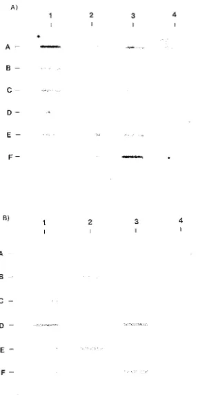
3.4 RESULTS
The 1.1 kb PBP2a gene probe was labelled with either 32
P or nonradioactive digoxigenin-dUTP and hybridised to S.aureus DNA on a nylon membrane (Table 3.1, pg 44). The results using the 32
P labelled mec gene probe are shown in Figure 3.la. The results using the digoxigenin-dUTP labelled mec gene probe are shown in Figure 3.lb.
The results using either probe were in total agreement.
All the 'high level multiply resistant' MRSA strains (MIC 2::64 µg/ml) hybridised with the mec gene fragment (lanes 2E to 4A). This included the PN MRSA strain (lane 4A).
All the 'low level singularly resistant' MRSA strains (MICs 8 to 32 µg/rnl) hybridised the mec gene probe (lanes IA to 2B).
The two 'borderline methicillin resistant' strains (defined by their ability to grow in 4 µg/ml oxacillin (resistant (NCCLS 1990)) but inability to grow in 8 µg/ml of methicillin (sensitive (NCCLS 1990)) did not hybridise with the mec gene fragment (lanes 2C and 2D).
PBP2a gene fragment (Lane 4B).
3.5 DISCUSSION
All of the 'low level singularly resistant' MRSA strains (MIC 8 to 32 µg/ml) hybridised with the mec gene fragment, which implies that the mec gene is responsible for resistance to methicillin in these isolates.
The two 'borderline' MRSA strains did not hybridise with the mec gene showing that the mec gene is not responsible for the low level of resistance to oxacillin in this group.
In general, there was a good (in fact perfect) correlation between the MIC at which a strain is considered resistant (;:::8µg/ml) and possession of the mec gene. As all of the S.aureus strains with MICs ~8 µg/ml hybridised with the mec gene, while all of the S.aureus strains with MICs s;4 µg/ml did not hybridise with the mec gene probe.
The AMSSA strain (MIC = 4 µg/ml) also did not hybridise with the PBP2a gene fragment. The 1.1 kb PBP2a gene probe covers most of the structural sequence for the PBP2a gene. Therefore if the AMSSA strain was derived in vivo from the PN MRSA its sensitivity was not due to an easily reversible point mutation or to a small deletion, but rather must be due to the deletion of at least 1.1 kilobases which covers most of the coding region for PBP2a. It follows from this that the AMSSA strain is no more likely than any other methicillin sensitive strain to develop resistance to methicillin following exposure to the drug.
Deletion of such a large amount of DNA is not improbable as all methicillin resistant strains that have been converted to sensitivity in vitro to date have lost the entire mec gene plus all or part of the mec gene complex. The size of the deletion in these strains varied considerably but could be up to 200 kb in size. In most cases one or both of the deletion endpoints occurred at or near an insertion sequence or a transposon attachment site (Inglis et al 1990, Wada et al 1991).
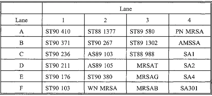
Table 3.2.
Order of S.aureus isolates on the Nylon membrane
Lane
Lane 1 2 3 4
A ST90 410 ST88 1377 ST89 580 PN MRSA
B ST90 371 ST90 267 ST89 1302 AMSSA
C ST90 236 AS89 103 ST88 988 SAl
D ST90 211 AS89 105 MRSAT SA2
E ST90 176 ST90 380 MRSAG SA4
3
•
A
B
-
c-
DE
-F-
•
B)
1 2 3 4
A
B
-c-
D
-E - ,_,._
F-
-Figure 3.1. Hybridisation of S.aureus DNA with a) 32
[image:54.597.163.460.75.654.2]CHAPTER 4
COMPARISON OF THE AMSSA STRAIN WITH THE PN MRSA AND
OTHER METHICILLIN RESISTANT AND SENSITIVE ISOLATES
4.1 INTRODUCTION
4.2 MATERIALS
Phage Typing and Reverse Phage Typing
Typing Broth
Difeo Nutrient Broth NaCl
Adjust pH to 7 .2 - 7.4 Distilled water to
Standard Typing Agar Oxoid Nutrient Broth No.2 NaCl
Distilled water (100°c) Oxoid Agar No. I 1% CaC12
Adjust pH to 7.4 - 7 .6
20 g 5g
1000 ml
120 g 30 g 6000 ml 42 g 240 ml
Dispense into 200 ml aliquots and autoclave for 15 minutes at 121 °c.
Preparation of S.aureus DNA
Lysis of S.aureus bacteria
Luria Glucose Broth plus Tris-HCl NaCl
Tryptone (Bacto) Yeast Extract (Difeo) Glucose (0.2% W N) pH to 7-7.5 with 5M NaOH
IM Tris-HCl (pH 7.5) Distilled water to
TES (Tris-EDTA-Saline) IM Tris-HCl (pH 7.8) 5M NaCl
0.2M EDTA Distilled water to
4.0 g 8.0 g
4.0 g
1.6 g
0.4 ml 16.0 ml 800.0 ml
15.0 ml 5.0 ml
TES plus 2.5M NaCl IM Tris-HCl (pH 7.8) 0.2M EDTA
NaCl
Distilled water to
Lysostaphin lmg/ml Lysostaphin (Sigma) Sterile distilled water to
10% Sarkosy 1
Sarkosyl
Distilled water to
0.2 M EDTA
EDTA (disodium salt) pH to 8.0 with NaOH Distilled water to
RN ase 2mg/ml RNase (Sigma) Distilled water to
DNA Extraction
Phenol/Chloroform/Isoamyl alcohol solution A 25:24:1 solution, respectively, was prepared.
15.0 ml 12.5 ml 73.05 g 500.0 ml
5 mg 5.0 ml
10 g 100.0 ml
7.44 g
200.0 ml
0.02 g 10.0 ml
1/10 the volume of STE Buffer was then bubbled through the mixture.
STE (Saline-Tris-EDTA) Buffer 5.0MNaCL
I.OM Tris-HCl (pH 7.5) 0.2M EDTA (pH 7.2) Distilled water to
Chloroform/Isoamyl alcohol
A 24: 1 solution, respectively was prepared.
3 M Sodium acetate Sodium acetate Distilled water to
Agarose Gel Electrophoresis
Tris Borate Buffer (lOx TBB) Trizma Base
EDTA (disodium salt) Boric Acid
adjust pH to 8.2
Distilled water to
lx TBB lOx TBB
Distilled water to
1% Agarose Agarose
lx TBB to
Bromophenol Blue Dye Bromophenol Blue Glycerol
Distilled water to
Ethidium bromide 10 mg/ml Ethidium bromide
Distilled water to Store in a dark bottle.
24.61 g 100.0 ml
108.0 g 9.3 g 55.0 g
1000.0 ml
50.0 ml 500.0 ml
0.5 g 50.0 ml
0.05 g 40.0 ml 50.0 ml
4.3 METHODS
4.3.1 COMPARISON OF S.AUREUS ISOLATES USING PHAGE TYPING
AND REVERSE PHAGE TYPING
Phage Typing of S.aureus isolates
Colonies of isolates to be typed were selected from Blood Agar plates, inoculated into 3 ml of Typing broth and incubated for 5 hours in a 37°c waterbath. Using a sterile pasteur pipette the cultures were flooded onto the surface of Standard Typing agar plates. The excess liquid was removed and the plates were dried at room temperature for 20 minutes. Twenty three S.aureus bacteriophage from the Basic International Set (Table 4.1 ), at lO0x Routine Test Dilution, were applied to each plate using a Lidwell Applicator (Blair and Williams 1961 ). The plates were incubated at 30°c for 18 hours.
Phage Typing of S.aureus isolates following Heat Shock
If a culture showed no significant lysis with any phage the isolate was retyped following heat shock.
Table 4.1.
International Basic Set of Phage for Typing Staphylococcus aureus
(Blair and Williams 1961)
Lytic Phage Numbers
Group
I
29
52
52A
79
II
3A
3C
55
71
III
6
42E
47
53
III