This is a repository copy of dOCRL maintains immune cell quiescence in Drosophila by regulating endosomal traffic.
White Rose Research Online URL for this paper: http://eprints.whiterose.ac.uk/126085/
Version: Accepted Version
Article:
del Signore, Steven J, Biber, Sarah A, Lehmann, Katherine S et al. (5 more authors) (2017) dOCRL maintains immune cell quiescence in Drosophila by regulating endosomal traffic. PLoS Genetics. ISSN 1553-7404
https://doi.org/10.1371/journal.pgen.1007052
eprints@whiterose.ac.uk https://eprints.whiterose.ac.uk/
Reuse
Items deposited in White Rose Research Online are protected by copyright, with all rights reserved unless indicated otherwise. They may be downloaded and/or printed for private study, or other acts as permitted by national copyright laws. The publisher or other rights holders may allow further reproduction and re-use of the full text version. This is indicated by the licence information on the White Rose Research Online record for the item.
Takedown
If you consider content in White Rose Research Online to be in breach of UK law, please notify us by
PLOS Genetics
dOCRL maintains immune cell quiescence in Drosophila by regulating endosomal
traffic
--Manuscript
Draft--Manuscript Number: PGENETICS-D-17-00662
Full Title: dOCRL maintains immune cell quiescence in Drosophila by regulating endosomal
traffic
Short Title: dOCRL restricts immune activation in Drosophila
Article Type: Research Article
Section/Category: General
Keywords: Drosophila; OCRL; Lowe Syndrome; endosome; Toll; innate immunity
Corresponding Author: Avital Rodal
Brandeis University
Waltham, UNITED STATES Corresponding Author's Institution: Brandeis University
First Author: Steven J Del Signore
Order of Authors: Steven J Del Signore
Sarah A Biber Katherine S. Lehmann Tania L Eskin
Sean T Sweeney Avital Rodal
Abstract: Lowe Syndrome is a developmental disorder characterized by eye, kidney, and
neurological pathologies, and is caused by mutations in the phosphatidylinositol-5-phosphatase OCRL. OCRL plays diverse roles in endocytic and endolysosomal trafficking, cytokinesis, and ciliogenesis, but it is unclear which of these cellular functions underlie specific patient symptoms. Here, we show that mutation of
Drosophila OCRL causes cell-autonomous activation of hemocytes, which are cells of the innate immune system. Among many cell biological defects in docrl mutant hemocytes, we pinpointed the cause of innate immune activation to reduced Rab11-dependent recycling traffic and concomitant amplification of Rab7-Rab11-dependent late endosome traffic. Activation is associated with mis-sorting of the Toll ligand Spåtzle, excessive secretion of Spåtzle into hemolymph, and Toll pathway activation. Thus, docrl regulation of endosomal traffic maintains innate immune cells in a poised, but quiescent state, suggesting mechanisms by which endosomal misregulation of signaling may contribute to symptoms of Lowe syndrome.
Suggested Reviewers: Jonathan Kagan
Children's Hospital Boston
jonathan.kagan@childrens.harvard.edu Innate Immunity
Neal Silverman
University of Massachusetts Medical School Neal.Silverman@umassmed.edu
Innate Immunity in Drosophila Martin Lowe
University of Manchester
pete.cullen@bristol.ac.uk Endosomal traffic Arnaud Echard Institut Pasteur
arnaud.echard@pasteur.fr Lowe Syndrome Cell Biology Opposed Reviewers:
Additional Information:
Question Response
Financial Disclosure
Please describe all sources of funding that have supported your work. This information is required for submission and will be published with your article, should it be accepted. A complete funding statement should do the following:
Include grant numbers and the URLs of any funder's website. Use the full name, not acronyms, of funding institutions, and use initials to identify authors who received the funding.
Describe the role of any sponsors or funders in the study design, data collection and analysis, decision to publish, or preparation of the manuscript. If the funders had no role in any of the above, include this sentence at the end of your statement: "The funders had no role in study design, data collection and analysis, decision to publish, or preparation of the manuscript."
However, if the study was unfunded, please provide a statement that clearly indicates this, for example: "The author(s) received no specific funding for this work."
* typeset
Stocks and reagents were obtained from the Bloomington Drosophila Stock Center (NIH P40OD018537), the Drosophila Genetics Resource Center (NIH 2P40OD010949-10A1), and the Developmental Studies Hybridoma Bank. This work was supported by the Lowe Syndrome Association (A.A.R.), National Institutes of Health T32 NS007292 (S.J.D), and Medical Research Council (UK) MR/M013596/1 (STS). The funders had no role in study design, data collection and analysis, decision to publish, or preparation of the manuscript.
Competing Interests
You are responsible for recognizing and disclosing on behalf of all authors any competing interest that could be perceived to bias their work,
acknowledging all financial support and any other relevant financial or non-financial competing interests.
Do any authors of this manuscript have
Evaluation of Competing Interests)?
If yes, please provide details about any and all competing interests in the box below. Your response should begin with this statement: I have read the journal's policy and the authors of this manuscript have the following competing interests:
If no authors have any competing interests to declare, please enter this statement in the box: "The authors have declared that no competing interests exist."
* typeset Data Availability
PLOS journals require authors to make all data underlying the findings described in their manuscript fully available, without restriction and from the time of
publication, with only rare exceptions to address legal and ethical concerns (see the PLOS Data Policy and FAQ for further details). When submitting a manuscript, authors must provide a Data Availability Statement that describes where the data underlying their manuscript can be found.
Your answers to the following constitute your statement about data availability and will be included with the article in the event of publication. Please note that simply stating data available on request from the author is not acceptable. If, however, your data are only available upon request from the author(s), you must answer No to the first question below, and explain your exceptional situation in the text box provided.
Do the authors confirm that all data underlying the findings described in their manuscript are fully available without restriction?
Yes - all data are fully available without restriction
Please describe where your data may be found, writing in full sentences. Your answers should be entered into the box below and will be published in the form you provide them, if your manuscript is accepted. If you are copying our sample text below, please ensure you replace any instances of XXX with the appropriate details.
If your data are all contained within the paper and/or Supporting Information files, please state this in your answer below. For example, All relevant data are within the paper and its Supporting Information files.
If your data are held or will be held in a public repository, include URLs,
accession numbers or DOIs. For example, All XXX files are available from the XXX
database (accession number(s) XXX, XXX)." If this information will only be available after acceptance, please indicate this by ticking the box below. If neither of these applies but you are able to provide details of access elsewhere, with or without limitations, please do so in the box below. For example:
Data are available from the XXX
Institutional Data Access / Ethics Committee for researchers who meet the criteria for access to confidential data.
Data are from the XXX study whose authors may be contacted at XXX.
* typeset
Avital A. Rodal, PhD Biology Department
Assistant Professor Rosenstiel Basic Medical Sciences Research Center 415 South Street, Mailstop 029
Waltham, Massachusetts 02454-9110 Email: arodal@brandeis.edu
3/31/17
Dear editors,
We are submitting our manuscript, entitled ÔdOCRL maintains immune quiescence by regulating endosomal trafficÕ, by Del Signore et al. for consideration as an article in PLOS Genetics. Lowe Syndrome is a congenital disease characterized by severe brain, eye, and kidney pathologies, and is caused by mutations in the phosphoinositide 5-phosphatase OCRL. Though in vitro studies have identified diverse roles for OCRL in ciliogenesis, endocytosis, endosomal trafficking, and cytokinesis, it has remained a mystery which of these cell biological processes contribute to specific symptoms in Lowe Syndrome patients, and how this leads to tissue and organ-level defects in disease.
In the attached manuscript, we use Drosophila to perform the first in vivo mechanistic cell biological studies to illuminate the specific contributions of OCRL cellular functions to organism-level physiology. Surprisingly, we found that mutation of the Drosophila OCRL homolog docrl caused a profound activation of the innate immune system. This phenotype had previously been observed in several endosomal trafficking mutants, but it has remained unclear which immune cells and tissues contribute to this phenotype, and which endosomal trafficking steps and immune-related cargoes are involved. Here, we show that the docrl phenotype is cell autonomous to macrophage-like hemocytes, and among diverse trafficking phenotypes in these cells, we were able to ascribe immune activation specifically to defective late/recycling endosomal transport, associated with mis-sorting of the immune-stimulating Toll ligand Spz. Our results are particularly intriguing in light of recent studies in mammals that implicate Toll-like signaling in astrocyte-mediated inflammation and seizure susceptibility, which is a significant neurological symptom of Lowe Syndrome. Overall, our findings define novel mechanisms by which membrane traffic controls a conserved signaling pathway, show that tissue-level defects in a pleiotropic disease like Lowe Syndrome can be linked to specific cell biological processes, and open a new avenue of investigation into Lowe Syndrome pathogenesis.
We believe that our findings should be of broad interest to cell and developmental biologists working on membrane traffic, signaling and innate immunity. We know of no financial or other conflicts of interest influencing our results. None of the data have been submitted to another journal for publication. We recommend the following reviewers with expertise in the areas of OCRL function, membrane trafficking, and immune activation:
Jon Kagan jonathan.kagan@childrens.harvard.edu Neal Silverman Neal.Silverman@umassmed.edu Martin Lowe martin.p.lowe@manchester.ac.uk Pete Cullen pete.cullen@bristol.ac.uk
Arnaud Echard arnaud.echard@pasteur.fr
Thank you again for considering our manuscript. Best regards,
dOCRL maintains immune cell quiescence by regulating
endosomal traffic
Steven J. Del Signore1¶, Sarah A. Biber1¶, Katherine S. Lehmann1, Tania L. Eskin1,
Sean T. Sweeney2, and Avital A. Rodal1,* 1
Rosenstiel Basic Medical Sciences Research Center, Department of Biology
Brandeis University, Waltham, MA 02453 1
Department of Biology, University of York, York YO10 5DD, UK.
¶co-first authors
*Corresponding author:
Avital A. Rodal
Rosenstiel Basic Medical Sciences Research Center
Brandeis University
Tel. 781-736-2459
Email: arodal@brandeis.edu
ABSTRACT
Lowe Syndrome is a developmental disorder characterized by eye, kidney, and
neurological pathologies, and is caused by mutations in the
phosphatidylinositol-5-phosphatase OCRL. OCRL plays diverse roles in endocytic and endolysosomal
trafficking, cytokinesis, and ciliogenesis, but it is unclear which of these cellular
functions underlie specific patient symptoms. Here, we show that mutation of Drosophila
OCRL causes cell-autonomous activation of hemocytes, which are cells of the innate
immune system. Among many cell biological defects in docrl mutant hemocytes, we
pinpointed the cause of innate immune activation to reduced Rab11-dependent
recycling traffic and concomitant amplification of Rab7-dependent late endosome traffic.
Activation is associated with mis-sorting of the Toll ligand SpŒtzle, excessive secretion
of SpŒtzle into hemolymph, and Toll pathway activation. Thus, docrl regulation of
endosomal traffic maintains innate immune cells in a poised, but quiescent state,
suggesting mechanisms by which endosomal misregulation of signaling may contribute
to symptoms of Lowe syndrome.
KEY WORDS
OCRL, Lowe Syndrome, phosphoinositide, hemocyte, innate immunity, Toll, Spz,
Drosophila, endosome
INTRODUCTION
Lowe syndrome is an X-linked disorder caused by mutations in the
phosphoinositide-5-phosphatase OCRL (Oculocerebrorenal Syndrome of Lowe). Lowe Syndrome patients
display renal proximal tubule dysfunction, glaucoma, cataracts, and neurological
phenotypes such as cognitive and behavioral impairments, hypotonia, and epilepsy [1].
OCRL encodes a 901 amino acid protein with an N-terminal Pleckstrin Homology (PH)
domain bearing clathrin-binding motifs, a central phosphoinositide-5-phosphatase
domain (with preference for PI(4,5)P2 and PI(3,4,5)P3), as well as an
ASPM-SPD2-hydin (ASH) domain and a catalytically inactive Rho GTPase activating (RhoGAP)
domain that each mediate interactions with membrane-associated proteins such as Rab
GTPases, IPIP27A/B, and APPL [2]. OCRL localizes to multiple membrane
compartments and is involved in a range of cell biological processes, including
clathrin-mediated endocytosis [3-5], intracellular trafficking [6-9], actin cytoskeleton regulation
[5, 10, 11], ciliogenesis [12], and cytokinesis [10, 13]. However, it remains unclear
precisely how these diverse cellular requirements contribute to tissue and organ level
pathology in Lowe Syndrome patients. A redundant gene, INPP5B, may partially
compensate for loss of OCRL, complicating studies in vertebrate systems [12, 14, 15].
By contrast, Drosophila expresses only a single homolog of OCRL, CG3573/dOCRL
[13], and may therefore be a useful model for understanding the functions of OCRL in
complex tissues in vivo. dOCRL is required for cytokinesis in cultured S2 cells [13], but
its functions have not yet been examined in vivo.
Membrane traffic plays critical roles in regulating signal transduction in many
developmental contexts. Signaling cargoes, including both ligands and receptors, are
rapidly trafficked through the endocytic system, changing their signaling activities en
route [16]. Therefore, mis-regulation of membrane trafficking pathways can lead to
respond quickly and effectively to infection. Mutants in a variety of components of the
endosomal trafficking system exhibit hyperactivation of the immune response and
increased hemocyte abundance [17-21], but it has remained unclear which specific
immune tissues or pathways are altered or how this leads to hemocyte activation. Here
we show that dOCRL controls endosomal traffic in Drosophila larval hemocytes to
restrict Toll-associated innate immune activation.
RESULTS
docrl is required to maintain immune quiescence
To investigate the role of dOCRL in vivo, we generated null alleles by excision of
a P element from the viable, fertile line docrlEY15890 (Fig S1A) and isolated two null
alleles, docrl∆3 and docrl∆4, which lacked the dOCRL protein product, and were larval or
pupal lethal when homozygous (Fig S1B,C). Lethality was specific to docrl, as it was
not complemented by a deficiency removing the docrl locus, and was rescued by a
docrl-containing genomic fragment (Fig S1C). Upon dissecting docrl mutant larvae, we
observed a striking (5-10 fold) increase in the numbers of circulating hemocytes (Fig
1A-B), which are macrophage-like cells that mediate innate immune responses.
Notably, docrlΔ3 larvae still accumulated excess hemocytes and died as larvae and
pupae when raised under axenic conditions (Fig S1C), suggesting that immune
activation was not due to over-sensitivity to pathogens. docrl mutants exhibited few
actively dividing cells (marked by phosphorylated histone H3, Fig S2A), suggesting that
excess hemocytes do not arise from increased cell division, but instead may reflect
reduced hemocyte turnover [22]. Though we detected cytokinetic defects in docrl
mutant hemocytes (Fig S2B-C), as previously described in cultured cells [10, 13], this
appeared to be insufficient to counteract the excess hemocyte phenotype. Finally, both
a docrl-containing genomic fragment and GAL4-UAS-driven dOCRL-EGFP restored
hemocyte numbers to control levels, indicating that this phenotype is specific to loss of
docrl (Figs. 1A,B).
We also observed the frequent presence of large melanotic masses in the
posterior larval body cavity (Fig 1C-D). Such masses often occur in mutants with
To test if this was the case in docrl mutants, we visualized genetically marked
hemocytes (He-GAL4 driving UAS-GFP.nls) directly through the cuticles of live larvae.
Melanotic masses were indeed surrounded by GFP-positive blood cells (Fig 1E).
However, excess hemocytes were observed in docrl mutant larva with or without
melanotic masses, suggesting that the underlying phenotype in docrl mutants is
hemocyte over-abundance.
We next asked which of several innate immune-implicated tissues require
dOCRL to limit hemocyte number: hemocytes themselves, the lymph gland (the site of
hemocyte precursor maturation [24]), the fat body (which mediates the majority of
antimicrobial peptide expression [25]), and nephrocytes (which mediate clearance of
immune-suppressing serpins [26]). Hemocyte number was rescued cell-autonomously
by driving dOCRL-eGFP (but not by control mCD8-GFP) in hemocytes with He-Gal4
(Fig 1B,F). By contrast, expression of dOCRL-EGFP in the fat body, with the strong
driver Lsp2-GAL4, did not restore hemocyte numbers to wild type levels (Fig 1F).
Further, expression of dOCRL-EGFP by the driver Dot-GAL4, which expresses at high
levels in salivary glands, lymph gland, and nephrocytes and only at low levels in
hemocytes [27], did not significantly rescue hemocyte abundance (Fig 1F). Therefore,
dOCRL is required cell-autonomously in mature hemocytes to restrict hemocyte
number.
docrl mutants exhibit altered hemocyte differentiation and activation
Larval hemocyte types include plasmatocytes, which are small macrophage-like
cells; crystal cells, which control melanization of foreign bodies; and lamellocytes, which
are large, banana-shaped cells involved in encapsulation of foreign bodies. The majority
of circulating hemocytes are plasmatocytes, while lamellocytes are rare in unstimulated
larvae [28]. However, when we examined hemocyte composition by immunostaining
with the antibodies P1 (which labels plasmatocytes) and L1 (which labels lamellocytes),
we found that 21% of hemocytes in docrl mutant larvae were L1-positive (Fig 2A,B).
Surprisingly, 62% of these L1-positive cells appeared morphologically similar to
plasmatocytes (Fig 2C), suggesting these cells may represent plasmatocytes or
differentiation was fully rescued by a single chromosomal copy of dOCRL, but only
partially rescued by expression of dOCRL-EGFP with the He-GAL4 driver (Fig 2B),
suggesting that lamellocyte differentiation was not completely cell autonomous to
He-GAL4-expressing cells, or that it may arise from the 20% of hemocytes that do not
express this GAL4 driver [30].
One major feature of hemocyte activation is a dramatic increase in actin filament
polymerization [17, 31]. We visualized F-actin by phalloidin staining and measured an
approximately 20-fold increase in F-actin intensity in docrl mutants relative to controls
(Fig 2D,E). Increased F-actin assembly corresponded to a significantly spikier
morphology in docrl mutant hemocytes (Fig 2F), also consistent with hemocyte
activation [31]. These cytoskeletal phenotypes were rescued cell autonomously by
expressing dOCRL-EGFP with the He-GAL4 driver (Fig 2E,F). Together, these data
indicate that hemocytes in docrl mutants exhibit cell autonomous hyper-activation and
partially cell autonomous hyper-differentiation, in addition to greatly increased
abundance.
dOCRL regulates PIP2 homeostasis in diverse endosomal compartments
To investigate the role of docrl in hemocyte physiology, we examined dOCRL
localization in hemocytes by live imaging of endogenously tagged dOCRL
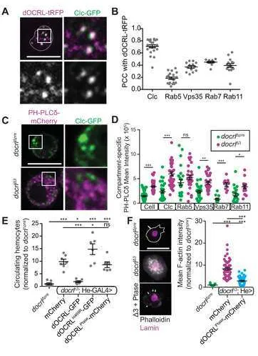
(dOCRL-TagRFPT [32]), which localized to discrete puncta at the hemocyte plasma membrane
and throughout the cytoplasm (Fig 3A). dOCRL-TagRFPT colocalized most strongly
with an He-Gal4-driven GFP tagged clathrin light chain (Clc), and moderately
colocalized other compartment markers (Fig 3B, S3A), including endogenously tagged
YFP-Rab5 (early endosomes), YFP-Rab7 (late endosomes), YFP-Rab11 (recycling
endosomes), and He-GAL4-driven Vps35-GFP (a component of the endosomal
cargo-sorting retromer complex, which has itself previously been implicated in restricting
innate immune activation [17, 21]). Interestingly, dOCRL exhibited a qualitatively
different pattern of association with different compartments. While dOCRL localized
strongly with Clc and diffusely with Rab5 early endosomes and Rab11 recycling
endosomes, it exhibited a complementary association with Vps35, and accumulated in
To test the functional requirement for this broad distribution of dOCRL, we
examined the localization of the primary dOCRL substrate PIP2 in control and mutant
hemocytes by live imaging of an mCherry-tagged PH domain of PLCδ, which
specifically binds PI(4,5)P2 [33]. In control hemocytes, PH-PLCδ localized at low levels
to the plasma membrane and in discrete intracellular puncta (Fig 3C, Fig S3B). By
contrast, in docrlΔ3 mutant hemocytes, PH-PLCδ accumulated at higher levels both at
the plasma membrane and in intracellular puncta. At the level of the whole cell, we
observed a significant increase of mean PH-PLCδ intensity relative to control, perhaps
due to stabilization of the reporter in the presence of excess PIP2 (Fig 3C). To better
analyze whether aberrant PIP2 associated with a specific endosomal compartment, we
compared the accumulation of PH-PLCδ to fluorescently labeled endosomal markers.
Levels of PH-PLCδ were increased in all compartments examined, similar to the effect
seen in whole hemocytes (Fig 3C-D, Fig S3B). Together, these data suggest that
dOCRL is required to maintain PIP2 homeostasis in diverse endosomal compartments.
To test how the dOCRL phosphatase activity contributes to its role in hemocytes,
we performed rescue experiments in the docrl mutant using He-Gal4-mediated
expression of wild-type dOCRL, a phosphatase-inactive dOCRL (dOCRLH469R,
corresponding to a Lowe Syndrome mutation [34]), or the dOCRL phosphatase domain
alone [35]. In contrast to full-length dOCRL, which rescued hemocyte abundance (Fig
1B,F, 3E), hemocyte-specific expression of either phosphatase-dead dOCRL or the
phosphatase domain alone did not suppress excess hemocyte numbers (Fig 3E),
though, based on GFP and mCherry fluorescence, they were expressed as well as the
wild-type transgene and much more highly than fully rescuing, endogenously tagged
dOCRL. Expression of the phosphatase domain alone partially rescued F-actin levels
(Fig 3F), though actin still accumulated at plasma membrane ruffles and in intracellular
puncta. Together, these results indicate that phosphoinositide homeostasis is required
for innate immune quiescence and that the phosphatase activity of dOCRL, together
with contributions from other dOCRL domains, is critically involved in this process.
To determine the consequences of loss of docrl on endosomal trafficking, we first
assayed the abundance and morphology of endosomal compartments. Consistent with
the broad increases in PIP2 that we observed in docrl mutant hemocytes, we found
defects in compartment abundance, morphology, or behavior throughout the endosomal
system (Fig 4A-B, S4A). In control hemocytes, Clc, Rab5, and Vps35 compartments
formed well-distributed medium-sized puncta, while in docrl mutant cells these
compartments fragmented into small punctae at the cell periphery, and also densely
accumulated in the perinuclear region. We also observed loss of larger Vps35-positive
tubulo-vesicular structures. Further, Rab5 endosomes were less motile in docrl∆3
hemocytes than in controls (Fig S4A). The Rab7 compartment was strikingly enlarged
in docrl mutant hemocytes compared to controls (Fig 4A), and the level of endogenous
Rab7 was significantly increased (Fig 4B). We did not detect a marked change in
Rab35 compartment structure or distribution in docrl mutants.
To assess whether these defects in membrane compartment structure correlated
with changes in function, we first examined scavenger receptor-mediated endocytosis
and phagocytosis, both of which are highly dependent on membrane trafficking and are
critical to the functional capacity of hemocytes [36-39]. We tested scavenger
receptor-mediated endocytosis by measuring internalized maleylated BSA [40] at confocal slices
through the cell interior. At all time points after pulse-chase with mBSA, docrl mutant
hemocytes exhibited reduced internalization (Fig 4C). Next, we tested phagocytosis by
measuring the internalization of E. coli. docrl hemocytes readily internalized
Alexa-488-labeled E. coli, though there was a small but significant decrease in the mean number of
bacteria internalized in the population of cells (Fig S4C). However, docrl mutants are
enriched for lamellocytes (Fig 2B) and this cell type is less phagocytically active [28],
potentially reducing the mean number of bacteria per cell. Indeed, when we excluded
from our analyses the hemocytes that took up no E. coli, docrl mutants were identical to
controls (Fig S4C). These results suggest that dOCRL is not critically required for
phagocytosis in hemocytes, though they do not exclude a role in phagosome
maturation.
Finally, we tested whether docrl mutant hemocytes exhibited defects in
allows detection of both nonacidic autophagosomes (coincident mCherry and GFP) as
well as acidic autolyososomes (mCherry alone, due to quenching of GFP) [41]. docrl
mutant hemocytes exhibited an increase in mCherry-GFP co-localization, indicating a
failure to fuse autophagosomes with the lysosome, as well as a marked increase in both
GFP and mCherry fluorescence, suggesting either upregulation of autophagy and/or a
failure of lysosomal degradation (Fig 4D). Thus, docrl is strongly required for proper
regulation of autophagosome-lysosome flux in hemocytes.
Specific endosomal defects contribute to docrl mediated immune activation
We next explored which of these dysfunctional endolysosomal compartments
could account for docrl-induced immune activation, by individually disrupting them in
otherwise wild type animals. We first inhibited the internalization step of endocytosis, by
expressing dominant negative versions of clathrin heavy chain and dynamin, as well as
a temperature sensitive dynamin mutant (raised at the restrictive temperature of 29¼C,
at which uptake of mBSA was blocked (Fig S5)). None of these manipulations caused
melanotic masses or excess hemocyte abundance (Fig 5A). OCRL interacts with
Rab35 during both early endocytosis and the abscission step of cytokinesis [3, 10].
However, we did not detect changes in immune activation following expression of
activated (not shown) or dominant negative Rab35 constructs (Fig 5A). Finally, we
found that hemocyte-specific expression of the dOCRL phosphatase domain, which
does not rescue innate immune activation (Fig 3E), robustly rescued mBSA uptake in
docrl mutants (Fig 5B). Taken together, these data indicate that defects in the
internalization step of endocytosis are unlikely to account for innate immune activation
in docrl mutants. Similarly, previous work has shown that mutants affecting canonical
autophagy do not cause innate immune defects [18], suggesting that changes in
autophagy that we observe in docrl mutants (Fig 4D) are unlikely to account for innate
immune phenotypes.
We next tested the role of post-endocytic endosomal sorting in innate immune
activation. It has previously been shown that hemocyte-specific depletion of Rab5 and
Rab11 increases circulating hemocyte concentration [19], suggesting again that innate
these results and extend the analysis to include additional Rab proteins, we expressed
constitutively active or dominant negative Rab5, 7, and 11 constructs in hemocytes with
he-GAL4. Inhibition of Rab5 (and to a milder degree inhibition of Rab11) each led to an
increase in circulating hemocyte number (Fig 5A). We further hypothesized that
activation of Rab7 would promote a similar increase in hemocyte number, consistent
with the expanded Rab7 compartment in docrl mutants. Indeed, hemocyte specific
activation of Rab7 also led to an increase in hemocyte number (Fig 5A). Thus,
manipulating endosomal traffic mediated by Rab5, Rab7, and Rab11 recapitulates the
docrl mutant phenotype.
We next tested additional components of post-endocytic endosomal sorting.
Retromer mediates endosomal sorting, and is composed of a cargo-selective trimer of
Vps35, Vps26, and Vps29. Vps35 mutants have previously been shown to exhibit
immune activation [17, 21], and we found that mutations in Vps26 similarly caused
melanotic mass formation (Fig 5C). Vps35 and Vps26 associate with endosomes via
distinct membrane binding SNX1 containing ÒSNX-BARÓÕ or SNX3 containing
complexes, likely with distinct localization and functions [42]. To clarify the requirement
for retromer in innate immunity, we analyzed snx1 and snx3 mutants. We detected
frequent melanotic masses in snx1, but not snx3 larvae (Fig 5C), suggesting that
specific loss of SNX-BAR retromer underlies immune activation in vps35 and vps26
retromer mutants, and supporting the model that endosomal sorting underlies innate
immune defects.
Finally, we asked if hemocyte-specific manipulations of Rab5, Rab7, and Rab11
could enhance or rescue the docrl mutant phenotype. Expression of dominant negative
Rab5 and Rab11 caused early larval lethality in docrl mutants, but not in controls,
suggesting that docrl mutants are sensitized to defects in a Rab5-Rab11 trafficking
route. Strikingly, increasing Rab11 activity by overexpressing a constitutively active
transgene (Rab11CA) significantly reduced hemocyte number toward normal levels (Fig
5D). However, Rab5 activation failed to rescue hemocyte abundance, suggesting that
docrl-mediated trafficking defects may occur downstream of Rab5, but upstream of
Rab11. Finally, inhibition of Rab7 activity by overexpressing a dominant negative
hemocytes. Neither Rab11CA nor Rab7DN rescue restored docrl mutant adult viability,
suggesting that lethality may require more complete rescue, or that it arises from a
distinct function of dOCRL. Taken together, these results suggest that the membrane
trafficking defect most salient to the docrl innate immune phenotype is mis-direction of
endosomal traffic from a Rab11-dependent route towards a Rab7-dependent route.
docrl is required to restrict Toll signaling
We next explored which innate immune regulatory pathway might be disrupted
by endosomal mis-sorting in docrl mutants. Immunity in Drosophila is mediated by
several signaling pathways, including Toll and IMD, which promote expression of
various target antimicrobial peptide and signaling genes [43]. We found that the Toll
targets Drs and IM1 were elevated in docrl mutants, while the IMD-specific target Dipt
was not (Fig 6A). Additionally, we measured increased levels of the Toll pathway
transcription factor Dorsal (Dl) (Fig S6A), consistent with the previously observed
induction of Dl expression by Toll activity [43, 44].
We next investigated potential mechanisms by which misregulation of endosomal
traffic in docrl mutants could lead to Toll pathway activation. We detected mild but
significant differences in Toll receptor levels and distribution, using He-GAL4-driven
Toll-Venus (Fig 6B). Further, the Toll adaptor protein MyD88, which binds PIP2 [45]
relocalized from the plasma membrane in controls to intracellular punctae in docrl
mutants (Fig 6C). Finally, we examined the Toll ligand SpŠtzle (Spz), which is secreted
by hemocytes [46], and can exert both autocrine and endocrine functions in tissues
including the fat body. He-GAL4-driven Spz-GFP accumulated to greater levels, and
with a more punctate distribution in docrl mutant hemocytes relative to controls (Fig
6D). Further, we found increased levels of endogenous Spz in docrl mutant cell-free
hemolymph (Fig 6E). To ask whether this excess Spz release underlies innate immune
activation in docrl mutants, we took advantage of the rescue of excess hemocytes by
Rab11CA. Because Rab11CA autonomously suppresses hemocyte activation, we
reasoned that this rescue should also suppress the docrl-dependent Toll signaling
defects. Indeed, we found that constitutively active Rab11 caused dramatic
increase in intracellular Spz-GFP corresponded to a decrease in secreted Spz-GFP, as
assayed by Western blot of cell-free hemolymph (Fig 6G). Together, these data suggest
that a Rab11-dependent sorting route may suppress innate immune activation by
retaining Spz in the cell. This association between Rab11 rescue of the docrl mutant
phenotype and redistribution of Spz suggest that dOCRL-dependent membrane
trafficking may maintain immune quiescence by regulating the localization, abundance,
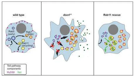
and release of the Toll ligand Spz (Fig 7).
DISCUSSION
Here we describe a requirement for the Lowe Syndrome phosphoinositide
phosphatase dOCRL in maintaining innate immune quiescence in Drosophila. Among
the many cellular functions of dOCRL, our results suggest that innate immune activation
arises from defective endosomal trafficking of Toll pathway components. These data
shed new light not only on OCRL function, but also on the contribution of membrane
trafficking to innate immunity, and suggest new avenues for investigating the diverse
symptoms of Lowe Syndrome.
dOCRL regulates endosomal sorting to maintain immune quiescence
OCRL has been implicated in numerous membrane trafficking events, including
multiple steps of endocytosis and endolysosomal traffic, autophagy, ciliogenesis, and
cytokinesis [2]. It has remained an open question whether individual cell and tissue level
pathologies in Lowe Syndrome emerge from defects at specific cellular locations, or
from a combination of OCRL-dependent functions. Though we found that docrl mutant
hemocytes exhibit many of these cellular defects, our data strongly suggest that
endosomal membrane trafficking is the primary role of dOCRL in regulating innate
immune activation. First, cytokinesis defects are unlikely to account for innate immune
activation, since relatively few docrl mutant hemocytes fail cytokinesis (Fig S2A), and
this would be predicted to reduce rather than increase total hemocyte number. Second,
defects in the internalization step of endocytosis are unlikely to cause innate immune
activation, since independently inhibiting this process via clathrin and dynamin did not
mutants using the dOCRL phosphatase domain (Fig 5B) did not suppress hemocyte
abundance (Fig 3E). Third, autophagy defects are unlikely to account for innate immune
activation, as canonical atg loss-of-function mutants do not cause a melanotic mass
phenotype [18]. Instead, we found that recapitulating the trafficking defects of docrl
mutants by manipulating retromer or endosomal Rab GTPases was sufficient to
produce immune activation. Further, the converse manipulation of Rab11 and Rab7
GTPases could rescue the docrl-induced hemocyte phenotype. Together, our results
suggest that the role of dOCRL in maintaining innate immune quiescence is most critical
in post-endocytic endolysosomal traffic, and adds to a growing list of OCRL functions in
these compartments [9, 47, 48].
dOCRL functions cell autonomously in hemocytes to control Toll signaling
Innate immune activation in Drosophila depends on cross talk among multiple
tissues, including hemocytes [46], the fat body [46, 49], muscles [50], and epithelia such
as the gut and epidermis [51]. Our results suggest that cell-autonomous Toll activation
promotes excess hemocyte accumulation. This accumulation is not due to excessive
proliferation (Fig S2A), and may reflect the role of Toll-Dl signaling in suppressing
apoptosis via diap1 expression [22]. Prior studies have identified a similar
hemocyte-specific role for endosomal GTPases in the control of hemocyte number [19]. It remains
to be determined whether immune activation in other membrane trafficking mutants,
including retromer (Fig 5C) [17, 21] and Atg6 [18], reflect defects in hemocytes or other
immune relevant tissues. Indeed, the ESCRT complex members hrs and myopic (mop)
are both required to promote Tl signaling and to generate a normal immune response in
the Drosophila fat body [49]. Further, defective membrane balance in the fat body, via
either impaired endocytosis or increased secretion, can lead to an immune response
very similar to that described here [52]. While our results do not exclude a contribution
of other tissues to docrl mediated immune activation, our rescue experiments argue
strongly that hemocytes are the initiating tissue with regard to this phenotype, and that
Toll signaling is intimately implicated.
Few studies to date have investigated the role of trafficking in Toll signaling in
regulated at specific cellular compartments [53]. Indeed, work in mammalian cells
suggests that endosomal sorting may regulate the localization of cytokine receptors to
prevent spurious Toll-like receptor activation [54]. Our work suggests several
nonexclusive mechanisms by which changes in endosomal trafficking in docrl mutant
hemocytes might drive Toll-mediated immune activation. First, the Toll receptor itself
localizes to endosomal compartments, and though we only observed subtle changes in
its distribution in docrl mutants (Fig 6B), the possibility remains that its sorting or
turnover is misregulated, resulting in amplified signaling. Second, the Toll adaptor
MyD88 binds PIP2, and is mislocalized from the plasma membrane to intracellular
compartments in docrl mutants (Fig 6C). Third, and perhaps most intriguing, our data
suggest that hemocyte-autonomous redistribution and increased secretion of the Toll
ligand Spz could contribute to aberrant pathway activation and subsequent immune
response. We found docrl mutant hemocytes exhibited increased levels of both
intracellular and secreted Spz. Strikingly, we found that Rab11-mediated rescue of docrl
innate immune activation correlated with increased intracellular retention and thus
diminished secretion of Spz, strongly suggesting that Spz release is a primary driver of
innate immune activation (Fig 6D-G). Taken together, our data suggest that Spz is
normally degraded by an endolysosomal pathway. In docrl mutants, this pathway is
defective (Fig 4A, D), leading to accumulation of Spz and its release into the
hemolymph, promoting immune activation. Aberrant fusion of Spz-containing
compartments with the plasma membrane may arise directly from altered endosomal
membrane composition in docrl mutants: Previous studies have shown that PIP2-rich
membranes are favored for plasma membrane fusion [55], and the membrane
composition of late endosomes correlates with plasma membrane or lysosome fusion
[56, 57]. Finally, activation of Rab11 may rescue aberrant Spz release by redirecting it
from secretion into hemolymph to a canonical recycling pathway. In the future, it will be
interesting to determine whether and how similar changes in Spz trafficking may
participate in normal immune responses.
Relevance to Lowe Syndrome
system to suppress spurious activity. One interesting hypothesis is that innate immune
phenotypes might underlie epilepsy and cystic brain lesions in Lowe Syndrome patients
[58]. There is strong evidence for a link between inflammation, innate immunity, and
epilepsy [59]. Most strikingly, a recent report implicates that early immune challenge by
LPS in mice promotes an astrocytic TLR4-MyD88 signaling pathway that enhances
excitatory synaptogenesis and subsequent seizure susceptibility [60]. Several studies
also suggest a direct link between OCRL, neuroinflammation, and seizure. Zebrafish
ocrl mutants feature cystic lesions in the brain that are enriched in glia, and exhibit
seizure susceptibility [14]. Further, pilocarpine seizure induction in mice led to
decreased accumulation of OCRL in hippocampal astrocytes [61]. Though it remains
unclear in this case whether these changes are pathological or compensatory, it will be
important to investigate the link between OCRL and seizure in these mammalian
systems. Together, these data raise the possibility that loss of OCRL causes seizures in
humans due to immune activation in the brain, by mechanisms similar to those we have
uncovered in Drosophila. Thus, our finding that dOCRL acts specifically in immune cells
to restrain innate immune activation provides a novel line of inquiry into the
pathogenesis of the symptoms in Lowe Syndrome patients.
EXPERIMENTAL PROCEDURES
Statistical methods
All graphs show mean ± SEM. Statistical significance was calculated with Prism
software (GraphPad, La Jolla, CA) as follows for specific datasets: We used one-way
ANOVA followed by pairwise Tukey's test (Figs 1A, 1F, 3E, 4B, 4D (intensity), 5A, 5D,
6F, S4C, S5), or Kruskal-Wallis test with post-hoc DunnÕs test (Figs 1B, 1F, 2E, 2F, 3B,
4C, 5B, 6A, 6G, S6 (whole cell & nuclear Dl)). For comparisons between two groups,
we utilized Student's t test (Figs 6C-E, S6A (% nuclear)) or Mann-Whitney U test (Fig
3D (PCC), 6B). Chi squared tests were used to calculate differences from expected
distributions of cell types. In all cases *p < 0.05, **p < 0.01, ***p < 0.001.
Drosophila larvae were cultured using standard media at low density at 25¼C for all
experiments, unless noted otherwise. To generate docrl mutants, P[EPgy2]EY15890
(732 bp upstream of the docrl start codon) was mobilized using a ∆2-3 transposase, in
the mus309 mutant background for docrl∆pre, docrl∆1, docrl∆2, and docrl∆3 [62]. 600
candidate lines were screened by PCR to identify deletions, which were subsequently
sequenced to determine precise molecular coordinates. Line docrl∆pre contains a
sequence-verified precise excision of the P-element. dOCRL and VPS35 were cloned
using Gateway technology (Life Technologies, Inc) into pBI-UASc-gateway-eGFP [63].
Constructs were injected into flies (Genetic Services Inc. Cambridge, MA), using Φc31
recombinase at the Attp40 locus [64].
Additional fly strains used have been previously described (and where noted with BL
stock numbers, are available at the Bloomington Drosophila Stock Center) as follows.
Alleles: vps26 (BL26623), snx1∆2 and snx3∆1 [65], Df(X)ED6565 (BL9299), Dup(X)DC402
[66]. Drivers: He-Gal4 (BL8699, [30]), Lsp2-Gal4 [67], Dot-Gal4 (BL6902, [27]).
Endogenously labeled proteins: dOCRLT-STEP [32] and Rab GTPases [68]. Expression
lines: Spz-GFP [69], Toll-Venus [70], GFP-mCherry-Atg8a [41],
UAS-Clc-GFP (BL7109, Henry Chang), UAS-OCRLptase [35]., UAS-PHPLC-cherry (BL51658,
[71]), UAS-shiTS (BL44222), UAS-shiK44A (BL5811), UAS-lacZ (BL1777), UAS-Rab11QL
[72], rab11N124I [73]. Remaining UAS-Rab constructs including YFP-Rab5QL (BL9773),
rab7QL (BL9779), rab7SN (BL9778), rab5SN (BL42704), rab35SN (BL9820) were
described in [74].
Hemocyte extraction, quantification and immunocytochemistry
Wandering 3rd instar larvae were used for all hemocyte experiments. Hemolymph was
extracted from individual larva or groups of 2-5 larvae. Larvae were collected into PBS,
quickly washed with 70% ethanol and then rinsed three times in PBS. Hemolymph was
collected by tearing the larval cuticle into PBS with 0.01% phenylthiourea. For absolute
hemocyte counts (Fig 1A-B), hemolymph was loaded onto each side of a disposable
hemocytometer (Incyto C-Chip DHC-N01) and allowed to settle for 30 minutes in a
moist chamber before quantification of GFP positive cells using a 20x objective on an
immunocytochemistry, hemolymph was extracted into PBS from 2 or more pooled
larvae, and placed in each well of a multi-chamber microscope slide (Thermo Scientific
Nunc Lab-Tek II 8-chamber slides). Each well, containing an independent collection of
hemocytes from pooled larvae, represents a single sample. Hemocytes were allowed to
settle for 60 minutes in a moist chamber at room temperature and then fixed for 10
minutes in 4% formaldehyde in PBS, washed in PBS, and then permeabilized, stained
with primary and secondary antibodies and washed with PBX (PBS with 0.1%
Triton-x-100). Slides were mounted with Mowiol with DABCO. Cells were imaged by either
confocal or widefield microscopy, and counted from at least 6 fields of view per sample.
Antibodies
A fragment of dOCRL encoding amino acids 1-179 was cloned into pGEX-6P
(GE Healthcare). E. coli strain BL21(DE3) expressing this construct was grown to log
phase at 37¼C, then induced for 3 h at 37¼C with 0.4 mM IPTG. Cells were lysed in PBS
(phosphate-buffered saline, pH 7.4) supplemented with 0.5 mM DTT, 0.5% Triton-X100,
0.5 mg/ml pepstatin, leupeptin and aprotonin and 1 mM PMSF. Lysates were purified on
glutathione agarose (GE Healthcare), washed 4 times with 50 ml of PBS with 0.5 mM
DTT, and GST was cleaved from dOCRL1-178 at 4¼ overnight with a ~1:50 molar ratio of
Precision Protease (GE Healthcare). Supernatant containing the cleaved protein was
further purified by gel filtration on a Superose 12 10/30 column equilibrated in PBS with
0.5 mM DTT. Purified protein was flash frozen in liquid nitrogen at a final concentration
of 0.3 mg/ml, and sent to Cocalico, Inc. for injection into rabbits. Serum from Rabbit #18
was affinity purified against GST-dOCRL1-178, which was expressed as above, purified
on a Profinia system (Biorad) with a glutathione affinity column, and eluted with
glutathione according to manufacturerÕs instructions. Glutathione eluates were gel
filtered into PBS on a Sephacryl S-200 16/60 column (GE Healthcare), and conjugated
to an Aminolink immobilization column (Thermo-Pierce) using the cyanoborohydride
method, according to manufacturerÕs instructions. 2 mL of α-dOCRL serum from Rabbit
#18 were incubated for 2 h on the resin, then washed 5 times with 2 mL PBS, and
eluted with 0.1 M glycine, pH 2.5. The eluate was then neutralized by adding Tris pH 9.0
anti-L1 and P1 (gift of Istvan Ando) anti-Lamin Dm0 (DSHB ADL67.10), anti-Spz (gift of
S. Goto), anti-MyD88 (gift of S. Wasserman), anti-actin (DSHB JLA20), anti-pH3
(Abcam ab5176).
Phagocytosis and endocytosis assays
To measure phagocytosis, hemocytes were extracted and spread on slides for 30 min
as described above. Cells were then washed twice with PBS, and incubated for the
indicated times with 100 µl PBS containing 6x10⁶ particles Alexa 488-labeled E. coli
(Life Technologies). Cells were then washed quickly 5 times with 500 µl PBS before
fixing and imaging as above.
Receptor-mediated and bulk endocytosis were measured essentially as previously
described [40, 75]. Specifically, hemocytes were extracted and spread for 5 minutes on
slides as described for immunocytochemistry, then pulsed for 45 sec with 5ug/mL
Cy5-labeled maleylated BSA (see below) in M1 medium (150 mM NaCl, 5 mM KCl, 1 mM
CaCl2, 1 mM MgCl2, 20 mM HEPES, pH 6.9; supplemented with BSA (1.5 mg/ml) and
D-glucose (2 mg/ml)), and chased with M1 medium at room temperature. Cells were
then fixed and imaged as above. Cy5-labeled maleylated BSA (Cy5-mBSA) was
prepared as described in [76] and labeled with bis-functional Cy5 according to
manufacturerÕs instructions (GE Healthcare)).
Immunoblots of whole larva and hemolymph extracts
Whole 3rd instar larvae were homogenized in Laemmli sample buffer (20 µl/larvae) and
boiled for 1 min. 10 µl (the equivalent of 0.5 larvae) were loaded for immunoblotting.
Larval hemolymph was isolated as follows: A ~2mm slit was made in the bottom of a
500uL Eppendorf tube and the cap was removed. The indicated number of larvae were
pierced at their posterior end by a pair of forceps and added to the prepared tube on
ice. The tube was placed in a 1.7mL Eppendorf tube and spun at 1000xG for 10 sec. 50
µl ice cold PBS with 0.01% phenylthiourea was added to hemolymph and centrifuged at
5000 x g for 5 min. 25 µl solution was reserved as cell free hemolymph, and boiled for 1
min in 25 µl Laemmli sample buffer. Boiled samples were centrifuged at 14,000 x g for 1
labeled with Alexa 680-conjugated secondary antibodies, and detected on a LI-COR
Odyssey infrared detection system.
Imaging and image analysis
Confocal imaging was conducted on an Andor Revolution spinning disk system
consisting of a Nikon Ni-E upright microscope, equipped with 40x (n.a. 1.3), 60X (n.a.
1.4), and 100X (n.a. 1.45) oil immersion objectives, a Yokogawa CSU-W1 spinning disk
head, and an Andor iXon 897U EMCCD camera. Confocal imaging was used to acquire
data related to protein subcellular localization and abundance (Figs 2, 3A-D, 4, 6).
Widefield imaging was conducted on a Ni-E inverted microscope equipped with a
Spectra-X LED light engine and a Zyla sCMOS camera, and a 60X (n.a. 1.4) objective.
Widefield imaging was used to measure cell relative cell counts and actin accumulation
(Fig 1F, 3E-F, 5). Images were collected using Nikon Elements AR software.
Fluorescence microscopy image processing and analysis was performed in FIJI
(National Institutes of Health, Bethesda, MD). Fluorescence intensity measurements
(area, perimeter, mean and integrated intensity) were performed on sum intensity
projections. Plasma membrane ratio of MyD88 was calculated as follows: Cell profiles of
single Z sections taken at the approximate midpoint of the cell body were thresholded in
FIJI, and the resulting cell mask was divided into three circumferential regions of
approximately equal area. The mean fluorescence intensity in the outer ring (which
captured the plasma membrane signal) was divided by the mean fluorescence intensity
in the adjacent ring (which captured a representative section of cytoplasm). Pearson R
was calculated using Coloc2 (FIJI) on 3D cell image stacks.
Live imaging
1-3 wandering third instar larvae/genotype were bled directly into 50uL M1
medium supplemented with BSA (1.5mg/ml) and D-glucose (2 mg/ml). Hemocytes were
allowed to settle 5 minutes, and then coverslips were affixed to a glass slide by double
sided tape, which simultaneously served as a bridge (3M). A single experiment was
considered to be the aggregate of all cells imaged of a single genotype during a single
on the same day (over the course of ~3 hours) imaged with identical settings.
REFERENCES
1. Lewis RA, Nussbaum RL, Brewer ED. Lowe Syndrome. 1993. Epub 2012/02/23. doi: NBK1480 [bookaccession]. PubMed PMID: 20301653.
2. Mehta ZB, Pietka G, Lowe M. The Cellular and Physiological Functions of the Lowe Syndrome Protein OCRL1. Traffic. 2014;15(5):471-87. Epub 2014/02/07. doi: 10.1111/tra.12160. PubMed PMID: 24499450.
3. Cauvin C, Rosendale M, Gupta-Rossi N, Rocancourt M, Larraufie P, Salomon R, et al. Rab35 GTPase Triggers Switch-like Recruitment of the Lowe Syndrome Lipid Phosphatase OCRL on Newborn Endosomes. Curr Biol. 2016;26(1):120-8. Epub 2016/01/05. doi: S0960-9822(15)01434-7 [pii]
10.1016/j.cub.2015.11.040. PubMed PMID: 26725203.
4. Erdmann KS, Mao Y, McCrea HJ, Zoncu R, Lee S, Paradise S, et al. A role of the Lowe syndrome protein OCRL in early steps of the endocytic pathway. Dev Cell. 2007;13(3):377-90. Epub 2007/09/04. doi: S1534-5807(07)00304-8 [pii]
10.1016/j.devcel.2007.08.004. PubMed PMID: 17765681; PubMed Central PMCID: PMC2025683.
5. Nandez R, Balkin DM, Messa M, Liang L, Paradise S, Czapla H, et al. A role of OCRL in clathrin-coated pit dynamics and uncoating revealed by studies of Lowe syndrome cells. Elife. 2014;3. Epub 2014/08/12. doi: 10.7554/eLife.02975. PubMed PMID: 25107275.
6. Billcliff PG, Noakes CJ, Mehta ZB, Yan G, Mak L, Woscholski R, et al. OCRL1 engages with the F-BAR protein pacsin 2 to promote biogenesis of membrane-trafficking intermediates. Mol Biol Cell. 2016;27(1):90-107. Epub 2015/10/30. doi: mbc.E15-06-0329 [pii]
10.1091/mbc.E15-06-0329. PubMed PMID: 26510499; PubMed Central PMCID: PMC4694765.
7. Choudhury R, Diao A, Zhang F, Eisenberg E, Saint-Pol A, Williams C, et al. Lowe syndrome protein OCRL1 interacts with clathrin and regulates protein trafficking between endosomes and the trans-Golgi network. Mol Biol Cell. 2005;16(8):3467-79. Epub 2005/05/27. doi: E05-02-0120 [pii]
10.1091/mbc.E05-02-0120. PubMed PMID: 15917292; PubMed Central PMCID: PMC1182289.
8. Noakes CJ, Lee G, Lowe M. The PH domain proteins IPIP27A and B link OCRL1 to receptor recycling in the endocytic pathway. Mol Biol Cell. 2011;22(5):606-23. Epub 2011/01/15. doi: mbc.E10-08-0730 [pii]
9. De Leo MG, Staiano L, Vicinanza M, Luciani A, Carissimo A, Mutarelli M, et al. Autophagosome-lysosome fusion triggers a lysosomal response mediated by TLR9 and controlled by OCRL. Nat Cell Biol. 2016. Epub 2016/07/12. doi: ncb3386 [pii]
10.1038/ncb3386. PubMed PMID: 27398910.
10. Dambournet D, Machicoane M, Chesneau L, Sachse M, Rocancourt M, El Marjou A, et al. Rab35 GTPase and OCRL phosphatase remodel lipids and F-actin for successful cytokinesis. Nat Cell Biol. 2011;13(8):981-8. Epub 2011/06/28. doi: ncb2279 [pii]
10.1038/ncb2279. PubMed PMID: 21706022.
11. Faucherre A, Desbois P, Nagano F, Satre V, Lunardi J, Gacon G, et al. Lowe syndrome protein Ocrl1 is translocated to membrane ruffles upon Rac GTPase activation: a new perspective on Lowe syndrome pathophysiology. Hum Mol Genet. 2005;14(11):1441-8. Epub 2005/04/15. doi: ddi153 [pii]
10.1093/hmg/ddi153. PubMed PMID: 15829501.
12. Luo N, Kumar A, Conwell M, Weinreb RN, Anderson R, Sun Y. Compensatory Role of Inositol 5-Phosphatase INPP5B to OCRL in Primary Cilia Formation in Oculocerebrorenal Syndrome of Lowe. PLoS One. 2013;8(6):e66727. Epub 2013/06/28. doi: 10.1371/journal.pone.0066727
PONE-D-13-11031 [pii]. PubMed PMID: 23805271; PubMed Central PMCID: PMC3689662.
13. Ben El Kadhi K, Roubinet C, Solinet S, Emery G, Carreno S. The inositol 5-phosphatase dOCRL controls PI(4,5)P2 homeostasis and is necessary for cytokinesis. Curr Biol. 2011;21(12):1074-9. Epub 2011/06/11. doi: S0960-9822(11)00557-4 [pii]
10.1016/j.cub.2011.05.030. PubMed PMID: 21658948.
14. Ramirez IB, Pietka G, Jones DR, Divecha N, Alia A, Baraban SC, et al. Impaired neural development in a zebrafish model for Lowe syndrome. Hum Mol Genet. 2012;21(8):1744-59. Epub 2012/01/03. doi: ddr608 [pii]
10.1093/hmg/ddr608. PubMed PMID: 22210625; PubMed Central PMCID: PMC3313792.
15. Bothwell SP, Chan E, Bernardini IM, Kuo YM, Gahl WA, Nussbaum RL. Mouse model for Lowe syndrome/Dent Disease 2 renal tubulopathy. J Am Soc Nephrol. 2011;22(3):443-8. Epub 2010/12/25. doi: ASN.2010050565 [pii]
10.1681/ASN.2010050565. PubMed PMID: 21183592; PubMed Central PMCID: PMC3060438.
16. Shilo BZ, Schejter ED. Regulation of developmental intercellular signalling by intracellular trafficking. EMBO J. 2011;30(17):3516-26. Epub 2011/09/01. doi: emboj2011269 [pii]
17. Korolchuk VI, Schutz MM, Gomez-Llorente C, Rocha J, Lansu NR, Collins SM, et al. Drosophila Vps35 function is necessary for normal endocytic trafficking and actin cytoskeleton organisation. J Cell Sci. 2007;120(Pt 24):4367-76. Epub 2007/12/07. doi: 120/24/4367 [pii]
10.1242/jcs.012336. PubMed PMID: 18057029.
18. Shravage BV, Hill JH, Powers CM, Wu L, Baehrecke EH. Atg6 is required for multiple vesicle trafficking pathways and hematopoiesis in Drosophila. Development. 2013;140(6):1321-9. Epub 2013/02/15. doi: dev.089490 [pii]
10.1242/dev.089490. PubMed PMID: 23406899; PubMed Central PMCID: PMC3585664.
19. Jean S, Cox S, Schmidt EJ, Robinson FL, Kiger A. Sbf/MTMR13 coordinates PI(3)P and Rab21 regulation in endocytic control of cellular remodeling. Mol Biol Cell. 2012;23(14):2723-40. Epub 2012/06/01. doi: mbc.E12-05-0375 [pii]
10.1091/mbc.E12-05-0375. PubMed PMID: 22648168; PubMed Central PMCID: PMC3395661.
20. Khadilkar RJ, Rodrigues D, Mote RD, Sinha AR, Kulkarni V, Magadi SS, et al. ARF1-GTP regulates Asrij to provide endocytic control of Drosophila blood cell homeostasis. Proc Natl Acad Sci U S A. 2014;111(13):4898-903. Epub 2014/04/08. doi: 1303559111 [pii]
10.1073/pnas.1303559111. PubMed PMID: 24707047; PubMed Central PMCID: PMC3977295.
21. Zhou B, Yun EY, Ray L, You J, Ip YT, Lin X. Retromer promotes immune quiescence by suppressing Spatzle-Toll pathway in Drosophila. J Cell Physiol. 2014;229(4):512-20. Epub 2013/12/18. doi: 10.1002/jcp.24472. PubMed PMID: 24343480.
22. Matova N, Anderson KV. Drosophila Rel proteins are central regulators of a robust, multi-organ immune network. J Cell Sci. 2010;123(Pt 4):627-33. Epub 2010/02/11. doi: 123/4/627 [pii]
10.1242/jcs.060731. PubMed PMID: 20145002; PubMed Central PMCID: PMC2818199.
23. Minakhina S, Steward R. Melanotic mutants in Drosophila: pathways and phenotypes. Genetics. 2006;174(1):253-63. Epub 2006/07/04. doi: genetics.106.061978 [pii]
10.1534/genetics.106.061978. PubMed PMID: 16816412; PubMed Central PMCID: PMC1569781.
24. Crozatier M, Meister M. Drosophila haematopoiesis. Cell Microbiol. 2007;9(5):1117-26. Epub 2007/03/31. doi: CMI930 [pii]
25. Schmid MR, Anderl I, Vesala L, Vanha-aho LM, Deng XJ, Ramet M, et al. Control of Drosophila blood cell activation via Toll signaling in the fat body. PLoS One. 2014;9(8):e102568. Epub 2014/08/08. doi: 10.1371/journal.pone.0102568
PONE-D-14-13634 [pii]. PubMed PMID: 25102059; PubMed Central PMCID: PMC4125153.
26. Soukup SF, Culi J, Gubb D. Uptake of the necrotic serpin in Drosophila melanogaster via the lipophorin receptor-1. PLoS Genet. 2009;5(6):e1000532. Epub 2009/06/27. doi: 10.1371/journal.pgen.1000532. PubMed PMID: 19557185; PubMed Central PMCID: PMC2694266.
27. Kimbrell DA, Hice C, Bolduc C, Kleinhesselink K, Beckingham K. The Dorothy enhancer has Tinman binding sites and drives hopscotch-induced tumor formation. Genesis. 2002;34(1-2):23-8. Epub 2002/09/27. doi: 10.1002/gene.10134. PubMed PMID: 12324942.
28. Honti V, Csordas G, Kurucz E, Markus R, Ando I. The cell-mediated immunity of Drosophila melanogaster: hemocyte lineages, immune compartments, microanatomy and regulation. Dev Comp Immunol. 2014;42(1):47-56. Epub 2013/06/27. doi: S0145-305X(13)00168-7 [pii]
10.1016/j.dci.2013.06.005. PubMed PMID: 23800719.
29. Honti V, Csordas G, Markus R, Kurucz E, Jankovics F, Ando I. Cell lineage tracing reveals the plasticity of the hemocyte lineages and of the hematopoietic compartments in Drosophila melanogaster. Mol Immunol. 2010;47(11-12):1997-2004. Epub 2010/05/21. doi: S0161-5890(10)00156-2 [pii]
10.1016/j.molimm.2010.04.017. PubMed PMID: 20483458.
30. Zettervall CJ, Anderl I, Williams MJ, Palmer R, Kurucz E, Ando I, et al. A directed screen for genes involved in Drosophila blood cell activation. Proc Natl Acad Sci U S A. 2004;101(39):14192-7. Epub 2004/09/24. doi: 10.1073/pnas.0403789101
0403789101 [pii]. PubMed PMID: 15381778; PubMed Central PMCID: PMC521135.
31. Williams MJ, Wiklund ML, Wikman S, Hultmark D. Rac1 signalling in the Drosophila larval cellular immune response. J Cell Sci. 2006;119(Pt 10):2015-24. Epub 2006/04/20. doi: jcs.02920 [pii]
10.1242/jcs.02920. PubMed PMID: 16621891.
32. Koles K, Yeh AR, Rodal AA. Tissue-specific tagging of endogenous loci in Drosophila melanogaster. Biol Open. 2015;5(1):83-9. Epub 2015/12/25. doi: bio.016089 [pii]
10.1242/bio.016089. PubMed PMID: 26700726; PubMed Central PMCID: PMC4728311.
10.1016/j.bbalip.2015.02.013. PubMed PMID: 25732852; PubMed Central PMCID: PMC4380703.
34. Lin T, Orrison BM, Leahey AM, Suchy SF, Bernard DJ, Lewis RA, et al. Spectrum of mutations in the OCRL1 gene in the Lowe oculocerebrorenal syndrome. Am J Hum Genet. 1997;60(6):1384-8. Epub 1997/06/01. doi: S0002-9297(07)64230-X [pii]
10.1086/515471. PubMed PMID: 9199559; PubMed Central PMCID: PMC1716142.
35. Guglielmi G, Barry JD, Huber W, De Renzis S. An Optogenetic Method to Modulate Cell Contractility during Tissue Morphogenesis. Dev Cell. 2015;35(5):646-60. Epub 2016/01/19. doi: S1534-5807(15)00685-1 [pii]
10.1016/j.devcel.2015.10.020. PubMed PMID: 26777292; PubMed Central PMCID: PMC4683098.
36. Manaka J, Kuraishi T, Shiratsuchi A, Nakai Y, Higashida H, Henson P, et al. Draper-mediated and phosphatidylserine-independent phagocytosis of apoptotic cells by Drosophila hemocytes/macrophages. J Biol Chem. 2004;279(46):48466-76. Epub 2004/09/03. doi: 10.1074/jbc.M408597200
M408597200 [pii]. PubMed PMID: 15342648.
37. Kurucz E, Markus R, Zsamboki J, Folkl-Medzihradszky K, Darula Z, Vilmos P, et al. Nimrod, a putative phagocytosis receptor with EGF repeats in Drosophila plasmatocytes. Curr Biol. 2007;17(7):649-54. Epub 2007/03/17. doi: S0960-9822(07)01018-4 [pii]
10.1016/j.cub.2007.02.041. PubMed PMID: 17363253.
38. Abrams JM, Lux A, Steller H, Krieger M. Macrophages in Drosophila embryos and L2 cells exhibit scavenger receptor-mediated endocytosis. Proc Natl Acad Sci U S A. 1992;89(21):10375-9. Epub 1992/11/01. PubMed PMID: 1438223; PubMed Central PMCID: PMC50341.
39. Franc NC, Heitzler P, Ezekowitz RA, White K. Requirement for croquemort in phagocytosis of apoptotic cells in Drosophila. Science. 1999;284(5422):1991-4. Epub 1999/06/18. doi: 7588 [pii]. PubMed PMID: 10373118.
40. Guha A, Sriram V, Krishnan KS, Mayor S. Shibire mutations reveal distinct dynamin-independent and -dependent endocytic pathways in primary cultures of Drosophila hemocytes. J Cell Sci. 2003;116(Pt 16):3373-86. Epub 2003/07/15. doi: 10.1242/jcs.00637
116/16/3373 [pii]. PubMed PMID: 12857788.
41. Nezis IP, Shravage BV, Sagona AP, Lamark T, Bjorkoy G, Johansen T, et al. Autophagic degradation of dBruce controls DNA fragmentation in nurse cells during late Drosophila melanogaster oogenesis. J Cell Biol. 2010;190(4):523-31. Epub 2010/08/18. doi: jcb.201002035 [pii]
42. van Weering JR, Cullen PJ. Membrane-associated cargo recycling by tubule-based endosomal sorting. Semin Cell Dev Biol. 2014;31:40-7. Epub 2014/03/20. doi: S1084-9521(14)00043-3 [pii]
10.1016/j.semcdb.2014.03.015. PubMed PMID: 24641888.
43. Lemaitre B, Meister M, Govind S, Georgel P, Steward R, Reichhart JM, et al. Functional analysis and regulation of nuclear import of dorsal during the immune response in Drosophila. EMBO J. 1995;14(3):536-45. Epub 1995/02/01. PubMed PMID: 7859742; PubMed Central PMCID: PMC398111.
44. Qiu P, Pan PC, Govind S. A role for the Drosophila Toll/Cactus pathway in larval hematopoiesis. Development. 1998;125(10):1909-20. Epub 1998/06/18. PubMed PMID: 9550723.
45. Marek LR, Kagan JC. Phosphoinositide binding by the Toll adaptor dMyD88 controls antibacterial responses in Drosophila. Immunity. 2012;36(4):612-22. Epub 2012/04/03. doi: S1074-7613(12)00097-0 [pii]
10.1016/j.immuni.2012.01.019. PubMed PMID: 22464168; PubMed Central PMCID: PMC3354765.
46. Shia AK, Glittenberg M, Thompson G, Weber AN, Reichhart JM, Ligoxygakis P. Toll-dependent antimicrobial responses in Drosophila larval fat body require Spatzle secreted by haemocytes. J Cell Sci. 2009;122(Pt 24):4505-15. Epub 2009/11/26. doi: jcs.049155 [pii]
10.1242/jcs.049155. PubMed PMID: 19934223; PubMed Central PMCID: PMC2787462.
47. Oltrabella F, Pietka G, Ramirez IB, Mironov A, Starborg T, Drummond IA, et al. The Lowe syndrome protein OCRL1 is required for endocytosis in the zebrafish pronephric tubule. PLoS Genet. 2015;11(4):e1005058. Epub 2015/04/04. doi: 10.1371/journal.pgen.1005058
PGENETICS-D-14-02253 [pii]. PubMed PMID: 25838181; PubMed Central PMCID: PMC4383555.
48. Vicinanza M, Di Campli A, Polishchuk E, Santoro M, Di Tullio G, Godi A, et al. OCRL controls trafficking through early endosomes via PtdIns4,5P(2)-dependent regulation of endosomal actin. EMBO J. 2011;30(24):4970-85. Epub 2011/10/06. doi: emboj2011354 [pii]
10.1038/emboj.2011.354. PubMed PMID: 21971085; PubMed Central PMCID: PMC3242071.
49. Huang HR, Chen ZJ, Kunes S, Chang GD, Maniatis T. Endocytic pathway is required for Drosophila Toll innate immune signaling. Proc Natl Acad Sci U S A. 2010;107(18):8322-7. Epub 2010/04/21. doi: 1004031107 [pii]