This is a repository copy of
Cell wall evolution and diversity
.
White Rose Research Online URL for this paper:
http://eprints.whiterose.ac.uk/83724/
Version: Published Version
Article:
Fangel, JU, Ulvskov, P, Knox, JP et al. (4 more authors) (2012) Cell wall evolution and
diversity. Frontiers in Plant Science, 3. 152. ISSN 1664-462X
https://doi.org/10.3389/fpls.2012.00152
Reuse
Unless indicated otherwise, fulltext items are protected by copyright with all rights reserved. The copyright exception in section 29 of the Copyright, Designs and Patents Act 1988 allows the making of a single copy solely for the purpose of non-commercial research or private study within the limits of fair dealing. The publisher or other rights-holder may allow further reproduction and re-use of this version - refer to the White Rose Research Online record for this item. Where records identify the publisher as the copyright holder, users can verify any specific terms of use on the publisher’s website.
Takedown
If you consider content in White Rose Research Online to be in breach of UK law, please notify us by
Cell wall evolution and diversity
Jonatan U. Fangel1*, Peter Ulvskov1, J. P. Knox2, Maria D. Mikkelsen1, Jesper Harholt1, Zoë A. Popper3and William G. T. Willats1
1Department of Plant Biology and Biotechnology, Faculty of Life Sciences, University of Copenhagen, Frederiksberg, Denmark 2Centre for Plant Sciences, Faculty of Biological Sciences, University of Leeds, Leeds, UK
3School of Natural Sciences, National University of Ireland, Galway, Ireland
Edited by:
Jose Manuel Estevez, University of Buenos Aires, Argentina
Reviewed by:
Michael G. Hahn, University of Georgia, USA
Malcolm O’Niell, University of Georgia, USA
*Correspondence:
Jonatan U. Fangel, Department of Plant Biology and Biotechnology, Faculty of Life Sciences, University of Copenhagen, Thorvaldsensvej 40, Frederiksberg 1871, Denmark. e-mail: [email protected]
Plant cell walls display a considerable degree of diversity in their compositions and molec-ular architectures. In some cases the functional significance of a particmolec-ular cell wall type appears to be easy to discern: secondary cells walls are often reinforced with lignin that provides durability; the thin cell walls of pollen tubes have particular compositions that enable their tip growth; lupin seed cell walls are characteristically thickened with galactan used as a storage polysaccharide. However, more frequently the evolutionary mechanisms and selection pressures that underpin cell wall diversity and evolution are unclear. For diverse green plants (chlorophytes and streptophytes) the rapidly increasing availability of transcriptome and genome data sets, the development of methods for cell wall analyses which require less material for analysis, and expansion of molecular probe sets, are providing new insights into the diversity and occurrence of cell wall polysac-charides and associated biosynthetic genes. Such research is important for refining our understanding of some of the fundamental processes that enabled plants to colonize land and to subsequently radiate so comprehensively. The study of cell wall structural diversity is also an important aspect of the industrial utilization of global polysaccharide bio-resources.
Keywords: biomechanics, carbohydrate microarrays, CAZy, diversity, monoclonal antibodies, evolution, glycome, plant cell wall
INTRODUCTION
Plant cell walls are multifunctional polysaccharide-rich fibrous composites in which polymers interact to form load-bearing struc-tures embedded in a polysaccharide matrix (Bacic et al., 1988; Fry, 2004). Cells in the growing parts of plants are bound by “primary walls” in which the load bearing function is performed primarily by cellulose microfibrils. Models of the plant cell wall typically depict the microfibrils cross-linked with hemicelluloses, including mannans, xylans, mixed-linkage glucans (MLG), and xyloglucans. This network is then further embedded in a matrix of pectic polysaccharides including homogalacturonan (HG), rhamnogalacturonan-I (RG-I), rhamnogalacturonan-II (RG-II), and xylogalacturonan (Fry, 2004; Mohnen, 2008; Caffall and Mohnen, 2009;Harholt et al., 2010). However, this conventional description of primary walls that emphasizes tethering glycans as indispensible “load-bearing” structures may need revising as discussed inScheller and Ulvskov (2010). Primary cell walls estab-lish the foundations for cell shape and resist the tensile forces exerted by turgor pressure. They must also be capable of con-trolled expansion to enable cell growth. In non-growing plant tissues, some cells are typically surrounded by “secondary walls” whose primary role is to resist compressive force and since cell expansion is not required, these walls are often reinforced with lignin (Hepler et al., 1970; Carpita and Gibeaut, 1993; Boerjan et al., 2003; Cosgrove, 2005). Although these descriptions serve to describe many plant cell walls in broad terms, they are gener-alizations and are primarily based on investigations of the cell
Fangel et al. Cell wall evolution and diversity
abiotic stresses as well as developmental cues (Sarkar et al., 2009; Sørensen et al., 2010).
WHY STUDY CELL WALL DIVERSITY?
The study of cell wall glycomes across the plant kingdom is impor-tant for developing our understanding of cell wall structures and functions, for understanding cell wall and plant evolution, and for optimizing the utilization of the largest source of biomass on earth. Plants emerged onto land around 470 million years ago and have since colonized a large proportion of the Earth’s surface (Kenrick and Crane, 1997; Waters, 2003; Niklas and Kutschera, 2010). The transition to land was a pivotal event in the history of life which resulted in the formation of new habitats and ecosys-tems and had profound effects on atmospheric chemistry. Cell walls have played significant roles in these epochal evolutionary events but our current understanding of many aspects of cell wall structures and their evolution is limited (Niklas, 2004;Popper and Tuohy, 2010;Sørensen et al., 2010). Improving our understand-ing will contribute to a wider understandunderstand-ing of plant evolution and phylogenetic relationships and may provide knowledge about past environments and insight into how plants might respond to predicted climate change scenarios.
The study of cell wall evolution is based largely upon the sur-veying of cell wall diversity (Popper, 2008;Sørensen et al., 2010; Popper et al., 2011). Only by doing this is it possible to cor-relate changes in plant physiology, morphology or habit with corresponding innovations in cell wall biology. A study of cell wall diversity across the plant kingdom also has other benefits. Cell wall polysaccharides are an immensely valuable renewable bio-resource and have numerous industrial applications. Timber, fibers, paper, functional ingredients (e.g., pectins from flowering plants and alginates and carrageenans from algae), and nutraceu-ticals, and first and second generation biofuels are predominantly cell wall-based (Bacic et al., 1988; Willats et al., 2006; Pauly and Keegstra, 2010). In contrast to nucleotide sequences and pro-teins, polysaccharides cannot readily be synthesized and so must be sourced from nature. Currently we use only a minute fraction of the global cell wall glycome and a comprehensive inventory of available polysaccharides may reveal valuable new molecules and materials with novel uses. The analysis of diverse cell wall compositions and architectures might also provide inspiration for current efforts aimed at the targeted modification of cell walls, notably for energy crops. However, surveying of cell walls across the plant kingdom is a daunting undertaking which as described below, entails many significant challenges and requires a multi-disciplinary approach. This is in large part because polysac-charides are not directly encoded by genome sequence; multiple enzymes are required to synthesize the activated sugar residues, linkages and many wall components undergo extensive modifi-cations including methylation, esterification/deesterification, and acetylation as well as the addition of single or blocks of sugar residues.
CHALLENGES IN SURVEYING CELL WALL DIVERSITY
The specific genes and enzymes that lead to synthesis of specific cell wall components has yet to be fully elucidated. Furthermore, in the majority of cases several enzymes are required to synthesize
have. Some have flowers and leaves, some do not. Additionally, altered growth conditions may affect the expression and struc-ture of cell wall components within the same species (Iraki et al., 1989a,b,c). Such considerations are important because we know that some cell wall components can be very selectively distributed throughout a plant (see section Fine mapping of cell wall diversity and heterogeneity at the cellular and subcellular levels) and can easily be missed. Interpretation of various analyses is a further important challenge. Considerable caution is required consider-ing the near impossibility of truly inclusive samplconsider-ing. If positively identified by multiple methods then a particular polysaccharide can be regarded as “present,” but unless all parts of a plant have been sampled (including all developmental stages) then failure to identify a particular polysaccharide should be interpreted as the presence of that polysaccharide being “unknown” rather than “absent.” When investigating cell wall evolution it is also impor-tant to consider polysaccharides that may occur at such low levels that they may be regarded as functionally unimportant in other studies. For example, although glycosyl linkage and ICP-MS data suggest RG-II or RG-II-like oligosaccharide occurs at very low lev-els in avascular bryophytes, less than 1% of that in angiosperms (Matsunaga et al., 2004), its presence in these plants would nev-ertheless be significant in terms of the evolution of underlying biosynthetic mechanisms.
A MULTI-LEVEL APPROACH TOWARDS UNDERSTANDING CELL WALL DIVERSITY AND EVOLUTION
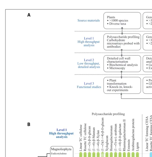
The authors have adopted a multi-level strategy for mapping cell wall polysaccharide and genetic diversity in order to gain insight into underlying evolutionary mechanisms (Figure 1A). The first level consists of primary screens for cell wall polysac-charides based on carbohydrate microarrays probed with mAbs and CBMs (Moller et al., 2007). This level is limited by the availability of characterized mAbs and CBMs and their ability to recognize the numerous epitopes which occur in the vari-ous plant cell wall components. Obvivari-ously the ideal would be to have as much coverage as possible of all the epitopes that exist within cell wall components. However, this is not the current situation and there are some notable wall components, such as RG-II, for which an effective mAb has yet to be gen-erated. In parallel to carbohydrate microarrays, genomes and transcriptomes are mined to identify cell wall-related GTs. The second level of analysis seeks to obtain more detailed informa-tion about certain polysaccharides and genes from subsets of plants. These analyses are performed using established meth-ods for polysaccharide analysis and gene cloning and sequencing. A third level is aimed at obtaining definitive information and the functions of genes, protein, and polysaccharides. In some cases genes are expressed and biochemical activities of GTs deter-mined. Figure 1B shows some preliminary data from primary screens of polysaccharides and cell wall-related genes. The com-bined analysis of data sets can provide insight into the timing and mechanism of certain evolutionary events. For example,
Xue and Fry (2012)have suggested that MLGs are restricted to horsetails based on the results obtained when diverse monilophyte cell walls were treated with lichenase, an enzyme that specifi-cally fragments these (1→3)(1→4)-β-D-glucans. In contrast, we
have obtained evidence for MLGs inSelaginella moellendorffiiand selected Charophycean green algae (CGA) using microarray-based polysaccharide screening and lichenase treatments (Harholt et al., 2012; Fangel and Willats, unpublished).Since a genome sequence is available forS. moellendorffiiit is possible to establish with con-fidence that this plant does not contain orthologs of theCSLFand CSLHgenes (Harholt et al., 2012) that are implicated in the syn-thesis of (1→3)(1→4)-β-D-glucan in Poales species (Burton et al., 2006;Doblin et al., 2009). These data provide good evidence that (1→3)(1→4)-β-D-glucan has evolved at least twice by convergent evolution.
GENOME AND TRANSCRIPTOME MINING FOR CELL WALL-RELATED GLYCOSYL TRANSFERASES
“At least one GT for each glycosidic linkage” is axiomatic for our bioinformatic analysis of cell wall biogenesis. Rare dual function GTs are known from animals and plants, with CslA as an exam-ple of a GT that can utilize two different GDP-sugars as substrate. Accepting the axiom of one GT per glycosidic linkage should per-mit inference of the minimum number of GTs involved in cell wall biosynthesis. However, this is not possible for two reasons. Firstly, the evolution of flowering plants was accompanied by very substantial gene duplication. Differentiation of complex tissues in angiosperms calling for separate regulation in space and time of the same catalytic activity is a likely contributing factor.Mitchell et al. (2007)combined this line of thinking with expression analysis and proposed that certain clades of family GT61 should comprise genes involved in synthesis of a polymer of particular importance in grasses, a prediction that was recently proven correct (Anders et al., 2012). The large repertoire of GTs of the mossPhyscomitrella patens, despite being non-vascular (so having non-lignified tissues only) and also having diverged prior to the gene duplication events associated with the evolution of flowering plants, cautions us not to generalize this principle excessively. The second reason why the number of GTs cannot be inferred from the number of dif-ferent linkages is that the biosynthesis of some polysaccharides has turned out to be much more complicated than anticipated from the polysaccharide structure; xylan biosynthesis, reviewed by Scheller and Ulvskov (2010), provides a striking example. In dicots,
BOX 1 | Text Box entitled CAZy.
Fangel et al. Cell wall evolution and diversity
Polysaccharide profiling Gene/transcript mining
Chlorophyta Charophyta Anthocerotophyta Bryophyta Marchantiophyta Coniferophyta
Cycadophyta Ginkgophyta Gnetophyta
Lycopodiophyta Pteridophyta Magnoliophyta
Eudicotyledons Monocotyledons
Rosette TC
cellulose
(1→4)-β-Mannan (1→4)-β-Xylan
(1→3)-β-D
-glucan
(1→3)(1->4)-β-D
-glucan
Xyloglucan Homogalacturonan (1→4)-β- Rhamnogalacturonan II Extensin
D
-galactan
(1→5)-α-L
-arabinan
Linear TC
cellulose
Arabinogalactan protein Lignin Rosette
TC forming CESA
CALS CSLA CSLC CSLD CSLF CSLH CSLE CSLJ CSLB CSLG
Linear
TC forming CESA
CSLK GAUT RRA GT31
1 1
2 3
4 5 5 5 5 5 6
7 5
8
9 9
10 11
11 11
11 11
12
13 14 Level 1
High throughput analysis
Plants
•>1000 species
•Diverse taxa
Genes
• >30 genomes
• >250 transcriptomes
Level 1
High throughput analysis
Polysaccharide profiling Carbohydrate
microarrays probed with antibodies
Gene/transcript mining
•>30 genomes
•>250 transcriptomes
Level 2
Low throughput, detailed analysis
Detailed cell wall characterisation
•Biochemical analysis
•Microscopy
Level 3
Functional studies
•Plant transformation
•Knock-in, knock-out experiments
•Protein expression
•Glycosyl transferase activity assays Detailed genetic analysis
•Gene sequencing
•Expression studies
Source materials
A
B
FIGURE 1 | (A)A multi-level approach is required to assess the occurrence of cell wall polysaccharides and related genes throughout the plant kingdom. (B)Selected data from primary screens of polysaccharide occurrence in diverse plant species and results from mining genomes and transcriptomes for cell wall-related GTs. The dots at the bottom of the bars indicate the most basal plant group in which that particular polysaccharide or gene sequence has so far been identified. The bars indicate that it is assumed in most cases, once evolved, a gene or polysaccharide persists throughout evolution and is present in later diverging species. However, this is by no means universal. For example, the dashed bar for (1→3)(1→4)-β-D-glucan indicates that the occurrence of this polysaccharide is intermittent throughout the plant kingdom and has arisen by convergent evolution more than once. In some cases the presence of cell wall components is equivocal. For example, lignin
has been tentatively identified in certain Charophycean green algae but the most basal group in which it has been definitively identified is the Lycopodiophyta. A clade of putative ancestral CSLs that are common ancestors to theCSLAs andCSLCs has been identified in certain Chlorophyte algae byYin et al. (2009). These genes share homology with bothCSLAs and
[image:5.842.50.552.58.574.2]GTs from two different families (GT43 and GT47) are implicated in synthesizing the xylan backbone totaling eight GTs for the synthe-sis of one linkage. Pectin biosynthesynthe-sis is predicted to require at least 67 enzymes, GTs plus methyl- and acetyl transferases (Mohnen, 2008). Too few of these have been identified so far to judge whether Nature’s approach to pectin biosynthesis is lean, or expansive as with xylan. These limitations notwithstanding, it has proven fruit-ful to mine genomes for their GT repertoire. This is usually done using the CAZy database as a foundation, SeeBox 1. The CAZy database is the most extensive database of GTs and contains GTs from all three kingdoms. By using a global approach including the whole CAZy database in the screen, more remote orthologies can be discovered, exemplified by the discovery of a mannosylglyc-erate synthase of GT78 inS. moellendorffii(Scheller et al., 2010). But CAZy is not complete and GTs may be found outside CAZy and certain activities are hard to account for within the limit of the present CAZy database (Hansen et al., 2012). The CAZy driven approach has been applied to poplar (Geisler-Lee et al., 2006) and most recently toBrachypodium distachyon(Vogel et al., 2010) and S. moellendorffii(Banks et al., 2011) also leading to a CAZy-based naming convention for putative GTs that can be assigned to a gene family but not to a function.
Applying the CAZy-based classification of putative GTs across large phylogenetic distances can yield evolutionary relevant infor-mation as exemplified by the case for convergent evolution of MLG. CAZy may also be employed as a support for gene discov-ery efforts asEgelund et al. (2007)did using homology between Chlamydomonas and Arabidopsis genes in clade A of family GT77 to infer the function of the Reduced Residual Arabinose (RRA) genes as encoding putative extensin arabinosyl trans-ferases. This annotation is not proven but was corroborated by Velasquez et al. (2011). Extensin along with mannan are to the authors’ knowledge the only known shared cell wall components between Chlorophycean green algae and Arabidopsis(Figure 1;
Estevez et al., 2009).
No CGA has yet been sequenced which is unfortunate given that cell wall analysis strongly suggests that the common ancestor of all plants, with xyloglucan and the pectic polymers typical of vascular plants, was a CGA (Figure 1). A number of EST datasets (Timme and Delwiche, 2010;Wodniok et al., 2011;Timme et al., 2012) are available and may be subjected to the same CAZy-based analysis as full genomes, albeit less safely. Our unpublished observations of this nature, in combination with recent phylogenetic analyses based on genomic data, lead us to propose that genera likePenium andColeochaeterepresent the earliest versions of a higher plant cell wall while species likeChara, which superficially looks more advanced, has diverged substantially from the main evolution-ary path leading to terrestrial plants and hence is a less attractive model for tracing the evolutionary history of the plants cell wall (Timme et al., 2012).
FINE MAPPING OF CELL WALL DIVERSITY AND HETEROGENEITY AT THE CELLULAR AND SUBCELLULAR LEVELS
[image:6.842.307.550.61.439.2]Cellulose is widely distributed in most cell walls but it is clear that the known structural diversity of the polysaccharides of the hemicellulose and pectic groups is regulated both taxonomically and also in relation to cell type and cell wall microstructures within
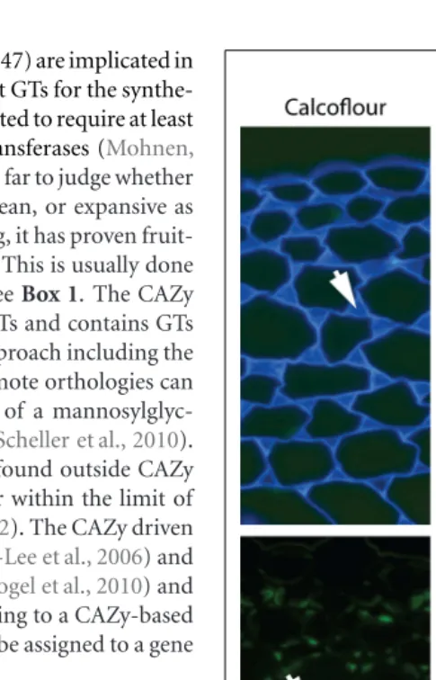
FIGURE 2 | Architectural heterogeneity in primary cell walls.Equivalent transverse sections of the cortical region of tobacco stems with immunofluorescence imaging of two arabinan epitopes (mAbs LM6 and LM13), two xylan epitopes (mAb LM10 and CBM15) and one xyloglucan epitope (mAb LM15). Arrows indicate intercellular matrix shared by adjacent cells. Xylan and xyloglucan epitopes are shown after removal of pectic homogalacturonan. Pectic arabinan, xylan, and xyloglucan structural features all display spatial heterogeneity in relation to cell wall thickening and intercellular regions. mAb, monoclonal antibody; CBM,
carbohydrate-binding module. Scale bar=100µm.
tissues. The use of mAbs and CBMs forin situ analyses of cell walls remains a key approach to determine wall molecular archi-tectures and their heterogeneities (Burton et al., 2010; Pattathil et al., 2010;Lee et al., 2011). The use of the same molecular probes in glycomic analysis is a very useful and complementary activity in which specific oligosaccharide structural features can be stud-ied widely in terms of occurrence and biochemistry in addition to cellular locations.
Fangel et al. Cell wall evolution and diversity
of HG in primary cell walls and (an example from the subcel-lular level) the absence of pectic galactan from pit fields, we do not have a good understanding of the cellular distributions of all polymers for most plant cell types. Even for Arabidop-sisa systematicin situassessment of the major polymers in the cell walls of the major organs has not yet been achieved. Some can be predicted – such as those indicated above but the dis-tributions of RG-I and other pectic epitopes or hemicelluloses cannot yet be predicted with certainty. In situanalyses of cell wall structures are made more complex but also more revealing in that as probe sets for specific polysaccharides are extended more cell wall diversity and heterogeneity can be uncovered. For example, this is the case for pectic HG (Willats et al., 2001; Parre and Geitmann, 2005; Wolf et al., 2009) and also for pec-tic arabinan (Verhertbruggen et al., 2009) where a mAb for linear arabinan detects arabinan substructures in restricted intercellular regions of parenchyma as shown inFigure 2. This also applies to xylan structures in the same parenchyma (Hervé et al., 2009). What is the functional basis for polysaccharide fine structure of both xylans and arabinans being so intimately spatially reg-ulated in relation to factors such as cell adhesion (Figure 2)? Is this to be a paradigm for non-cellulosic wall polymers for which we currently have one or a limited number of probes such as pectic galactan and xyloglucan? The next few years will see more detailed systematic assessments of molecular archi-tectures. This will be in conjunction with enzymatic and/or chemical pre-treatments that are in some cases required to opti-mize polysaccharide detection and in the case of polysaccharide masking (in which one polymer class blocks access to another poly-mer class) indicates important features of cell wall architectures
reflecting protein access (Vreeland et al., 1984;Marcus et al., 2010; Davies et al., 2012).
Clearly we face many challenges in understanding cell wall evo-lution not least of which include sufficient sampling followed by appropriate synthesis and interpretation of large data sets includ-ing diverse information such as gene and protein sequence data as well as sugar linkages and epitope distributions. However, as cell and tissue molecular architectures are documented another major issue that is brought into focus is the function of individ-ual wall components and of the entire cell walls, which can vary enormously with respect to quantitative and qualitative composi-tion. Why do some cell walls have xyloglucan, xylan, and mannan hemicelluloses in distinct spatial distributions as for example in the extensively studied tobacco stem system as shown inFigure 2? How are these heterogeneities integrated into a functional whole in terms of wall properties and functions? In vitroanalyses of composites formed from cellulose, pectins, and xyloglucans have yielded invaluable information about the properties of some wall components (Chanliaud et al., 2002). However, the structural and compositional complexity of naturally occurring cell walls means that anin vitroapproach cannot reasonably be applied to investi-gate the functional properties of the full diversity of extant walls. In vivomethods of investigating wall biomechanics, at the tissue and lower levels, have been developed (Spatz et al., 1998;Burget, 2006) facilitating an improved understanding of how walls are assembled and the fine detail of wall domains and their nanome-chanical properties are likely to emerge within the next few years as detailedin situanalyses are combined with genetic and enzy-matic interventions. Integrating this knowledge will be a major challenge and is an exciting frontier for cell wall biology.
REFERENCES
Abercrombie, J. M., O’Meara, B. C., Moffatt, A. R., and Williams, J. H. (2011). Developmental evolution of flowering plant pollen tube cell walls: callose synthase (CalS) gene expres-sion patterns.EvoDevo2, 14. Anders, N., Wilkinson, M. D.,
Love-grove, A., Freeman, J., Tryfona, T., Pellny, T. K., Weimar, T., Mortimer, J. C., Stott, K., Baker, J. M., Defoin-Platel, M., Shewry, P. R., Dupree, P., and Mitchell, R. A. C. (2012). Glyco-syl transferases in family 61 mediate arabinofuranosyl transfer onto xylan in grasses. Proc. Natl. Acad. Sci. U.S.A.17, 989–993.
Bacic, A., Harris, A. J., and Stone, B. A. (1988). “Structure and function of plant cell walls,” in
The Biochemistry of Plants, ed J. Preiss (New York: Academic Press), 297–371.
Banks, J. A., Nishiyama, T., Hasebe, M., Bowman, J. L., Gribskov, M., dePam-philis, C., Albert, V. A., Aono, N., Aoyama, T., Ambrose, B. A., Ashton, N. W., Axtell, M. J., Barker, E., Barker, M. S., Bennetzen, J. L., Bonawitz, N. D., Chapple, C., Cheng, C., Correa, L. G., Dacre, M., DeBarry, J., Dreyer,
I., Elias, M., Engstrom, E. M., Estelle, M., Feng, L., Finet, C., Floyd, S. K., Frommer, W. B., Fujita, T., Gramzow, L., Gutensohn, M., Harholt, J., Hat-tori, M., Heyl, A., Hirai, T., Hiwatashi, Y., Ishikawa, M., Iwata, M., Karol, K. G., Koehler, B., Kolukisaoglu, U., Kubo, M., Kurata, T., Lalonde, S., Li, K., Li, Y., Litt, A., Lyons, E., Man-ning, G., Maruyama, T., Michael, T. P., Mikami, K., Miyazaki, S., Mori-naga, S., Murata, T., Mueller-Roeber, B., Nelson, D. R., Obara, M., Oguri, Y., Olmstead, R. G., Onodera, N., Petersen, B. L., Pils, B., Prigge, M., Rensing, S. A., Riaño-Pachón, D. M., Roberts, A. W., Sato, Y., Scheller, H. V., Schulz, B., Schulz, C., Shakirov, E. V., Shibagaki, N., Shinohara, N., Ship-pen, D. E., Sørensen, I., Sotooka, R., Sugimoto, N., Sugita, M., Sumikawa, N., Tanurdzic, M., Theissen, G., Ulvskov, P., Wakazuki, S., Weng, J. K., Willats, W. W., Wipf, D., Wolf, P. G., Yang, L., Zimmer, A. D., Zhu, Q., Mitros, T., Hellsten, U., Loqué, D., Otillar, R., Salamov, A., Schmutz, J., Shapiro, H., Lindquist, E., Lucas, S., Rokhsar, D., and Grigoriev, I. V. (2011). The Selaginellagenome identifies genetic changes associated
with the evolution of vascular plants.
Science332, 960–963.
Boerjan, W., Ralph, J., and Baucher, M. (2003). Lignin biosynthesis. Annu. Rev. Plant. Biol.54, 519–546. Burget, I. (2006). Exploring the
micromechanical design of plant cell walls.Am. J. Bot.93, 1391–1401. Burton, R. A., Wilson, S. M., Hrmova,
M., Harvey, A. J., Shirley, N. J., Medhurst, A., Stone, B. A., New-bigin, E. J., Bacic, A., and Fincher, G. B. (2006). Cellulose synthase-like CslF genes mediate the synthe-sis of cell wall (1,3;1,4)-β-D-glucans.
Science311, 1940–1942.
Burton, R. A., Gidley, M. J., and Fincher, G. B. (2010). Heterogeneity in the chemistry, structure and function of plant cell walls.Nat. Chem. Biol.6, 724–732.
Caffall, K. H., and Mohnen, D. (2009). The structure, function, and biosynthesis of plant cell wall pectic polysaccharides.Carbohydr. Res.344, 1879–1900.
Cantarel, B. L., Coutinho, P. M., Rancurel, C., Bernard, T., Lom-bard, V., and, Henrissat, B. (2009). The Carbohydrate-Active EnZymes database (CAZy): an expert resource
for glycogenomics.Nucleic Acids Res.
37, 233–238.
Carpita, N. C., and Gibeaut, D. M. (1993). Structural models of primary cell walls in flowering plants: con-sistency of molecular structure with the physical properties of the walls during growth.Plant J.3, 1–30. Chanliaud, E., Burrows, K. M.,
Jeron-imidis, G., and Gidley, M. J. (2002). Mechanical properties of primary cell wall analogues.Planta215, 989–996. Cosgrove, D. (2005). Growth of the plant cell wall.Nat. Rev. Mol. Cell. Biol.6, 850–861.
Davies, L. J., Lilley, C. J., Knox, J. P., and Urwin, P. E. (2012). Syncytia formed by adult femaleHeterodera schachtii
onArabidopsis thalianaroots have a distinct cell wall molecular architec-ture.New Phytol. (in press). Dhugga, K. S. (2012). Biosynthesis
of non-cellulosic polysaccharides of plant cell walls. Phytochemistry 74, 8–19.
transgenic Arabidopsis. Proc. Natl. Acad. Sci. U.S.A.106, 5996–6001. Domozych, D. S., Sørensen, I., and
Willats, W. G. T. (2009). The distri-bution of cell wall polymers during antheridium development and sper-matogenesis in the Charophycean green alga,Chara corallina.Ann. Bot.
104, 1045–1056.
Egelund, J., Obel, N., Ulvskov, P., Geshi, N., Pauly, M., Bacic, A., and Petersen, B. L. (2007). Molecular character-ization of twoArabidopsis thaliana
glycosyltransferase mutants, rra1 and rra2, which have a reduced resid-ual arabinose content in a polymer tightly associated with the cellulosic wall residue. Plant Mol. Biol. 64, 439–451.
Endler, A., and Persson, S. (2011). Cellulose synthases and synthesis in
Arabidopsis.Mol. Plant4, 199–211. Estevez, J. M., Fernández, P. V., Kasulin,
L., Dupree, P., and Ciancia, M. (2009). Chemical andin situ char-acterization of macromolecular com-ponents of the cell walls from the green seaweedCodium fragile. Glyco-biology19, 212–228.
Feingold, D. S., and Avigad, G. (1980). “Sugar nucleotide transformations in plants,” inThe Biochemistry of Plants: A Comprehensive Treatise, eds P. K. Stumpf and E. E. Conn (New York: Academic Press), 101–170. Fry, S. C. (2001).The Growing Plant Cell
Wall: Chemical and Metabolic Analy-sis, Reprint edition. New Jersey: The Blackburn Press.
Fry, S. C. (2004). Primary cell wall metabolism: tracking the careers of wall polymers in living plant cells.
New Phytol.161, 641–675. Fry, S. C., Mohler, K. E., Nesselrode, B.
H. W. A., and Franková, L. (2008). Mixed-linkageβ-glucan: xyloglucan endotransglucosylase, a novel wall-remodelling enzyme from Equisetum (horsetails) and charophytic algae.
Plant J.55, 240–252.
Geisler-Lee, J., Geisler, M., Coutinho, P. M., Segerman, B., Nishikubo, N., Takahashi, J., Aspeborg, H., Djerbi, S., Master, E., Andersson-Gunneras, S., Sundberg, B., Karpinski, S., Teeri, T. T., Kleczkowski, L. A., Henris-sat, B., and Mellerowicz, E. J. (2006). Poplar carbohydrate-active enzymes. Gene identification and expres-sion analyses. Plant Physiol. 140, 946–962.
Goubet, F., Jackson, P., Deery, M. J., and Dupree, P. (2002). Polysaccharide analysis using carbohydrate gel elec-trophoresis: a method to study plant cell wall polysaccharides and polysac-charide hydrolases. Anal. Biochem.
300, 53–68.
Hansen, S. F., Harholt, J., Oikawa, A., and Scheller, H. V. (2012). Plant glycosyltransferases beyond CAZy: a perspective on DUF fam-ilies. Front. Plant Sci. 3:59. doi: 10.3389/fpls.2012.00059
Harholt, J., Suttangkakul, A., and Vibe Scheller, H. (2010). Biosynthesis of pectin.Plant Physiol.153, 384–395. Harholt, J., Sørensen, I., Fangel, J.
U., Roberts, A., Willats, W. G. T., Scheller, H. V., Bent Larsen Petersen, B. L., Banks, J. A., and Ulvskov, P. (2012). The glycosyl-transferase repertoire of the spike-mossSelaginella moellendorffiiand a comparative study of the cell wall structure. PLoS ONE 7, e35846. doi:10.1371/journal.pone.0035846 Hepler, P. K., Fosket, D. E., and
New-comb, E. H. (1970). Lignification during secondary wall formation in
Coleus: an electron microscopy study.
Am. J. Bot.57, 85–96.
Hervé, C., Rogowski, A., Gilbert, H. J., and Knox, J. P. (2009). Enzy-matic treatments reveal differential capacities for xylan recognition and degradation in primary and sec-ondary plant cell walls.Plant J.58, 413–422.
Iraki, N. M., Bressan, R. A., Hasegawa, P. M., and Carpita, N. C. (1989a). Alteration of the physical and chem-ical structure of the primary cell wall of growth-limited plant cells adapted to osmotic stress.Plant Physiol. 91, 39–47.
Iraki, N. M., Singh, N., Bressan, R. A., and Carpita, N. C. (1989b). Cell walls of tobacco cells and changes in composition associated with reduced growth upon adaptation to water and saline stress.Plant Physiol.91, 48–53. Iraki, N. M., Bressan, R. A., and Carpita, N. C. (1989c). Extracellular polysac-charides and proteins of tobacco cell cultures and changes in composi-tion associated with growth-limiting adaptation to water and saline stress.
Plant Physiol.91, 54–61.
Kenrick, P., and Crane, P. R. (1997). The origin and early evolution of plants on land.Nature389, 33–39. Lee, K. J. D., Marcus, S. E., and Knox, J.
P. (2011). Plant cell wall biology: per-spectives from cell wall imaging.Mol. Plant4, 212–219.
Lerouxel, O., Choo, T. S., Seveno, M., Usadel, B., Faye, L., Lerouge, P., and Pauly, M. (2002). Rapid structural phenotyping of plant cell wall mutants by enzymatic oligosac-charide fingerprinting.Plant Physiol.
130, 1754–1763.
Mackie, W., and Sto, R. D. P. R. (1968). The occurrence of mannan microfib-rils in the green algaeCodium fragile
and Acetabularia crenulata. Planta
253, 249–253.
Marcus, S. E., Blake, A. W., Benians, T. A. S., Lee, K. J. D., Poyser, C., Don-aldson, L., Leroux, O., Rogowski, A., Petersen, H. L., Boraston, A., Gilbert, H. J., Willats, W. G. T., and Knox, J. P. (2010). Restricted access of proteins to mannan polysaccharides in intact plant cell walls.Plant J.64, 191–203. Matsunaga, T., Ishii, T., Matsumoto,
S., Higuchi, M., Darvill, A., Alber-sheim, P., and O’Neill, M. A. (2004). Occurrence of the primary cell wall polysaccharide rhamnogalacturonan II in pteridophytes, lycophytes, and bryophytes: implications for the evo-lution of vascular plants.Plant Phys-iol.134, 339–351.
Mitchell, R. A., Dupree, P., and Shewry, P. R. (2007). A novel bioinformatics approach identifies candidate genes for the synthesis and feruloylation of arabinoxylan.Plant Physiol. 144, 43–53.
Mohnen, D. (2008). Pectin structure and biosynthesis.Curr. Opin. Plant. Biol.11, 266–277.
Moller, I., Sørensen, I., Bernal, A. J., Blaukopf, C., Lee, K., Øbro, J., Pet-tolino, F., Roberts, A., Mikkelsen, J. D., Knox, J. P., Bacic, A., and Willats, W. G. T. (2007). High-throughput mapping of cell-wall polymers within and between plants using novel microarrays.Plant J.50, 1118–1128. Mouille, G., Robin, S., Lecomte, M.,
Pagant, S., and Höfte, H. (2003). Classification and identification of Arabidopsis cell wall mutants using Fourier-Transform InfraRed (FT-IR) microspectroscopy. Plant J.35, 393–404.
Niklas, K. J. (2004). The cell walls that bind the tree of life. BioScience54, 831–841.
Niklas, K. J., and Kutschera, U. (2010). The evolution of the land plant life cycle.New Phytol.185, 27–41. Parre, E., and Geitmann, A. (2005).
Pectin and the role of the physical properties of the cell-wall in pollen tube growth ofSolanum chacoense.
Planta220, 582–592.
Park, S., Szumlanski, A. L., Gu, F., Guo, F., and Nielsen, E. (2011). A role for CSLD3 during cell-wall synthe-sis in apical plasma membranes of tip-growing root-hair cells.Nat. Cell. Biol.13, 973– 980.
Pattathil, S., Avci, U., Baldwin, D., Swennes, A. G., McGill, J. A., Pop-per, Z., Bootten, T., Albert, A., Davis, R. H., Chennareddy, C., Dong, R., O’Shea, B., Rossi, R., Leoff, C., Freshour, G., Narra, R., O’Neil, M., York, W. S., and Hahn, M. G. (2010). A comprehensive toolkit of
plant cell wall glycan-directed mono-clonal antibodies.Plant Physiol.153, 514–525.
Pauly, M., and Keegstra, K. (2010). Plant cell wall polymers as precursors for biofuels.Curr. Opin. Plant Biol.13, 305–312.
Popper, Z. A. (2008). Evolution and diversity of green plant cell walls.
Curr. Opin. Plant Biol.11, 286–292. Popper, Z. A., and Tuohy, M. G. (2010).
Beyond the green: understanding the evolutionary puzzle of plant and algal cell walls.Plant Physiol.153, 373–383. Popper, Z. A., Michel, G., Hervé, C., Domozych, D. S., Willats, W. G., Tuohy, M. G., Kloareg, B., and Sten-gel, D. B. (2011). Evolution and diver-sity of plant cell walls: from algae to flowering plants.Annu. Rev. Plant. Biol.62, 567–590.
Qu, Y., Egelund, J., Gilson, P. R., Houghton, F., Gleeson, P. A., Schultz, C. J., and Bacic, A. (2004). Identi-fication of a novel group of puta-tive Arabidopsis thaliana β -(1,3)-galactosyltransferases. Plant Mol. Biol.68, 43–59.
Roberts, A. W., Roberts, E. M., and Delmer, D. P. (2002). Cellulose Syn-thase (CesA) Genes in the green alga
Mesotaenium caldariorum.Eukaryot. Cell1, 847–855.
Sarkar, P., Bosneaga, E., and Auer, M. (2009). Plant cell walls through-out evolution: towards a molecular understanding of their design princi-ples.J. Exp. Bot.60, 3615–3635. Scheible, W. R., and Pauly, M.
(2004). Glycosyltransferases and cell wall biosynthesis: novel players and insights.Curr. Opin. Plant. Biol.7, 285–295.
Scheller, H. V., and Ulvskov, P. (2010.) Hemicelluloses. Annu. Rev. Plant. Biol.61, 263–289.
Scheller, H. V., Ulvskov, P., and Harholt, J. (2010). Stress-tolerant plants expr-essing mannosylglycerate-producing enzymes. US National Stage Patent Application No. 13/318,661. WIPO Patent WO/2010/129574A1. Avail-able at: http://www.sumobrain.com/ patents/wipo/Stress-tolerant-plants-expressing-mannosylglycerate/ WO2010129574A1.pdf
Scherp, P., Grotha, R., and Kutschera, U. (2001). Occurrence and phylogenetic significance of cytokinesis-related callose in green algae, bryophytes, ferns and seed plants. Plant Cell Reports20, 143–149.
Sørensen, I., Domozych, D., and Willats, W. G. (2010). How have plant cell walls evolved? Plant Physiol. 153, 366–372.
Fangel et al. Cell wall evolution and diversity
possibilities for pectin. Carbohydr. Res.344, 1872–1878.
Sørensen, I., Pettolino, F. A, Bacic, A., Ralph, J., Lu, F., O’Neill, M. A, Fei, Z., Rose, J. K. C., Domozych, D. S., and Willats, W. G. T. (2011). The charo-phycean green algae provide insights into the early origins of plant cell walls.Plant J.68, 201–211. Spatz, H.-C., Rowe, N., Speck, T.,
and Daviero, V. (1998). Biomechan-ics of hollow stemmed Sphenopsids: II. Calamites – to have or not to have secondary xylem.Rev. Palaeobot. Palynol.102, 63–77.
Timme, R. E., and Delwiche, C. F. (2010). Uncovring the evolutionary origin of plant molecular processes: comparison of Coleochaete (Coleo-chaetales) and Spirogyra (Zygne-matales) transcriptomes.BMC Plant Biol.10, 96. doi: 10.1186/1471-2229-10-96
Timme, R. E., Bachvaroff, T. R., and Del-wiche, C. F. (2012). Broad phyloge-nomic sampling and the sister lineage of land plants.PLoS ONE7, e29696. doi:10.1371/journal.pone.0029696 Tsekos, I. (1999). The sites of
cellu-lose synthesis in algae: diversity and evolution of cellulose-synthesizing enzyme complexes. J. Phycol. 35, 635–655.
Velasquez, S. M., Ricardi, M. M., Dorosz, J. G., Fernandez, P. V., Nadra, A. D., Pol-Fachin, L., Egelund, J., Gille, S., Harholt, J., Ciancia, M., Verli, H., Pauly, M., Bacic, A., Olsen, C. E., Ulvskov, P., Petersen, B. L, Somerville, C., Iusem, N. D., and Estevez, J. M.(2011).O-Glycosylated cell wall proteins are essential in root hair growth.Science332, 1401–1403. Verhertbruggen, Y., Marcus, S. E., Haeger, A., Verhoef, R., Schols, H. A., McCleary, B. V., McKee, L., Gilbert, H. J., and Knox, J. P. (2009). Developmental complexity of arabi-nan polysaccharides and their pro-cessing in plant cell walls.Plant J.59, 413–425.
Vogel, J. P., Garvin, D. F., Mock-ler, T. C., Schmutz, J., Rokhsar, D., Bevan, M. W., Barry, K., Lucas, S., Harmon-Smith, M., Lail, K., Tice, H., Schmutz, J., Grimwood, J., McKenzie, N., Bevan, M. W., Huo, N., Gu, Y. Q., Lazo, G. R., Anderson, O. D., Vogel, J. P., You, F. M., Luo, M. C., Dvorak, J., Wright, J., Febrer, M., Bevan, M. W., Idziak, D., Hasterok, R., Garvin, D. F., Lindquist, E., Wang, M., Fox, S. E., Priest, H. D., Filichkin, S. A., Givan, S. A., Bryant, D. W., Chang, J. H., Mockler, T. C., Wu, H., Wu, W., Hsia, A. P., Schnable, P. S., Kalya-naraman, A., Barbazuk, B., Michael, T. P., Hazen, S. P., Bragg, J. N., Laudencia-Chingcuanco, D., Vogel, J. P., Garvin, D. F., Weng, Y., McKenzie, N., Bevan, M. W., Haberer, G., Span-nagl, M., Mayer, K., Rattei, T., Mitros, T., Rokhsar, D., Lee, S. J., Rose, J. K., Mueller, L. A., York, T. L., Wicker, T., Buchmann, J. P., Tanskanen, J., Schul-man, A. H., Gundlach, H., Wright, J., Bevan, M., de Oliveira, A. C., Maia Lda, C., Belknap, W., Gu, Y. Q., Jiang, N., Lai. J., Zhu, L., Ma, J., Sun, C., Pritham, E., Salse, J., Murat, F., Abrouk, M., Haberer. G., Spannagl. M., Mayer, K., Bruggmann, R., Mess-ing, J., You, F. M., Luo, M. C., Dvorak, J., Fahlgren, N., Fox, S. E., Sullivan, C. M., Mockler, T. C., Carrington, J. C., Chapman, E. J., May, G. D., Zhai, J., Ganssmann, M., Gurazada, S. G., German, M., Meyers, B. C., Green, P. J., Bragg, J. N., Tyler, L., Wu, J., Gu, Y. Q., Lazo, G. R., Laudencia-Chingcuanco, D., Thomson, J., Vogel, J. P., Hazen, S. P., Chen, S., Scheller, H. V., Harholt, J., Ulvskov, P., Fox, S. E., Filichkin, S. A., Fahlgren, N., Kimbrel, J. A., Chang, J. H., Sullivan, C. M., Chapman, E. J., Carrington, J. C., Mockler, T. C., Bartley, L. E., Cao, P., Jung, K. H., Sharma, M. K., Vega-Sanchez, M., Ronald, P., Dard-ick, C. D., De Bodt, S., Verelst, W., Inzé, D., Heese, M., Schnittger, A., Yang, X., Kalluri, U. C., Tuskan, G. A.,
Hua, Z., Vierstra, R. D., Garvin, D. F., Cui, Y., Ouyang, S., Sun, Q., Liu, Z., Yilmaz, A., Grotewold, E., Sibout, R., Hematy, K., Mouille, G., Höfte, H., Michael, T., Pelloux, J., O’Connor, D., Schnable, J., Rowe, S., Harmon, F., Cass, C. L., Sedbrook, J. C., Byrne, M. E., Walsh, S., Higgins, J., Bevan, M., Li, P., Brutnell, T., Unver, T., Budak, H., Belcram, H., Charles, M., Chalhoub, B., and Baxter, I. (2010). Genome sequencing and analysis of the model grassBrachypodium dis-tachyon.Nature463, 763–768. Vreeland, V., Slomich, M., and Laetsch,
W. M. (1984). Monoclonal antibodies as molecular probes for cell wall anti-gens of the brown algaFucus.Planta
163, 506–517.
Waters, E. R. (2003). Molecular adap-tation and the origin of land plants.
Mol. Phylogenet. Evol.29, 456–463. Weng, J.-K., Li, X., Stout, J., and
Chap-ple, C. (2008). Independent origins of syringyl lignin in vascular plants.
Proc. Nat. Acad. Sci. U.S.A. 105, 7887–7892.
Willats, W. G. T., Knox, J. P., and Mikkelsen, J. D. (2006). Pectin: new insights into an old polymer are start-ing to gel.Trends Food Sci. Technol.
17, 97–104.
Willats, W. G. T., Orfila, C., Limberg, G., Buchholt, H. C., van Alebeek, G.-J. W. M., Voragen, A. G. J., Marcus, S. E., Christensen, T. M. I. E., Mikkelsen, J. D., Murray, B. S., and Knox, J. P. (2001). Modulation of the degree and pattern of methyl-esterification of pectic homogalacturonan in plant cell walls: implications for pectin methyl esterase action, matrix prop-erties and cell adhesion.J. Biol. Chem.
276, 19404–19413.
Wodniok, S., Brinkmann, H., Glöck-ner, G., Heidel, A. J., Philippe, H., Melkonian, M., and Becker, B. (2011). Origin of land plants: do conjugat-ing green algae hold the key?BMC Evol. Biol.11, 104. doi: 10.1186/1471-2148-11-104
Wolf, S., Mouille, G., and Pelloux, J. (2009). Homogalacturonan methyl-esterification and plant development.
Mol. Plant2, 851–860.
Xue, X., and Fry, S. C. (2012). Evolu-tion of mixed-linkage (1→3, 1→4)-β-D-glucan (MLG) and xyloglu-can in Equisetum (horsetails) and other monilophytes.Ann. Bot.109, 873–886.
Yin, Y., Chen, H., Hahn, M. G., Mohnen, D., and Xu, Y. (2010). Evo-lution and function of the plant cell wall synthesis-related glycosyltrans-ferase family 8. Plant Physiol. 153, 1729–1746.
Yin, Y., Huang, J., and Xu, Y. (2009). The cellulose synthase superfamily in fully sequenced plants and algae. BMC Plant Biol. 9, 99. doi: 10.1186/1471-2229-9-99
Conflict of Interest Statement: The authors declare that the research was conducted in the absence of any com-mercial or financial relationships that could be construed as a potential con-flict of interest.
Received: 20 March 2012; accepted: 18 June 2012; published online: 06 July 2012.
Citation: Fangel JU, Ulvskov P, Knox JP, Mikkelsen MD, Harholt J, Popper ZA and Willats WGT (2012) Cell wall evolution and diversity. Front. Plant Sci.3:152. doi:
10.3389/fpls.2012.00152
This article was submitted to Frontiers in Plant Physiology, a specialty of Frontiers in Plant Science.