DNA mismatch repair gene
MSH6
implicated
in determining age at natural menopause
John R.B. Perry
1,2,3,4,{,∗, Yi-Hsiang Hsu
5,6,{, Daniel I. Chasman
7,8, Andrew D. Johnson
9,10,
Cathy Elks
4, Eva Albrecht
11, Irene L. Andrulis
14,15, Jonathan Beesley
16, Gerald S. Berenson
17,
Sven Bergmann
18,19, Stig E. Bojesen
20, Manjeet K. Bolla
22, Judith Brown
22, Julie E. Buring
7,8,
Harry Campbell
24, Jenny Chang-Claude
25, Georgia Chenevix-Trench
16, Tanguy Corre
18,19,
Fergus J. Couch
26, Angela Cox
28, Kamila Czene
29, Adamo Pio D’adamo
31, Gail Davies
32,33,34,
Ian J. Deary
32,33, Joe Dennis
22, Douglas F. Easton
22, Ellen G. Engelhardt
35,
Johan G. Eriksson
37,38,39,40,41, To
˜ nu Esko
42,43,44, Peter A. Fasching
45, Jonine D. Figueroa
46,
Henrik Flyger
21, Abigail Fraser
47, Montse Garcia-Closas
48, Paolo Gasparini
31, Christian Gieger
11,
Graham Giles
49,50, Pascal Guenel
51, Sara Ha¨gg
29,52, Per Hall
29, Caroline Hayward
53, John Hopper
49,
Erik Ingelsson
52, kConFab investigators
54, Sharon L.R. Kardia
55, Katherine Kasiman
29,
Julia A. Knight
14, Jari Lahti
39,56, Debbie A. Lawlor
47, Patrik K.E. Magnusson
29, Sara Margolin
30,
Julie A. Marsh
57, Andres Metspalu
44, Janet E. Olson
27, Craig E. Pennell
57, Ozren Polasek
58,
Iffat Rahman
29, Paul M. Ridker
7,8, Antonietta Robino
31, Igor Rudan
24, Anja Rudolph
25,
Andres Salumets
59,60, Marjanka K. Schmidt
35,36, Minouk J. Schoemaker
48, Erin N. Smith
61,
Jennifer A. Smith
55, Melissa Southey
62, Doris Sto
¨ ckl
12,63, Anthony J. Swerdlow
48,
Deborah J. Thompson
22, Therese Truong
51, Sheila Ulivi
64, Melanie Waldenberger
13, Qin Wang
22,
Sarah Wild
24, James F Wilson
24, Alan F. Wright
34, Lina Zgaga
24, ReproGen Consortium,
Ken K. Ong
4,23, Joanne M. Murabito
9,65, David Karasik
5and Anna Murray
1,∗1
University of Exeter Medical School, Exeter, UK,2Wellcome Trust Centre for Human Genetics, University of Oxford, Oxford, UK,3Department of Twin Research and Genetic Epidemiology, King’s College London, London, UK,4Medical Research Council (MRC) Epidemiology Unit, Institute of Metabolic Science, Addenbrooke’s Hospital, Cambridge, UK,
5
Hebrew SeniorLife Institute for Aging Research and Harvard Medical School, Boston, MA, USA,6Molecular and Integrative Physiological Sciences Program, Harvard School of Public Health, Boston, MA, USA,7Division of Preventive Medicine, Brigham and Women’s Hospital, 900 Commonwealth Avenue East, Boston MA 02215, USA,8Harvard Medical School, Boston, MA, USA,9The National Heart Lung and Blood Institute’s Framingham Heart Study, Framingham, MA, USA,10NHLBI Cardiovascular Epidemiology & Human Genomics Branch, Bethesda, MD, USA,11Institute of Genetic Epidemiology,12Institute of Epidemiology II and13Research Unit of Molecular Epidemiology, Helmholtz Zentrum Mu¨nchen – German Research Center for Environmental Health, Neuherberg, Germany,14Lunenfeld-Tanenbaum Research Institute, Mount Sinai Hospital, Toronto, ON, Canada,15Department of Molecular Genetics, University of Toronto, Toronto, ON, Canada,16Department of Genetics, Queensland Institute of Medical Research, Brisbane, QLD, Australia,17Department of Epidemiology, Tulane University, New Orleans, LA, USA,18Department of Medical Genetics, University of Lausanne, Lausanne, Switzerland,19Swiss Institute of Bioinformatics, Lausanne, Switzerland,
20
Department of Clinical Biochemistry and the Copenhagen General Population Study, Herlev Hospital, Copenhagen University Hospital, Copenhagen, Denmark,21Department of Breast Surgery, Herlev Hospital, Copenhagen University
†These authors contributed equally to the work.
∗To whom correspondence should be addressed. Email: john.perry@mrc-epid.cam.ac.uk (J.R.B.P.), A.Murray@exeter.ac.uk (A.M.)
#The Author 2013. Published by Oxford University Press.
This is an Open Access article distributed under the terms of the Creative Commons Attribution License (http://creativecommons.org/licenses/by/3.0/), which permits unrestricted reuse, distribution, and reproduction in any medium, provided the original work is properly cited.
Hospital, Copenhagen, Denmark, 22Centre for Cancer Genetic Epidemiology and Department of Public Health and Primary Care, University of Cambridge, Cambridge, UK,23Department of Paediatrics, University of Cambridge, Cambridge, UK,24Centre for Population Health Sciences, University of Edinburgh, Edinburgh EH8 9AG, UK,25Division of Cancer Epidemiology, German Cancer Research Center, Heidelberg, Germany,26Departments of Laboratory Medicine and Pathology, and Health Science Research,27Department of Health Sciences Research, Mayo Clinic, Rochester, MN, USA,28CR-UK/YCR Sheffield Cancer Research Centre, Department of Oncology, University of Sheffield, UK,29Medical Epidemiology and Biostatistics, Karolinska Institutet, Stockholm, Sweden,30Department of Oncology-Pathology, Karolinska Institutet, Stockholm, Sweden,31Institute for Maternal and Child Health, IRCCS ‘Burlo Garofolo’, University of Trieste, Trieste, Italy,32Centre for Cognitive Ageing and Cognitive Epidemiology, 33Department of Psychology and
34
MRC Human Genetics Unit, Institute of Genetics and Molecular Medicine, University of Edinburgh, Edinburgh, UK,
35
Division of Psychosocial Research and Epidemiology and 36Division of Molecular Pathology, The Netherlands Cancer Institute, Amsterdam, The Netherlands,37Department of General Practice and Primary Health Care, University of Helsinki, Helsinki, Finland,38National Institute for Health and Welfare, Helsinki, Finland,39Folkha¨lsan Research Centre, Helsinki, Finland,40University Central Hospital, Unit of General Practice, Helsinki, Finland,41Vasa Central Hospital, Vasa, Finland,42Divisions of Endocrinology, Children’s Hospital, Boston, MA, USA,43Broad Institute, Cambridge, MA, USA,
44
Estonian Genome Center, University of Tartu, 51010 Tartu, Estonia,45Department of Gynecology and Obstetrics, University Hospital Erlangen, Friedrich-Alexander University Erlangen-Nuremberg, Erlangen, Germany,46Division of Cancer Epidemiology & Genetics, National Cancer Institute, Maryland, USA,47School of Social and Community Medicine, MRC Centre for Causal Analyses in Translational Epidemiology, University of Bristol, Bristol, UK,48Divisions of Breast Cancer Research and of Genetics and Epidemiology, and the Breakthrough Breast Cancer Research Centre, The Institute of Cancer Research, London, UK,49Centre for Molecular, Environmental, Genetic and Analytic Epidemiology, Melbourne School of Population Health, The University of Melbourne, Melbourne, VIC, Australia,50Cancer Epidemiology Centre, The Cancer Council Victoria, Melbourne, VIC, Australia,51Environmental Epidemiology of Cancer, Inserm U1018, Villejuif, France,52Department of Medical Sciences, Molecular Epidemiology and Science for Life Laboratory, Uppsala University, Uppsala, Sweden,53Institute of Genetics and Molecular Medicine, University of Edinburgh, Western General Hospital, Edinburgh EH4 2XU, UK,54Peter MacCallum Cancer Centre, Melbourne, VIC, Australia,55Department of Epidemiology, University of Michigan, Ann Arbor, MI, USA,56Institute of Behavioural Science, University of Helsinki, Helsinki, Finland,57School of Women’s and Infants’ Health, University of Western Australia, Australia,58Department of Public Health, Faculty of Medicine, University of Split, Croatia,59Department of Obstetrics and Gynecology, University of Tartu, 51014 Tartu, Estonia,60Competence Centre on Reproductive Medicine and Biology, 50410 Tartu, Estonia,
61
Department of Pediatrics and Rady Children’s Hospital, University of California San Diego, La Jolla, CA 92093, USA,
62
Genetic Epidemiology Laboratory, Department of Pathology, The University of Melbourne, Melbourne, VIC, Australia,
63
Department of Obstetrics and Gynaecology, Campus Grosshadern, Ludwig-Maximilians-University, Munich,
Germany,64Institute for Maternal and Child Health, IRCCS ‘Burlo Garofolo’, Trieste, Italy,65Section of General Internal Medicine, Department of Medicine, Boston University School of Medicine, Boston, MA, USA
Received August 23, 2013; Revised November 19, 2013; Accepted December 6, 2013
INTRODUCTION
Female reproductive lifespan starts just prior to menarche, the onset of the first menstrual bleed and finishes when oocyte supply becomes exhausted at menopause. Both processes are governed by genetic and non-genetic factors and the timing of these events is associated with multiple adverse health out-comes, including breast cancer, cardiovascular disease, osteo-porotic fractures and infertility (1). Recent genome-wide association studies (GWAS) have identified 32 loci involved in age at menarche (2) and 17 with age at natural menopause (3): however none of the variants identified to date overlap. Epi-demiological studies also do not strongly support a role for over-lapping aetiology in the processes governing timing of menarche and menopause (4–7). Age at menarche has decreased signifi-cantly in recent history and this has been thought to be largely due to increased levels of childhood obesity (8–10). The role of adiposity in regulating menarche timing is supported by genetic studies which have reported that many genes involved in the regulation of fat mass are also associated with timing of menarche (11–13). Secular changes in menopause age are more controversial with individual studies reporting conflicting findings (14,15). The correlation between ages at menopause and menarche is also controversial, but larger studies show a modest correlation between the two phenotypes (16) and as both events involve the same organ system, it is conceivable that there are common physiological processes involved, which may be influ-enced by genetic and environmental factors.
The length of reproductive lifespan has been associated with several adverse health outcomes, particularly breast cancer. Genes involved in regulating reproductive lifespan in humans have not been described to date. There are two ways in which reproductive lifespan can be altered: either total length, or it can be temporally shifted, either earlier or later. These shifts in lifespan would not be detected if the outcome measured was the length of reproductive lifespan. It is possible that both menarche and menopause could occur early, yet the time period between the two events could be normal. In order to capture the features of this phenotype and investigate the under-lying genetic aetiology, we used a bivariate GWAS method to identify genetic loci associated with both age at menopause and menarche in either direction. This study incorporated
GWAS data from theReproGenconsortium meta-analyses of
87 802 individuals for menarche and 38 968 individuals for menopause (2,17).
RESULTS
Genetic correlation between traits
We performed a restricted maximum likelihood (REML) bivari-ate analysis (18,19) within theWomen’s Genome Health Study
(total sampleN¼21 505) to test for genome-wide genetic cor-relation between timing of menarche and menopause. Using 329 966 autosomal SNPs we observed a positive correlation of
rgenetic¼0.138 (P¼0.04, s.e 0.068). This result remained similar after adjustment for the top 10 principal components of population stratification.
Bivariate meta-analysis
The bivariate meta-analysis for menarche and menopause generated two signals with genome-wide significantP values
,5×1028 and a further four independent signals with P
values,1×1027(Table1). We assessed the association with each of the individual traits of the top bivariate signal plus SNPs in linkage disequilibrium (LD) with the best SNP. Of the six top hits, for four signals either the top SNP, or a SNP in LD with the top SNP (hapmapr2.0.05), had been previously iden-tified in the GWAS for one of the traits individually. The stron-gest bivariate signal was nearGAB2, a known locus for menarche
(2) and SNPs nearFSHB,SYCP2LandPRRC2Awere known
loci for menopause (3). There were two signals nearRPAIN
andMSH6, which had not been previously reported for either trait and hadPvalues of 1×1027and 3×1027, respectively in the bivariate analysis. We did not have sufficiently robust stat-istical evidence for any locus being associated with both meno-pause and menarche and thus influencing reproductive lifespan. An ingenuity pathway analysis of the top six signals (www.ingenuity.com) found an enrichment in the ovarian cancer signalling pathway (P¼7.67×1024), with three of the six genes closest to the top variants being in that pathway (FSHB,GAB2andMSH6).
Replication of top signals in individual traits
[image:3.612.43.558.605.693.2]In order to increase our power to detect SNPs associated with both traits, we increased our sample size for the top six loci. We tested the top 6 signals in additionalin silicoreplication cohorts, including up to 28 470 individuals from 22 studies for menarche and up to 19 851 individuals from 22 studies for
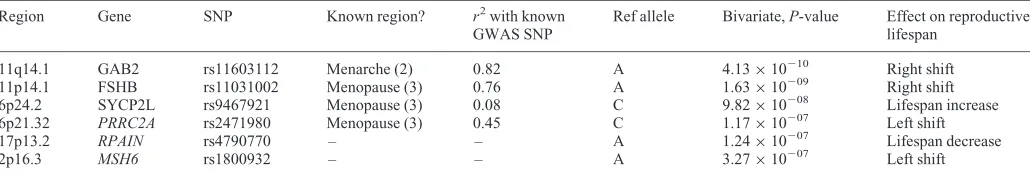
Table 1. Results of bivariate analysis
Region Gene SNP Known region? r2with known
GWAS SNP
Ref allele Bivariate,P-value Effect on reproductive
lifespan
11q14.1 GAB2 rs11603112 Menarche (2) 0.82 A 4.13×10210 Right shift
11p14.1 FSHB rs11031002 Menopause (3) 0.76 A 1.63×10209 Right shift
6p24.2 SYCP2L rs9467921 Menopause (3) 0.08 C 9.82×10208 Lifespan increase
6p21.32 PRRC2A rs2471980 Menopause (3) 0.45 C 1.17×10207 Left shift
17p13.2 RPAIN rs4790770 – – A 1.24×10207 Lifespan decrease
2p16.3 MSH6 rs1800932 – – A 3.27×10207 Left shift
Effect on reproductive lifespan gives the effect directions for both menarche and menopause associations; right shift¼menarche increasing allele is menopause
increasing allele (and vice versa for left shift), reproductive lifespan increase¼menarche decreasing allele same as menopause increasing allele (vice versa for
menopause (Table2). One of the six bivariate SNPs from the dis-covery analysis reached genome-wide significance in the
com-bined meta-analysis for menopause alone (rs1800932, P¼
2×1029). This variant was in theMSH6gene on chromosome 2 and was associated with a 1.3 months (se¼0.38) reduction in menopause age per common allele in the replication cohorts alone (allele frequency¼0.83) (Fig.1).
Functional significance of novel variant for age at menopause
We investigated the functional significance of the novel locus associated with age at menopause by determining whether the top variant, or SNPs in LD with it, were associated with expres-sion levels of any genes in the genome. We accessed data from four tissue types (monocytes, whole blood, liver and lung) and in all tissues rs1800932 or rs3136247 (r2¼0.95) was associated with expression of MSH6 (P¼3.9×1026 –3.1×10220). In monocytes, whole blood and liver, rs3136247 was the SNP most strongly associated with expression ofMSH6, but in lung tissue the best SNP was rs2047681 (r2¼0.6 with rs3136247).
SNPs in LD with top bivariate signals
In addition to the top bivariate SNP, we chose to also replicate the strongest SNP from each individual trait GWAS, which was in LD (hapmapr2.0.8) with the lead bivariate SNP (Table 2). In the individual menarche GWAS five independent SNPs in LD with a bivariate signal were more strongly associated with menarche than the lead bivariate SNP. Following the replication stage, none of the five SNPs reached genome-wide significance in the combined analysis of discovery and replication data, but statistical evidence increased for a known menopause locus in
PRRC2A being associated with age at menarche following replication, with the Pvalue strengthening from 2×1024to 5×1025. In the univariate menopause GWAS five SNPs in LD with top bivariate SNPs gave a stronger association and were taken forward for replication, but the only SNPs that gave a stronger association following replication were the three previously known menopause signals.
DISCUSSION
Female reproductive lifespan starts at menarche and ends at menopause and we therefore performed a bivariate analysis of the two traits. Two signals reached genome-wide significance in this analysis, but these were both heavily driven by low
P values in one of the individual traits. GAB2 is a known signal for menarche and FSHB for menopause (2,3). We sought to improve the statistical association for the second trait by in silico replication, but for both variants the statistical evidence got weaker. We also took four bivariate signals forward for additional replication, which were just below the genome-wide significance threshold, but none were robustly associated in both traits. The most promising bivariate signal after replication was associated with a left shift in reproductive lifespan and was in the HLA III region on chromosome 6 (nearPRRC2Agene), which is a known signal for menopause and reachedP¼5×1025for menarche, following replication.
This genomic region has also been associated with other Tabl
e 2 . Fol low-up of top si x biva riate sign als for associat ion wi th indiv idua l traits Gene Top biva riate SNP
Menarche (P
)
Menopause (P
) Top Me narche SNP in region Top Meno pause SN P in regio n SNP r 2
Discovery (P
phenotypes from GWAS scans, e.g. type 1 diabetes, multiple sclerosis and lupus (http://www.genome.gov/gwastudies/). This would be a good candidate to follow-up in additional cohorts with menarche data and if substantiated would provide evidence for an immunological pathway involved in both pheno-types. Despite finding no individual locus associated with both age at menarche and age at menopause, the positive although modest genetic correlation (rgenetic¼0.138) suggests that common genetic loci do exist, likely with very subtle effects. Our pathway analysis also suggests that there be one or two path-ways of shared aetiology between the two traits, with many others uniquely involved.
Future studies could include increasing the sample size in the discovery meta-analysis or replication of the top signals in add-itional cohorts. This is important since most of our participating studies were Caucasian, therefore other ethnic groups need to be similarly analysed. Studies are ongoing which include indivi-duals of multiple ethnicities, which should narrow down the as-sociation further. Also, other than adjusting for birth year in menarche analysis, we did not correct for environmental factors which may influence reproductive life span (such as early obesity or adult-age smoking). A further limitation of our study is that the cohorts included in the analysis were predomin-antly the same as those used in the meta-analyses for the individ-ual traits (2,3). Thus, finding that four of the six top hits for
reproductive lifespan had been reported previously for one of the individual traits, was not necessarily surprising, as these had very lowPvalues in the original analysis. One of the limita-tions of the bivariate meta-analysis is that very strong signals in individual traits can drive the bivariate statistic over theP,5× 10-8 threshold. The method is therefore best applied to signals that are sub-genome-wide significant in individual traits, or to use in pathway analyses than can highlight biological processes common to both traits.
We identified a novel variant associated with age at natural
menopause which is a synonymous exonic SNP in theMSH6
gene on chromosome 2. The SNP had a minor allele frequency of18% and the rare allele increased menopause age by1.3 months per allele. MSH6 heterodimerizes with MSH2 to form the MutS alpha protein complex, which is involved in mismatch repair (MMR), predominantly of single base mismatches and small dinucleotide insertion/deletion loops (20). It forms a complex with MutL alpha which is a heterodimer of PMS1
and MLH1. Germline mutations inMSH6,MSH2 andMLH1
[image:5.612.92.516.76.381.2]have been associated with hereditary non-polyposis colorectal cancer (HNPCC) (21).MSH6mutations are rarer than mutations in the other two genes, being found in 10 – 20% of HNPCC cases, who often have an atypical presentation and increased predispos-ition to endometrial cancer (22,23). No role forMSH6in repro-ductive ageing has been described previously, but other genes
Figure 1.Regional plot illustrating the strength of association (2log10P) versus hg19 position. The purple diamond represents the lead SNP in the combined analysis
of replication and discovery data. Other dots, coloured according to the degree of pairwise linkage disequilibrium, represent single SNP test statistics for discovery
stage only, including rs1800932, which was the SNP with lowestPvalue in the discovery analysis. The lower panels show structures of genes; layered ENCODE
histone modification marks (H3K4Me1 which marks enhancers; H3K4Me3 which marks promoters and H3K27Ac which marks active regulatory regions) and
linkage disequilibrium pairwise correlation (r2) derived from the CEU population in the 1000 Genomes Project, where white corresponds tor2¼0 and black to
involved in DNA repair were identified in the recent GWAS for
age at menopause, including EXO1, UIMC1, MCM8 and
POLG(3).EXO1 has 5′– 3′exonuclease activity and interacts with several of the MMR proteins, including MSH2, MLH1, MSH3, PCNA and WRN for its role in MMR and recombination (24). UIMC1 recruits BRCA1 to DNA damage sites and initiates G2/M checkpoint control (25). MCM8 is a member of a family of DNA replication complex proteins and is thought to have a role in meiotic double-strand break repair (26). Finally, POLG is re-sponsible for the replication and repair of the mitochondrial genome (27). Thus, variation in DNA repair processes, including single nucleotide, double-strand and mitochondrial DNA repair, appear to play a crucial role in determining age at natural meno-pause. A recent paper showed accumulation of double-strand DNA breaks in human follicles with age, with concomitant
downregulation of key DNA repair genes BRCA1, MRE11,
Rad51andATM, providing evidence that these processes play a functional role in ovarian ageing (28). It has also been reported that carriers of germline mutations in MMR genes, namely
BRCA1andBRCA2 are at increased risk of early menopause (29–32). We demonstrated that rs1800932 is an expression quantitative trait locus forMSH6, with the rarer allele being asso-ciated with increased levels of mRNA. Thus, the lower expres-sion ofMSH6is associated with earlier menopause, consistent with the work of Titus et al. (28), where downregulation of DNA repair genes was associated with ovarian ageing. The effect of the MSH6 variant on menopause age is relatively small and only explains a small proportion of the variance in menopause age. There are thus likely to be many more undiscov-ered genetic variants responsible for determining age at natural menopause.
The mechanism by which DNA repair influences timing of menopause is yet to be determined, but it is conceivable that damage to DNA of meiotic cells, if not repaired effectively, would result in the activation of apoptotic pathways to prevent transmission of potentially deleterious mutations to offspring. It is known that in non-meiotic cells DNA damage results in increased apoptosis and thus it is conceivable that similar
pro-cesses affect germ cells. Mouse models of theBRCA1MMR
gene have marked depletion of germ cells (33). Oocytes are lost throughout female life until menopause, predominantly by apoptosis. Any increase in apoptosis would therefore be expected to exhaust the oocyte pool prematurely and result in earlier menopause. We have described a novel association between a key DNA MMR gene and age at menopause, implicat-ing MMR as a key process involved in determinimplicat-ing reproductive lifespan.
MATERIALS AND METHODS
Estimation of genetic correlation between menarche and menopause
We used our largest individual study the Women’s Genome
Health Study (WGHS) (total sample N¼23 294) to test for genome-wide genetic correlation between timing of menarche and menopause. The total joint contribution of common SNPs to both age of menarche and age of menopause in the WGHS was estimated using the REML method implemented in GCTA (18,19). The genetic-related matrix in the WGHS was
calculated using 329 966 autosomal SNPs with minor allele fre-quency.0.01. For analysis of the joint heritability, this matrix was pruned to include only women with relatedness estimate
,0.025, leaving a total of 21 505 individuals with age of menar-che and 11 025 with age at natural menopause.
GWA studies for menarche and menopause
Full details of the individual GWAS can be found in the original publications, but briefly: the menarche GWAS included 32 studies, comprising 87 802 women and the menopause GWAS included 22 studies, comprising 38 968 women: all were of White European ancestry. Nineteen studies were included in both GWAS meta-analyses, although the number of women from each study differed (N¼71 942 for menarche andN¼33 460 for menopause). Women with recalled age at menarche between 9 and 17 years were included in the analysis. Age at natural menopause was defined as the age at the last menstrual period that occurred naturally between the ages of 40 and 60 years. Women were excluded with menopause due to hysterec-tomy and/or bilateral ovariechysterec-tomy, or chemotherapy/irradiation, if validated by medical records, and women using HRT before menopause. All studies were approved by local ethics committees and all participants provided written informed consent. Each study performed genome-wide association testing for age at menarche or menopause across2.5 million imputed SNPs, based on linear re-gression under an additive genetic model. Individual study data were meta-analysed using the METAL software package; genomic control (GC) adjustments were applied. We considered
P-values of,5×1028to indicate genome-wide significance.
Discovery bivariate GWAS meta-analysis
We performed multi-phenotype GWAS meta-analyses with ag-gregate data (Z test statistics) from each univariate GWAS meta-analysis (inverse-variance meta-analysis with GC con-trols, as described above) of age at menarche and natural menopause using our newly developed algorithm, empirical-weighted linear-combined test statistics (eLC) (34,35). Briefly, eLC directly combined correlated test statistics obtained from univariate GWAS meta-analyses with a weighted sum of univariate test statistics to empirically maxi-mize the overall association signals and also to account for the phenotypic correlation between menarche and menopause. Our eLC approach is expressed as
SeLC=
k
1
[max(|Tk|,c)∗|Tk|]
covariance matrix S of univariate test statistics using the sample covariance matrix of the test statistics of all SNPs from univariate GWAS analyses as an approximation.S:
Var(Z1) Cov(Z1,Z2)
Cov(Z1,Z2) Var(Z2)
whereZ1consists of unbiased univariate test statistics of all the SNPs for the first trait on genome-wide scale, so doesZ2. On the other hand, S can be estimated by using generalized least squares from individual-level data. Bivariate P-values of
,5×1028were considered genome-wide significant with po-tential pleiotropic effects, except when one of the individual trait P values was lower than the bivariate P value. The
eLC method is implemented in eLX package using C++and
is available at https://sites.google.com/site/multivariateyih-sianghsu/.
Replication strategy
All SNPs with aPvalue of,1×1027in the bivariate analysis were taken forward for replication in 22 additional cohorts with
in silico data, which included data from the iCOGs meta-analysis, which incorporated 16 individual studies. We also determined for each SNP whether any SNPs in LD with the lead SNP (,1 Mb away andr2.0.8) were more strongly associated with each of the traits in the individual menarche or menopause GWAS studies. There were 10 proxy SNPs with a lowerPvalue in the individual trait and these were also taken forward forin silico replication. Therefore, in total 16 SNPs were tested in replication cohorts (Supplementary Material, Table S1).
Access to data is available via the GREAT database https:// ifar-great.hsl.harvard.edu/ or by contacting the authors directly.
eQTL analysis
The novel genome-wide associated SNP for age at menopause (rs1800932) and SNPs in LD with this SNP (r2.0.9) were searched against a multi-tissue eQTL database of expression of SNP results (3). In four of the tissues [monocytes (36), blood (37), lung (38) and liver (39)] there was an eQTL for
MSH6and for three of these (monocytes, blood and liver) the SNP most strongly associated with expression of MSH6 was our top GWAS SNP or one in strong LD with it (r2.0.95).
SUPPLEMENTARY MATERIAL
Supplementary Material is available atHMGonline.
ACKNOWLEDGEMENTS
The authors are very grateful to the study participants and staff from all cohorts involved in this study. We acknowledge Kathy Lunetta/Doug Kiel for helpful comments. See supplemen-tary methods for extended acknowledgements.
Conflict of Interest statement. None
FUNDING
J.R.B.P is a Sir Henry Wellcome Postdoctoral Research Fellow (092447/Z/10/Z). D.A.L. and A.F. work in a centre that receives infrastructure funding from the UK Medical Research Council (G0600705) and A.F. is funded by a UK Medical Research Council Post-doctoral research fellowship (0701594). Further information on funding for individual studies is provided in supplementary information. Funding to pay the Open Access publication charges for this article was provided by the Well-come Trust.
REFERENCES
1. Hartge, P. (2009) Genetics of reproductive lifespan.Nat. Genet.,41,
637 – 638.
2. Elks, C.E., Perry, J.R., Sulem, P., Chasman, D.I., Franceschini, N., He, C.,
Lunetta, K.L., Visser, J.A., Byrne, E.M., Cousminer, D.L.et al.(2010)
Thirty new loci for age at menarche identified by a meta-analysis of
genome-wide association studies.Nat. Genet.,42, 1077– 1085.
3. Stolk, L., Perry, J.R., Chasman, D.I., He, C., Mangino, M., Sulem, P.,
Barbalic, M., Broer, L., Byrne, E.M., Ernst, F.et al.(2012) Meta-analyses
identify 13 loci associated with age at menopause and highlight DNA repair
and immune pathways.Nat. Genet.,44, 260 – 268.
4. van Noord, P.A., Dubas, J.S., Dorland, M., Boersma, H. and te Velde, E. (1997) Age at natural menopause in a population-based screening cohort: the
role of menarche, fecundity, and lifestyle factors.Fertil. Steril.,68, 95 – 102.
5. Cooper, G.S., Baird, D.D. and Darden, F.R. (2001) Measures of menopausal status in relation to demographic, reproductive, and behavioral
characteristics in a population-based study of women aged 35 – 49 years.Am.
J. Epidemiol.,153, 1159– 1165.
6. Otero, U.B., Chor, D., Carvalho, M.S., Faerstein, E., Lopes Cde, S. and Werneck, G.L. (2010) Lack of association between age at menarche and
age at menopause: Pro-Saude Study, Rio de Janeiro, Brazil.Maturitas,67,
245 – 250.
7. Snieder, H., MacGregor, A.J. and Spector, T.D. (1998) Genes control the cessation of a woman’s reproductive life: a twin study of hysterectomy and
age at menopause.J. Clin. Endocrinol. Metab.,83, 1875– 1880.
8. Euling, S.Y., Herman-Giddens, M.E., Lee, P.A., Selevan, S.G., Juul, A., Sorensen, T.I.A., Dunkel, L., Himes, J.H., Teilmann, G. and Swan, S.H. (2008) Examination of US puberty-timing data from 1940 to 1994 for secular
trends: panel findings.Pediatrics,121, S172– S191.
9. Harris, M.A., Prior, J.C. and Koehoorn, M. (2008) Age at menarche in the Canadian population: secular trends and relationship to adulthood BMI.
J. Adolesc. Health,43, 548 – 554.
10. Morris, D.H., Jones, M.E., Schoemaker, M.J., Ashworth, A. and Swerdlow, A.J. (2011) Secular trends in age at menarche in women in the UK born
1908– 93: results from the Breakthrough Generations Study.Paediatr.
Perinat. Epidemiol.,25, 394 – 400.
11. Speliotes, E.K., Willer, C.J., Berndt, S.I., Monda, K.L., Thorleifsson, G.,
Jackson, A.U., Lango Allen, H., Lindgren, C.M., Luan, J., Magi, R.et al.
(2010) Association analyses of 249,796 individuals reveal 18 new loci
associated with body mass index.Nat. Genet.,42, 937 – 948.
12. Dunger, D.B., Ahmed, M.L. and Ong, K.K. (2005) Effects of obesity on
growth and puberty.Best Pract. Res. Clin. Endocrinol. Metab.,19, 375 – 390.
13. Wang, W., Zhao, L.-J., Liu, Y.-Z., Recker, R.R. and Deng, H.-W. (2006) Genetic and environmental correlations between obesity phenotypes and age
at menarche.Int. J. Obes.,30, 1595– 1600.
14. Hoel, D.G., Wakabayashi, T. and Pike, M.C. (1983) Secular trends in the distributions of the breast cancer risk factors—menarche, first birth, menopause, and weight—in Hiroshima and Nagasaki, japan.
Am. J. Epidemiol.,118, 78 – 89.
15. Nichols, H.B., Trentham-Dietz, A., Hampton, J.M., Titus-Ernstoff, L., Egan, K.M., Willett, W.C. and Newcomb, P.A. (2006) From menarche to menopause: trends among US women born from 1912 to 1969.
Am. J. Epidemiol.,164, 1003 – 1011.
16. Henderson, K.D., Bernstein, L., Henderson, B., Kolonel, L. and Pike, M.C. (2008) Predictors of the timing of natural menopause in the multiethnic
17. Stolk, L., Perry, J.R., Chasman, D.I., He, C., Mangino, M., Sulem, P.,
Barbalic, M., Broer, L., Byrne, E.M., Ernst, F.et al.(2012) Meta-analyses
identify 13 loci associated with age at menopause and highlight DNA repair
and immune pathways.Nat. Genet.,44, 260 – 268.
18. Lee, S.H., Yang, J., Goddard, M.E., Visscher, P.M. and Wray, N.R. (2012) Estimation of pleiotropy between complex diseases using single-nucleotide polymorphism-derived genomic relationships and restricted maximum
likelihood.Bioinformatics,28, 2540 – 2542.
19. Yang, J., Lee, S.H., Goddard, M.E. and Visscher, P.M. (2011) GCTA: a tool
for genome-wide complex trait analysis.Am. J. Hum. Genet.,88, 76 – 82.
20. Kunkel, T.A. and Erie, D.A. (2005) DNA mismatch repair.Annu. Rev.
Biochem.,74, 681 – 710.
21. Peltomaki, P. (2001) Deficient DNA mismatch repair: a common etiologic
factor for colon cancer.Hum. Mol. Genet.,10, 735 – 740.
22. Berends, M.J., Wu, Y., Sijmons, R.H., Mensink, R.G., van der Sluis, T., Hordijk-Hos, J.M., de Vries, E.G., Hollema, H., Karrenbeld, A., Buys, C.H.
et al.(2002) Molecular and clinical characteristics of MSH6 variants: an
analysis of 25 index carriers of a germline variant.Am. J. Hum. Genet.,70,
26 – 37.
23. Win, A.K., Lindor, N.M., Winship, I., Tucker, K.M., Buchanan, D.D.,
Young, J.P., Rosty, C., Leggett, B., Giles, G.G., Goldblatt, J.et al.(2013)
Risks of colorectal and other cancers after endometrial cancer for women
with Lynch syndrome.J. Natl Cancer Inst.,105, 274 – 279.
24. Amin, N.S., Nguyen, M.N., Oh, S. and Kolodner, R.D. (2001)
exo1-Dependent mutator mutations: model system for studying functional
interactions in mismatch repair.Mol. Cell. Biol.,21, 5142 – 5155.
25. Wang, B., Matsuoka, S., Ballif, B.A., Zhang, D., Smogorzewska, A., Gygi, S.P. and Elledge, S.J. (2007) Abraxas and RAP80 form a BRCA1 protein
complex required for the DNA damage response.Science,316, 1194 – 1198.
26. Lutzmann, M., Grey, C., Traver, S., Ganier, O., Maya-Mendoza, A.,
Ranisavljevic, N., Bernex, F., Nishiyama, A., Montel, N., Gavois, E.et al.
(2012) MCM8- and MCM9-deficient mice reveal gametogenesis defects and
genome instability due to impaired homologous recombination.Mol. Cell,
47, 523 – 534.
27. Trifunovic, A., Wredenberg, A., Falkenberg, M., Spelbrink, J.N., Rovio,
A.T., Bruder, C.E., Bohlooly, Y.M., Gidlof, S., Oldfors, A., Wibom, R.et al.
(2004) Premature ageing in mice expressing defective mitochondrial DNA
polymerase.Nature,429, 417 – 423.
28. Titus, S., Li, F., Stobezki, R., Akula, K., Unsal, E., Jeong, K., Dickler, M.,
Robson, M., Moy, F., Goswami, S.et al.(2013) Impairment of
BRCA1-Related DNA Double-Strand Break Repair Leads to Ovarian Aging
in Mice and Humans.Sci. Transl. Med.,5, 172ra121.
29. Finch, A., Valentini, A., Greenblatt, E., Lynch, H.T., Ghadirian, P., Armel,
S., Neuhausen, S.L., Kim-Sing, C., Tung, N., Karlan, B.et al.(2013)
Frequency of premature menopause in women who carry a BRCA1 or
BRCA2 mutation.Fertil. Steril.,99, 1724– 1728.
30. Lin, W.T., Beattie, M., Chen, L.M., Oktay, K., Crawford, S.L., Gold, E.B., Cedars, M. and Rosen, M. (2013) Comparison of age at natural menopause in BRCA1/2 mutation carriers with a non-clinic-based sample of women in
northern California.Cancer,119, 1652– 1659.
31. Rzepka-Gorska, I., Tarnowski, B., Chudecka-Glaz, A., Gorski, B., Zielinska, D. and Toloczko-Grabarek, A. (2006) Premature menopause in
patients with BRCA1 gene mutation.Breast Cancer Res. Treat.,100, 59 – 63.
32. Oktay, K., Kim, J.Y., Barad, D. and Babayev, S.N. (2010) Association of BRCA1 mutations with occult primary ovarian insufficiency: a possible explanation for the link between infertility and breast/ovarian cancer risks.
J. Clin. Oncol.,28, 240 – 244.
33. Moslehi, R., Singh, R., Lessner, L. and Friedman, J.M. (2010) Impact of
BRCA mutations on female fertility and offspring sex ratio.Am. J. Hum.
Biol.,22, 201 – 205.
34. Hsu, Y.H., Mclean, R., Newton, E., Hanna, M., Cupples, L.A. and Kiel, D. (2012) Muscle, fat and bone connections: genetic risk factors of sarcopenic-obesity and dynapenic-sarcopenic-obesity and their consequent risks of osteoporotic
fractures. (ASBMR Annual Meeting Presentation).J. Bone Miner. Res.,27
(Suppl 1).http://www.asbmr.org/Meetings/AnnualMeeting/AbstractDetail. aspx?aid=f518434d-0e99-4f8f-858e-238f3fd451c5(accessed 16 December 2013).
35. Hsu, Y.H. (2013) Identifying pleiotropic genetic effects: a two-stage approach using genome-wide association meta-analysis data, in press. https://sites.google.com/site/multivariateyihsianghsu/.
36. Zeller, T., Wild, P., Szymczak, S., Rotival, M., Schillert, A., Castagne, R.,
Maouche, S., Germain, M., Lackner, K., Rossmann, H.et al.(2010) Genetics
and beyond—the transcriptome of human monocytes and disease
susceptibility.PLoS One,5, e10693.
37. Fehrmann, R.S., Jansen, R.C., Veldink, J.H., Westra, H.J., Arends, D.,
Bonder, M.J., Fu, J., Deelen, P., Groen, H.J., Smolonska, A.et al.(2011)
Trans-eQTLs reveal that independent genetic variants associated with a complex phenotype converge on intermediate genes, with a major role for the HLA.PLoS Genet.,7, e1002197.
38. Hao, K., Bosse, Y., Nickle, D.C., Pare, P.D., Postma, D.S., Laviolette, M.,
Sandford, A., Hackett, T.L., Daley, D., Hogg, J.C.et al.(2012) Lung eQTLs
to help reveal the molecular underpinnings of asthma.PLoS Genet.,8,
e1003029.
39. Greenawalt, D.M., Dobrin, R., Chudin, E., Hatoum, I.J., Suver, C.,
Beaulaurier, J., Zhang, B., Castro, V., Zhu, J., Sieberts, S.K.et al.(2011) A
survey of the genetics of stomach, liver, and adipose gene expression from a