R E S E A R C H
Open Access
The effect of bone marrow microenvironment on
the functional properties of the therapeutic bone
marrow-derived cells in patients with acute
myocardial infarction
Johanna A Miettinen
1*, Riikka J Salonen
1,2, Kari Ylitalo
1, Matti Niemelä
1, Kari Kervinen
1, Marjaana Säily
1,
Pirjo Koistinen
1, Eeva-Riitta Savolainen
3, Timo H Mäkikallio
1, Heikki V Huikuri
1and Petri Lehenkari
2Abstract
Background:Treatment of acute myocardial infarction with stem cell transplantation has achieved beneficial effects in many clinical trials. The bone marrow microenvironment of ST-elevation myocardial infarction (STEMI) patients has never been studied even though myocardial infarction is known to cause an imbalance in the acid-base status of these patients. The aim of this study was to assess if the blood gas levels in the bone marrow of STEMI patients affect the characteristics of the bone marrow cells (BMCs) and, furthermore, do they influence the change in cardiac function after autologous BMC transplantation. The arterial, venous and bone marrow blood gas concentrations were also compared.
Methods:Blood gas analysis of the bone marrow aspirate and peripheral blood was performed for 27 STEMI patients receiving autologous stem cell therapy after percutaneous coronary intervention. Cells from the bone marrow aspirate were further cultured and the bone marrow mesenchymal stem cell (MSC) proliferation rate was determined by MTT assay and the MSC osteogenic differentiation capacity by alkaline phosphatase (ALP) activity assay. All the patients underwent a 2D-echocardiography at baseline and 4 months after STEMI.
Results:As expected, the levels of pO2, pCO2, base excess and HCO3were similar in venous blood and bone marrow. Surprisingly, bone marrow showed significantly lower pH and Na+and elevated K+levels compared to arterial and venous blood. There was a positive correlation between the bone marrow pCO2and HCO3levels and MSC osteogenic differentiation capacity. In contrast, bone marrow pCO2and HCO3levels displayed a negative correlation with the proliferation rate of MSCs. Patients with the HCO3level below the median value exhibited a more marked change in LVEF after BMC treatment than patients with HCO3level above the median (11.13 ± 8.07% vs. 2.67 ± 11.89%, P = 0.014). Conclusions:Low bone marrow pCO2and HCO3levels may represent the optimal environment for BMCs in terms of their efficacy in autologous stem cell therapy in STEMI patients.
Keywords:Blood gas analysis, Bone marrow stem cells, Cell therapy, Mesenchymal stem cells, Myocardial infarction
Background
Acute myocardial infarction (AMI) is the major cause of congestive heart failure and subsequent mortality in the developed countries. Despite the major advances in treat-ment methods, myocardial infarction usually causes irre-versible damage to heart muscle. Cell therapy based on
autologous stem cell transplantation and potential myo-cardial regeneration has been the focus of many clinical trials for more than a decade. These trials have yielded contradictory results which have been proposed to be attributable to the heterogeneity of the study designs [1]. A recent experimental study showed that donor myocar-dial infarction impaired the therapeutic potential of bone marrow cells in mice and it was hypothesized that this might explain why human cell therapy trials have not matched the success achieved in rodent experiments [2]. * Correspondence: johanna.miettinen@oulu.fi
1
Department of Internal Medicine, Institute of Clinical Medicine, University of Oulu, P.O. Box 5000, Kajaanintie 50, Oulu FIN-90014, Finland
Full list of author information is available at the end of the article
Moreover, it has not been previously studied if the bone marrow microenvironment of patients with ST-elevation myocardial infarction (STEMI) can affect the functional-ity of the cells used in autologous cell therapy. Myocar-dial infarction has been shown to result in metabolic acidosis [3] and base excess in the venous blood has proved to be an independent predictor for intra-intensive care unit mortality of STEMI patients [4]. Since the blood gases measured from venous blood have been shown to correlate with bone marrow measurements [5], the acid-base status of STEMI patients likely affects also the microenvironment of bone marrow. After a careful literature review, we were not able to find any previous data on bone marrow physiological conditions in adults. Thus, the aim of the present study was to assess if the blood gas or electrolyte levels in the bone marrow of STEMI patients can influence the characteristics of the bone marrow cells and, subsequently, the success of autologous stem cell transplantation. We also compared arterial, venous and bone marrow blood gas and electro-lyte concentrations in patients with STEMI. This study is part of a pilot study (FINCELL II), which followed the original FINCELL (FINnish stem CELL) trial [6].
Methods
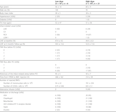
We examined a consecutive series of STEMI patients who were admitted to the University Hospital of Oulu, Finland, or to the central hospitals of Kajaani, Rovaniemi or Kemi, Finland, between April 2008 and August 2009. Inclusion criteria were age 18-79 years, acute STEMI and depressed left ventricular function (LVEF < 55%) measured by echo-cardiography 2-5 days after STEMI. Exclusion criteria were unwillingness or inability to provide informed con-sent, NYHA class IV, psychological or physiological unsuitability for participation in the study, inaccessibility for follow-up due to geographical or other reasons, severe leukopenia or thrombocytopenia, hepatic or renal dysfunc-tion or evidence for malignant disease. Patient characteris-tics are shown in Table 1. Written informed consent was obtained from patients within 5 days after STEMI. The study protocol conformed to the Declaration of Helsinki and was approved by the Ethical Committee of the North-ern Ostrobothnia Hospital District. The original FINCELL trial is registered at http://www.clinicaltrials.gov with registration number NCT00363324.
Study design
The day of acute STEMI was defined as day 0. On days 1-5, patients were randomly assigned in a double-blinded fashion, either to the group receiving the small cell dose (10 ml, mean cell count 5 × 10⁷, n = 6) or to the group receiving the high cell dose (160 ml, mean cell count 7 × 10⁸, n = 21). The patients and investigators performing the cell injection procedures and analysis of data were
unaware of the randomization throughout the study. Bone marrow aspiration, collection and preparation of cells were performed in the morning preceding the intra-coronary injection of BMCs, which was performed 4-14 days post-STEMI.
Cell preparation, administration and culturing
BMCs were collected in the morning of the cell administration day under local anesthesia and sterile conditions. 0.5-1 ml sample was aspirated from one-two iliac crest into a RAPIDLyte® heparinized syringe and the blood gas and electrolyte levels were analysed
using an i-STAT® Point-of-Care System (Abbot
Laboratories, Illinois, USA). 10 ml or 160 ml of bone marrow was aspirated and placed into tubes containing heparinized phosphate buffered saline (PBS; Gibco, Paisley, UK). The bone marrow cells were then trans-ported to a culture laboratory dedicated to aseptic cell manipulation for autologous stem cell transplantation. The mononuclear cell fraction containing stem and progenitor cells was isolated by Ficoll density gradient centrifugation, washed twice with PBS and resus-pended with 50% autologous serum in heparinized sal-ine or with heparinized salsal-ine alone (total volume of 10 ml). The cells were kept at +4°C until use. The BMC separation procedure took approximately 3 hours and the cells were administered within 24 hours to the target coronary artery. One aliquot of the cell sample was subjected to quality-control procedures, i.e. micro-bial culture for sterility and flow cytometer analysis for CD34+ cell counting and determination of cell viability in the accredited laboratory of the Oulu University Hospital, which is subjected to both outside and inside quality control. The validity of the cell preparation sys-tem was assessed as described previously [6]. One
ali-quot of the BMC fraction was plated into 25 cm2
tissue culture flask and cultured in 5 ml medium con-taining alpha MEM (Gibco) buffered with 20 mM HEPES (Gibco) and containing 10% heat-inactivated fetal calf serum (FCS; Bioclear, Netherlands), 100 U/ml penicillin, 0.1 mg/ml streptomycin, 2 mM L-glutamine
(Gibco). Cells were cultured at +37°C in 5% CO2 and
95% air. After one day, the medium was changed and non-attached cells were washed away. The attached cells (mesenchymal stem cells, MSCs) were cultured in a flask and the medium was replaced two times per week until near-confluence. The cells were passed two times before the analyses. In the cell counting analyses, the cells were washed with PBS and adherent cells detached using trypsin-EDTA solution (Gibco).
Flow cytometric analysis of cell surface antigens
criteria panel of cell surface markers for hMSCs pro-posed by International Society for Cellular Therapy [8] was evaluated from all MSC lines analyzed, and each of the lines met the criteria. The percentage values of posi-tive MSC markers were CD90 99.81 ± 0.23%, CD105 99.58 ± 0.37%, CD73 99.66 ± 0.28% and HLA-ABC 97.58 ± 3.06%. The percentage value of negative markers (CD14, CD34, CD45, CD19 and HLA-DR) was 1.80 ± 0.58%. Thus, the marker analysis showed that the cell cultures contained almost purely MSCs.
MTT proliferation assay
[image:3.595.57.539.101.563.2]MSCs were cultured in 96-well plates in six replicates. 500 cells/well were plated and half of the medium was changed twice a week. Cell proliferation was measured by MTT (3-(4,5-Dimethylthiazol-2-yl)-2,5-diphenyltetrazo-lium bromide) assay after 1, 4, 7, and 14 days culture. The medium was removed and MTT reagent (Sigma-Aldrich, St. Louis, MO, USA) (0,5 mg/ml in medium) was added to the cells and incubated for 2 h at +37°C in 5% CO2and 95% air. After incubation, the MTT solution Table 1 Patients characteristics
Low dose (5 × 10⁷), n = 6
High dose (7 × 10⁸), n = 21
Age (years) 56 ± 10 60 ± 9
Male sex (%) 100 95
Body mass index 28 ± 5 28 ± 3
Hypertension [n(%)] 2 (33) 5 (24)
Diabetes [n(%)] 0 1 (5)
TnI max (μg/L) 52 ± 47 51 ± 56
Infarct-related vessel [n(%)]
LAD 3 (50) 6 (29)
CX 0 0
RCA 3 (50) 14 (67)
LOM I 0 1 (5)
LVEF at baseline (%) 47.4 ± 9.2 44.4 ± 6.3
LVEF at 4 months’follow-up (%) 50.5 ± 13.2 52.3 ± 11.6
TIMI flow before PCI [n(%)]
0 2 (33) 4 (19)
1 1 (17) 2 (10)
2 1 (17) 3 (14)
3 1 (17) 9 (43)
TIMI flow after PCI [n(%)]
1 0 0
2 1 (17) 2 (10)
3 4 (33) 17 (81)
%Stenosis of the infarct-related artery before PCI 95 ± 5 93 ± 7
Time from STEMI to BMC injection (h) 268 ± 162 214 ± 94
Number of injected BMCs
Number of mononuclear cells (× 106) 54 ± 22 701 ± 254
Number of CD34+ cells (× 106) 0.75 ± 0.46 8.9 ± 5.1
Adenosine infusion [n(%)] 0 9 (43)
Medication at discharge [n(%)]
ASA 6 (100) 20 (95)
Clopidogrel 6 (100) 21 (100)
Beta-blocker 6 (100) 21 (100)
ACE inhibitor/AT II receptor blocker 6 (100) 21 (100)
Statin 6 (100) 18 (86)
Diuretic 2 (33) 9 (43)
was removed and 100 μl/well of dimethyl sulfoxide (DMSO; Sigma-Aldrich) was added. The absorbance of the reduced form of MTT was measured at 550 nm and 650 nm (background) in a plate reader (Victor 2, Wallac Oy, Turku, Finland).
Alkaline phosphatase activity assay
Human MSCs were seeded at 10000 cells/well into 24-well plates in four replicate 24-wells and were cultured with the medium described above and another four replicates in a medium containing also 100 mM dexamethasone (Sigma, St. Louis, MO, USA), 10 mMb-glycerol (Sigma) and 0.05 mM ascorbic acid (Sigma). After 3 weeks, cul-tured cells were assayed in the following way: the assay buffer containing 0.1% Triton X-100, pH 7.6, was added to each well, and the plates were frozen. After thawing, alkaline phosphatase (ALP) activity was determined using 0.1 mM 4-p-nitrophenylphosphate (Sigma-Aldrich) as the substrate and absorbance was read at 405 nm in a plate reader (Victor 2, Wallac Oy, Turku, Finland). Each sample was measured in duplicate. The protein content of the wells was determined by the BIO-RAD Protein Assay (Bio-Rad Laboratories, Richmond, California, USA). The enzyme activities were expressed as units/mg protein.
Blood sampling, blood gas and biochemical laboratory analyses
All the patients underwent venous and arterial blood sampling on the day of cell transplantation. pH, pO2, pCO2, HCO3, BE, Na+ and K+ levels were measured in the central laboratory of Oulu University Hospital.
Measurement of left ventricular ejection fraction
A 2-dimensional echocardiogram was performed 2-5 days after STEMI and at 4 months after STEMI. The LVEF was measured by an experienced investigator in the core laboratory, unaware of the patient’s treatment assignment, using the technique described previously [9].
Statistical analysis
Variables are expressed as means ± SD, or medians with interquartile range with skewed data. Paired samplest-test was used to determine the statistical significance of the difference in laboratory values between bone marrow and arterial/venous blood. Correlation analyses between bone marrow and arterial/venous blood laboratory values were conducted using paired samples correlation. In other ana-lyses, Pearson’s correlation or Spearman’s correlation with skewed data was used. Analysis-of-variance (ANOVA) was used in the between-group comparisons. The variance analyses of the LVEF change in HCO3, BE and potassium subgroups (above/below median) were corrected with the cell dose group (low/high dose). All P values are two-tailed and statistical significance was set at P < 0.05. Analyses were performed with SPSS software, version 14.0.
Results
Blood gas and electrolyte analyses of bone marrow, venous and arterial blood
The levels of blood gases (pO2, pCO2), serum
bicarbo-nate (HCO3), base excess (BE), pH and electrolytes
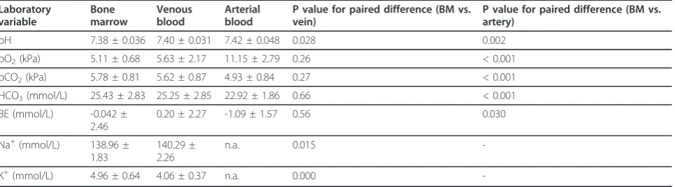
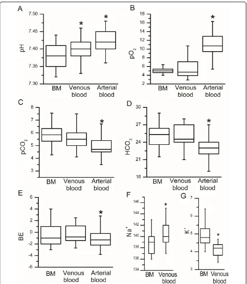
sodium (Na+) and potassium (K+) were measured from venous, arterial and bone marrow blood samples (Table 2, Figure 1). All laboratory values measured dif-fered significantly between bone marrow aspirate and arterial blood. In addition, a significant difference was detected between bone marrow aspirate and venous blood in pH, sodium and potassium levels (Table 2). The levels of pO2, pCO2, BE and HCO3 were similar in venous blood and bone marrow.
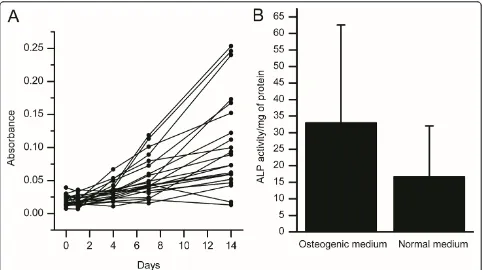
[image:4.595.58.541.588.722.2]Association of bone marrow blood gas and electrolyte levels with the functional properties of bone marrow cells The bone marrow mesenchymal stem cell (MSC) prolif-eration rate was determined by MTT assay (absorbance/ time in days) (Figure 2A). The absorbance reading was plotted against time and the slope of the line was used as the measure of cell proliferation (average slope of all
Table 2 Laboratory analyses of bone marrow, venous and arterial blood
Laboratory variable
Bone marrow
Venous blood
Arterial blood
P value for paired difference (BM vs. vein)
P value for paired difference (BM vs. artery)
pH 7.38 ± 0.036 7.40 ± 0.031 7.42 ± 0.048 0.028 0.002
pO2(kPa) 5.11 ± 0.68 5.63 ± 2.17 11.15 ± 2.79 0.26 < 0.001
pCO2(kPa) 5.78 ± 0.81 5.62 ± 0.87 4.93 ± 0.84 0.27 < 0.001
HCO3(mmol/L) 25.43 ± 2.83 25.25 ± 2.85 22.92 ± 1.86 0.66 < 0.001
BE (mmol/L) -0.042 ±
2.46
0.20 ± 2.27 -1.09 ± 1.57 0.56 0.030
Na+(mmol/L) 138.96 ±
1.83
140.29 ± 2.26
n.a. 0.015
-K+(mmol/L) 4.96 ± 0.64 4.06 ± 0.37 n.a. 0.000
measurements 0.0076 ± 0.0086). The MSC differentiation capacity was determined by ALP activity assay (u/mg of protein in basic/differentiation medium) (Figure 2B). The associations of bone marrow blood gas and electrolyte
[image:5.595.57.540.86.641.2]levels with BM cell viability (87.9 ± 8.7%), MSC prolifera-tion rate and the differentiaprolifera-tion capacity of MSCs with bone marrow blood gas and electrolyte levels are pre-sented in Table 3. Bone marrow pCO2and HCO3levels
exhibited a negative correlation with the proliferation rate of mesenchymal stem cells. In contrast, there was a positive correlation between the bone marrow pCO2and HCO3levels and MSC ALP activity. This can be inter-preted as better osteoblast differentiation capacity of
MSCs when bone marrow pCO2 and HCO3 levels are
high. However, none of the biochemical laboratory values measured was associated with the viability of cells in the bone marrow aspirate.
Association of bone marrow blood gas and electrolyte levels with the functional recovery of the heart after cell therapy
The association of the functional recovery of the heart after cell therapy with bone marrow blood gas and
electrolyte levels was evaluated by studying the correla-tions of the blood gases with the change of LVEF
between baseline and the 4 months’ follow-up. There
was a trend towards a negative correlation between
LVEF change and bone marrow HCO3 level as well as
the BE level and a positive correlation between LVEF change and potassium level, but these associations did not reach statistical significance (Table 4).
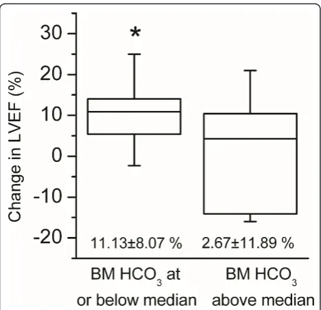
In a further analysis, the patient population was divided into two subgroups according to their HCO3, BE and potassium values, respectively, and the cell dose group was used as a covariate in the analyses. Patients with the HCO3level below the median value exhibited a more marked change in LVEF after stem cell treatment
[image:6.595.57.540.88.358.2]than patients with the HCO3 level above the median
Figure 2Tests for bone marrow-derived mesenchymal stem cell (MSC) proliferation and differentiation.(A) MTT proliferation assay. (B) Alkaline phosphatase (ALP) activity test of MSCs in normal growth medium and osteogenic differentiation medium.
Table 3 Association of bone marrow blood gas and electrolyte levels with functional properties of bone marrow cells
Laboratory variable CD34+ cell viability MSC proliferation rate MSC osteoblast differentiation
Correlation coefficient P-value Correlation coefficient P-value Correlation coefficient P-value
pH 0.093 0.67 0.23 0.31 -0.41 0.095
pO2(kPa) 0.009 0.97 0.16 0.48 -0.040 0.88
pCO2(kPa) 0.002 0.99 -0.50 0.023 0.57 0.013
HCO3(mmol/L) 0.085 0.69 -0.43 0.052 0.48 0.043
BE (mmol/L) 0.083 0.70 -0.41 0.066 0.34 0.17
Na+(mmol/L) 0.10 0.64 0.27 0.24 -0.015 0.95
K+(mmol/L) 0.147 0.50 0.012 0.96 0.037 0.88
[image:6.595.59.539.614.735.2](11.13 ± 8.07% vs. 2.67 ± 11.89%, P = 0.014) (Figure 3). No significant differences were found in the LVEF change between BE or potassium subgroups (data not shown). In addition, the cell dose did not have a signifi-cant effect on the LVEF change (p = 0.067).
In an attempt to clarify the individual differences in the levels of bone marrow blood gases, the associations of several other parameters (e.g. age, sex, body mass index, hypertension, hypercholesterolemia, diabetes, heart failure, severity of CAD, medication at discharge) with the bone marrow blood gas values were tested. The
bone marrow BE and HCO3 levels were found to be
higher in patients using diuretic drugs at the time of hospital discharge than in patients with no diuretic medication (BE 1.10 ± 2.81 vs. -0.87 ± 1.81 mmol/L, P
= 0.043 and HCO326.69 ± 3.46 vs. 24.54 ± 1.89 mmol/ L, P = 0.056, respectively). In addition, the patients using diuretics experienced a smaller improvement in the LVEF than patients without diuretic medication (1.81 ± 12.42 vs. 10.24 ± 7.41%-units, respectively, P = 0.036). No significant associations were found between any other patient characteristics and the bone marrow blood gases (P = NS in each analysis, data not shown).
Discussion
The results of the present study demonstrate that there is a significant difference in blood gas and electrolyte levels between bone marrow and arterial blood in patients with STEMI. In addition, a significant difference was found between bone marrow aspirate and venous blood in pH, sodium and potassium levels. In contrast, the levels of pO2, pCO2, BE and HCO3 were similar in venous blood and bone marrow. In a further blood gas analysis, the proliferation rate of bone marrow-derived MSCs was associated with the pCO2 and HCO3levels in the bone marrow. The MSC proliferation was more
effi-cient when bone marrow pCO2 and HCO3 levels were
low. It was also shown that a low HCO3 level in bone marrow was associated with better functional recovery of the heart after myocardial infarction treated with intracoronary injection of autologous BM cells. Patients with an HCO3level below the median value experienced a more marked improvement of LVEF than patients
with the HCO3 level above the median value. In
con-trast, the osteogenic differentiation potential of the BM-derived MSCs was shown to be better when bone mar-row pCO2 and HCO3levels were high.
One objective of this study was to compare arterial, venous and bone marrow blood gas and electrolyte con-centrations in STEMI patients. As far as we are aware, only one previous study has assessed the levels of blood gases and electrolytes in bone marrow and venous blood in humans [5]. Grisham et al. studied children with acute leukemia or other hematological disorders. In those patients, a significant correlation was found between venous and bone marrow samples for pH, pCO2, HCO3, BE and sodium concentration, but no pre-dictable relationship was observed for pO2 and potas-sium concentration. In the present study, pCO2, BE and
HCO3also correlated between bone marrow and venous
[image:7.595.55.290.123.242.2]blood as well as bone marrow and arterial blood. How-ever, pH, sodium and potassium levels differed signifi-cantly between venous blood and bone marrow. In the study of Grisham et al. the actual differences between bone marrow and venous blood were not assessed. However, they also noticed that the potassium levels dif-fered significantly between bone marrow and venous blood. They considered that this could be due to the traumatic nature of specimen collection and the high Table 4 Association of bone marrow blood gas and
electrolyte levels with the change of LVEF after cell therapy
Laboratory variable Change of LVEF (%-points)
Correlation coefficient P-value
pH 0.063 0.77
pO2(kPa) 0.16 0.45
pCO2(kPa) -0.31 0.13
HCO3(mmol/L) -0.37 0.070
BE (mmol/L) -0.36 0.082
Na+(mmol/L) -0.26 0.23
K+(mmol/L) 0.39 0.059
[image:7.595.57.291.460.685.2]negative pressure generated in aspirating marrow evok-ing hemolysis [5]. A portion of the cells inevitably break during the bone marrow aspiration but we think that the cell breakage does not fully explain the difference in potassium concentration between bone marrow and venous blood because we found a difference also in the sodium concentration. Instead, it may be a true finding being related to the different activity of sodium-potas-sium pump.
Bicarbonate is alkaline, and it is one of the compo-nents that maintain acid-base homeostasis in the body. Most of the CO2 in the body is converted into carbonic acid (H2CO3), which rapidly forms bicarbonate (HCO3-). The pH in the body depends on the pCO2 and the level of HCO3-. In this study MSC proliferation was found to be more efficient when the bone marrow aspirate pCO2
and HCO3 levels were low. The pH in the bone marrow
was not directly associated to the proliferation rate but this may be due to other compensatory buffering mechanisms, such as hemoglobin, which is known to stabilize the pH value. It is also known that active inflammation creates an acidic environment [10]. In the present study, the levels of pCO2and HCO3displayed a nonsignificant negative correlation with bone marrow interleukin (IL)-6 levels (data not shown). Thus, the
levels of pCO2 and HCO3 may have declined in an
attempt to buffer the acidosis caused by inflammatory processes. We have previously shown that inflammation (mediated by tumour necrosis factor alpha) enhances the proliferation of MSCs and activates many immuno-suppressive pathwaysin vitro[7]. Thus, if inflammation is more extensive in the bone marrow of the patients with lower levels of pCO2 and HCO3, it can be postu-lated to have an influence on the resident MSCs and enhance also their in vitro proliferation. On the con-trary, the osteogenic differentiation potential of the BM
cells was found to be better when bone marrow pCO2
and HCO3 levels were high. This result is in line with previous studies which have shown that acidic environ-ment impairs osteoblast function,[11,12] reduces osteo-blast alkaline phosphatase (ALP) activity [13,14] and inhibits osteogenic differentiation of MSCs [14,15].
Several previous trials have shown that the number of bone marrow CD34+ cells does not correlate with the change of LVEF after cell transplantation [16-19]. In the present study, the viability of CD34+ cells was not asso-ciated with the pCO2and HCO3 levels. In addition, the
observation that low bone marrow pCO2 and HCO3 is
beneficial for BM-derived MSC proliferation but not for the osteoblast differentiation suggests that maintaining the stemness of MSCs or some other cell lineage is dominant in this kind of environment. It has been pre-viously shown that hMSCs cultured in hypoxic conditions (2% oxygen) display significantly improved
expansion characteristics while maintaining their multi-lineage potential [20]. On the other hand, hypoxia as such can lead to unfavorable effects in the cultured cells, such as chromosomal aberrations [21]. Obviously, one of the most important functions of the bone mar-row microenvironment is to support the stemness of the resident BMCs and protect the infinitely dividing cell population from oxygen derived free radicals that could theoretically promote the cancer propagation of these cells. It has also been shown that bone marrow contains many different microenvironments in which MSCs are the prominent cell components; these cells play many important roles including controlling hematopoiesis [22,23]. Based on our results, it seems that the optimal environment for BM cells in this respect is associated
with a low bone marrow pCO2 value and reduced
HCO3 levels which support the stemness of BM cells
making them more efficient for cell therapy use and also more capable of undergoing intrinsic repair mechanisms. However, the results of this study are still preliminary findings and the importance of bone mar-row microenvironment on MSC functionality after two
MSC passages in culture media with 5% CO2 and 95%
air, which are completely different conditions compared to the bone marrow of STEMI patients, remain to be elucidated. Further tests in cell cultures are currently ongoing and will later provide us more insights about the effect of different environmental factors of the bone marrow on the functionality of the BM cells.
bone marrow mesenchymal cells is different in aged (> 50 years old) patients compared to young (< 18 years) and umbilical cord blood mesenchymal stem cells [29]. In the present study, the age of the patients was not associated with the parameters describing the metabolic state of bone marrow or the viability of BMCs. Heart failure, hypercholesterolemia, diabetes or hypertension were not associated to these parameters either. How-ever, this may be due to the small patient number in each disease group. Further studies with larger patient populations are needed to clarify the association between these diseases and the bone marrow metabolic state. There were also attempts to collect samples from healthy hip fracture patients and patients undergoing spinal surgery to compare their bone marrow blood gas levels with STEMI patients. However, there were serious technical difficulties in the measurement of the thick bone marrow sample taken from the open bone and
also arterial blood ‘contamination’. Thus, the
comparison of blood gas values between healthy subjects and patients with myocardial infarction remains to be elucidated in future studies.
The LVEF change between baseline and follow-up visit has been the most widely used endpoint to evaluate the effectiveness of stem cell transplantation after myocar-dial infarction [1]. In the present study, patients with
the bone marrow HCO3 level below the median value
experienced a more marked improvement of LVEF after stem cell treatment than their counterparts with an HCO3 level above the median value. This clinical trial was designed to be a dose-response study but no differ-ence in the LVEF recovery could be observed between the groups receiving low or high dose of BMCs in this pilot phase. Thus, it is not likely that the differences in the cell doses would have affected the EF results of the present substudy. However, because of the low patient number, the results of this study have to be interpreted
[image:9.595.58.538.330.712.2]as descriptive rather than confirmatory when
considering the importance of the blood gas values of the bone marrow on the clinical outcome of BMC trea-ted STEMI patients. More studies with higher patient number are needed to confirm these preliminary findings.
The bone marrow BE and HCO3 levels were found to
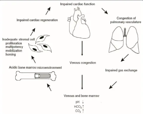
be significantly higher in patients using diuretic drugs at the time of hospital discharge than in patients with no diuretic medication. The patients using diuretics also experienced less extensive improvement in the LVEF than the patients with no diuretic medication. The use of diuretic drugs is usually associated with post-STEMI hypertension or to venous or pulmonary congestion, and it is these conditions that most likely are affecting the blood gas values rather than the medication itself. Based on the results of the present study, we have cre-ated a hypothetical model describing the associations between heart, pulmonary vasculature, peripheral circu-lation and bone marrow after an ischemic event (Figure 4). In this model, the impaired cardiac function leads to venous or pulmonary congestion making bone marrow more acidic and, as a buffering effect, there are eleva-tions in the pCO2 and HCO3 concentrations in the per-ipheral circulation as well as in the bone marrow. The acidic bone marrow microenvironment also impairs the function of the resident stromal cells which leads to impaired cardiac regeneration after myocardial infarc-tion. The course of events creates a vicious cycle causing even more impairment of cardiac function. This idea is supported by a previous experimental work which showed that donor myocardial infarction impaired the therapeutic potential of bone marrow cells in mice [2]. However, based on the results of the present study, we cannot be sure if it is the change in cardiac, venous or pulmonary function that triggers this process. Thus, further studies with a higher number of patients will be needed to clarify the pathways which are responsible for the individual differences in acid-base status and the metabolic state of the bone marrow in STEMI patients. We believe that this study suggests that we should not ignore the role of bone marrow physiology as a regula-tor of BMC-mediated tissue repair, both therapeutic and spontaneous.
Conclusions
There is a significant difference in blood gas and elec-trolyte levels between bone marrow and arterial blood in patients with STEMI. There are also significant differ-ences in the pH, sodium and potassium levels between bone marrow aspirate and venous blood. Even though bone marrow acid-base status and blood gas levels resemble more closely venous blood than its arterial counterpart, the bone marrow seems to form a unique
compartment for maintenance and storage of stromal cells, similarly to that reported in the context of the hematopoietic stem cell niche. The proliferation rate of bone marrow-derived MSCs is more efficient when
bone marrow pCO2 and HCO3 levels are low. In
con-trast, the osteogenic differentiation potential of the
BM-derived MSCs is better when bone marrow pCO2 and
HCO3 levels are high. The STEMI patients with lower
HCO3 levels experience a more marked change in
LVEF. Moreover, patients using diuretic drugs at the time of hospital discharge tended to have higher bone
marrow pCO2 and HCO3 levels and these patients
experienced a poorer improvement in the LVEF after stem cell therapy, most probably because of the underly-ing cardiac or venous congestion affectunderly-ing also the bone marrow metabolic state. In this respect, low bone mar-row pCO2and HCO3 levels represent the optimal envir-onment for BM cells in terms of their efficacy in autologous stem cell therapy in STEMI patients.
Acknowledgements
We would like to thank Ms. Kirsi Kvist-Mäkelä, Ms. Päivi Kastell, Ms. Pirkko Huikuri, Ms. Päivi Koski, and Ms. Sari Kaarlenkaski for their excellent assistance in this study.
This study was supported by grants from the Medical Council of the Academy of Finland, Helsinki, Finland and Finnish Foundation for Cardiovascular Research, Helsinki, Finland, and to Dr. Miettinen; grant from Ida Montin Foundation, Helsinki, Finland.
Author details
1Department of Internal Medicine, Institute of Clinical Medicine, University of
Oulu, P.O. Box 5000, Kajaanintie 50, Oulu FIN-90014, Finland.2Department of Anatomy and Cell Biology, Institute of Biomedicine, University of Oulu, P.O. Box 5000, Kajaanintie 50, Oulu FIN-90014, Finland.3Department of Clinical Chemistry, Institute of Diagnostics, University of Oulu, P.O. Box 5000, Kajaanintie 50, Oulu FIN-90014, Finland.
Authors’contributions
JM participated in the design of the study, made laboratory analyses and cell culturing, acquired the data, analysed and interpreted the data, performed statistical analyses and drafted the manuscript.
RS made laboratory analyses and cell culturing, acquired the data, analysed and interpreted the data and helped to draft the manuscript. KY conceived and designed the study, chose and examined the patients, performed 2D-echocardiography and other medical interventions for the study patients, injected the cells, acquired the data and analysed and interpreted the data. MN and KK chose and examined the patients, performed
Competing interests
The authors declare that they have no competing interests.
Received: 28 November 2011 Accepted: 2 April 2012 Published: 2 April 2012
References
1. George JC:Stem cell therapy in acute myocardial infarction: a review of clinical trials.Transl Res2010,155:10-19.
2. Wang X, Takagawa J, Lam VC, Haddad DJ, Tobler DL, Mok PY, Zhang Y, Clifford BT, Pinnamaneni K, Saini SA, Su R, Bartel MJ, Sievers RE, Carbone L, Kogan S, Yeghiazarians Y, Hermiston M, Springer ML:Donor myocardial infarction impairs the therapeutic potential of bone marrow cells by an interleukin-1-mediated inflammatory response.Sci Transl Med2011, 3:100ra90.
3. Kirby BJ, McNicol MW:Acid-base status in acute myocardial infarction.
Lancet1966,2:1054-1056.
4. Lazzeri C, Valente S, Chiostri M, Picariello C, Gensini GF:Evaluation of acid-base balance in ST-elevation myocardial infarction in the early phase: a prognostic tool?Coron Artery Dis2010,21:266-272.
5. Grisham J, Hastings C:Bone marrow aspirate as an accessible and reliable source for critical laboratory studies.Ann Emerg Med1991,20:1121-1124. 6. Huikuri HV, Kervinen K, Niemelä M, Ylitalo K, Säily M, Koistinen P,
Savolainen ER, Ukkonen H, Pietilä M, Airaksinen JK, Knuuti J, Mäkikallio TH: Effects of intracoronary injection of mononuclear bone marrow cells on left ventricular function, arrhythmia risk profile, and restenosis after thrombolytic therapy of acute myocardial infarction.Eur Heart J2008, 29:2723-2732.
7. Miettinen JA, Pietilä M, Salonen RJ, Ohlmeier S, Ylitalo K, Huikuri HV, Lehenkari P:Tumor necrosis factor alpha promotes the expression of immunosuppressive proteins and enhances the cell growth in a human bone marrow-derived stem cell culture.Exp Cell Res2011,317:791-801. 8. Dominici M, Le Blanc K, Mueller I, Slaper-Cortenbach I, Marini F, Krause D,
Deans R, Keating A, Prockop D, Horwitz E:Minimal criteria for defining multipotent mesenchymal stromal cells. The International Society for Cellular Therapy position statement.Cytotherapy2006,8:315-317. 9. Huikuri HV, Tapanainen JM, Lindgren K, Raatikainen P, Mäkikallio TH, Juhani
Airaksinen KE, Myerburg RJ:Prediction of sudden cardiac death after myocardial infarction in the beta-blocking era.J Am Coll Cardiol2003, 42:652-658.
10. Falchuk KH, Goetzl EJ, Kulka JP:Respiratory gases of synovial fluids. An approach to synovial tissue circulatory-metabolic imbalance in rheumatoid arthritis.Am J Med1970,49:223-231.
11. Bushinsky DA:Stimulated osteoclastic and suppressed osteoblastic activity in metabolic but not respiratory acidosis.Am J Physiol1995,268: C80-C88.
12. Frick KK, Bushinsky DA:In vitro metabolic and respiratory acidosis selectively inhibit osteoblastic matrix gene expression.Am J Physiol1999, 277:F750-F755.
13. Brandao-Burch A, Utting JC, Orriss IR, Arnett TR:Acidosis inhibits bone formation by osteoblasts in vitro by preventing mineralization.Calcif Tissue Int2005,77:167-174.
14. Disthabanchong S, Radinahamed P, Stitchantrakul W, Hongeng S, Rajatanavin R:Chronic metabolic acidosis alters osteoblast differentiation from human mesenchymal stem cells.Kidney Int2007,71:201-209. 15. Chen T, Zhou Y, Tan WS:Influence of lactic acid on the proliferation,
metabolism, and differentiation of rabbit mesenchymal stem cells.Cell Biol Toxicol2009,25:573-586.
16. Britten MB, Abolmaali ND, Assmus B, Lehmann R, Honold J, Schmitt J, Vogl TJ, Martin H, Schachinger V, Dimmeler S, Zeiher AM:Infarct remodeling after intracoronary progenitor cell treatment in patients with acute myocardial infarction (TOPCARE-AMI): mechanistic insights from serial contrast-enhanced magnetic resonance imaging.Circulation
2003,108:2212-2218.
17. Fernandez-Aviles F, San Roman JA, Garcia-Frade J, Fernandez ME, Penarrubia MJ, de la Fuente L, Gomez-Bueno M, Cantalapiedra A, Fernandez J, Gutierrez O, Sanchez PL, Hernandez C, Sanz R, Garcia-Sancho J, Sanchez A:Experimental and clinical regenerative capability of human bone marrow cells after myocardial infarction.Circ Res2004,95:742-748.
18. Lunde K, Solheim S, Aakhus S, Arnesen H, Abdelnoor M, Egeland T, Endresen K, Ilebekk A, Mangschau A, Fjeld JG, Smith HJ, Taraldsrud E, Grogaard HK, Bjornerheim R, Brekke M, Muller C, Hopp E, Ragnarsson A, Brinchmann JE, Forfang K:Intracoronary injection of mononuclear bone marrow cells in acute myocardial infarction.N Engl J Med2006, 355:1199-1209.
19. Miettinen JA, Ylitalo K, Hedberg P, Jokelainen J, Kervinen K, Niemelä M, Säily M, Koistinen P, Savolainen ER, Ukkonen H, Pietilä M, Airaksinen KE, Knuuti J, Vuolteenaho O, Mäkikallio TH, Huikuri HV:Determinants of functional recovery after myocardial infarction of patients treated with bone marrow-derived stem cells after thrombolytic therapy.Heart2010, 96:362-367.
20. Grayson WL, Zhao F, Bunnell B, Ma T:Hypoxia enhances proliferation and tissue formation of human mesenchymal stem cells.Biochem Biophys Res Commun2007,358:948-953.
21. Ueyama H, Horibe T, Hinotsu S, Tanaka T, Inoue T, Urushihara H, Kitagawa A, Kawakami K:Chromosomal variability of human mesenchymal stem cells cultured under hypoxic conditions.J Cell Mol Med2012,16:72-82.
22. Trentin JJ:Determination of bone marrow stem cell differentiation by stromal hemopoietic inductive microenvironments (HIM).Am J Pathol
1971,65:621-628.
23. Askmyr M, Sims NA, Martin TJ, Purton LE:What is the true nature of the osteoblastic hematopoietic stem cell niche?Trends Endocrinol Metab2009, 20:303-309.
24. Vasa M, Fichtlscherer S, Aicher A, Adler K, Urbich C, Martin H, Zeiher AM, Dimmeler S:Number and migratory activity of circulating endothelial progenitor cells inversely correlate with risk factors for coronary artery disease.Circ Res2001,89:E1-E7.
25. Tepper OM, Galiano RD, Capla JM, Kalka C, Gagne PJ, Jacobowitz GR, Levine JP, Gurtner GC:Human endothelial progenitor cells from type II diabetics exhibit impaired proliferation, adhesion, and incorporation into vascular structures.Circulation2002,106:2781-2786.
26. Dimmeler S, Leri A:Aging and disease as modifiers of efficacy of cell therapy.Circ Res2008,102:1319-1330.
27. Heeschen C, Lehmann R, Honold J, Assmus B, Aicher A, Walter DH, Martin H, Zeiher AM, Dimmeler S:Profoundly reduced neovascularization capacity of bone marrow mononuclear cells derived from patients with chronic ischemic heart disease.Circulation2004,109:1615-1622. 28. Kissel CK, Lehmann R, Assmus B, Aicher A, Honold J, Fischer-Rasokat U,
Heeschen C, Spyridopoulos I, Dimmeler S, Zeiher AM:Selective functional exhaustion of hematopoietic progenitor cells in the bone marrow of patients with postinfarction heart failure.J Am Coll Cardiol2007, 49:2341-2349.
29. Pietilä M, Palomäki S, Lehtonen S, Ritamo I, Valmu L, Nystedt J, Laitinen S, Leskelä HV, Sormunen R, Pesälä J, Nordstrom K, Vepsäläinen A, Lehenkari P: Mitochondrial Function and Energy Metabolism in Umbilical Cord Blood-and Bone Marrow-Derived Mesenchymal Stem Cells.Stem Cells Dev2012, 21:575-588.
doi:10.1186/1479-5876-10-66
Cite this article as:Miettinenet al.:The effect of bone marrow microenvironment on the functional properties of the therapeutic bone marrow-derived cells in patients with acute myocardial infarction.
Journal of Translational Medicine201210:66.
Submit your next manuscript to BioMed Central and take full advantage of:
• Convenient online submission
• Thorough peer review
• No space constraints or color figure charges
• Immediate publication on acceptance
• Inclusion in PubMed, CAS, Scopus and Google Scholar
• Research which is freely available for redistribution