R E S E A R C H
Open Access
Improved methods for haemozoin quantification
in tissues yield organ-and parasite-specific
information in malaria-infected mice
Katrien Deroost
1, Natacha Lays
1, Sam Noppen
2,3, Erik Martens
1, Ghislain Opdenakker
1and Philippe E Van den Steen
1*Abstract
Background:Despite intensive research, malaria remains a major health concern for non-immune residents and travelers in malaria-endemic regions. Efficient adjunctive therapies against life-threatening complications such as severe malarial anaemia, encephalopathy, placental malaria or respiratory problems are still lacking. Therefore, new insights into the pathogenesis of severe malaria are imperative. Haemozoin (Hz) or malaria pigment is produced during intra-erythrocytic parasite replication, released in the circulation after schizont rupture and accumulates inside multiple organs. Manyin vitroandex vivoimmunomodulating effects are described for Hz butin vivodata are limited. This study aimed to improve methods for Hz quantification in tissues and to investigate the
accumulation of Hz in different organs from mice infected withPlasmodiumparasites with a varying degree of virulence.
Methods:An improved method for extraction of Hz from tissues was elaborated and coupled to an optimized, quantitative, microtiter plate-based luminescence assay with a high sensitivity. In addition, a technique for measuring Hz by semi-quantitative densitometry, applicable on transmitted light images, was developed. The methods were applied to measure Hz in various organs of C57BL/6 J mice infected withPlasmodium bergheiANKA, P. bergheiNK65 orPlasmodium chabaudiAS. The used statistical methods were the Mann–WhitneyUtest and Pearsons correlation analysis.
Results:Most Hz was detected in livers and spleens, lower levels in lungs and kidneys, whereas sub-nanomolar amounts were observed in brains and hearts from infected mice, irrespectively of the parasite strain used.
Furthermore, total Hz contents correlated with peripheral parasitaemia and were significantly higher in mice with a lethalP. bergheiANKA orP. bergheiNK65-infection than in mice with a self-resolvingP. chabaudiAS-infection, despite similar peripheral parasitaemia levels.
Conclusions:The developed techniques were useful to quantify Hz in different organs with a high reproducibility and sensitivity. An organ-specific Hz deposition pattern was found and was independent of the parasite strain used. Highest Hz levels were identified in mice infected with lethal parasite strains suggesting that Hz accumulation in tissues is associated with malaria-related mortality.
Keywords:Chemo-luminescence, Densitometry, Haemozoin quantification, Malaria pigment,Plasmodium
* Correspondence:Philippe.VandenSteen@rega.kuleuven.be 1
Laboratory of Immunobiology, Rega Institute, University of Leuven, Leuven, Belgium
Full list of author information is available at the end of the article
Background
With growing world populations in tropical countries, every year more people become exposed to malaria. As many areas of endemic malaria transmission overlap with regions of poverty, direct and indirect burdens of this infectious disease are important. In particular, a small percentage of the malaria-infected patients develop life-threatening complications such as severe malarial anaemia, encephalopathy, placental malaria or respira-tory problems, even if they have similar peripheral para-sitaemia levels compared to patients with mild or asymptomatic malaria [1]. Efficient adjunct therapies against these immunopathological complications are still not available. Therefore, studying the mechanisms of disease development in severe malaria is paramount.
Inside the red blood cell (RBC), almost 80% of the haemoglobin (Hb) is degraded, which means that high amounts of toxic haem (which is rapidly oxidized to hae-min) are liberated in the food vacuole of the parasite capable of generating reactive oxygen species and dam-aging cell membranes and proteins [2,3]. As a detoxifica-tion mechanism, the parasite biocrystallizes the haemin molecules into insoluble haemozoin (Hz) crystals or malaria pigment [4,5]. When the schizont ruptures, Hz is released into the circulation and is rapidly removed by phagocytes inside several organs. Multiple in vitro and
ex vivo pro-inflammatory and immunosuppressive effects of Hz have been described (reviewed in [4-7]). However, fewin vivodata about the effects of Hz on the immune system exist. As large amounts of Hz are pro-duced during infection and accumulate inside multiple organs, Hz may be important for the progress towards malaria-associated pathologies. This hypothesis is further strengthened by the fact that abundant Hz has been observed in brains [8-10] and placentas [11-13] from malaria patients with cerebral and placental complica-tions, respectively. In addition, Hz was detected in brains of mice with cerebral symptoms [14,15] and in lungs of mice with malaria-associated acute respiratory distress syndrome (MA-ARDS) [16].
Experimental mouse models offer useful tools to study malaria-related disease mechanisms. Depending on the mouse-parasite combination, different aspects of human malaria can be mimicked and investigated, even if these models are not exact replicas and should thus be extrapo-lated with caution. In this study, C57BL/6 J mice were infected with three different parasite strains with a varying degree of pathogenicity. ThePlasmodium berghei ANKA
(PbANKA)parasite induces typical symptoms of cerebral malaria (CM), such as paralysis or coma and mice suc-cumb within seven to nine days. In this mouse model, the pathology critically depends on activation of leukocytes, including CD8+T cells, and a local inflammatory reaction [1]. Although sequestration ofPlasmodium falciparumin
the brain is strongly associated with CM in patients, it is unclear whether specific cyto-adherence of PbANKA occurs in the brain [17]. However, local parasite accumu-lation in the brain is thought to be an important feature of this model [18]. Mice infected with Plasmodium berghei
NK65 (PbNK65)do not develop such an encephalopathy but rather die from severe respiratory problems between nine and eleven days post-infection [16]. This respiratory pathology closely resembles human MA-ARDS, as in both mice and patients leukocytes (predominantly macro-phages and lymphocytes) and infected RBCs (iRBCs) ac-cumulate in the lungs, resulting in the disruption of endothelial barriers, severe edema and intra-alveolar hya-line membrane formation [16,19,20]. Plasmodium cha-baudiAS (PcAS)-infections are self-limiting and the mice are able to recover. Moreover, C57BL/6 mice mount a protective immune response against PcAS-parasites mediated by phagocytes, CD4+ T cells and specific anti-bodies, which is very similar to the immune response gen-erated againstP. falciparumin humans [21].
To investigate the organ-specific Hz deposition in these three mouse models, novel techniques are described in the present study to accurately quantify the Hz content in tissues. These methods were implemented to compare the amount of Hz between various organs and between simi-lar organs from mice infected with parasites of different pathogenicity. Most Hz was found in livers and spleens. Far less Hz was detected in lungs and kidneys, whereas limited amounts of Hz were observed in hearts and brains, irrespectively of the parasite species. In addition, more Hz was found in mice infected with PbNK65 or PbANKA compared withPcAS-infected mice despite of similar per-ipheral parasitaemia levels.
Methods
Chemical products
All chemicals were purchased from Sigma-Aldrich (Bornem, Belgium), unless otherwise stated.
Mice and parasites
microscopic analysis after Giemsa staining. Mice were sacrificed at the indicated time points after infection and blood was removed by heart puncture. Mice were perfused with Dulbecco’s phosphate-buffered saline (PBS) (Lonza, Verviers, Belgium) to remove circulating iRBCs from the organs. Livers, spleens, kidneys, lungs, hearts and brains were removed, weighed and stored at -80°C until further analysis. A part of the liver, spleen, lung and kidney was embedded in Tissue-Tek O.C.T. Compound (Sakura Finetek Europe B.V., Alphen aan den Rijn, The Netherlands) and frozen in liquid nitrogen-cooled isopentane for histological analysis. All experiments were approved by the local ethical commit-tee (License LA121251, Belgium).
Haemozoin quantification in organ cryosections by densitometric analysis
Cryosections with a thickness of 7 μm were prepared from frozen livers, spleens, lungs and kidneys and imaged by light microscopy. Transmitted light images were taken through a 20x/0.8 Plan-Apochromat object-ive of an Axiovert 200 M microscope equipped with an AxioCam MRm camera (Zeiss, Göttingen, Germany). For Hz quantification on liver sections, images were obtained from two rows of three consecutive fields. The densitometric analysis was performed with the AxioVi-sion 4.6 software with a home-written script and the relative quantity of Hz/μm2was calculated with the for-mula as shown in Figure 1G. The densitometric value (DV) of a pixel reflects the intensity of transmitted light at this position in the section. The densitometric back-ground (DB) was determined in each picture by calculat-ing the mean DV of all pixels with a DV above an empirically determined threshold. To test the linearity of the densitometric method to measure Hz on cryosec-tions, gelatin blocks containing different concentrations of synthetic Hz (sHz) were analysed. sHz was prepared as described previously [22] and was homogenized and added in different concentrations to a 10% gelatin-PBS solution at 37°C in a 24-well plate. Upon solidification on ice (to prevent sedimentation of sHz), sHz-containing gelatin blocks were cut out of the wells, em-bedded in O.C.T and frozen at -80°C. For each concen-tration, five to six images were analysed from one section/sHz block and this was done for two blocks.
Haem quantification by colorimetric analysis and haem-enhanced luminescence
The Fe-ions in the haemin molecules that constitute the Hz crystal are in the oxidized state (Fe3+). Therefore, a dilution series of a haematin stock solution (10 μM – 1.2 nM), prepared by dissolving haemin in 100 mM NaOH, 2% SDS and 3 mM EDTA, was used as a stand-ard to compare methods. In an alkaline environment,
haematin produces a brown colour measurable spectro-photometrically (Biotech Powerwave XS) at 405 nm [23]. Background absorbance was evaluated from a blank sample and subtracted from the measurements. The method for quantification by luminescence was based on the method of Schwarzer et al. [24] and was optimized for working in a 96-well plate and modified according to Yuan et al. [25]. Different concentrations of haematin were added in 96-well plates suitable for luminescence (Perkin Elmer, Waltham, MA, USA) and diluted in a so-lution containing NaOH and Na2CO3 (four volumes of 100 mM NaOH, 2% SDS and 3 mM EDTA and one volume of 1 M Na2CO3, pH 10.4) (final volume 50 μL). After addition of 100 μL luminol (100 μg/mL 3-aminophtalhydrazide) and 100μL of peroxide (7%tert -butyl hydroperoxide), both dissolved in the NaOH/ Na2CO3-solution, light emitted in the presence of Fe3+ (present in the haematin core) was measured during one second using a Thermo Luminoskan Ascent apparatus. Peroxide catalysis into oxygen by Fe3+is a fast reaction. Therefore, special care was taken to keep the time be-tween the addition of the peroxide and the luminescence measurements minimal and as similar as possible tween the different wells (maximum time deviation be-tween individual wells was eight seconds). A sigmoidal relationship between the haematin concentration and the luminescence (events/sec) was obtained. Background lu-minescence was evaluated from a blank sample and sub-tracted from the measurements. The above method for haem-enhanced luminescence was used for all measure-ments unless differently stated. The time-dependence of the luminescence signal was measured with 125 nM haematin in duplicate every ten seconds for eight min-utes during a kinetic reading without any other samples in the plate to allow fast repetitive reading of these two wells. A lag-time of twelve seconds existed between the addition of the peroxide and the start of the measure-ment, which was partly attributable to a shaking step.
Haemozoin determination in tissues and trophozoites
30 min to remove degraded tissue, free haem and Hb. After the third wash, the pellet (Hz) was dissolved and sonicated in 100 mM NaOH, 2% SDS and 3 mM EDTA to form haematin and centrifuged to pellet any remaining in-soluble material. To confirm that the isolated material was indeed Hz, it was examined for its birefringence character. For this purpose, isolated Hz that was washed three times as described above, was subsequently washed in distilled H2O to remove the salts, smeared on a glass slide and monitored by polarized light microscopy with a 40x/1.3 oil EC Plan-Neofluar objective of an Axiovert 200 M microscope.
To isolate Hz from trophozoites, heparinized blood was obtained by heart puncture from PbNK65-infected mice and trophozoites were cultivated ex vivoovernight and harvested as described [26]. After determination of the total red cell number and the percentage of iRBCs, Hz was extracted from the cells as described above but without proteinase K treatment and subsequently dis-solved as described.
The extracted Hz was measured in different dilutions with the above-mentioned protocol for haem quantifica-tion by luminescence. A diluquantifica-tion series of haematin (10μM–1.2 nM) was used as a standard. The unknown Hz concentration was calculated from the calibration curve of the haematin concentration (nM) versusluminescence (events/sec). Background luminescence was evaluated from a blank sample and subtracted from the measurements. The amount of Hz (fmol or pmol haematin/mg tissue) was multiplied with the total weight of the concerning organ and expressed as pmol or nmol haematin/organ. An accur-acy limit was estimated for each organ separately.
Statistical analysis
P-values for the differences between two groups were calculated with the Mann–Whitney U-test, using the GraphPad Prism software (GraphPad Software, San Diego, CA, USA). The same software was used for linear regression analyses and for calculating Spearman correl-ation coefficients. The slopes of the individual regression lines of the different groups were compared online [27]. A p-value less than 0.05 (p< 0.05) was taken as statisti-cally significant.
Results
Haemozoin detection by densitometric analysis
Pigment distribution was investigated on unstained cryo-sections from various organs of mice infected with
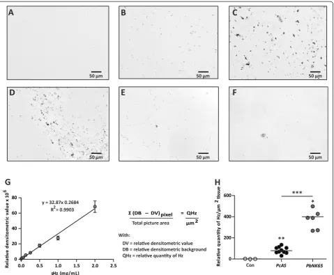
PbNK65 or PcAS and monitored with light microscopy. Hz was observed on transmitted light images from infected mouse organs as brown pigments and was ab-sent in organ sections from uninfected mice as is shown for the liver (Figure 1A–F). The pigment was found equally distributed throughout the liver (Figure 1B–C),
whereas in the spleen (Figure 1D), the lungs (Figure 1E) or the kidneys (Figure 1F), it was located in the red pulp, the interstitial tissue or clustered in presumably the glomeruli, respectively. Brains and hearts contained such low amounts of Hz that it was almost unnoticeable on unstained sections. To estimate the amount of Hz on organ cryosections, semi-quantitative densitometry with the AxioVision 4.6 software using a home-written script was applied. The linearity of this densitometric method was investigated on cryosections from gelatin-blocks with different concentrations of sHz. A broad linear rela-tionship was found between the sHz concentration and the obtained relative densitometric value (Figure 1G). The detected Hz signal was converted into the relative quantity of Hz/μm2tissue as calculated with the formula described in Figure 1G. This technique was used to measure Hz in liver cryosections from non-infected,
PcAS and PbNK65-infected mice. Despite similar mean peripheral parasitaemia levels in both groups of mice (PcAS 18%; PbNK65 12.7%; p= 0.4), significantly more Hz/μm2tissue was detected in livers ofPbNK65-infected mice compared with PcAS-infected mice (Figure 1H). This technique combines in-situ information with rela-tive quantification and can be used to measure Hz in organs with a high and evenly distributed Hz content such as livers. However, this method is poorly applicable for organs in which the pigment is not equally distribu-ted, e.g. spleen, lungs and kidneys. In brains and hearts, the amounts of Hz were too low to be quantified by densitometry.
Comparison of haem quantification by different techniques
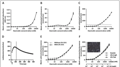
raising the concentrations of luminol and peroxide 100-fold. In this way, a detection limit around 100 nM was obtained (Figure 2C). Furthermore, the time-dependence of the luminescence signal was examined by measuring the emitted light of a single concentration during a kin-etic measurement. With the described protocol for haem-enhanced luminescence in a 96-well plate format, emitted light was measured when the luminescence sig-nal was almost maximal (Figure 2D). The sensitivity of this 96-well plate-based assay is significantly lower than the sensitivity of the cuvette-based method described by Schwarzeret al.[24]. However, the current 96-well based
[image:5.595.60.540.88.483.2]method has the advantage of a higher throughput and the sensitivity appeared sufficient to measure Hz in organ extracts. To extract Hz from organs, the method described by Sullivanet al.[15] was optimized. The most important modification was the addition of an overnight proteinase K digestion step that eliminated high back-ground signals e.g. in lung samples. After several wash steps to remove any free haemin, the Hz crystals were converted to free haematin by dissolving in a strong alka-line environment, so that the haematin concentration could be measured. Since the extraction procedure involved washing steps in the presence of SDS, possible
Figure 1Haemozoin detection by densitometric analysis.Transmitted light images (grey scale) were taken from unstained 7μm thick cryosections from livers of uninfected mice (A) and from mice infected withPcAS (B) orPbNK65 (C), fromPcAS-infected spleens (D), PbNK65-infected lungs (E) and kidneys (F). In panelG, the relative densitometric value obtained from cryosections of gelatin blocks with different concentrations of sHz were analyzed and the formula used to calculate the relative quantity of Hz/μm2is shown. In panelH, the relative Hz
quenching of the emitted light by SDS was investigated and had no effect on the luminescence catalyzed by haematin-Fe3+(Figure 2E).
As a final check up for the appropriateness of the assay for quantifying natural Hz, the concentration de-pendence of the haem-enhanced luminescence mea-sured with the plate reader was studied with natural Hz purified from trophozoites and from the liver. As shown in Figure 2F, haemin and Hz derived from tro-phozoites or livers behaved similarly in the assay con-firming the suitability of using haemin as a standard for deducing the Hz concentration in the samples. In addition, as birefringence is a typical feature of Hz [28], Hz isolated from the liver was spread on a glass slide and monitored by polarized light microscopy. This con-firmed that the isolated material consisted mainly of Hz (Figure 2F, inset).
Quantitative analysis of haemozoin in various organs of mice infected with different parasite species
[image:6.595.55.541.89.359.2]The optimized haem-enhanced luminescence technique was used to study differences in the amount of Hz be-tween various organs and bebe-tween the same organs of mice infected with parasites of different pathogenicity (PbANKA,PbNK65 orPcAS).Mice were sacrificed at the indicated times post-infection and perfused systemically. Even though no difference was found in the amount of Hz before and after perfusion (Additional file 1), it seemed more reasonable to apply perfusion on all samples tested. In this way, there could be no doubt that the detected Hz represented organ-trapped Hz and not Hz present in the circulation. Quantification of the total amount of Hz per organ revealed that most Hz was present in livers followed by spleens (Figure 3A–B). Far less Hz was detected in lungs and kidneys (Figure 3C–D), whereas subnanomolar
Figure 2Haem quantification by different techniques.Different concentrations of haematin (10μM–1.2 nM) were used to compare the sensitivity of previously described techniques to quantify haem in a 96-well based format. In panelA, haematin was measured with a
levels of Hz were found in brains and hearts, irrespectively of the parasite species used (Figure 3E–F). Livers, lungs, kidneys and hearts from PbNK65-infected mice nine to ten days post-infection contained significantly more Hz compared with the same organs from PbANKA-infected mice seven to eight days post-infection orPcAS-infected mice ten days post-infection, even though livers ofPc AS-infected mice were significantly larger (p< 0.0001 for liver weights between PcAS d10 and PbNK65 d9-10 and be-tweenPcAS d10 and PbANKA d7–8). In addition, lungs
and hearts from PbANKA-infected mice seven to eight days post-infection had significantly more Hz compared with the same organs from PcAS-infected mice after ten days of infection. The total amount of Hz was similar in spleens of PcAS and PbNK65-infected mice ten days post-infection (Figure 3B), although the amount of Hz/mg spleen tissue was six-fold lower in mice infected with
PcAS compared to PbNK65 (median value was 1164.4 pmol haematin/mg spleen for PbNK65 and 199.9 pmol haematin/mg spleen for PcAS; p< 0.0001). This was
compensated by the three- to four-fold larger spleen size in PcAS-infected mice (p< 0.0001). Furthermore, similar amounts of Hz were observed in brains fromPbNK65 and
PbANKA-infected mice, whereas less Hz was found in brains ofPcAS-infected mice.
Different total haemozoin levels inPcAS-infected mice andPlasmodium berghei-infected mice
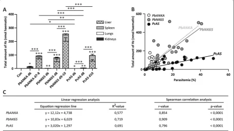
As brains and hearts contained only subnanomolar amounts of Hz, individual total Hz levels were calculated by making the sum of organ-specific Hz levels from liver, spleen, lungs and kidneys. Mice infected with parasites of theP. bergheistrains contained significantly more Hz than mice infected withPcAS-parasites (Figure 4A). Moreover, the total amounts of Hz correlated with the peripheral parasitaemia levels for all parasite strains (Figure 4B and C).
[image:7.595.59.541.87.394.2]PbANKA and PbNK65-parasites had a similar Hz-production pattern, i.e. comparable amounts of Hz at simi-lar peripheral parasitaemia levels. In contrast, Pc AS-infected mice seemed to have significantly less Hz in
Figure 3Quantification of haemozoin in tissues.C57BL/6 J mice were infected with 104PbANKA,PbNK65 orPcAS parasites or were left
relation with their parasitaemia compared with PbANKA orPbNK65 as indicated by the lower slope on the sion curve. The difference between the slopes of the regres-sion lines was significantly different betweenPbNK65 and
PcAS-infected mice (p< 0.0001).
Discussion
The first observations of black pigment in necroptic spleens and brains go back to the 18thCentury (reviewed in [5]). About 130 years later, a publication mentioned brown-grey colourations of brain, spleen and liver, which turned out to arise from pigment deposition. At first believed to be melatonin, it was later linked to a parasitic disease. Presently, manyin vitroandex vivo immunomo-dulating effects have been ascribed to Hz [4-7]. However, data about the fate and properties of Hz in thein vivo situ-ation are still scarce. Hz is released in the circulsitu-ation in considerable amounts after schizont rupture where it may interact with a whole range of different cell types. The ma-jority of the liberated Hz is presumably captured and pha-gocytosed by circulating and tissue resident monocytes/ macrophages in which it can persist for a long time. In
this way, Hz may be capable of causing considerable in-flammation that might progress to tissue injury. In this study, techniques for sensitively quantifying the amount of Hz in tissues were examined and the organ-specific Hz content was compared between parasite species with a varying degree of pathogenicity.
[image:8.595.58.539.91.361.2]As Hz crystals were observed on unstained cryosections from livers, spleens, lungs and kidneys, a technique for es-timating the amount of Hz in these sections by densito-metric analysis was developed. As Hz is proportionally distributed throughout the liver, the estimation of the amount of Hz by densitometry was quite reliable. In other organs, however, Hz was found in specific structures such as the red pulp in the spleen, the interstitial tissue in the lungs or presumably the glomeruli in the kidneys. This may in part be attributed to the differential localization of tissue-resident phagocytes. This implies that Hz distri-bution is a confounding factor for the accuracy of the Hz measurements on organ cryosections by densitom-etry. In addition, this technique is time-consuming, labour-intensive, semi-quantitative and not suitable for organs with a low Hz content and was thus not further
explored. Therefore, a more sensitive, analytical and quan-titative method for determining the Hz content in tissues was investigated. To isolate Hz from organs, a protocol described by Sullivan and colleagues [15] was modified. The main adaptation was the digestion of the homoge-nates with proteinase K. This digestion eliminated high background signals, which were presumably due to the binding of Hb to otherwise insoluble extracellular matrix components. Upon conversion of the isolated Hz into sol-uble haematin, a chemo-luminescence assay was used for quantification. This assay was based on the method of Schwarzeret al.[24] and adapted to microtiter plate for-mat. The obtained sensitivity with the optimized protocol was lower compared with the haem-enhanced lumines-cence assay described by Schwarzer et al. This was not due to quenching of the luminescence signal by SDS nor was it caused by the altered time frame during which the emitted light was measured (two seconds/sample versus
approximately ninety six seconds/plate), but probably ori-ginated from the use of different luminescence detector systems (cuvette systemversusmicroplate reader). Never-theless, the microplate-adjusted approach offers the ad-vantage of measuring several samples in varying concentrations simultaneously with a sensitivity that is op-timal for the quantification of Hz in malaria-infected organs.
As an application, the distribution of Hz throughout the body of infected mice was studied and compared between diverse parasite strains with varying pathogen-icity. Sullivan and colleagues already quantified the Hz content in brains, livers and spleens of mice [15,29] and in human placentas [11], but no detailed comparison between organs and between parasite species was described. Almost 95% of the total pool of Hz was found in livers and spleens. This was expected as large volumes of blood are filtered through these organs and both contain a vast population of tissue-resident mono-cytes/macrophages capable of rapidly removing the crystalline material from the circulation by means of phagocytosis. It was also important to consider the liver and spleen sizes when determining the total Hz amounts, as these sizes evolve in a different way during infection with different parasites (i.e. induction of hepa-tosplenomegaly by PcAS). As the absolute Hz concen-tration in the organs could be determined by the luminescence assay, this was easily taken into account by multiplication with the organ weights.
Furthermore, substantial amounts of Hz were detected in lungs of malaria-infected mice. In a new mouse model of MA-ARDS [16], considerable amounts of Hz were observed on histological sections of the lungs. By quanti-fying the Hz content in the lungs, significantly higher Hz levels were validated in lungs from P. berghei-infected mice (lung pathology) compared to PcAS-infected mice
(no lung pathology), indicating that Hz may have a role in the development of malaria-associated lung disease.
Low but detectable amounts of Hz were found in kid-neys, hearts and brains of malaria-infected mice. Most Hz was detected in kidneys and hearts fromPbNK65-infected mice ten days post-infection compared withPbANKA and
PcAS-infected mice seven to eight and ten days post-infection, respectively. However, a different pattern was observed in the brains, i.e. Hz was undetectable in brains from PcAS-infected mice whereas similar amounts of Hz were detected in brains ofPbNK65 andPbANKA-infected mice. A possible explanation for this difference is their di-verse parasite synchronicity. At the moment of sacrificing the mice and organ removal, thePcAS-parasites in the cir-culation were all in the ring and young trophozoite stage. As these developmental stages do not yet contain abun-dant Hz [3,10], it seemed reasonable that Hz was not detected in brains from mice infected with this parasite species. It is also possible that no Hz was detected because
PcAS-parasites may not sequester in the brains as is the case forPlasmodium vivax-infected erythrocytes [10]. On the contrary, several developmental stages of P. berghei
parasites are found in the circulation simultaneously and accumulation of P. bergheiin the brain is still a debated issue. The observation of similar brain Hz contents in
PbANKA andPbNK65-infected mice cannot be explained by their parasitaemia levels as significantly higher parasi-taemias were found in mice that were infected with
PbNK65 than in mice infected withPbANKA. The data however do suggest that Hz as such is not sufficient for the development of this immunopathology as Pb NK65-infected C57BL/6 J mice do not develop cerebral compli-cations [16]. These data are in contrast with data from Coban et al.[14] and Sullivanet al. [15] who found that brains from mice with cerebral pathology contained more Hz than healthy brains from infected mice. However, this may be explained by differences in the timing of analysis after infection and in the mouse or parasite strains used in the studies.
Organ-trapped Hz may originate from two sources. As free Hz is rapidly removed from the circulation, it is found either inside phagocytes or inside cyto-adhering iRBCs along the endothelial lining of the organs’ micro-vasculature. Systemic perfusion removes circulating iRBCs but not sequestering iRBCs or Hz inside resident phagocytes, although inadequate perfusion can result from obstruction due to organ-specific cyto-adherence and haemorrhages. Furthermore, it is still not completely clarified if sequestration by murine malaria parasites occurs and which organs are the main targets. Local parasite accumulation has been demonstrated in brains and lungs of PbANKA-infected mice suffering from cerebral symptoms [18,30], but no reports exist on
After calculating the total amount of Hz in the mice, it was found that PbANKA and PbNK65-infected mice contained similar amounts of Hz at comparable parasit-aemia levels. This suggests that both parasites produced similar amounts of Hz, or that their schizonts presum-ably consumed comparable amounts of Hb. On the con-trary, lower amounts of Hz were retrieved in Pc AS-infected mice despite of similar peripheral parasitaemia. Several explanations can be given for this finding.Pc AS-parasites may produce less Hz, e.g. by digesting less Hb or by using other haem detoxification mechanisms (transport of haem out of the food vacuole or anti-oxidative defense mechanisms of the parasite) or PcAS Hz could be more easily degraded. Interestingly, Noland
et al. [31] demonstrated that Hz crystals from different
Plasmodium species have different shapes and dimen-sions, supporting the notion that Hz from different spe-cies may have different properties. In addition, Hz contents are variable in RBC infected with different
Plasmodium falciparumstrains [32].
Another possibility is that peripheral parasitaemia, esti-mated by counting the percentage of iRBCs by micro-scopic analysis of Giemsa-stained blood smears, are not a true reflection of the total parasite biomass as they do not take sequestered parasites into account. Consequently, it is possible that PcAS-infected mice contain less Hz be-cause of lower total parasite burdens. These observations may well translate to the situation in human malaria, where various parasite species have different degrees of virulence. Total parasite biomass in P. falciparum infec-tions is higher than peripheral parasitaemia levels and the difference between these two parameters increases with disease severity [33]. Similarly, Hz-containing peripheral leukocytes are a marker for disease severity [34-36], and accumulation of Hz in brain micro-vessels is associated with a subtype of cerebral malaria [37]. No data are avail-able yet about total parasite burdens inP. vivax-infections and it is still questionable ifP. vivax-iRBCs can adhere to the endothelial micro-vascular lining. However, cytoadhe-sion of P. vivax-infected erythrocytes was demonstrated
in vitro [38] and, despite of the absence of sequestration in the brain [10], it was hypothesized that parasitized RBCs might sequester in lungs from patients withP. vivax
malaria [39]. Similarly, very little knowledge exists on the role of Hz inP. vivaxinfections.
Besides differences in pathogenicity, another interest-ing difference betweenP. bergheiandPcAS is thatPcAS can be cleared from the circulation in several mouse strains, including C57BL/6 mice, whereas PbANKA and
PbNK65 cannot. The amounts of Hz produced by these parasites may also contribute to these differences, as Hz is known to suppress macrophage activity in vitro [40] and in vivo [41]. Interestingly, Spaccapelo et al. found that plasmepsin 4-deficient PbANKA-parasites, which
produce less Hz, cause less immunopathology and are more easily cleared by some mouse strains [42].
Conclusions
This paper describes newly developed and improved methods for sensitive Hz quantification in mouse organs. Different amounts of Hz were detected in the analysed organs and total Hz contents were highest in mice that were infected with lethal parasite strains. Therefore, it is clear that these techniques will be valuable in the inves-tigation of a possible relationship between Hz and organ-specific malaria pathologies.
Additional file
Additional file 1:Effect of perfusion on the
organ-specific haemozoin content.To investigate the effect of perfusion on the organ-specific haemozoin content, mice were infected intraperitoneally with 104PbNK65 parasites. Ten days later, mice were
sacrificed, the right lung and kidney were pinched off, and the other organs were perfused with phosphate-buffered saline to remove circulating erythrocytes. The amount of Hz/mg tissue was quantified in both perfused and non-perfused lungs and kidneys with the modified 96-well plate based haem-enhanced luminescence assay. The total Hz content in lung and kidney was calculated by multiplying the amount of Hz/mg tissue with the organ weights. As shown in panel A and B, no difference was found in the amount of Hz in lungs and kidneys with or without perfusion. Nevertheless, perfusion was applied for all organs tested as it is conceptually more rational to remove circulating infected erythrocytes as a source of haemozoin. Each dot represents data of an individual mouse.
Abbreviations
CM, Cerebral malaria; DB, Densitometric background; DV, Densitometric value; Hb, Haemoglobin; Hz, Haemozoin; MA-ARDS, Malaria-associated acute respiratory distress syndrome;PbANKA,Plasmodium bergheiANKA;
PbNK65,Plasmodium bergheiNK65;PcAS,Plasmodium chabaudiAS; iRBC, Infected red blood cell; sHz, Synthetic haemozoin (beta-haematin).
Competing interests
The authors declare that they have no competing interests.
Authors’contributions
KD participated in the design of the study, performed the animal experiments, optimized the luminescence technique, analysed and interpreted the data, performed the statistical analysis and drafted the manuscript. NL participated in Hz analysis. SN operated the microscope and designed the script for the densitometric analysis of Hz on pictures from organ cryosections. EM participated in optimizing the luminescence technique. GO participated in study design, manuscript writing and provided critical support. PVDS conceived and participated in the design of the study, optimized the method for densitometric analysis of Hz in cryosections and participated in interpreting the data and drafting the manuscript. All authors read and approved the final manuscript.
Acknowledgements
We want to thank Prof. Jo Van Damme and Prof. Sofie Struyf for the usage of the Axiovert 200 M microscope and Prof. Johan Neyts for the usage of the Precellys homogenizer and the luminometer. Furthermore, we are grateful to Prof. E Schwarzer and Prof. P Arese (University of Turin, Italy) for helpful discussions.
Author details 1
Laboratory of Immunobiology, Rega Institute, University of Leuven, Leuven, Belgium.2Laboratory of Molecular Immunology, Rega Institute, University of Leuven, Leuven, Belgium.3Currently at the laboratory of Virology and Chemotherapy, Rega Institute, University of Leuven, Leuven, Belgium.
Received: 17 November 2011 Accepted: 7 March 2012 Published: 14 May 2012
References
1. Schofield L, Grau GE:Immunological processes in malaria pathogenesis.
Nat Rev Immunol2005,5:722–735.
2. Goldberg DE:Hemoglobin degradation.Curr Top Microbiol Immunol2005,
295:275–291.
3. Rosenthal PJ, Meshnick SR:Hemoglobin catabolism and iron utilization by malaria parasites.Mol Biochem Parasitol1996,83:131–139.
4. Shio MT, Kassa FA, Bellemare MJ, Olivier M:Innate inflammatory response to the malarial pigment hemozoin.Microbes Infect2010,12:889–899. 5. Hanscheid T, Egan TJ, Grobusch MP:Haemozoin: from melatonin pigment
to drug target, diagnostic tool, and immune modulator.Lancet Infect Dis 2007,7:675–685.
6. Schwarzer E, Skorokhod OA, Barrera V, Arese P:Hemozoin and the human monocyte–a brief review of their interactions.Parassitologia2008,50:143–145. 7. Arese P, Schwarzer E:Malarial pigment (haemozoin): a very active‘inert’
substance.Ann Trop Med Parasitol1997,91:501–516.
8. Grau GE, Mackenzie CD, Carr RA, Redard M, Pizzolato G, Allasia C, Cataldo C, Taylor TE, Molyneux ME:Platelet accumulation in brain microvessels in fatal pediatric cerebral malaria.J Infect Dis2003,187:461–466. 9. Dorovini-Zis K, Schmidt K, Huynh H, Fu W, Whitten RO, Milner D, Kamiza S,
Molyneux M, Taylor TE:The neuropathology of fatal cerebral malaria in malawian children.Am J Pathol2011,178:2146–2158.
10. Silamut K, Phu NH, Whitty C, Turner GD, Louwrier K, Mai NT, Simpson JA, Hien TT, White NJ:A quantitative analysis of the microvascular sequestration of malaria parasites in the human brain.Am J Pathol1999,155:395–410. 11. Sullivan AD, Nyirenda T, Cullinan T, Taylor T, Lau A, Meshnick SR:Placental
haemozoin and malaria in pregnancy.Placenta2000,21:417–421. 12. Ismail MR, Ordi J, Menendez C, Ventura PJ, Aponte JJ, Kahigwa E, Hirt R,
Cardesa A, Alonso PL:Placental pathology in malaria: a histological, immunohistochemical, and quantitative study.Hum Pathol2000,31:85–93. 13. Muehlenbachs A, Fried M, McGready R, Harrington WE, Mutabingwa TK,
Nosten F, Duffy PE:A novel histological grading scheme for placental malaria applied in areas of high and low malaria transmission.J Infect Dis 2010,202:1608–1616.
14. Coban C, Ishii KJ, Uematsu S, Arisue N, Sato S, Yamamoto M, Kawai T, Takeuchi O, Hisaeda H, Horii T, Akira S:Pathological role of Toll-like receptor signaling in cerebral malaria.Int Immunol2007,19:67–79. 15. Sullivan AD, Ittarat I, Meshnick SR:Patterns of haemozoin accumulation in
tissue.Parasitology1996,112(Pt 3):285–294.
16. Van den Steen PE, Geurts N, Deroost K, Van AI, Verhenne S, Heremans H, Van Damme J, Opdenakker G:Immunopathology and dexamethasone therapy in a new model for malaria-associated acute respiratory distress syndrome.Am J Respir Crit Care Med2010,181:957–968.
17. Craig AG, Grau GE, Janse C, Kazura JW, Milner D, Barnwell JW, Turner G, Langhorne J:The role of animal models for research on severe malaria.
PLoS Pathog2012,8:e1002401.
18. Haque A, Best SE, Unosson K, Amante FH, de Labastida F, Anstey NM, Karupiah G, Smyth MJ, Heath WR, Engwerda CR:Granzyme B expression by CD8+ T cells is required for the development of experimental cerebral malaria.J Immunol2011,186:6148–6156.
19. Mohan A, Sharma SK, Bollineni S:Acute lung injury and acute respiratory distress syndrome in malaria.J Vector Borne Dis2008,45:179–193. 20. Valecha N, Pinto RG, Turner GD, Kumar A, Rodrigues S, Dubhashi NG,
Rodrigues E, Banaulikar SS, Singh R, Dash AP, Baird JK:Histopathology of fatal respiratory distress caused byPlasmodium vivaxmalaria.Am J Trop Med Hyg2009,81:758–762.
21. Stephens R, Culleton RL, Lamb TJ:The contribution ofPlasmodium chabaudito our understanding of malaria.Trends Parasitol2012,28:73–82. 22. Geurts N, Martens E, Van Aelst I, Proost P, Opdenakker G, Van den Steen PE:
Beta-hematin interaction with the hemopexin domain of gelatinase B/MMP-9 provokes autocatalytic processing of the propeptide, thereby priming activation by MMP-3.Biochemistry2008,47:2689–2699.
23. Ncokazi KK, Egan TJ:A colorimetric high-throughput beta-hematin inhibition screening assay for use in the search for antimalarial compounds.Anal Biochem2005,338:306–319.
24. Schwarzer E, Turrini F, Arese P:A luminescence method for the quantitative determination of phagocytosis of erythrocytes, of malaria-parasitized erythrocytes and of malarial pigment.Br J Haematol1994,88:740–745. 25. Yuan J, Shiller AM:Determination of subnanomolar levels of hydrogen
peroxide in seawater by reagent-injection chemiluminescence detection.
Anal Chem1999,71:1975–1980.
26. Janse CJ, Ramesar J, Waters AP:High-efficiency transfection and drug selection of genetically transformed blood stages of the rodent malaria parasitePlasmodium berghei.Nat Protoc2006,1:346–356.
27. http://www.stattools.net/.
28. Lawrence C, Olson JA:Birefringent hemozoin identifies malaria.Am J Clin Pathol1986,86:360–363.
29. Levesque MA, Sullivan AD, Meshnick SR:Splenic and hepatic hemozoin in mice after malaria parasite clearance.J Parasitol1999,85:570–573. 30. Fonager J, Pasini EM, Braks JA, Klop O, Ramesar J, Remarque EJ, Vroegrijk IO,
van Duinen SG, Thomas AW, Khan SM, Mann M, Kocken CH, Janse CJ, Franke-Fayard BM:Reduced CD36-dependent tissue sequestration of Plasmodium-infected erythrocytes is detrimental to malaria parasite growth in vivo.J Exp Med2012,209:93–107.
31. Noland GS, Briones N, Sullivan DJ Jr:The shape and size of hemozoin crystals distinguishes diverse Plasmodium species.Mol Biochem Parasitol 2003,130:91–99.
32. Orjih AU, Fitch CD:Hemozoin production byPlasmodium falciparum:
variation with strain and exposure to chloroquine.Biochim Biophys Acta 1993,1157:270–274.
33. Dondorp AM, Desakorn V, Pongtavornpinyo W, Sahassananda D, Silamut K, Chotivanich K, Newton PN, Pitisuttithum P, Smithyman AM, White NJ, Day NP:Estimation of the total parasite biomass in acute falciparum malaria from plasma PfHRP2.PLoS Med2005,2:e204.
34. Nguyen PH, Day N, Pram TD, Ferguson DJ, White NJ:Intraleucocytic malaria pigment and prognosis in severe malaria.Trans R Soc Trop Med Hyg1995,89:200–204.
35. Amodu OK, Adeyemo AA, Olumese PE, Gbadegesin RA:Intraleucocytic malaria pigment and clinical severity of malaria in children.Trans R Soc Trop Med Hyg1998,92:54–56.
36. Hanscheid T, Langin M, Lell B, Potschke M, Oyakhirome S, Kremsner PG, Grobusch MP:Full blood count and haemozoin-containing leukocytes in children with malaria: diagnostic value and association with disease severity.Malar J2008,7::109.
37. Taylor TE, Fu WJ, Carr RA, Whitten RO, Mueller JS, Fosiko NG, Lewallen S, Liomba NG, Molyneux ME:Differentiating the pathologies of cerebral malaria by postmortem parasite counts.Nat Med2004,10:143–145. 38. Carvalho BO, Lopes SC, Nogueira PA, Orlandi PP, Bargieri DY, Blanco YC,
Mamoni R, Leite JA, Rodrigues MM, Soares IS, Oliveira TR, Wunderlich G, Lacerda MV, del Portillo HA, Araujo MO, Russell B, Suwanarusk R, Snounou G, Renia L, Costa FT:On the cytoadhesion ofPlasmodium vivax-infected erythrocytes.J Infect Dis2010,202:638–647.
39. Anstey NM, Handojo T, Pain MC, Kenangalem E, Tjitra E, Price RN, Maguire GP: Lung injury in vivax malaria: pathophysiological evidence for pulmonary vascular sequestration and posttreatment alveolar-capillary inflammation.J Infect Dis2007,195:589–596.
40. Schwarzer E, Turrini F, Ulliers D, Giribaldi G, Ginsburg H, Arese P:
Impairment of macrophage functions after ingestion ofPlasmodium falciparum-infected erythrocytes or isolated malarial pigment.J Exp Med 1992,176:1033–1041.
41. Scorza T, Magez S, Brys L, De Baetselier P:Hemozoin is a key factor in the induction of malaria-associated immunosuppression.Parasite Immunol 1999,21:545–554.
42. Spaccapelo R, Janse CJ, Caterbi S, Franke-Fayard B, Bonilla JA, Syphard LM, Di Cristina M, Dottorini T, Savarino A, Cassone A, Bistoni F, Waters AP, Dame JB, Crisanti A:Plasmepsin 4-deficientPlasmodium bergheiare virulence attenuated and induce protective immunity against experimental malaria.Am J Pathol2010,176:205–217.
doi:10.1186/1475-2875-11-166