Open Access
Research
Combined IL-21 and Low-Dose IL-2 therapy induces anti-tumor
immunity and long-term curative effects in a murine melanoma
tumor model
Hong He
1, Preya Wisner
1, Guojun Yang
1, Hong-Ming Hu
1, Dan Haley
1,
William Miller
1, Aisling O'Hara
1, W Gregory Alvord
2, Christopher H Clegg
3,
Bernard A Fox
1, Walter J Urba
1and Edwin B Walker*
1Address: 1Robert W. Franz Cancer Research Center, Earle A. Chiles Research Institute, Providence Portland Medical Center, Portland OR, USA, 2DMS-National Cancer Institute, Frederick MD, USA and 3Immunology Research, ZymoGenetics, Seattle WA, USA
Email: Hong He - hong.he@providence.org; Preya Wisner - Kpreyaa@gmail.com; Guojun Yang - guojun.yang@providence.org; Hong-Ming Hu - hhu@providence.org; Dan Haley - daniel.haley@providence.org; William Miller - william.miller@providence.org; Aisling O'Hara - aohara@liai.org; W Gregory Alvord - gwa@css.ncifcrf.gov; Christopher H Clegg - cleggc@zgi.com;
Bernard A Fox - foxb@foxlab.org; Walter J Urba - walter.urba@providence.org; Edwin B Walker* - edwin.walker@providence.org * Corresponding author
Abstract
Background: In vivo studies have recently demonstrated that interleukin 21 (IL-21) enhances the anti-tumor function of T-cells and NK cells in murine tumor models, and the combined use of IL-21 and IL-15 has resulted in prolonged tumor regression and survival in mice with previously established tumors. However, the combined anti-tumor effects of IL-21 and low dose IL-2 have not been studied even though IL-2 has been approved for human use, and, at low dose administration, stimulates the proliferation of memory T cells, and does not significantly increase antigen-induced apoptosis or regulatory T cell (Treg) expansion. This study examined whether recombinant IL-21 alone or in combination with low-dose IL-2 could improve the in vivo anti-tumor function of naïve, tumor-antigen specific CD8+ T cells in a gp100
25–33 T cell receptor transgenic pmel murine
melanoma model.
Methods: Congenic C57BL/6 (Ly5.2) mice bearing subcutaneous B16F10 melanoma tumors were sublethally irradiated to induce lymphopenia. After irradiation naive pmel splenocytes were adoptively transferred, and mice were immunized with bone marrow-derived dendritic cells pulsed with human gp10025–33 (hgp10025–33). Seven days after vaccination groups of mice received 5
consecutive days of intraperitoneal administration of IL-2 alone (20 × 103 IU), IL-21 alone (20 µg)
or IL-21 and IL-2. Control animals received no cytokine therapy.
Results: IL-21 alone and IL-2 alone both delayed tumor progression, but only IL-21 significantly augmented long-term survival (20%) compared to the control group. However, combination therapy with IL-21 and IL-2 resulted in the highest long-term (>150 days) tumor-free survival frequency of 46%. Animals that were tumor-free for > 150 days demonstrated tumor-specific protection after rechallenge with B16F10 melanoma cells. At peak expansion (21 days post vaccination), the combination of IL-21 plus IL-2 resulted in a 2- to 3-fold higher absolute number of circulating tumor antigen-specific pmel CD8+ T cells than was stimulated by IL-2 or IL-21 alone.
Published: 13 June 2006
Journal of Translational Medicine 2006, 4:24 doi:10.1186/1479-5876-4-24
Received: 10 April 2006 Accepted: 13 June 2006 This article is available from: http://www.translational-medicine.com/content/4/1/24
© 2006 He et al; licensee BioMed Central Ltd.
Pmel CD8+ T cells were predominantly partitioned into central memory (CD62L+/CD127+) or
effector-memory (CD62L-/CD127+) phenotypes by day 28-post vaccination in IL-21 + IL-2 treated
mice.
Conclusion: These observations support the potential use of IL-21 and low-dose IL-2 therapy in combination with a tumor-antigen vaccine and lymphopenic conditioning in future cancer clinical trials to maintain high numbers of anti-tumor memory CD8+ T cells with the potential to sustain
long term tumor regression and survival.
Background
IL-21 has potent immunomodulatory effects on T cells and NK cells [1]. However, the current understanding of the effects of IL-21 on the regulation of both antigen-inde-pendent homeostatic and antigen-stimulated prolifera-tion and activaprolifera-tion of naive and memory CD8+ T cells is
confounded by conflicting data. IL-21 has been reported both to synergize with IL-15 to increase homeostatic anti-gen-independent proliferation of naive and memory CD8+ T cells [2,3], and to inhibit IL-15 induced
homeo-static proliferation of memory T cells [4]. While evidence suggests IL-21 enhances primary antigen-stimulated T-cell proliferation and functional activation [3-6], there is disa-greement over whether this effect occurs during priming [5], or during the late expansion phase of antigen-specific proliferation [6]. Recent data suggests IL-21 primarily increases the frequency and absolute number of in vitro stimulated antigen-specific CD8+ T cells by enhancing the
rate of proliferation rather than by reducing apoptosis [5]. Alternatively, other studies indicate IL-21 maintains increased numbers of antigen-stimulated effector and long-term memory CD8+ T cells by reducing apoptosis
while maintaining a low rate of cell division [6]. These conflicting data may be attributed to differences in the in vitro and in vivo models employed in the individual stud-ies. However, they suggest that much work remains in the effort to elucidate the mechanism of IL-21 regulation of T-cell immune responses, including any potential role it may play in the regulation of T-cell mediated anti-tumor immunity through the induction of functionally active tumor-specific effector and memory T cells (Reviewed in [1]).
In vivo data demonstrate that IL-21 enhances the anti-tumor function of T-cells and NK cells. Several studies have shown that genetically engineered IL-21-secreting murine tumors activate potent NK and CD8+ T cell
medi-ated anti-tumor responses [7-9]. IL-21 was also compared to IL-2 and IL-15 injected intraperitoneally (IP) subse-quent to challenge with the ova-expressing E.G7 thy-moma in either naive C57BL/6 mice, or in mice infused with OT-1 transgenic CD8+ T cells [6]. In both models,
IL-2 and IL-15 delayed tumor growth, but only IL-IL-21 resulted in significant prevention of tumor progression and improved survival beyond 50 days from tumor challenge.
The combination of IL-21 and IL-15 was also adminis-tered IP subsequent to human gp10025–33 (hgp10025–33) peptide vaccination of lymphopenic, tumor-bearing (B16F10 melanoma) C57BL/6 mice [3]. Before vaccina-tion mice received in vitro stimulated (IVS) pmel trans-genic (Tg) CD8+ T cells, which have a T-cell receptor
specific for the murine H-2Db restricted gp100
25–33
pep-tide of the B16F10 melanoma associated gp100 protein, and strongly cross react with hgp10025–33 [10]. The com-bination of IL-21 + IL-15 resulted in prolonged tumor regression and survival out to 32 days. Mice treated with the vaccine plus IL-15 or IL-21 alone all died of tumor within 32 days of treatment. No data was presented dem-onstrating long-term survival (>60 days) of mice treated with IL-21 + IL-15. Notably, IL-2 was not tested in vivo in combination with IL-21 in this study primarily because the combination of these two cytokines failed to drive antigen-independent homeostatic proliferation of murine CD8+ splenocytes in vitro [3], and the concern that
high-dose IL-2 has been shown to decrease memory CD8+ T-cell
function by inducing regulatory T-cells and increasing antigen-driven apoptosis [11-14]. However, other data have demonstrated that the in vitro maintenance of anti-gen-stimulated murine T cells in low-dose IL-2 resulted in the proliferative expansion of cells with a memory pheno-type similar to that induced by IL-15; these cells were capable of long-term survival in vivo, and potent prolifer-ative and functional responses after secondary in vivo challenge [15]. Thus, treatment with 21 + low-dose IL-2 after the adoptive transfer of pmel Tg CD8+ T cells into
mice with established B16 melanoma tumors (pmel Tg/ B16 model) might yield anti-tumor effects comparable to or greater than those observed with IL-21+IL-15.
The pmel Tg/B16 melanoma model has been employed repeatedly in recent studies to model immunotherapeutic strategies for treatment of pre-existing disease. Most of these experiments have focused on the adoptive transfer of antigen-educated pmel CD8+ splenocytes stimulated in
vitro with hgp10025–33 peptide and IL-2, and the
(≥ 100 days) with IL-2 alone [16,17], IL-15 alone [18], or the combination of IL-15 and IL-21 [3] – except in mice with small tumors (≤ 10 mm2) at the time of treatment, or
after the adoptive transfer of very large numbers (107) of
IVS pmel CD8+ T cells [16]. The adoptive transfer of highly
activated IVS T-cells may not produce long-term anti-tumor curative effects due to functional and proliferative exhaustion [19-21]. This conclusion is supported by a series of provocative experiments using the pmel Tg/B16 model in which the adoptive transfer of naive or "early" IVS-effector pmel CD8+ T cells into lymphopenic
tumor-bearing recipient mice resulted in much more durable tumor regression following hgp10025–33 vaccination and IL-2 therapy compared to the poor therapeutic effects observed using IVS expanded, highly stimulated interme-diate or late-stage effector T cells [22]. Thus, as described herein, the adoptive transfer of naïve, tumor antigen-spe-cific CD8+ T cells in this model prior to vaccine and IL-21
+ low-dose IL-2 therapy may provide a better opportunity to study methods of generating both effector and long-term anti-tumor memory T-cell function than can be achieved with highly activated IVS T cells.
There have been few published studies describing the anti-tumor effects of recombinant IL-21 in conjunction with hgp10025–33 vaccination in B16F10 tumor-bearing lym-phopenic mice receiving naive pmel Tg CD8+ T cells. There
have, to our knowledge, been no reports describing the potential synergy of IL-21 + low-dose IL-2 in this or other tumor models. Herein we describe the long-term curative effect of combined IL-21 + low-dose IL-2 cytokine therapy in lymphopenic, tumor-bearing C57BL/6 mice infused with naive pmel splenocytes and vaccinated with the hgp10025–33 melanoma peptide.
Materials and methods
Mice
Congenic C57BL/6 (Ly5.2) mice (Charles River Laborato-ries, Inc. NCI-Frederick) bred at the Earle A. Chiles Research Institute (EACRI) served as recipient mice for tumor inoculation and pmel Tg CD8+ T-cell adoptive
transfer in all experiments. Pmel Tg mice express a TCR specific for an H-2Db epitope (gp100
25–33) of the
melanoma-associated gp100 protein on the C57BL/6 (Ly5.1) background [10,16]. Virtually all (>90%) CD8+T
cells in pmel Tg mice were Vβ13+, and were also
distin-guishable by a monoclonal antibody specific for a point mutation of the CD45 epitope (CD45.2). Pmel Tg mice were the generous gift of Dr. Nicholas Restifo (Surgery Branch, NCI-NIH) and were bred at EACRI. Male or female mice (10–16 weeks) were used in separate experi-ments. All mice were treated according to the regulations and guidelines of the Institutional Animal Care and Use Committee.
Tumor cell lines, tumor inoculation and in vivo measurement
The B16 F10 melanoma and 3LL (Lewis Lung) cell lines were obtained from ATCC. Both cell lines were main-tained in complete medium consisting of RPMI 1640 sup-plemented with 10% heat-inactivated fetal bovine serum (Biofluids, Rockville, MD), 0.03% L-glutamine, 100 µg/ ml streptomycin, 100 µg/ml penicillin, 50 µg/ml gen-tamicin sulfate and 50 mmol 2-mercaptoethanol. Tumor was established by injecting mice subcutaneously in the flank with 2 × 10⁵ B16 F10 melanoma cells in 0.1 ml of
phosphate buffered balanced salt solution (PBS). In tumor rechallenge experiments mice were injected subcu-taneously in the opposite flank with 5.0 × 10⁵ B16 F10
melanoma cells or 5.0 × 10⁴ 3LL tumor cells in 0.1 ml of
PBS. Tumor growth was monitored three times a week by measurement of two perpendicular diameters using a dig-ital caliper. The products of the perpendicular diameters plotted for multiple animals are presented as the mean mm2 ± SEM. Mice were sacrificed when tumors exceeded
200 mm2. Survival was analyzed by using Kaplan-Meier
statistics.
Adoptive cell transfer
Splenocytes from pmel transgenic mice were depleted of erythrocytes with ACK lysing buffer, and 4 × 10⁶ were
adoptively transferred via I.V. injection into C57BL/6 (Ly5.2) mice. Pmel spleen cells contained on average 20%–25% (~1 × 10⁶) naive CD45.2+ CD8+ T cells.
Peptides
The H-2Db-restricted hgp100
25–33 peptide,
(KVPRN-QDWL) was used as the CD8+ immunogen [16], and the
I-Ab-restricted epitope of Plasmodium falciparum
(NANPNVDPNANP), hereafter referred to as "NV", was used as a CD4+directed helper peptide [23,24]in all
exper-iments. The peptides were made by Invitrogen Inc. and purified by reverse-phase high-performance liquid chro-matography. Purity of >99% was confirmed by mass spec-trometry.
Preparation of dendritic cells
Bone marrow-derived murine dendritic cells (DCs) were generated as described previously [25]. Briefly, cells from the femur of C57BL/6 mice were grown at 1 × 10⁶ cells/ml
in RPMI 1640 complete medium supplemented with 25 ng/ml of murine GM-CSF (PeproTech, Rocky Hill, NJ). Fresh medium supplemented with GM-CSF was added on day 3, and all loosely adherent cells were transferred to petri dishes on day 6. Three days later, nonadherent and loosely adherent cells were harvested, washed, and frozen in 10% DMSO and 90% FCS in liquid nitrogen at 107 cells
times with PBS before injection. DC purity and matura-tion were confirmed by flow cytometry analysis after staining with antibodies against MHC class I (H-2Db,
H-2Kb), class II (I-Ab), CD11c, CD40, CD80, CD86. (BD
Bio-sciences PharMingen, San Diego, CA.)
Vaccine and cytokine therapy regimen
After B16F10 tumor cells were injected subcutaneously on day 0, groups of 5–10 mice per test group were sublethally irradiated (500 cGy) 7 days later to induce lymphopenia. All animals were then infused IV with 4 × 10⁶ pmel
splen-ocytes, which equated to the transfer of 8 × 10⁵ - 1 × 10⁶
naive CD45.2+/CD8+ pmel T cells. Immediately after cell
transfer, mice were immunized subcutaneously with 2 × 10⁶ C57BL/6 DCs pulsed with hgp100
25–33 (1 µg/ml) and
NV peptide (1 µg/ml). The hgp10025–33 and NV peptide pulsed DC immunization was the standard vaccine in all experiments. Seven days after pmel T cell transfer and vac-cination, mice received 5 consecutive days (days 14–18 post tumor inoculation) of IP injection of recombinant human IL-2 at 20 × 103 IU/dose (Chiron Corporation,
Emoryville, CA), murine IL-21 at 20 µg/dose (ZymoGe-netics Corporation, Seattle, WA), or the combination of IL-2 and IL-21 at the same dosage used for single cytokine injections.
Flow cytometry
Except where noted, cell surface staining of peripheral blood lymphocytes and spleen cells was performed using BD Biosciences PharMingen (San Diego, CA) reagents. Single-cell suspensions were incubated with anti-mouse CD16/32 (eBioscience, San Diego, CA) to block Fc recep-tors. Cells were then stained with allophycocyanin (APC) conjugated anti-CD8 and fluorescein isothiocyanate (FITC)-labeled CD45.2, phycoerythrin (PE) anti-CD62L and PE-Cy7 anti-CD127 (eBiosciences). Data were acquired on a FACS Calibur (BD Biosciences, San Jose, CA.) and analyzed using Cellquest Pro software (version 4.0.1). Peripheral blood and tissue derived-lymphocytes were analyzed by direct ex vivo interrogation of fresh sam-ples. The frequency of pmel T cells was determined by measuring the frequency of total gated CD8+ T cells, which
were also CD45.2+. CD8+ T cells from recipient Ly5.2 mice
could be distinguished from donor pmel CD8+ T-cells
since recipient T cells did not express the CD45.2 point mutation, and thus were not stained with the anti-CD45.2 antibody. "Flow-Count" fluorospheres (Beckman-Coul-ter; Miami, Fl.) were added to each sample before event acquisition to determine the absolute cell count according to the manufacturer's instructions. A minimum of 8000 "Flow Count" beads were counted for each sample col-lected. The absolute number of pmel CD8+ T cells per µl
of blood was determined by first calculating the ratio of beads counted by flow divided by the total number of beads added to the sample, and multiplying this ratio
times the known volume of the test sample of blood; this determined the fraction of the total test sample volume (in µls) analyzed. The total sample volume analyzed (in
µls) was then divided into the total analysis pmel cell count to give pmel+CD8+ T cells/µl.
Cytokine flow cytometry (CFC) analysis was performed by stimulating 106 spleen cells suspended in 250 ul of
complete medium in a microtiter well (96 well plate) with hgp10025–33 peptide (1 ug/ml). Splenocytes were cultured with antigen and brefeldin A (10 ug/ml) for 5 hours at 37°C. Cells were washed twice in PBS and incubated with mouse CD16/32 (FcR-blocking) monoclonal anti-body (eBiosciences). Cells were then stained with APC-conjugated CD8 and FITC-labeled CD45.2 (eBiosciences) antibodies for 30 minutes at 4°C, washed 2× in PBS, and fixed and permeablized with 100 ul Cytofix/Cytoperm™ buffer/well (BD Biosciences Pharmingen) for15 minutes at room temperature. Cells were washed 2× in Per-mWash™ buffer (BD Biosciences Pharmingen) and resus-pended in 100 ul of PermWash™ buffer. PE-conjugated anti-IL-2, IFNγ or TNFγ specific antibodies (BD Bio-sciences Pharmingen) were added at optimal dilution and incubated for one hour at 4°C; PE-conjugated isotype controls were used to determine non-specific cytoplasmic background staining. Cells were washed 2× with PBS and analyzed by flow cytometry. Cytokine positive cell fre-quencies were determined for pre-gated CD8+/
CD45.2+pmel T cells.
Statistical analysis
Phenotype data in this study were analyzed using repeated measures analysis of variance (ANOVA), analysis of covar-iance (ANCOVA), linear hierarchical mixed-effects regres-sion models, nonparametric (distribution free) tumor growth analyses, and simple and advanced graphical tech-niques [26-28]. Animal survival data were analyzed with standard log-rank statistics and Kaplan-Meier plots. Pair wise a posteriori comparisons among treatment conditions were performed with standard post hoc tests (i.e. Tukey's test). Interpretations of tumor growth results from follow-up tests were consistent with those obtained from global analyses. Hence, for simplicity we report probability (p) values obtained from follow-up nonparametric Wilcoxon tests at specific time points. All tests were two-sided; prob-ability values less than 0.05 were considered significant.
Results
Combined IL-21 + IL-2 therapy significantly enhanced anti-tumor immunity
Seven days after subcutaneous injection of 2 × 10⁵ B16F10
tumor inoculation) of IL-2 alone, IL-21 alone or both cytokines. The optimal dose of IL-21 (20 µg/dose) was established previously [6]; and the optimal IL-2 dose (20 × 103 IU/injection) was established by titration (data not
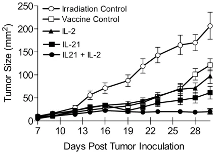
shown). Figure 1 shows the cumulative mean tumor size from three separate experiments during the first four weeks following tumor inoculation (≥ 15 mice/group). On day 14, just prior to cytokine administration, the mean tumor size for all vaccinated mice (N = 73) was 26 mm2 (± 2.4 mm). Thus, all vaccinated mice had
estab-lished disease prior to cytokine therapy. Global analysis showed that each therapy, including the vaccine alone, reduced the rate of tumor growth compared to the irradi-ation only control. By day 21, vaccinirradi-ation combined with
IL-21 + IL-2 treatment inhibited tumor growth signifi-cantly better than vaccination alone (p = 0.0013) and vac-cination combined with IL-2 (p = 0.0044) or with IL-21 (p = 0.044). By day 28, vaccination combined with 21+ IL-2 again inhibited tumor growth significantly better than vaccination alone (p < 0.0001) and vaccination combined with IL-2 (p = 0.0006) or with IL-21 (p = 0.0033). By day 30, vaccination combined with IL-21 + IL-2 similarly inhibited tumor growth significantly better than vaccina-tion alone (P < 0.0001), but produced higher statistically significant inhibition than vaccination combined with IL-2 (p = 0.000IL-2) or with IL-IL-21 (p = 0.00IL-24) when compared to day 28. Mice in the irradiation only (lymphopenic) control group exhibited rapid tumor growth; the mean
[image:5.612.89.511.131.433.2]B16F10 melanoma tumor growth in hgp10025–33 vaccinated and cytokine treated lymphopenic mice
Figure 1
B16F10 melanoma tumor growth in hgp10025–33 vaccinated and cytokine treated lymphopenic mice. Tumor size is presented as the mean (mm2) ± SEM of the cumulative analysis of replicate mice (≥ 15) for each test group from 3 separate experiments.
Cytokines were administered for 5 consecutive days beginning on day 14 when the average tumor size for vaccinated mice from all test groups in 3 experiments was 26 mm2 ± 2.4 mm (N = 73 mice). By day 30 IL-21 + low-dose IL-2 treated mice
showed significant inhibition of tumor growth compared to the vaccine control (p < 0.0001), or compared to vaccinated mice treated with IL-2 (p = 0.0002) or IL-21 only (p = 0.0024).
7
10
13
16
19
22
25
28
0
50
100
150
200
250
IL21 + IL-2
IL-21
IL-2
Vaccine Control
Irradiation Control
Days Post Tumor Inoculation
Tum
or
Si
z
e
(m
m
tumor size for all animals reached > 200 mm2 30 days
after tumor inoculation. Similarly, unvaccinated tumor-bearing control mice which were irradiated and treated with IL-21 or IL-21 + IL-2 only had comparable rapid tumor growth (data not shown). Mice treated with IL-21 + IL-2 after vaccination were the only animals that experi-enced no significant increase in tumor size (Figure 1).
IL-21 + IL-2 improved long-term tumor-free survival Having shown that treatment with IL-21+ low-dose IL-2 significantly delayed early tumor growth, we compared the effects of the three different cytokine regimens on long-term tumor regression and overall survival beyond 30 days. Figure 2 shows individual tumor growth curves and long-term survival for all mice in each test group col-lected from three separate experiments. The large majority of tumor-bearing mice that received sublethal irradiation only (Figure 2A) had progressing tumors ≥ 200 mm2 by
day 28. Control animals that received sublethal irradia-tion and the hgp10025–33 vaccine exhibited a short delay in tumor progression, but >80% of the mice had tumors ≥
200 mm2 by day 35. There were no long-term survivors
among mice whose treatment did not include a cytokine (Figure 2A, B). Approximately half of the mice treated with low dose IL-2 demonstrated a modest delay of tumor growth to ≥ 200 mm2 until approximately day 42, and
there was one long-term survivor (Figure 2C). IL-21 treat-ment resulted in an even longer delay (Figure 2D) in tumor progression, with 50% of animals reaching day 49 with tumors < 200 mm2, and 3/15 mice from the three
experiments survived long-term. Mice treated with both IL-21 and IL-2 exhibited the best anti-tumor response. Among 24 mice from three separate experiments approxi-mately 63% survived to day 49 with tumors < 200 mm2,
and 46% (11/24) remained tumor free 63 days after tumor inoculation (Figure 2E). The cumulative long-term survival of mice from all groups from three experiments followed out to 150 days post tumor inoculation showed that IL-21 + IL-2 treated mice had significantly better sur-vival (46%) compared to all other groups (Figure 2F). Combined therapy with IL-21 and IL-2 resulted in improved survival compared to vaccination only (p <<
Rate of tumor growth and long-term survival in tumor-bearing mice after hgp10025–33 vaccination and cytokine therapy
Figure 2
Rate of tumor growth and long-term survival in tumor-bearing mice after hgp10025–33 vaccination and cytokine therapy. Cumu-lative analysis of tumor growth rates are depicted for all replicate mice from 3 separate experiments in which mice were irra-diated only (A), irrairra-diated and vaccinated (B), or irrairra-diated, vaccinated and treated with IL-2 (C), IL-21 (D), or IL-21 + IL-2 (E). Both IL-21 and IL-21 + IL-2 therapy delayed tumor progression in >50% of treated mice until day 42 while tumors in control and 2 treated mice all progressed more rapidly. 21 + 2 treated mice had a significant survival advantage (46%) over IL-21 (p = 0.035), or IL-2 (p << 0.0001) treated mice; and IL-IL-21 therapy alone (20% survival) was significantly better than the vac-cine only (p = 0.03) (F).
Irradiation Control
0 35 70
0 50 100 150 200 0/18 Vaccine Control
0 35 70
0 50 100 150 200 0/18 IL-21
0 35 70
0 50 100 150 200 3/15 IL-2
0 35 70
0 50 100 150 200 1/16
IL-21 + IL-2
0 35 70
0 50 100 150 200 11/24
Days Post Tumor Inoculation
T u m o r S ize (m m 2)
B
C
A
D
E
F
0 25 50 75 100
0 25 50 75
100 Irradiation CntrlVaccine Cntrl IL-2 IL-21 IL-21+IL-2 P e rc en t S u rv iv al // 150 Irradiation Control
0 35 70
0 50 100 150 200 0/18 Vaccine Control
0 35 70
0 50 100 150 200 0/18 IL-21
0 35 70
0 50 100 150 200 3/15 IL-2
0 35 70
0 50 100 150 200 1/16
IL-21 + IL-2
0 35 70
0 50 100 150 200 11/24
Days Post Tumor Inoculation
T u m o r S ize (m m 2)
B
C
A
D
E
F
0 25 50 75 100
0 25 50 75
0.0001), vaccination plus IL-2 (p << 0.0001), or vaccina-tion plus IL-21 (p = 0.035). The IL-21 only effect was sig-nificantly better than the vaccine only control (p = 0.030), but there was no significant difference between the long-term survival response to IL-21 alone and IL-2 alone (p = 0.80). All surviving mice in the IL-21 + IL-2 test group were tumor-free at day 150-post tumor inoculation.
Tumor-Specific Protective Immunity in Long-Term Survivor Mice
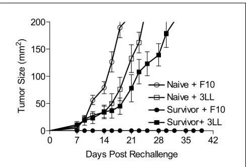
To determine whether long-term tumor-specific protec-tive immunity developed after treatment with 21 + IL-2, mice surviving tumor free for 150 days were subse-quently rechallenged subcutaneously with 5 × 10⁵ B16F10
tumor cells or 5 × 10⁴ 3LL tumor cells. Tumor size was
monitored three times per week out to 39 days (Figure 3). All 4 mice injected with control 3LL cells developed tumor (mean size >150 mm2) by day 32, while all 5 mice
chal-lenged with B16F10 were tumor-free to day 39, and remained so for another 150 days (data not shown). Addi-tional control groups included naive congenic C57BL/6 mice inoculated with B16F10 (5 mice) or 3LL (5 mice) tumor cells. The mean tumor size for both tumors in naive control mice reached 200 mm2 before day 25. Thus, the
regimen of adoptive cell transfer with lymphopenic con-ditioning and tumor antigen vaccination followed by IL-21 + IL-2 therapy was curative in mice with established tumors, and produced a long-term tumor-specific mem-ory response capable of protecting against a secondary challenge with the original tumor.
IL-21 + IL-2 therapy induced high numbers of circulating pmel CD8+ T cells
[image:7.612.60.549.88.419.2]Representative data from one of three experiments dem-onstrated that IL-21 + IL-2 therapy in the pmel Tg/B16F10 model induces higher absolute numbers of circulating Tumor-specific protective immunity in long-term survivor mice
Figure 3
Tumor-specific protective immunity in long-term survivor mice. IL-21 + IL-2 treated mice that survived disease free for 150 days were rechallenged with B16F10 (5 × 10⁵) or 3LL(5 × 10⁴) tumor cells. All 3LL challenged mice (4/4) exhibited mean tumor
growth to ≥ 200 mm2 by day 32; all B16F10 challenged mice (5/5) were protected. Naïve mice challenged with the same
B16F10 (5/5) or 3LL (5/5) tumor cells had rapid mean tumor growth to ≥ 200 mm2 by day 25.
0
7
14
21
28
35
42
0
50
100
150
200
Survivor+ 3LL
Survivor + F10
Naive + F10
Naive + 3LL
Days Post Rechallenge
T
u
m
o
r S
iz
e
(m
m
pmel CD8+ T cells than either IL-2 or IL-21 therapy alone
(Figure 4). Pooled blood from 5–10 mice in each cytokine group and the vaccine only control were stained for CD8/ CD127/CD62L/CD45.2 expression, and the absolute number of circulating pmel CD8+ T cells was determined
using counting beads as described. Analysis of variance (ANOVA) was performed on cell counts made at weekly intervals from 7–28 days post vaccine administration from pooled samples for each group; there was a signifi-cant difference between test groups (p = 0.029). A follow-up (post hoc) Tukey test showed that the absolute number of circulating pmel CD8+ T cells in peripheral blood was
significantly higher after combined cytokine therapy than following IL-2 (p < 0.05), or IL-21 (p < 0.05) therapy alone at 14, 21, and 28 days after vaccination. The
expan-sion of circulating pmel cells appeared to peak at day 21 and go through a contraction phase from day 21 to day 28 in mice treated with IL-21+ IL-2, and for mice in the vac-cine only control group. Cell numbers remained relatively constant from day 14–28 for mice treated with 21 or IL-2 alone.
Cell surface phenotype of pmel CD8+ T cells from
cytokine-treated mice
Detailed pmel CD8+ T cell subset analysis (CD8/CD127/
CD62L/CD45.2) was performed on pooled peripheral blood samples from mice used in the experiment described in Figure 4. Recent reports have identified CD127 (IL-7α receptor) expression as an important descriptor for long-lived memory T cells which may arise
Quantitation of absolute numbers of pmel CD8+ T cells
Figure 4
Quantitation of absolute numbers of pmel CD8+ T cells. Pooled blood from mice in the vaccine control and all three cytokine
experimental groups (≥ 5 mice/group) was analyzed by flow cytometry to determine the absolute number of circulating pmel-1 CD8+ T cells/µL of blood. Vaccinated mice treated with IL-21+ IL-2 had the highest total pmel CD8+ cell numbers at all time
points. Data are shown from a representative experiment and show that IL-21 + IL-2 resulted in significantly higher circulating pmel CD8+ T cell numbers than was produced by IL-2 (p = 0.05), or IL-21 (p = 0.05) alone.
0
5
10
15
20
25
30
0
25
50
75
100
Vaccine Control
IL-2
IL-21
IL-2 + IL-21
Days Post hgp100 Vaccine
P
m
el
C
ount
/
early after in vivo priming [29-34]. These studies also indi-cated that CD62L and CD127 staining partitioned anti-gen-specific cells into two distinct memory T cell subsets which, by functional criteria, were similar to central mem-ory T cells (Tcm) with a CD62L+/CD127+ phenotype, and
effector memory (Tem) cells with a CD62L-/CD127+
phe-notype [32,33]. Effector T cells (Te) were CD62L-/CD127
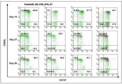
-and were characterized by poor proliferation response, immediate effector function, and high levels of antigen-induced apoptosis [33]. Figure 5 shows dot plots of CD127 versus CD62L staining of pre-gated CD8+/
CD45.2+ peripheral blood pmel T cells obtained on days
14, 21 and 28 from each cytokine therapy group and vac-cine only control mice. The frequency of circulating pmel CD8+ T cells with a Tcm phenotype on day 14 was no
dif-ferent in IL-2, and IL-21 + IL-2 treated mice or in the vac-cine controls. Only IL-21 treated mice showed a small
increase in Tcm frequency, which was 15% higher than that produced in control mice. Tem frequencies following treatment with IL-2 or with IL-21 + IL-2 were 30% and 63% higher respectively than in control mice. The fre-quencies of both Tcm and Tem pmel CD8+ T cells
decreased on day 21 for all experimental groups com-pared to day 14 values. The day 21 frequency of Tem pmel CD8+ T cells (CD62L-/CD127+) was similar for all test
groups except IL-21 + IL-2, which maintained approxi-mately a 70% higher Tem percentage. The frequencies of Tcm (CD62L+/CD127+) on day 21 indicated IL-2
sus-tained a slightly higher percentage than the control (22%); IL-21 stimulation resulted in a 54% higher fre-quency of Tcm CD8+ T cells compared to the control, and
IL-21 + IL-2 treated mice maintained a Tcm frequency 35% higher than the control. By day 28 the control and IL-2 induced Tcm frequencies were equal to each other but
[image:9.612.59.553.87.425.2]CD27 vs CD62L 2 parameter phenotype analysis of pmel CD8+ T cells
Figure 5
CD27 vs CD62L 2 parameter phenotype analysis of pmel CD8+ T cells. Lymphocytes from pooled blood (5–10 mice/group) in
a representative experiment were analyzed for CD127 and CD62L staining. Flow cytometry phenotype analysis of pmel CD8+
T cells on days 14, 21 and 28 post vaccination indicated higher frequencies of antigen specific Tcm (CD62L+/CD127+) were
induced at all time points by IL-21 alone compared to the control and IL-2 stimulated PBMCs; IL-21 + IL-2 stimulated compara-bly high Tcm frequencies on days 21 and 28. IL-21+IL-2 stimulated the highest frequency of pmel CD8+ Tem (CD62L-/CD127+)
at all time points.
higher than on day 21, while the IL-21 effect (38% increase) and the IL-21 + IL-2 effect (25% increase) on Tcm expression were both similarly elevated compared to the control and 2 induced percentages. Combined IL-21 and low-dose IL-2 therapy sustained the circulating Tem frequency at a level that was 2-fold higher than the control group or the effect induced by IL-2 alone or IL-21 alone. The cell density plots for IL-21+IL-2 on all three
days also suggested an overall higher absolute number of circulating Tcm and Tem CD8+/CD45.2+ T cells than was
induced by any other treatment. Generally, lower percent-ages of circulating effector cells (CD62L-/CD127-) were
present at day 21 and day 28 for IL-21 and IL-21+IL-2 treated mice compared to both IL-2 treated animals and control mice. The upper left quadrant of each 2-parameter histogram contained CD62L+/CD127- T cells, and their
[image:10.612.57.551.88.537.2]Quantitation of absolute numbers of Tem and Tcm CD8+ T cells
Figure 6
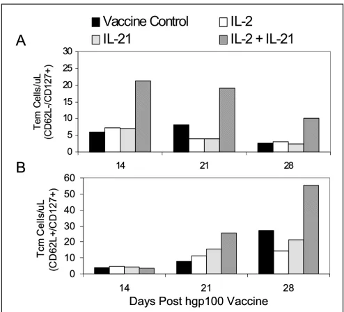
Quantitation of absolute numbers of Tem and Tcm CD8+ T cells. PBMCs were examined by flow cytometry to determine the
absolute number of Tem (CD62L-/CD127+) pmel CD8+ T cells (A), and Tcm (CD62L+/CD127+) pmel CD8+ T cells (B) on days
14, 21, and 28 after vaccination and cytokine treatment. IL-21 + IL-2 supported the highest level of circulating Tem cells on all three days compared to all other experimental groups (p = 0.011), and stimulated the greatest increase of Tcm cells on days 21 and 28 (p = 0.029). Data was collected from pooled blood from ≥ 5 mice in each test group.
0
5
10
15
20
25
30
14
21
28
Te
m C
e
lls
/u
L
(C
D
62L
-/
C
D
127
+
)
Vaccine Control
IL-2
IL-21
IL-2 + IL-21
0
10
20
30
40
50
60
14
21
28
T
c
m
Ce
lls
/u
L
(C
D
62L+
/C
D
127+
)
A
B
Days Post hgp100 Vaccine
0
5
10
15
20
25
30
14
21
28
Te
m C
e
lls
/u
L
(C
D
62L
-/
C
D
127
+
)
Vaccine Control
IL-2
IL-21
IL-2 + IL-21
0
10
20
30
40
50
60
14
21
28
T
c
m
Ce
lls
/u
L
(C
D
62L+
/C
D
127+
)
A
B
frequency was generally lower in the IL-21 and IL-21+IL-2 treated animals. Presently there is no correlated functional data to determine the lineage relationship of this popula-tion to Tcm or Tem T cells or their funcpopula-tional properties. Overall the data in Figure 5 suggest IL-21+IL-2 induced higher frequencies of Tem cells compared to IL-2 or IL-21 alone on all three days; and by the early contraction phase of the pmel CD8+ T cell response (day 28) IL-21 and IL-21
+ low-dose IL-2 induced equally higher percentages of tumor-specific Tcm cells than were stimulated in the con-trol or IL-2 treated mice.
IL-21+ IL-2 induced proliferation of Tem and Tcm pmel CD8+ T cells
The frequencies of Tcm and Tem T cells on days 14, 21 and 28 (Figure 5), and the total number of circulating pmel cells determined for each group (Figure 4) were subse-quently used to calculate the absolute number of Tem (Figure 6A) and Tcm (Figure 6B) pmel CD8+ T cells for
each experimental group. Tem numbers on each day were significantly higher for IL-21+ IL-2 treated mice than for all other groups (p = 0.011) as determined by ANCOVA, and declined from day 14 to day 28 (Figure 6A). By con-trast, the absolute number of Tcm cells for all groups was equally low at day 14 and increased incrementally for each group over time (Figure 6B). IL-21 + IL-2 induced Tcm cell numbers increased at a significantly higher rate compared to all other test groups (p = 0.029) as deter-mined by ANCOVA. Thus, IL-21+ IL-2 treatment main-tained Tem absolute numbers at all time points that were
2- to 4-fold greater than those produced by the vaccine control or by IL-2 or IL-21 therapy alone. Similarly, Tcm absolute numbers induced by IL-21+ IL-2 were 2- to 4-fold higher than those stimulated in any other test group by day 28. Recent data have demonstrated that the adop-tive transfer of pmel CD8+ Tcm (characterized as CD62L+/
CCR7+), or "early effector" T cells (CD62Ldim/CD127+/
CCR7+) produced a potent anti-tumor response
subse-quent to tumor-antigen hgp10025–33 vaccination and high-dose IL-2 therapy in the pmel Tg/B16 therapeutic model [22,35]. The data in Figure 6B suggest IL-21+ low-dose IL-2 in vivo therapy promotes the long-term mainte-nance of CD8+ T cells with a similar Tcm (CD62L+/
CD127+) or "early effector" – like phenotype (CD62L
dim/-/CD127+).
Functional phenotype of CD8+ T cells from IL-21 + IL-2
treated tumor regressor mice
By day 42 previously regressing tumors in a subset of IL-21 + IL-2 treated mice began to grow again, reaching a mean tumor size of 25–50 mm2. All tumors of this size on
day 42–49 continued to progress to ≥ 200 mm2 by day 63,
whereupon the mice were sacrificed. However, approxi-mately 50% of all IL-21+ IL-2 treated mice in three exper-iments did not exhibit tumor growth, and by day 42–49 manifested complete regression of the tumor. These ani-mals remained tumor free out to >150 days. Day 42 splen-ocytes from two "progressor", and from two "regressor" mice treated with IL-21 + IL-2 were pooled separately, and were stimulated in vitro with hgp10025–33; cells were then
Cytokine flow cytometry analysis of the functional phenotype of pmel CD8+ splenocytes from IL-21 + IL-2 treated mice
Figure 7
Cytokine flow cytometry analysis of the functional phenotype of pmel CD8+ splenocytes from IL-21 + IL-2 treated mice. Spleen
cells were pooled from 2 mice with progressing tumors (≥ 50 mm2) and from 2 mice with regressing tumors (<10 mm2) 42
days after vaccination. Cytokine flow cytometry analysis shows the absolute number of pmel CD8+ T cells per 106 splenocytes
(A), and the frequency of pmel CD8+ cells (B) that are positive for IL-2, IFNγ, and TNFγ production. Data show mice with
regressing tumors had 4.7-fold, 4.8-fold and 2.5-fold more IL-2+, IFN-γ+, and TNF-α+ pmel CD8+ T cells respectively, and
higher frequencies of Tc1 cytokine+ cells than progressor mice.
Absolute # of Cytokine+ Pmel CD8+T cells per 1 x10⁶ Splenocytes
0 2 4 6 8 10 12
IL-2 IFN-gamma TNF-alpha
Cy to k in e + /p m e l
+ c
e
lls
(x
1
0
3 ) Progressor
Regressor
% of Cytokine+ Pmel-1 CD8 T Cells
0 10 20 30 40 50 60
IL-2 IFN-gamma TNF-alpha
% o f Cyto ki n e
+ p
m e l-1 CD8 T Ce lls Progressor Regressor
A
B
Absolute # of Cytokine+ Pmel CD8+T cells per 1 x10⁶ Splenocytes
0 2 4 6 8 10 12
IL-2 IFN-gamma TNF-alpha
Cy to k in e + /p m e l
+ c
e
lls
(x
1
0
3 ) Progressor
Regressor
% of Cytokine+ Pmel-1 CD8 T Cells
0 10 20 30 40 50 60
IL-2 IFN-gamma TNF-alpha
% o f Cyto ki n e
+ p
analyzed for IFNγ, IL-2 and TNF-α production using a standard five-hour CFC assay. After IVS cells were fixed, permeablized, and stained with CD8, CD45.2 and cytokine-specific antibodies. Spleens from regressor mice had more IFNγ, IL-2 and TNF-α positive CD8+/CD45.2+
pmel T cells than spleens from progressor animals (Figure 7A). Regressor mice had 4.7-fold, 4.8-fold, and 2.5-fold more IL-2+, IFNγ+ and TNF-α+ pmel CD8+ T cells,
respec-tively, than progressor mice with rapidly growing tumors. The data in Figure 7A indicates that tumor regression in mice receiving IL-21+ IL-2 therapy was associated with an increase in the absolute number of pmel CD8+ T cells
pro-ducing Tc1 cytokines compared to the numbers found in mice with growing tumors. The presence of higher num-bers of Tc1 cytokine+ CD8+ T cells in regressor compared
to progressor mice was attributable to both a 2-fold greater absolute number of pmel CD8+ T cells in regressor
mice than were measured in progressor animals (data not shown) and to higher frequencies of pmel CD8+ T cells
which were positive for Tc1 cytokines in regressor mice at 42 days (Figure 7B).
Discussion and conclusions
We report for the first time the anti-tumor effects of a treatment strategy that combines lymphopenic condition-ing, adoptive transfer of naive antigen-specific CD8+ T
cells, tumor antigen-specific vaccination, and cytokine treatment with IL-21 + low-dose IL-2. Using a well estab-lished murine model of pre-existing disease, the pmel Tg/ B16 model, our results show that combined IL-21 + low-dose IL-2 therapy delayed B16F10 melanoma growth in the majority of mice, and resulted in a significant increase in tumor-free survival (46%) out to ≥ 150 days compared to mice treated with IL-21 or IL-2 alone. Surviving mice exhibited tumor-specific protective immunity since they resisted B16F10 rechallenge but succumbed to the unre-lated 3LL tumor. These data are in contrast to other studies in the pmel Tg/B16 model, which employed the adoptive transfer of highly activated IVS pmel CD8+ T cells
(stimu-lated with hgp10025–33 peptide and IL-2 or IL-15) rather than naive pmel T cells [3,17,18]. After lymphopenic con-ditioning and T-cell transfer to tumor-bearing mice, ther-apy was administered in these studies using different combinations of hgp10025–33 vaccination and cytokine
treatment. Thus, Lou et al [17] vaccinated with hgp10025–
33 peptide-pulsed DCs, and administered high-dose IL-2
(1.2 × 10⁶ IU/day) for three days beginning the day of
vac-cination. Klebanoff, et al [18] vaccinated with a recom-binant fowl pox virus encoding hgp10025–33 (rFPhgp100) after adoptive transfer of IVS pmel CD8+ T cells grown in
IL-2 or in IL-15 cultures. Follow-on cytokine therapy con-sisted of high-dose IL-2 (1.2 × 10⁶ IU/day) for three days.
In the only published study using IL-21 in the pmel Tg/ B16 model Zeng and coworkers [3] also vaccinated with rFPhgp10025–33 following adoptive transfer of IL-2
cul-tured pmel T cells, and animals were treated over three days with IL-15 (20 µg/day), IL-21 (20 µg/day), or both cytokines at the same doses. None of these treatment strat-egies resulted in long-term inhibition of tumor growth beyond 30–35 days after treatment unless very large num-bers (4–6 × 10⁶) of IVS pmel CD8+ T cells were adoptively
transferred [17]. Furthermore, in contrast to our results, none of these studies presented data describing long-term tumor-free survival ≥ 150 days with any combination of transferred T cells, vaccine administration and cytokine therapy. Another group compared the ability of IL-2, IL-15 or IL-21 to augment anti-tumor immunity in C57BL/6 mice challenged with the OVA-expressing E.G7 thymoma [6]. Cytokine therapy was initiated 48 hours after tumor inoculation – well before the establishment of vascular-ized tumors, and was administered every other day for two weeks thereafter. In this model IL-21 (20 µg/day) did result in prolonged tumor-free survival out to 100 days in approximately 25–30% of treated mice. Neither low-dose IL-2 (2 × 10³ IU and 20 × 103 IU/day) nor IL-15 (5 µg and
50 µg/day) produced a survival frequency that was as high. All animals that survived beyond 100 days were pro-tected from rechallenge with E.G7 tumor cells. Impor-tantly, this report also showed that the 2-week cytokine administration could begin as late as day 12 following tumor inoculation and still produce a therapeutic response [6]. This study also demonstrated the ability of IL-21 to augment immunity to the OVA-expressing E.G7 tumor in the absence of previously antigen-educated CD8+ T cells. However, the experimental design did not
test the anti-tumor effects of IL-21 in a model of estab-lished bulky disease such as the pmel Tg/B16 system, in which the cognate tumor antigen is a relatively weak self-tumor antigen rather than a strongly immunogenic for-eign protein. To date only the combined therapeutic effects of IL-21+ low-dose IL-2 reported here have resulted in long-term survival of tumor-free mice in the pmel Tg/ B16 model. As noted, in most of the previous studies using the pmel Tg/B16 melanoma model very high doses (1.2 × 10⁶ IU/day) of IL-2 were used in combination with
the adoptive transfer of large numbers (up to 107) of
highly activated IVS pmel CD8+ T cells [16-18] However,
treatment was initiated after very large, bulky (50–100 mm2) tumors were established. By contrast, cytokine
ther-apy was initiated in our experiments when vascularized tumors were smaller (26 mm2). Long-term survival using
our current IL-21 + IL-2 therapeutic model may decrease with larger established tumors; experiments designed to test and optimize IL-21 +low-dose IL-2 therapy in mice with larger vascularized tumors are ongoing.
The H-2Db-restricted NV peptide was used in all
pmel CD8+ T cell numbers over that produced by
hgp10025–33 alone (unpublished data – H.M. Hu, EACRI). CD4 helper T cell function in our DC-based vaccine sys-tem may have been provided by fetal calf serum (FCS) proteins/peptides associated with the DCs in the vaccine – both DCs and the B16F10 melanoma cells used in the tumor inoculum were cultured in medium containing FCS. The potential for FCS antigen-induced CD4 T cell activation is also suggested by other hgp10025–33-pulsed DC vaccine studies in the pmel Tg/B16 model in which anti-tumor therapeutic effect was achieved in the absence of any known source of CD4-specific antigen [17].
There is limited information on IL-21-mediated changes in the cell surface and functional phenotype of CD8+
T-cells associated with tumor immunity. In this report, we show that IL-21 alone and IL-21+ low-dose IL-2 increased the in vivo frequency of Tcm T cells as defined by concom-itant CD62L+/CD127+ staining[29], compared to the
vac-cine only control or to cells from IL-2-treated mice (Figure 5). IL-21 + IL-2 treatment also sustained the highest per-centage of Tem (CD62L-/CD127+) pmel CD8+ T cells [29]
compared to any other test group. This observation corre-lated with the result that IL-21+ IL-2 therapy also pro-duced the highest absolute number of circulating pmel CD8+ T cells during the beginning (day 14), peak (day 21)
and end (day 28) of the hgp10025–33 induced expansion of pmel CD8+ T cells (Figure 4). As a consequence, IL-21 +
IL-2 therapy produced the highest absolute number of cir-culating pmel CD8+ Tem cells at each time point of the
expansion and early contraction phases of the anti-hgp10025–33 immune response, and also resulted in the highest absolute number of Tcm T cells in the peripheral blood (Figure 6). Although Tem absolute numbers decreased from day 14 to day 28, and Tcm numbers increased over this same period the data do not indicate if this is attributable in anyway to innate differences in IL-21 + IL-2 induced Tem vs Tcm proliferation. Future in vitro and in vivo studies will examine the relative proliferative potential of cognate antigen-driven purified populations of pmel CD8+ Tcm and Tem cells. Notably, the
tumor-spe-cific protective memory response observed in long-term survivor mice was also associated with high frequencies of Tcm in the spleen and lymph nodes. Polychromatic (8 color) flow cytometry analysis of pmel CD8+ splenocytes
from IL-21 + IL-2 treated long-term survivor mice (> 150 days) indicated approximately 10%–12% of all CD8+ T
cells in the spleen and lymph nodes were pmel CD8+ T
cells, and 43% and 70% of these T cells in the lymph nodes and spleen respectively expressed a Tcm phenotype (CD62L+/CD127+/CD27+/CD28+)(unpublished data).
Recent data have demonstrated that enriched pmel CD8+
Tcm T cells conferred a potent in vivo anti-tumor recall response upon adoptive transfer – leading to the eradica-tion of large established tumors in the pmel Tg/B16
model [35]. Thus, as described herein, IL-21 + IL-2 ther-apy favored the in vivo expansion and maintenance of such tumor antigen-specific Tcm T cells from naive pmel CD8+ T cell precursors which were capable of providing
long-term protective immunity. Preliminary phenotype analysis of IL-21 + low-dose IL-2 and hgp10025–33 IVS pmel splenocytes similarly indicates the combination of both cytokines results in the proliferation of pmel Tg CD8+ T cells with a Tcm (CD62L+/CD127+/CD27+/
CD28+) phenotype (unpublished data – EACRI). This
fur-ther suggests that IL-21 + low-dose IL-2 might be effective in supporting the cognate antigen-driven in vitro expan-sion of rare self-tumor antigen Tcm CD8+ T cells from
autologous PBMCs – perhaps in concert with procedures to remove tumor antigen-specific regulatory T cell effects. In addition to increasing tumor-antigen directed expan-sion and maintenance of Tcm CD8+ T cells, data presented
here demonstrates that IL-21 + IL-2 therapy also stimu-lated the expression of anti-tumor memory CD8+ T cells
with a Tc1 cytokine functional phenotype in mice with regressing tumors. By day 42 post-tumor inoculation IL-21 + IL-2 treated tumor-regressor mice had much higher absolute numbers of IL-2+, IFNγ+ and TNF-α+ pmel CD8+
splenocytes than IL-21 + IL-2 treated tumor-progressor mice with rapidly growing tumors (Figure 7). The relative absence of CD8+ splenocytes with a Tc1 cytokine
func-tional phenotype in tumor progressor mice may be attrib-utable in part to a simple lack of sustained B16F10 tumor antigen stimulation. Previous published data from our institute using the pmel Tg/B16 model has demonstrated loss of MHC class I and gp100 expression in a high per-centage of tumors in experimental animals with progres-sive disease due to immunoediting[36]. Overall our data suggest that IL-21 + low-dose IL-2-mediated tumor regres-sion and long-term survival was associated with elevated absolute numbers of tumor antigen-specific Tem and Tcm CD8+ T cells with a Tc1 functional phenotype.
IL-21 + low-dose IL-2 induction of increased in vivo expression of Tc1 cytokine+ Tcm and Tem CD8+ T cells is a
central observation for any putative mechanistic explana-tion of the anti-tumor therapeutic synergy produced using both cytokines. This observation examined in the context of recent studies describing the broad inhibitory effects of IL-21 on B lymphocytes, NK cells and DCs (Reviewed in [37]) suggests a possible mechanism for the IL-21+ low-dose IL-2 induced expansion of Tcm CD8+ T cells similar
as an inducer of antigen stimulated IFNγ and CTL func-tion in CD8+ T cells [4,8,44,45], our unpublished data in
both murine and human in vitro experiments indicate IL-21 alone does not drive high levels of cognate antigen stimulated CTL proliferation. In vivo data presented here suggest that 21 works best in concert with low-dose IL-2 to support both optimal anti-tumor effector CTL func-tion and expansion of CD8+ memory T cells.
Paradoxi-cally, IL-21 + low-dose IL-2 treatment produced unexpectedly low frequencies of circulating effector pmel CD8+ T cells (Figure 4) – perhaps due to cell trafficking to
the primary tumor and sites of metastatic disease. Ongo-ing experiments are directed at the in situ analysis of the phenotype, cell number, and functional properties of tumor invasive pmel CD8+ T cells in 21 + low dose
IL-2 treated mice. Such studies should shed light on whether combined cytokine therapy induces trafficking of increased numbers of antigen-specific cytolytic CD8+ T
cells to the tumor site. Taken together, these observations suggest a dual role for IL-21 in dampening ongoing innate and adaptive immune responses (as might occur during the contraction phase of antigen-specific immunity), while concomitantly augmenting the expansion of anti-gen-driven long-term memory T cells. Recently published data demonstrate IL-21 treated cultures of antigen-stimu-lated cells produce CD8+ T cells with increased TCR
bind-ing affinity [5]. Other data show the IL-21 receptor (IL-21R) is upregulated upon TCR engagement [46,47]. Thus, Th2-derived IL-21 may "rescue" memory T cells by block-ing further antigen-driven effector T cell differentiation through presently unknown mechanisms, while simulta-neously increasing antigen binding affinity. The resulting increase in binding of low-levels of residual antigen (present during the contraction phase) could increase IL-21R expression and up-regulate increased IL-21 binding to memory T cells – thus further skewing the shift to mem-ory T cells. Memmem-ory CD8+ T cells in turn express amplified
levels of CD122 [48], which may facilitate increased high affinity IL-2R formation if other IL-2R chains (CD25 and CD132) are available, and potentially increase binding of the low levels of IL-2 which may be present during immu-nological contraction. The combination of the "arrested" Tcm memory phenotype (maintained by IL-21), and low dose IL-2-induced proliferation of these cells could result in increased Tcm cell numbers. Thus, 21 + low-dose IL-2 therapy may favor the enhanced expansion of long-term memory (Tcm) CD8+ T cells through augmented memory
T cell "rescue" and expansion mechanisms similar to those which may normally be present during the late traction phase of an ongoing immune response. This con-cept is supported by the results of our study and other reports [6,8], which suggest IL-21 induced tumor immu-nity is most effective when IL-21 is administered several days (4–12 days) after initial tumor-antigen activation of T cells. The late acting anti-tumor effects of IL-21, and, in
our study, IL-21 + IL-2 therapy suggests IL-21 may be act-ing directly on tumor antigen educated CD8+ T cells rather
than modulating early APC and/or T cell function associ-ated with primary tumor antigen stimulation. Preliminary in vitro studies with purified naïve and hgp10025–33 stim-ulated pmel CD8+ T cells support this conclusion (data
not shown).
In summary, the results of this study suggest that IL-21 + low-dose IL-2 cytokine therapy may "rescue" and aug-ment proliferation of tumor antigen-specific memory CD8+ T cells, and thus provide an important new strategic
component for effective cancer immunotherapy. The combined use of both cytokines may be most effective in the context of lymphopenic conditioning, tumor antigen-specific vaccination, and the adoptive transfer of autolo-gous tumor-specific Tcm or "early" effector T cells. Ongo-ing work at our institute continues to focus on developOngo-ing procedures for the IL-21 + low-dose IL-2-directed in vivo and in vitro expansion and maintenance of such Tcm and "early" effector subpopulations from small numbers of normally tolerized autologous self-tumor antigen-specific precursors.
Competing interests
Christopher Clegg is a scientist employed by Zymogenet-ics – all other authors declare that they have no competing interests.
Authors' contributions
HH designed the in vivo experiments and worked with PW and DH to perform the flow cytometry analysis of PBMC and splenocyte samples. GY assisted in all aspects of tumor inoculation, lymphopenic conditioning, tumor measurement and tissue harvest. HMH, CHC, and AOH provided key reagents and assisted in the design of con-trols which utilized these reagents in the experiments. WM was directly involved in drafting and revising the manuscript, and WGA performed all the statistical analy-sis. EBW designed and supervised all aspects of the exper-imental strategy and acquisition of data, and, with BAF and WJU, was involved in data interpretation and the crit-ical review and revision of the manuscript.
Acknowledgements
This study was supported in part by NIH grant 1 R21CA101325-02 and by grants from the Providence Portland Medical Foundation, and the M.J. Mur-dock Charitable Trust, and the Chiles Foundation.
References
1. Leonard WJ, Spolski R: Interleukin-21: a modulator of lymphoid proliferation, apoptosis and differentiation. Nat Rev Immunol
2005, 5(9):688-698.
Web-ster P, Whitmore T, Maurer M, Kaushansky K, Holly RD, FoWeb-ster D:
Interleukin 21 and its receptor are involved in NK cell expan-sion and regulation of lymphocyte function. Nature 2000,
408(6808):57-63.
3. Zeng R, Spolski R, Finkelstein SE, Oh S, Kovanen PE, Hinrichs CS, Pise-Masison CA, Radonovich MF, Brady JN, Restifo NP, Berzofsky JA, Leonard WJ: Synergy of IL-21 and IL-15 in regulating CD8+ T cell expansion and function. J Exp Med 2005, 201(1):139-148. 4. Kasaian MT, Whitters MJ, Carter LL, Lowe LD, Jussif JM, Deng B,
Johnson KA, Witek JS, Senices M, Konz RF, Wurster AL, Donaldson DD, Collins M, Young DA, Grusby MJ: IL-21 limits NK cell responses and promotes antigen-specific T cell activation: a mediator of the transition from innate to adaptive immu-nity. Immunity 2002, 16(4):559-569.
5. Li Y, Bleakley M, Yee C: IL-21 influences the frequency, pheno-type, and affinity of the antigen-specific CD8 T cell response.
J Immunol 2005, 175(4):2261-2269.
6. Moroz A, Eppolito C, Li Q, Tao J, Clegg CH, Shrikant PA: IL-21 enhances and sustains CD8+ T cell responses to achieve durable tumor immunity: comparative evaluation of 2, IL-15, and IL-21. J Immunol 2004, 173(2):900-909.
7. Ugai S, Shimozato O, Kawamura K, Wang YQ, Yamaguchi T, Saisho H, Sakiyama S, Tagawa M: Expression of the interleukin-21 gene in murine colon carcinoma cells generates systemic immu-nity in the inoculated hosts. Cancer Gene Ther 2003,
10(3):187-192.
8. Ma HL, Whitters MJ, Konz RF, Senices M, Young DA, Grusby MJ, Col-lins M, Dunussi-Joannopoulos K: IL-21 activates both innate and adaptive immunity to generate potent antitumor responses that require perforin but are independent of IFN-gamma. J Immunol 2003, 171(2):608-615.
9. Kishida T, Asada H, Itokawa Y, Cui FD, Shin-Ya M, Gojo S, Yasutomi K, Ueda Y, Yamagishi H, Imanishi J, Mazda O: Interleukin (IL)-21 and IL-15 genetic transfer synergistically augments thera-peutic antitumor immunity and promotes regression of metastatic lymphoma. Mol Ther 2003, 8(4):552-558.
10. Overwijk WW, Tsung A, Irvine KR, Parkhurst MR, Goletz TJ, Tsung K, Carroll MW, Liu C, Moss B, Rosenberg SA, Restifo NP: gp100/ pmel 17 is a murine tumor rejection antigen: induction of "self"-reactive, tumoricidal T cells using high-affinity, altered peptide ligand. J Exp Med 1998, 188(2):277-286.
11. Ku CC, Murakami M, Sakamoto A, Kappler J, Marrack P: Control of homeostasis of CD8+ memory T cells by opposing cytokines.
Science 2000, 288(5466):675-678.
12. Waldmann TA, Dubois S, Tagaya Y: Contrasting roles of IL-2 and IL-15 in the life and death of lymphocytes: implications for immunotherapy. Immunity 2001, 14(2):105-110.
13. Murakami M, Sakamoto A, Bender J, Kappler J, Marrack P:
CD25+CD4+ T cells contribute to the control of memory CD8+ T cells. Proc Natl Acad Sci USA 2002, 99(13):8832-8837. 14. Refaeli Y, Van Parijs L, London CA, Tschopp J, Abbas AK:
Biochem-ical mechanisms of IL-2-regulated Fas-mediated T cell apop-tosis. Immunity 1998, 8(5):615-623.
15. Manjunath N, Shankar P, Wan J, Weninger W, Crowley MA, Hieshima K, Springer TA, Fan X, Shen H, Lieberman J, von Andrian UH: Effec-tor differentiation is not prerequisite for generation of mem-ory cytotoxic T lymphocytes. J Clin Invest 2001, 108(6):871-878. 16. Overwijk WW, Theoret MR, Finkelstein SE, Surman DR, de Jong LA, Vyth-Dreese FA, Dellemijn TA, Antony PA, Spiess PJ, Palmer DC, Heimann DM, Klebanoff CA, Yu Z, Hwang LN, Feigenbaum L, Kruis-beek AM, Rosenberg SA, Restifo NP: Tumor regression and autoimmunity after reversal of a functionally tolerant state of self-reactive CD8+ T cells. J Exp Med 2003, 198(4):569-580. 17. Lou Y, Wang G, Lizee G, Kim GJ, Finkelstein SE, Feng C, Restifo NP,
Hwu P: Dendritic cells strongly boost the antitumor activity of adoptively transferred T cells in vivo. Cancer Res 2004,
64(18):6783-6790.
18. Klebanoff CA, Finkelstein SE, Surman DR, Lichtman MK, Gattinoni L, Theoret MR, Grewal N, Spiess PJ, Antony PA, Palmer DC, Tagaya Y, Rosenberg SA, Waldmann TA, Restifo NP: IL-15 enhances the in vivo antitumor activity of tumor-reactive CD8+ T cells. Proc Natl Acad Sci USA 2004, 101(7):1969-1974.
19. Yee C, Thompson JA, Byrd D, Riddell SR, Roche P, Celis E, Greenberg PD: Adoptive T cell therapy using antigen-specific CD8+ T cell clones for the treatment of patients with metastatic melanoma: in vivo persistence, migration, and antitumor
effect of transferred T cells. Proc Natl Acad Sci USA 2002,
99(25):16168-16173.
20. Dudley ME, Wunderlich J, Nishimura MI, Yu D, Yang JC, Topalian SL, Schwartzentruber DJ, Hwu P, Marincola FM, Sherry R, Leitman SF, Rosenberg SA: Adoptive transfer of cloned melanoma-reac-tive T lymphocytes for the treatment of patients with meta-static melanoma. J Immunother 2001, 24(4):363-373.
21. Dudley ME, Wunderlich JR, Yang JC, Hwu P, Schwartzentruber DJ, Topalian SL, Sherry RM, Marincola FM, Leitman SF, Seipp CA, Rogers-Freezer L, Morton KE, Nahvi A, Mavroukakis SA, White DE, Rosen-berg SA: A phase I study of nonmyeloablative chemotherapy and adoptive transfer of autologous tumor antigen-specific T lymphocytes in patients with metastatic melanoma. J Immu-nother 2002, 25(3):243-251.
22. Gattinoni L, Klebanoff CA, Palmer DC, Wrzesinski C, Kerstann K, Yu Z, Finkelstein SE, Theoret MR, Rosenberg SA, Restifo NP: Acquisi-tion of full effector funcAcquisi-tion in vitro paradoxically impairs the in vivo antitumor efficacy of adoptively transferred CD8+ T cells. J Clin Invest 2005, 115(6):1616-1626.
23. Nardin EH, Nussenzweig RS: T cell responses to pre-erythro-cytic stages of malaria: role in protection and vaccine devel-opment against pre-erythrocytic stages. Annu Rev Immunol
1993, 11:687-727.
24. Munesinghe DY, Clavijo P, Calle MC, Nussenzweig RS, Nardin E:
Immunogenicity of multiple antigen peptides (MAP) con-taining T and B cell epitopes of the repeat region of the P. falciparum circumsporozoite protein. Eur J Immunol 1991,
21(12):3015-3020.
25. Lutz MB, Kukutsch N, Ogilvie AL, Rossner S, Koch F, Romani N, Schuler G: An advanced culture method for generating large quantities of highly pure dendritic cells from mouse bone marrow. J Immunol Methods 1999, 223(1):77-92.
26. Crepeau H, Koziol J, Reid N, Yuh YS: Analysis of incomplete mul-tivariate data from repeated measurement experiments.
Biometrics 1985, 41:505-514.
27. Kozial J, Maxwell DA, Fukushima M, Colmeraurer M, Pilch YH: A dis-tribution-free test fro tumor-growth curve analyses with application to an animal tumor immuntherapy experiment.
Biometrics 1981, 37:383-390.
28. Milliken G, Johnson DE: Analysis of Messy Data. Volume I. Designed Experiments edn. New York; 1991.
29. Powell DJ Jr, Dudley ME, Robbins PF, Rosenberg SA: Transition of late-stage effector T cells to CD27+ CD28+ tumor-reactive effector memory T cells in humans after adoptive cell trans-fer therapy. Blood 2005, 105(1):241-250.
30. Schluns KS, Kieper WC, Jameson SC, Lefrancois L: Interleukin-7 mediates the homeostasis of naive and memory CD8 T cells in vivo. Nat Immunol 2000, 1(5):426-432.
31. Bradley LM, Haynes L, Swain SL: IL-7: maintaining T-cell memory and achieving homeostasis. Trends Immunol 2005, 26(3):172-176. 32. Huster KM, Busch V, Schiemann M, Linkemann K, Kerksiek KM, Wag-ner H, Busch DH: Selective expression of IL-7 receptor on memory T cells identifies early CD40L-dependent genera-tion of distinct CD8+ memory T cell subsets. Proc Natl Acad Sci USA 2004, 101(15):5610-5615.
33. Bachmann MF, Wolint P, Schwarz K, Jager P, Oxenius A: Functional properties and lineage relationship of CD8+ T cell subsets identified by expression of IL-7 receptor alpha and CD62L. J Immunol 2005, 175(7):4686-4696.
34. Kaech SM, Tan JT, Wherry EJ, Konieczny BT, Surh CD, Ahmed R:
Selective expression of the interleukin 7 receptor identifies effector CD8 T cells that give rise to long-lived memory cells. Nat Immunol 2003, 4(12):1191-1198.
35. Klebanoff CA, Gattinoni L, Torabi-Parizi P, Kerstann K, Cardones AR, Finkelstein SE, Palmer DC, Antony PA, Hwang ST, Rosenberg SA, Waldmann TA, Restifo NP: Central memory self/tumor-reac-tive CD8+ T cells confer superior antitumor immunity com-pared with effector memory T cells. Proc Natl Acad Sci USA 2005,
102(27):9571-9576.
36. Wang LX, Li R, Yang G, Lim M, O'Hara A, Chu Y, Fox BA, Restifo NP, Urba WJ, Hu HM: Interleukin-7-dependent expansion and per-sistence of melanoma-specific T cells in lymphodepleted mice lead to tumor regression and editing. Cancer Res 2005,
65(22):10569-10577.