Parainfluenza Virus 3 Blocks Antiviral Mediators Downstream of the
Interferon Lambda Receptor by Modulating Stat1 Phosphorylation
Kirsten C. Eberle,a,b,cJodi L. McGill,a*Timothy A. Reinhardt,aRandy E. Saccoa,b,c
Ruminant Diseases and Immunology Research Unit, National Animal Disease Center, Agricultural Research Service, U.S. Department of Agriculture, Ames, Iowa, USAa; Molecular, Cellular and Developmental Biology Graduate Program, Iowa State University, Ames, Iowa, USAb; Immunobiology Graduate Program, Iowa State University, Ames, Iowa, USAc
ABSTRACT
Parainfluenza viruses are known to inhibit type I interferon (IFN) production; however, there is a lack of information regarding
the type III IFN response during infection. Type III IFNs signal through a unique heterodimeric receptor, IFN-
R1/interleukin-10R2 (IL-R1/interleukin-10R2), which is primarily expressed by epithelial cells. Parainfluenza virus 3 (PIV-3) infection is highly restricted to the
airway epithelium. We therefore sought to examine type III IFN signaling pathways during PIV-3 infection of epithelial cells. We
used three strains of PIV-3: human PIV-3 (HPIV-3), bovine PIV-3 (BPIV-3), and dolphin PIV-1 (
Tursiops truncatus
PIV-1, or
TtPIV-1). Here, we show that message levels of IL-29 are significantly increased during PIV-3 infection, yet downstream antiviral
signaling molecules are not upregulated to levels similar to those of the positive control. Furthermore, in Vero cells infected with
PIV-3, stimulation with recombinant IL-29/-28A/-28B does not cause upregulation of downstream antiviral molecules,
suggest-ing that PIV-3 interferes with the JAK/STAT pathway downstream of the IFN-
R1/IL-10R2 receptor. We used Western blotting
to examine the phosphorylation of Stat1 and Stat2 in Vero cells and the bronchial epithelial cell line BEAS-2B. In Vero cells, we
observed reduced phosphorylation of the serine 727 (S727) site on Stat1, while in BEAS-2B cells Stat1 phosphorylation was
de-creased at the tyrosine 701 (Y701) site during PIV-3 infection. PIV-3 therefore interferes with the phosphorylation of Stat1
downstream of the type III IFN receptor. These data provide new evidence regarding strategies employed by parainfluenza
vi-ruses to effectively circumvent respiratory epithelial cell-specific antiviral immunity.
IMPORTANCE
Parainfluenza virus (PIV) in humans is associated with bronchiolitis and pneumonia and can be especially problematic in
in-fants and the elderly. Also seen in cattle, bovine PIV-3 causes respiratory infections in young calves. In addition, PIV-3 is one of a
number of pathogens that contribute to the bovine respiratory disease complex (BRDC). As their name suggests, interferons
(IFNs) are produced by cells to interfere with viral replication. Paramyxoviruses have previously been shown to block
produc-tion and downstream signaling of type I IFNs. For the first time, it is shown here that PIV-3 can induce protective type III IFNs in
epithelial cells, the primary site of PIV-3 infection. However, we found that PIV-3 modulates signaling pathways downstream of
the type III IFN receptor to block production of several specific molecules that aid in a productive antiviral response.
Impor-tantly, this work expands our understanding of how PIV-3 effectively evades host innate immunity.
P
arainfluenza virus 3 (PIV-3) causes a prominent respiratory
infection in both cattle and humans. The CDC reports that, in
humans, most children have antibodies against human PIV-3
(HPIV-3) by 5 years of age (
http://www.cdc.gov/parainfluenza
/hcp/clinical.html
). There is currently no vaccine available for
control of HPIV-3 infection; however, a few studies have
exam-ined the use of an attenuated bovine PIV-3 (BPIV-3) to protect
against HPIV-3 because of the homology between bovine and
hu-man strains (
1–3
). Given the lack of an efficacious vaccine for
HPIV-3, there is a critical need to understand the mechanisms of
HPIV-3-induced disease and the molecular pathways associated
with viral modulation of the host antiviral defenses.
Paramyoxviruses are negative-sense single-stranded RNA
vi-ruses which are part of the
Paramyxoviridae
family and
Paramyxo-virinae
subfamily (
4
). PIV-3 is found within the genus
Respirovi-rus
. PIV-3 is a respiratory virus that primarily infects the epithelial
cells of the lung. Symptoms of HPIV-3 infection include
bronchi-olitis and pneumonia, and HPIV-3 is especially problematic in
infants (
4
). Airway epithelial cells recognize viral infection
through pattern recognition receptors (PRRs): RIG-I like
recep-tors (RLRs) and Toll-like receprecep-tors (TLRs) (
5
). Viral RNA binds
to RLRs and TLRs, creating a signaling cascade to produce
proin-flammatory cytokines and interferons (IFNs). Type I and type III
IFNs are important in mounting a robust antiviral response by
inducing various IFN-stimulating genes (ISGs) (
6
).
Type I IFNs, particularly IFN-
␣
and -

, are known for their
antiviral role; however, many viruses have been shown to prevent
type I IFN production and responses. Paramyxoviruses in
partic-Received30 September 2015Accepted22 December 2015 Accepted manuscript posted online30 December 2015
CitationEberle KC, McGill JL, Reinhardt TA, Sacco RE. 2016. Parainfluenza virus 3 blocks antiviral mediators downstream of the interferon lambda receptor by modulating Stat1 phosphorylation. J Virol 90:2948 –2958.doi:10.1128/JVI.02502-15 .
Editor:S. Perlman
Address correspondence to Randy. E. Sacco, [email protected].
*Present address: Jodi L. McGill, Department of Diagnostic Medicine and Pathobiology, College of Veterinary Medicine, Kansas State University, Manhattan, Kansas, USA.
Copyright © 2016, American Society for Microbiology. All Rights Reserved.
on November 7, 2019 by guest
http://jvi.asm.org/
ular are known to block type I IFN production and downstream
signaling pathways (
7–16
). A newly described class of IFNs, the
type III IFNs or IFN-
s, were identified independently by two
groups and published in the same issue of
Nature Immunology
(
17
,
18
). The type III IFNs were termed interleukin-29
(IL-29)/IL-28A/IL-28B or IFN-1/IFN-2/IFN-3, respectively. IFN-s bind
a novel heterodimeric class II cytokine receptor, with IFN-
R1/
IL-28R␣
and IL-10R2/IL-10R
subunits (
19
). In some infections,
including hepatitis C, type I IFNs are used for treatment.
Never-theless, giving IFN-␣
to humans is problematic in itself because of
the lengthy list of adverse side effects (
http://www.hepatitis.va.gov
/provider/reviews/treatment-side effects.asp
). IFN-s may be
es-pecially beneficial during respiratory infections because the
IFN-R1 is more restricted to epithelial cells (
20
). Type III IFNs may
therefore be a useful treatment in HPIV-3 viral infection until an
efficacious vaccine is developed.
Like type I IFNs, the IFN-
s bind their distinct receptor to
induce Janus kinase/signal transducers and activators of
tran-scription (JAK/STAT) pathways (
17
,
18
). The JAK/STAT pathway
activated by either type I or III IFNs can turn on many ISGs to
control viral infection (
6
). The antiviral regulator protein kinase R
(PKR) is responsible for phosphorylating eukaryotic initiation
factor 2␣
(eIF2␣) to halt protein synthesis. OAS (2
=
-5
=
oligoad-enylate synthetase) activates RNase L, which, as the name suggests,
degrades viral RNA. The GTPase MxA (myxovirus resistance
pro-tein 1) mediates antiviral activity by controlling vesicle budding,
organogenesis, and cytokinesis (
6
). Finally, a multitude of ISGs
exist as antiviral mediators. Specifically, ISG56 has been shown to
be responsible for inhibiting protein synthesis during PIV
infec-tion (
21
).
Here, we examined type III IFN signaling during a PIV-3
in-fection. BEAS-2B cells, a bronchial epithelial cell line, were used
because the epithelial cells of the lung are the natural site of
infec-tion for PIV-3. Vero cells were used as a tool to study the IFN-
response independent of that of IFN-␣/
because they are unable
to produce type I IFNs (
22–24
). Here, we demonstrate that IL-29
message is upregulated during PIV-3 infection in both Vero and
BEAS-2B cells. Surprisingly, however, expression of the
down-stream antiviral molecules PKR, OAS, MxA, and ISG56 is not
induced to levels near those of the positive control in Vero cells.
This suggested that PIV-3 infection may be blocking downstream
signaling. Both the tyrosine 701 (Y701) and serine 727 (S727)
phosphorylation sites on Stat1 are necessary for full activation of
downstream antiviral molecules (
25–27
). A Western blot showed
that phosphorylation of S727 on Stat1 was reduced in the
PIV-3-infected Vero cells stimulated with type III IFNs, while Y701 was
phosphorylated. The reverse was true in the BEAS-2B cells:
pho-Stat1 (pStat1) S727 was phosphorylated normally, and
phos-phorylation of Y701 was decreased during PIV-3 infection. These
results suggest that PIV-3 reduces production of antiviral
mole-cules downstream of the type III IFN receptor by specifically
caus-ing reduced phosphorylation of Stat1.
MATERIALS AND METHODS
Cell culture and medium recipes.BEAS-2B cells (ATCC, CRL-9609) were grown as per ATCC’s recommendations. Briefly, flasks were coated in 0.01 mg/ml fibronectin (356008; Corning), 0.03 mg/ml bovine collagen type I (5005-B; Advanced Biomatrix), and 0.01 mg/ml bovine serum al-bumin (BSA) (A3059; Sigma). No antibiotics or antimycotics were used in the bronchial epithelial cell growth medium (BEGM), but BEGM was
supplemented with multiple growth factors (CC-3170; Lonza). Vero cells (ATCC, CCL-81) were grown in Eagle’s minimum essential medium (EMEM) (ATCC, 30-2003) supplemented with 1⫻ antibiotic-antimy-cotic (A5955; Sigma) and 10% heat-inactivated fetal bovine serum (FBS) (A11-265; PAA). Both cell lines were grown at 37°C in 5% CO2.
Viruses.The bovine parainfluenza virus 3 strain SF-4 was purchased from ATCC (VR-281). The dolphin isolate of parainfluenza virus ( Tursi-ops truncatusPIV-1, or TtPIV-1) was obtained from the Marine Mammal Health Program at the University of Florida (28). Both BPIV-3 and TtPIV-1 were propagated in primary bovine turbinate (BT) cells. The human parainfluenza virus 3 C243 strain used was purchased from ATCC (VR-93). HPIV-3 was grown in Vero cells. The HPIV-3 and BPIV-3 used here are commonly studied strains, which were both isolated in the 1950s. The TtPIV-1 isolate is a more recently described clinical strain that Eberle et al. concluded is actually a BPIV-3 (29). Therefore, all viruses used throughout the manuscript are considered PIV-3 viruses.
Recombinants and antibodies.The following recombinant cytokines were purchased from R&D Systems: IL-29 (catalog number 1598-IL), IL-28A (1587-IL), and IL-28B (5259-IL). Anti-human IL-29 (hIL-29)/ IFN-1 antibody was used for blocking experiments (AF1598; R&D Sys-tems). Western blotting was performed with the following antibodies from Cell Signaling: Stat1 (catalog number 9172), Stat2 (4594), phospho-Stat1 (Ser727) (9177), phospho-phospho-Stat1 (Tyr701) 58D6 (9167), and phos-pho-Stat2 (Tyr690) (4441). Anti-STAT2 (phosphorylated at Y690) anti-body from Abcam (ab53132) was used for the BEAS-2B cells.-Actin (ab82270-50; Abcam) was used as an internal control antibody. Goat anti-rabbit IgG(H⫹L), horseradish peroxidase (HRP) conjugate (31460; Thermo Fisher), was used as a secondary antibody for all Western blots.
Time course for real-time PCR.Virus was added to cells at a multi-plicity of infection (MOI) of 2 and left to incubate for 6, 12, 24, 48, or 72 h. A TLR3 agonist, poly(I·C) (P1530; Sigma), was used at 20g/ml. A 10-min Lipofectamine 3000 (L3000-015; Invitrogen) treatment with poly(I·C) was necessary in the BEAS-2B cells to see a positive cytokine response (9). Recombinant cytokine treatment with either IFN-␣ (11200-2; PBL) and IFN-(11415-1; PBL) at 500 U/ml or with IL-29/ -28A/-28B at 1g/ml was also used as a positive control. Recombinants were incubated for 24 h. At harvest, samples were collected in RLT lysis buffer (1015762; Qiagen) and 2-beta-mercaptoethanol (2-ME) (BP176; Sigma).
RNA extraction and cDNA synthesis.RNA extraction and cDNA synthesis have been described previously by Eberle et al. (29). Samples were collected in RLT lysis buffer (1015762; Qiagen) and 2-beta-mercap-toethanol (2-ME) (BP176; Sigma). Total RNA was isolated using an RNeasy Mini RNA Isolation kit (74106; Qiagen) per the manufacturer’s protocol. DNase digestion was achieved using a Qiagen RNase-Free DNase set (79254; Qiagen). After elution, RNaseOUT (10777; Invitrogen) was added to each sample. Eluted RNA was transcribed using random primers (48190; Invitrogen) and deoxynucleoside triphosphate (dNTP) mix (18427; Invitrogen), followed by 5⫻First-Strand Buffer, 0.1 M di-thiothreitol (DTT), and Superscript III reverse transcriptase (18080; In-vitrogen) per the manufacturer’s instructions.
Real-time PCR.Real-time PCR was described previously by Eberle et al. (29). Briefly, Power SYBR green PCR master mix (4367659; Invitro-gen), 10M both the forward and reverse primer (Integrated DNA Tech-nologies), distilled H2O (dH2O), and cDNA template were combined.
The amplification conditions were 95°C for 10 min, followed by 40 cycles of 95°C for 15 s and 60°C for 1 min, with a final dissociation step. Plates were run on an Applied Biosystems 7300 real-time PCR system. Relative gene expression was determined using the 2⫺⌬⌬CT(whereCT
is threshold cycle) method (30). Glyceraldehyde-3-phosphate dehy-drogenase (GAPDH) was used as a reference gene. Primer sequences used are listed inTable 1.
Recombinant cytokine treatment and blocking.Vero cells were pre-incubated with 1g/ml of IL-29/IL-28A/IL-28B for 24 h. For the blocking experiments, anti-IL-29 antibody was incubated with recombinant IL-29
on November 7, 2019 by guest
http://jvi.asm.org/
for 1 h at room temperature before being added to the cells for 24 h. After 24 h with the recombinant, virus was added on top of the IFN treatment and allowed to incubate for an additional 24 h. At the time of harvest, plates were placed at⫺80°C for a single freeze-thaw cycle. This allowed cell lysis and viral release. Serial dilutions in serum-free EMEM were made of lysates and transferred to Vero cell microtiter plates. The microtiter plates were read after 6 to 7 days at 37°C in 5% CO2. An endpoint titer in
units of 50% tissue culture infective doses (TCID50)/ml was calculated using the Reed-Muench method (31).
Western blotting.Protein was run on 10% Tris-glycine protein gels (EC6078BOX; Novex), followed by transfer to nitrocellulose using iBlot transfer stacks (IB3010-02). Antibody mixtures were made in 5% BSA and 0.1% Tween 20 in Tris-buffered saline (TBS) as per the recommendations of Cell Signaling for phospho-antibody use. After samples were blocked
FIG 1Real-time PCR of type I and type III IFNs in BEAS-2B cells infected with PIV-3. BEAS-2B cells were infected with dolphin (T), bovine (B), or human (H) PIV-3 for 6, 12, 24, or 48 h. Cells treated with 20g/ml poly(I·C) and 0.6% Lipofectamine 3000 for 10 min were used as a positive control (⫹). IL-28, IL-29, IFN-␣1 (IFNa.1), IFN-␣2 (IFNa.2), and IFN-(IFNb) message levels were measured by real-time PCR. The relative quantification (RQ) was calculated by normalization first to the GAPDH level to give a⌬CTvalue, followed by normalization to the mock level at a given time point (⌬⌬CT). The RQ or fold change
was then calculated as 2⫺⌬⌬CT. A significant change in levels from those in uninfected controls at a particular time point is denoted as follows: *,P⬍0.05;
[image:3.585.78.510.66.406.2]#,P⬍0.01;⫹,P⬍0.001.
TABLE 1Forward and reverse primer sequences used in real-time PCR assays
Target (cell line) Forward primer Reverse primer Reference IL-29 GAA GCC TCA GGT CCC AAT TC GAA GCC TCA GGT CCC AAT TC 45
IL-28 ACT GCA GCC ACT CCC CTC CAG AAC CTT CAG CGT CAG 45
IFN-␣1 CAG AGT CAC CCA TCT CAG CA CAC CAC CAG GAC CAT CAG TA 34
IFN-␣2 CTG GCA CAA ATG GGA AGA AT CTT GAG CCT TCT GGA ACT GG 34
IFN- AAG GCC AAG GAG TAC AGT C ATC TTC AGT TTC GGA GGT AA 45
PKR ACT TTT TCC TGG CTC ATC TC ACA TGC CTG TAA TCC AGC TA 46
OAS AGA AGG CAG CTC ACG AAA CC CCA CCA CCC AAG TTT CCT GTA 47
MxA (Vero) TTC AGC ACC TGA TGG CCT ATC TGG ATG ATC AAA GGG ATG TGG 48
MxA (BEAS-2B) ACC ACA GAG GCT CTC AGC AT CTC AGC TGG TCC TGG ATC TC 49
ISG56 CAG CAA CCA TGA GTA CAA AT AAG TGA CAT CTC AAT TGC TC 45
GAPDH ACT TTG GTA TCG TGG AAG GAC T GTA GAG GCA GGG ATG ATG TTC T 50
on November 7, 2019 by guest
http://jvi.asm.org/
[image:3.585.41.547.594.723.2]for 30 min at room temperature with StartingBlock (TBS) blocking buffer (37542; Thermo Fisher), antibodies were left to incubate at 4°C with con-stant shaking overnight. After vigorous washing with 0.1% Tween 20 in TBS, the secondary HRP antibody was added for 1 h at room temperature. The blots were washed as before and developed with SuperSignal West Dura Chemiluminescent Substrate (34076; Thermo Fisher). Images were developed using the medical film processor SRX-101A by Konica Minolta Medical and Graphic, Inc.
RESULTS
Real-time PCR of type I and type III IFNs in BEAS-2B cells
in-fected with PIV-3.
The epithelial cells of the lung are the primary
site of PIV-3 infection. The type III IFN response during PIV-3
infection has not been previously described. We therefore initially
sought to determine if type III IFNs were produced during PIV-3
infection in the human bronchial epithelial cell line BEAS-2B.
BEAS-2B cells were infected at an MOI of 2 with TtPIV-1, BPIV-3,
or HPIV-3, and expression levels of the type III IFNs (IL-28 and
IL-29) were measured by real-time PCR at multiple time points
after infection. In BEAS-2B cells, IL-29 message was induced to
levels similar to those of the positive control, poly(I·C), by 24 h
and continued through 48 h (
Fig. 1
). IL-28 expression was not
significantly upregulated during PIV-3 infection. IFN-
expres-sion in the BEAS-2B cells was significantly increased during
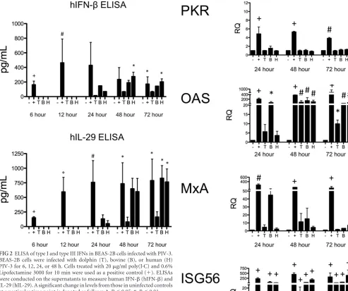
FIG 2ELISA of type I and type III IFNs in BEAS-2B cells infected with PIV-3. BEAS-2B cells were infected with dolphin (T), bovine (B), or human (H) PIV-3 for 6, 12, 24, or 48 h. Cells treated with 20g/ml poly(I·C) and 0.6% Lipofectamine 3000 for 10 min were used as a positive control (⫹). ELISAs were conducted on the supernatants to measure human IFN-(hIFN-) and IL-29 (hIL-29). A significant change in levels from those in uninfected controls at a particular time point is denoted as follows: *,P⬍0.05; #,P⬍0.01.
FIG 3Real-time PCR of antiviral molecules downstream of the IFN receptor in BEAS-2B cells infected with PIV-3. BEAS-2B cells were infected with dol-phin (T), bovine (B), or human (H) PIV-3 for 24, or 48, or 72 h. Cells treated with 20g/ml poly(I·C) and 0.6% Lipofectamine 3000 for 10 min were used as a positive control (⫹). Levels of downstream antiviral molecules (PKR, OAS, MxA, and ISG56) were measured by real-time PCR. The relative quantification (RQ) was calculated by normalization first to the GAPDH level to give a⌬CT
value, followed by normalization to the mock level at a given time point (⌬⌬CT). The RQ or fold change was then calculated as 2⫺⌬⌬CT. A significant
change in levels from those in uninfected controls at a particular time point is denoted as follows: *,P⬍0.05; #,P⬍0.01;⫹,P⬍0.001.
on November 7, 2019 by guest
http://jvi.asm.org/
[image:4.585.275.540.59.548.2] [image:4.585.42.535.67.480.2]HPIV-3 infection at 24 and 48 h (
Fig. 1
). IFN-␣
was not
signifi-cantly upregulated during PIV-3 infection in BEAS-2B cells.
ELISA of type I and type III IFNs in BEAS-2B cells infected
with PIV-3.
Enzyme-linked immunosorbent assays (ELISAs)
were run on cell culture supernatants to confirm our real-time
PCR results (
Fig. 2
). BEAS-2B cells produced significant levels of
IFN-
protein by 48 and 72 h postinfection with HPIV-3.
Con-centrations of IL-29 protein were also significantly upregulated by
72 h in response to BPIV-3 and HPIV-3 infection but not to
TtPIV-1 infection.
Real-time PCR of antiviral molecules downstream of the IFN
receptor in BEAS-2B cells infected with PIV-3.
PIV-3 infection
induced IL-29 production by BEAS-2B cells. Therefore, we next
wanted to confirm that the expression of downstream antiviral
molecules was induced as one would expect based on previous
reports (
Fig. 3
). In BEAS-2B cells, expression of OAS and ISG56
message increased significantly after infection with all of the
PIV-3s as early as 24 h postinfection. However, PKR and MxA
message levels were not upregulated, even by 72 h postinfection.
Examining type III IFN signaling in BEAS-2B cells using
Western blotting.
Type III IFNs signal through a unique receptor
but are thought to utilize the same JAK/STAT signaling pathways
as type I IFNs (
17
,
18
). Failure of the BEAS-2B cells to upregulate
expression of PKR and MxA in response to PIV-3 infection
sug-gested a blockade in the signaling pathways downstream of the
type I or type III receptors. We therefore wanted to determine the
effect of PIV-3 infection on the JAK/STAT signaling pathway.
BEAS-2B cells were infected with TtPIV-1, BPIV-3, or HPIV-3 at
an MOI of 2 for 24 h. After 24 h, the cells were treated with 1
g/ml
of recombinant IL-29/-28A/-28B for 30 min. Western blotting
was then used to examine the phosphorylation of Stat during
PIV-3 infection and subsequent recombinant cytokine treatment.
Stat1 has two critical phosphorylation sites: Y701 and S727. Both
sites are required to mediate downstream signaling (
25–27
).
Rep-resentative blots are depicted in
Fig. 4A
; densitometry
quantifica-tion is shown in
Fig. 4B
. As seen in
Fig. 4
, phosphorylation at Y701
on Stat1 is significantly reduced during PIV-3 infection compared
to the level in cells treated with IFN-
only, while phosphorylation
of the S727 site is unaffected. Importantly, calculations to
normal-ize values to those of Stat1 were based on the active form, Stat1-␣
because Stat1-

lacks the 38-amino-acid transcriptional
activa-tion domain (
32
). In addition, during HPIV-3 infection,
IFN--FIG 4Examining type III IFN signaling in BEAS-2B cells using Western blotting. Vero cells were infected with dolphin (T), bovine (B), or human (H) PIV-3 for 24 h. At 24 h postinfection, Vero cells were stimulated with recombinant IL-29/-28A/-28B (1g/ml) for 30 min to drive signaling downstream of the type III IFN receptor. (A) Western blotting was used to examine Stat1 and Stat2 phosphorylation during PIV-3 infection.-Actin was used as a loading control. (B) Densitometry quantification from Western blotting in BEAS-2B cells was calculated. The relative quantification from band intensity was calculated by its expression relative to that of the IFN--stimulated positive control (striped bars). This RQ was then normalized to the appropriate total Stat1 or Stat2 to give the percent pStat1 to total Stat1 or the percent pStat2 to total Stat2 value. A significant change in the level of phosphorylation from that in the IFN--treated positive control is denoted as follows: *,P⬍0.05; #,P⬍0.01.
on November 7, 2019 by guest
http://jvi.asm.org/
[image:5.585.94.494.65.405.2]induced phosphorylation at the Y690 site of Stat2 is significantly
reduced. Phosphorylation of Stat2 Y690 also appears to be
de-creased during BPIV-3 and TtPIV-1 infection, but these changes
are not significant.
Because BEAS-2B cells have the capacity to secrete both type I
and type III IFNs, it is difficult to dissect the effect of PIV-3
infec-tion on IL-29 signaling, independent of IFN-
␣
/

. In preliminary
experiments, we sought to knock down the expression of type I
IFN receptor (IFNAR1/IFNAR2) by small interfering RNA
(siRNA) treatment. After siRNA knockdown and restimulation
with recombinant IFN-
␣
/

, there was a 60 to 70% reduction in
expression levels of OAS, MxA, and ISG56 (data not shown).
However, Lipofectamine treatment alone caused untoward
changes in the JAK/STAT signaling pathway that would affect
any conclusions. Jensen et al. showed a similar phenomenon in
that multiple transfection reagents artificially upregulated
IFIT1, which is an antiviral molecule downstream of the IFN
receptor (
33
).
Real-time PCR of type III IFNs in Vero cells infected with
PIV-3.
We had observed intriguing results about the type III IFN
response in BEAS-2B cells during PIV-3 infection; however,
con-clusions made in the BEAS-2B cells are complicated by the
pres-ence of IFN-
␣
/

. We therefore chose to repeat these experiments
in a model independent of type I IFN. Vero cells were specifically
used because several previous studies have shown that they are not
able to produce type I IFNs (
22–24
). As in the BEAS-2B cells, Vero
cells were infected at an MOI of 2 with TtPIV-1, BPIV-3, or
HPIV-3, and expression levels of the type III IFNs (IL-28 and
IL-29) were measured by real-time PCR at multiple time points
after infection. As observed with the BEAS-2B cells, IL-29 message
was significantly increased in Vero cells by 48 h postinfection in
response to all virus strains (
Fig. 5
). Treatment with recombinant
IFN-
␣
/

was not able to significantly upregulate IL-29 or IL-28
message in Vero cells. IL-28 expression was not detectable in Vero
cells during PIV-3 infection; however, we were unable to establish
a positive control to confirm the capacity of these cells to produce
IL-28.
Real-time PCR of antiviral molecules downstream of the IFN
receptor in Vero cells infected with PIV-3 and stimulated with
recombinant type III IFNs.
We again wanted to examine
down-stream antiviral molecules that may be upregulated in the Vero
cells during PIV-3 infection (
Fig. 6
). Cells were infected with
TtPIV-1, BPIV-3, or HPIV-3, as described for the experiment
shown in
Fig. 5
, and expression of PKR, OAS, MxA, and ISG56
was measured by real-time PCR at multiple time points after
infection. As shown by the data in
Fig. 6
, expression of OAS was
upregulated in response to all three viruses within 48 h
postin-fection. ISG56 expression was also upregulated by 48 h
follow-ing HPIV-3 infection and by 72 h followfollow-ing BPIV-3 and
TtPIV-1 infection. However, PKR and MxA expression levels
FIG 5Real-time PCR of type III IFNs in Vero cells infected with PIV-3. Vero cells were infected with dolphin (T), bovine (B), or human (H) PIV-3 for 6, 12, 24, or 48 h. Cells were incubated with recombinant IFN-␣/at 500 U/ml for 24 h. IL-28 or -29 message levels were measured by real-time PCR. The relative quantification (RQ) was calculated by normalization first to the GAPDH level to give a⌬CTvalue, followed by normalization to the mock level at a given time
point (⌬⌬CT). The RQ or fold change was then calculated as 2⫺⌬⌬ CT. A
sig-nificant change from uninfected controls at a particular time point is denoted as follows: *,P⬍0.05; #,P⬍0.01. Striped bars, positive controls.
FIG 6Real-time PCR of antiviral molecules downstream of the IFN receptor in Vero cells infected with PIV-3. Vero cells were infected with dolphin (T), bovine (B), or human (H) PIV-3 for 24, or 48, or 72 h. Cells were incubated with recombinant IFN-␣/(500 U/ml) or IL-29/-28A/-28B (1g/ml) for 24 h. Levels of downstream antiviral molecules (PKR, OAS, MxA, and ISG56) were measured by real-time PCR. The relative quantification (RQ) was calculated by normalization first to the GAPDH level to give a⌬CTvalue, followed by
normalization to the mock level at a given time point (⌬⌬CT). The RQ or fold
change was then calculated as 2⫺⌬⌬CT. A significant change in levels from those
in the uninfected controls at a particular time point is denoted as follows: *,
P⬍0.05; #,P⬍0.01;⫹,P⬍0.001. Striped bars, positive controls.
on November 7, 2019 by guest
http://jvi.asm.org/
[image:6.585.316.524.64.401.2] [image:6.585.58.272.69.260.2]were not significantly induced in response to any of the virus
strains. Importantly, none of the downstream signaling
mole-cules were expressed at levels near those of the recombinant
type III or I IFN treatments used as positive controls (
Fig. 6
,
striped bars), suggesting an effect of PIV-3 on downstream
signaling pathways.
We sought to further determine if treatment with recombinant
type III IFN could overcome the blockade resulting from PIV-3
infection and induce signaling downstream of IFN-R1/IL-10R2.
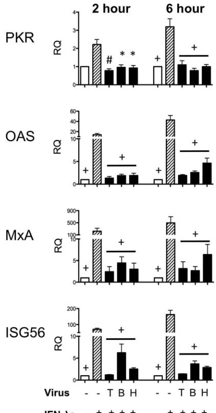
Therefore, Vero cells were infected with PIV-3 and 24 h
postinfec-tion treated with recombinant type III IFNs for 2 or 6 h. As seen
from the data in
Fig. 7
, downstream signaling molecules were still
not upregulated to levels near those of uninfected, IFN-
-treated
positive-control cells. Because recombinant type III IFN
treat-ment could not rescue the effect of virus infection on downstream
antiviral molecules, we hypothesized that PIV-3 infection directly
affects the JAK/STAT signaling pathway that is downstream of
IFN-
R1/IL-10R2.
Examining type III IFN signaling in Vero cells using Western
blotting.
IL-29 expression was upregulated in Vero cells infected
with PIV-3 (
Fig. 5
), while downstream antiviral signaling
mole-cules were not induced (
Fig. 6
). In addition, recombinant type III
IFN treatment of PIV-3-infected Vero cells failed to induce
ex-pression of downstream antiviral molecules to levels observed in
uninfected controls (
Fig. 7
). This suggested that, through an
un-known mechanism, PIV-3 inhibits the JAK/STAT signaling
path-way downstream of IFN-
R1/IL-10R2. Similarly to previous
ex-periments, Vero cells were infected with TtPIV-1, BPIV-3, or
HPIV-3 at an MOI of 2 for 24 h. After 24 h, the cells were treated
with recombinant type III IFN to drive signaling downstream of
the type III IFN receptor. In BEAS-2B cells we observed, by
West-ern blotting, reduced phosphorylation of pStat1 Y701 during
PIV-3 infection. In
Fig. 8
, we show that, in Vero cells,
phosphor-ylation is decreased at the S727 site on Stat1 during PIV-3
infec-tion. This reduced phosphorylation is specific to the S727 site
because the Y701 site of Stat1 is unaffected. As depicted by
densitometric quantitation in
Fig. 8B
, Stat1 phosphorylation at
S727 is significantly reduced during PIV-3 infection while
phosphorylation at the Y701 site significantly increased. In
ad-dition, levels of total Stat1 and Stat2 and of phosphorylation at
the tyrosine 690 site on Stat2 (pStat2 Y690) are not affected. To
calculate the relative quantification (RQ) of band intensity, the
level of pStat1 was normalized to that of Stat1-
␣
as previously
described (
32
).
Prophylactic treatment of Vero cells with type III IFNs
be-fore PIV-3 viral infection.
Because PIV-3 blocks signaling
down-stream of the type III IFN receptor, therapeutic treatment with
type III IFNs would not be useful. However, prophylactic
treat-ment with IL-29, IL-28A, and/or IL-28B in Vero cells does appear
effective against PIV-3 infection (
Fig. 9
, left panels). Vero cells
were treated with the type III IFNs, singly or pooled, for 24 h
before PIV-3 infection, and then the virus titer was measured 24 h
after PIV-3 infection. As seen in
Fig. 9
, recombinant type III IFN
treatment resulted in significantly reduced viral titers. These
re-sults are evidently independent of MOI in that MOIs of 2 and 0.2
were used. This antiviral response was specific to the recombinant
type III interferon added because 80
g/ml blocking antibody was
able to reverse the titer repression (
Fig. 9
, right panels).
DISCUSSION
Type I and type III IFNs are induced by viral sensors including
RLRs and TLRs. IFNs bind their respective receptors to activate
JAK/STAT signaling and produce downstream antiviral
mole-cules, including PKR, OAS, MxA, and ISGs. Parainfluenza virus
predominantly infects the airway epithelium. Because the type III
IFN receptor is primarily located on epithelial cells, we chose to
examine the IFN-
response to PIV-3 infection. While many
re-spiratory viruses, including rere-spiratory syncytial virus (RSV),
rhi-novirus, and influenza virus, have been shown to induce IFN-s
FIG 7Real-time PCR of antiviral molecules downstream of the IFN receptor in Vero cells stimulated with recombinant type III IFNs after PIV-3 infection. Vero cells were infected with dolphin (T), bovine (B), or human (H) PIV-3 for 24 h. At 24 h postinfection, Vero cells were stimulated with recombinant IL-29/-28A/-28B (1g/ml) for 2 or 6 hours. Levels of downstream antiviral mol-ecules (PKR, OAS, MxA, and ISG56) were measured by real-time PCR. The relative quantification (RQ) was calculated by normalization first to the GAPDH level to give a⌬CTvalue, followed by normalization to the mock level
at a given time point (⌬⌬CT). The RQ or fold change was then calculated as
2⫺⌬⌬CT. A significant change in levels from those in the IFN--treated positive
control at a particular time point is denoted as follows: *,P⬍0.05; #,P⬍0.01;
⫹,P⬍0.001.
on November 7, 2019 by guest
http://jvi.asm.org/
[image:7.585.314.525.65.469.2](
33–37
), this is the first report, to our knowledge, of PIV-3
induc-ing expression of type III IFNs and modulatinduc-ing the downstream
antiviral signaling pathways.
Here, we examined production of type III IFNs and antiviral
molecules downstream of IFN-R1/IL-10R2 during PIV-3
infec-tion. IL-29 message levels were significantly increased in BEAS-2B
cells as early as 24 h postinfection (
Fig. 1
). Other investigators have
shown that type I IFNs are necessary to activate type III IFNs, but
we did not observe the necessity of IFN-␣/
for IL-29 production
here (
38
). This can be seen from the data shown in
Fig. 5
, where
IL-29 message was significantly upregulated by 48 h postinfection
with PIV-3 in type I IFN-deficient Vero cells, and recombinant
IFN-␣/
alone (striped bars) did not stimulate IL-29 to a level
similar to that observed during virus infection. We measured
pro-tein levels of IL-29 in cell culture supernatant from BEAS-2B cells,
as shown in
Fig. 2
. Multiple IL-29 ELISAs were tested for use with
Vero cell supernatants; however, we found that none of the
anti-bodies were cross-reactive with Vero cell supernatants. It is
there-fore possible that PIV-3 could simply inhibit production of type
III IFN protein by an unknown mechanism in Vero cells, which
could, in turn, prevent induction of downstream antiviral
mole-cules. A significant increase in OAS and ISG56 levels was seen in
BEAS-2B cells infected with all three viruses, but this could be due
to type I IFNs (
Fig. 3
). Furthermore, downstream antiviral
mole-cules, including PKR, OAS, MxA, and ISG56, were not
upregu-lated in the Vero cells infected with PIV-3 compared to levels in
cells treated with recombinant type I or type III IFNs (
Fig. 6
,
striped bars).
There is clear evidence that PIV-3 interferes with signaling
downstream of the IFN-R1/IL-10R2 receptor.
Figure 7
showed
that Vero cells stimulated with recombinant type III IFNs at 24 h
postinfection with PIV-3 were unable to induce a robust antiviral
response. This was a strong indication that PIV-3 interferes with
the JAK/STAT signaling pathway downstream of the type III IFN
receptor. It is well known that paramyxoviruses block type I IFN
production (
7–16
). A few reports have studied the JAK/STAT
downstream signaling response during PIV-3 infection,
specifi-cally in response to type I IFNs.
By examining the phosphorylation downstream of IFN-
R1/
IL-10R2 through Western blotting, we specifically observed
de-creased phosphorylation of pStat1 Y701 in BEAS-2B cells (
Fig. 4
)
and a decrease in phosphorylation at the S727 site on Stat1 in Vero
FIG 8Examining type III IFN signaling in Vero cells using Western blotting. Vero cells were infected with dolphin (T), bovine (B), or human (H) PIV-3 for 24 h. At 24 h postinfection, Vero cells were stimulated with recombinant IL-29/-28A/-28B (1g/ml) for 30 min to drive signaling downstream of the type III IFN receptor. (A) Western blotting was used to examine Stat1 and Stat2 phosphorylation during PIV-3 infection.-Actin was used as a loading control. (B) Densitometry quantification from Western blotting in Vero cells was calculated. Relative quantification from band intensity was calculated by its expression relative to the level of the IFN--stimulated positive control. This RQ was then normalized to the level of the appropriate total Stat1 or Stat2 to give the percent pStat1 to total Stat1 or percent pStat2 to total Stat2 value. A significant change in phosphorylation levels from those in the IFN--treated positive control is indicated (*,P⬍0.05).
on November 7, 2019 by guest
http://jvi.asm.org/
[image:8.585.95.495.63.405.2]cells (
Fig. 8
) during PIV-3 infection. It is well known that both the
tyrosine and serine sites of Stat1 are necessary for full
transcrip-tional activation (
26
). Specifically, phosphorylation at the Y701
site has been shown to be vital because of its role in hetero- or
homodimerization of Stat1 (
26
,
32
). On the other hand, the S727
site must be phosphorylated to allow binding of transcriptional
coactivators such as CBP/p300 and MCM5 (
26
,
27
,
39
,
40
).
Phos-phorylation of the serine and tyrosine sites on Stat1 has been
shown to be independent of each other (
41
); here, we observed
reduced phosphorylation of Stat1 specific to S727 or Y701 in the
Vero or BEAS-2B cells, respectively. Because the Vero cells are
naturally unable to produce type I IFNs, it may be most
appropri-ate to base any conclusions on results obtained in the Vero cells.
Other paramyxoviruses have been shown to decrease total Stat1
and Stat2 levels to reduce the type I IFN antiviral response (
12
,
15
,
42
). As was observed in the BEAS-2B cells (
Fig. 4
) but not seen in
the Vero cells (
Fig. 8
), total Stat1-␣
and Stat2 may be slightly
reduced during HPIV-3 infection. However, any total Stat1 or
Stat2 reduction was accounted for during relative quantification
performed for graphical analysis (
Fig. 4B
and
8B
). It is of note that
in the BEAS-2B cell line a species-specific decrease in
phosphory-lation of Stat2 was observed during infection with HPIV-3 that
was not seen following infection with BPIV-3 or TtPIV-1 (
Fig. 4
).
We hypothesize that PIV-3 induces specific phosphatase
activ-ity to cause reduced phosphorylation of Stat1. The phosphatase
SHP-2 has previously been shown to dephosphorylate Stat1 at
both Y701 and S727 (
43
). In addition, it is plausible that
altera-tions in tyrosine kinases (Jak1/Tyk2) could occur during PIV-3
infection that, in turn, cause reduced phosphorylation of Stat1
(
44
). Future experiments will need to be conducted to examine the
specific mechanism of Stat1 dephosphorylation or reduced
phos-phorylation during PIV-3 infection.
Treatment of Vero cells with type III IFNs 24 h before PIV-3
infection was able to reduce viral titers (
Fig. 9
). Type III IFNs are
therefore able to prophylactically induce downstream antiviral
molecules to control viral growth in Vero cells. However, we show
for the first time that PIV-3 induces type III IFNs and identify a
novel mechanism by which PIV-3 inhibits production of the
downstream antiviral molecules PKR, OAS, MxA, and ISG56.
Downstream of the type III IFN receptor, PIV-3 causes reduced
phosphorylation of Stat1 to negatively affect the JAK/STAT
sig-naling pathway and reduce production of antiviral molecules.
ACKNOWLEDGMENTS
We acknowledge the excellent technical support of Theresa Waters and Tera Nyholm.
FIG 9Prophylactic treatment of Vero cells with type III IFNs before PIV-3 viral infection. Vero cells were prophylactically treated with recombinant IL-29/-28A/-28B, alone or pooled, at 1g/ml for 24 h before TtPIV-1, BPIV-3, or HPIV-3 infection at an MOI of 2 or 0.2 for an additional 24 h. After 48 h, titer plates were used to determine the effect of type III IFNs on virus growth. In the experiment shown in the right panels, IL-29 blocking antibody was added in increasing doses to show specificity of the viral inhibition by type III IFNs. At the highest blocking antibody dose of 80g/ml, a log(titer) similar to that of the control was observed. A significant reduction in log(titer) compared to that of the untreated control sample is denoted as follows: *,P⬍0.05; #,P⬍0.01;⫹,P⬍0.001.
on November 7, 2019 by guest
http://jvi.asm.org/
[image:9.585.114.476.67.396.2]FUNDING INFORMATION
DOD | Office of Naval Research (ONR) provided funding to Randy E Sacco under grant number N0001412IP20029.
The funders had no role in study design, data collection and interpreta-tion, or the decision to submit the work for publication.
REFERENCES
1.Karron RA, Thumar B, Schappell E, Surman S, Murphy BR, Collins PL, Schmidt AC.2012. Evaluation of two chimeric bovine-human parainflu-enza virus type 3 vaccines in infants and young children. Vaccine30:3975– 3981.http://dx.doi.org/10.1016/j.vaccine.2011.12.022.
2.Karron RA, Wright PF, Hall SL, Makhene M, Thompson J, Burns BA, Tollefson S, Steinhoff MC, Wilson MH, Harris DO.1995. A live atten-uated bovine parainfluenza virus type 3 vaccine is safe, infectious, immu-nogenic, and phenotypically stable in infants and children. J Infect Dis
171:1107–1114.http://dx.doi.org/10.1093/infdis/171.5.1107.
3.Schmidt AC, McAuliffe JM, Huang A, Surman SR, Bailly JE, Elkins WR, Collins PL, Murphy BR, Skiadopoulos MH.2000. Bovine parainfluenza virus type 3 (BPIV3) fusion and hemagglutinin-neuraminidase glycopro-teins make an important contribution to the restricted replication of BPIV3 in primates. J Virol74:8922– 8929.http://dx.doi.org/10.1128/JVI .74.19.8922-8929.2000.
4.Henrickson KJ.2003. Parainfluenza viruses. Clin Microbiol Rev16:242– 264.http://dx.doi.org/10.1128/CMR.16.2.242-264.2003.
5.Vareille M, Kieninger E, Edwards MR, Regamey N.2011. The airway epithelium: soldier in the fight against respiratory viruses. Clin Microbiol Rev24:210 –229.http://dx.doi.org/10.1128/CMR.00014-10.
6.Sadler AJ, Williams BR.2008. Interferon-inducible antiviral effectors. Nat Rev Immunol8:559 –568.http://dx.doi.org/10.1038/nri2314. 7.Andrejeva J, Childs KS, Young DF, Carlos TS, Stock N, Goodbourn S,
Randall RE.2004. The V proteins of paramyxoviruses bind the IFN-inducible RNA helicase, mda-5, and inhibit its activation of the IFN-beta promoter. Proc Natl Acad Sci U S A101:17264 –17269.http://dx.doi.org /10.1073/pnas.0407639101.
8.Bossert B, Marozin S, Conzelmann KK.2003. Nonstructural proteins NS1 and NS2 of bovine respiratory syncytial virus block activation of interferon regulatory factor 3. J Virol77:8661– 8668.http://dx.doi.org/10 .1128/JVI.77.16.8661-8668.2003.
9.Childs K, Stock N, Ross C, Andrejeva J, Hilton L, Skinner M, Randall R, Goodbourn S.2007. mda-5, but not RIG-I, is a common target for paramyxovirus V proteins. Virology359:190 –200.http://dx.doi.org/10 .1016/j.virol.2006.09.023.
10. Childs KS, Andrejeva J, Randall RE, Goodbourn S.2009. Mechanism of mda-5 Inhibition by paramyxovirus V proteins. J Virol83:1465–1473.
http://dx.doi.org/10.1128/JVI.01768-08.
11. Childs K, Randall R, Goodbourn S. 2012. Paramyxovirus V proteins interact with the RNA helicase LGP2 to inhibit RIG-I-dependent inter-feron induction. J Virol 86:3411–3421. http://dx.doi.org/10.1128/JVI .06405-11.
12. Elliott J, Lynch OT, Suessmuth Y, Qian P, Boyd CR, Burrows JF, Buick R, Stevenson NJ, Touzelet O, Gadina M, Power UF, Johnston JA.2007. Respiratory syncytial virus NS1 protein degrades STAT2 by using the elongin-cullin E3 ligase. J Virol81:3428 –3436.http://dx.doi.org/10.1128 /JVI.02303-06.
13. Kitagawa Y, Yamaguchi M, Zhou M, Nishio M, Itoh M, Gotoh B.
2013. Human parainfluenza virus type 2 V protein inhibits TRAF6-mediated ubiquitination of IRF7 to prevent TLR7-and TLR9-dependent interferon induction. J Virol87:7966 –7976.http://dx.doi .org/10.1128/JVI.03525-12.
14. Lu LL, Puri M, Horvath CM, Sen GC.2008. Select paramyxoviral V proteins inhibit IRF3 activation by acting as alternative substrates for in-hibitor ofB kinaseε(IKKe)/TBK1. J Biol Chem283:14269 –14276.http: //dx.doi.org/10.1074/jbc.M710089200.
15. Ramaswamy M, Shi L, Varga SM, Barik S, Behlke MA, Look DC.2006. Respiratory syncytial virus nonstructural protein 2 specifically inhibits type I interferon signal transduction. Virology344:328 –339.http://dx.doi .org/10.1016/j.virol.2005.09.009.
16. Yamaguchi M, Kitagawa Y, Zhou M, Itoh M, Gotoh B. 2014. An anti-interferon activity shared by paramyxovirus C proteins: inhibition of Toll-like receptor 7/9-dependent alpha interferon induction. FEBS Lett
588:28 –34.http://dx.doi.org/10.1016/j.febslet.2013.11.015.
17. Kotenko SV, Gallagher G, Baurin VV, Lewis-Antes A, Shen M, Shah NK, Langer JA, Sheikh F, Dickensheets H, Donnelly RP.2003. IFN-s mediate antiviral protection through a distinct class II cytokine receptor complex. Nat Immunol4:69 –77.http://dx.doi.org/10.1038/ni875. 18. Sheppard P, Kindsvogel W, Xu W, Henderson K, Schlutsmeyer S,
Whitmore TE, Kuestner R, Garrigues U, Birks C, Roraback J, Ostrander C, Dong D, Shin J, Presnell S, Fox B, Haldeman B, Cooper E, Taft D, Gilbert T, Grant FJ, Tackett M, Krivan W, McKnight G, Clegg C, Foster D, Klucher KM.2003. IL-28, IL-29 and their class II cytokine receptor IL-28R. Nat Immunol4:63– 68.http://dx.doi.org/10.1038/ni873. 19. Miknis ZJ, Magracheva E, Li W, Zdanov A, Kotenko SV, Wlodawer A.
2010. Crystal structure of human interferon-1 in complex with its high-affinity receptor interferon-R1. J Mol Biol404:650 – 664.http://dx.doi .org/10.1016/j.jmb.2010.09.068.
20. Sommereyns C, Paul S, Staeheli P, Michiels T. 2008. IFN-lambda (IFN-) is expressed in a tissue-dependent fashion and primarily acts on epithelial cells in vivo. PLoS Pathog4:e1000017.http://dx.doi.org/10.1371 /journal.ppat.1000017.
21. Andrejeva J, Norsted H, Habjan M, Thiel V, Goodbourn S, Randall RE.
2013. ISG56/IFIT1 is primarily responsible for interferon-induced changes to patterns of parainfluenza virus type 5 transcription and protein synthesis. J Gen Virol94:59 – 68.http://dx.doi.org/10.1099/vir.0.046797-0.
22. Emeny JM, Morgan MJ.1979. Regulation of the interferon system: evi-dence that Vero cells have a genetic defect in interferon production. J Gen Virol43:247–252.http://dx.doi.org/10.1099/0022-1317-43-1-247. 23. Osada N, Kohara A, Yamaji T, Hirayama N, Kasai F, Sekizuka T,
Kuroda M, Hanada K.2014. The genome landscape of the African green monkey kidney-derived Vero cell line. DNA Res21:673– 683.http://dx .doi.org/10.1093/dnares/dsu029.
24. Wathelet MG, Berr PM, Huez GA.1992. Regulation of gene expression by cytokines and virus in human cells lacking the type-I interferon locus. Eur J Biochem206:901–910.http://dx.doi.org/10.1111/j.1432-1033.1992 .tb16999.x.
25. Ooi EL, Chan ST, Cho NE, Wilkins C, Woodward J, Li M, Kikkawa U, Tellinghuisen T, Gale M, Saito T. 2014. Novel antiviral host factor, TNK1, regulates IFN signaling through serine phosphorylation of STAT1. Proc Natl Acad Sci U S A111:1909 –1914.http://dx.doi.org/10.1073/pnas .1314268111.
26. Wen Z, Zhong Z, Darnell JE, Jr.1995. Maximal activation of transcrip-tion by Statl and Stat3 requires both tyrosine and serine phosphorylatranscrip-tion. Cell82:241–250.http://dx.doi.org/10.1016/0092-8674(95)90311-9. 27. Zhang Y, Takami K, Lo MS, Huang G, Yu Q, Roswit WT, Holtzman
MJ.2005. Modification of the Stat1 SH2 domain broadly improves inter-feron efficacy in proportion to p300/CREB-binding protein coactivator recruitment. J Biol Chem280:34306 –34315.http://dx.doi.org/10.1074 /jbc.M503263200.
28. Nollens HH, Wellehan JF, Saliki JT, Caseltine SL, Jensen ED, Van Bonn W, Venn-Watson S.2008. Characterization of a parainfluenza virus iso-lated from a bottlenose dolphin (Tursiops truncatus). Vet Microbiol128:
231–242.http://dx.doi.org/10.1016/j.vetmic.2007.10.005.
29. Eberle KC, Neill JD, Venn-Watson SK, McGill JL, Sacco RE. 2015. Novel Atlantic bottlenose dolphin parainfluenza virus TtPIV-1 clusters with bovine PIV-3 genotype B strains. Virus Genes51:198 –208.http://dx .doi.org/10.1007/s11262-015-1224-7.
30. Livak KJ, Schmittgen TD.2001. Analysis of relative gene expression data using real-time quantitative PCR and the 2⫺⌬⌬CTmethod. Methods25:
402– 408.http://dx.doi.org/10.1006/meth.2001.1262.
31. Reed LJ, Muench J.1938. A simple method of estimating fifty percent endpoints. Am J Hyg27:493– 497.
32. Horvath CM, Darnell JE.1996. The antiviral state induced by alpha interferon and gamma interferon requires transcriptionally active Stat1 protein. J Virol70:647– 650.
33. Jensen K, Anderson JA, Glass EJ.2014. Comparison of small interfering RNA (siRNA) delivery into bovine monocyte-derived macrophages by transfection and electroporation. Vet Immunol Immunopathol158:224 – 232.http://dx.doi.org/10.1016/j.vetimm.2014.02.002.
34. Khaitov MR, Laza-Stanca V, Edwards MR, Walton RP, Rohde G, Contoli M, Papi A, Stanciu LA, Kotenko SV, Johnston SL. 2009. Respiratory virus induction of alpha-, beta- and lambda-interferons in bronchial epithelial cells and peripheral blood mononuclear cells. Allergy
64:375–386.http://dx.doi.org/10.1111/j.1398-9995.2008.01826.x. 35. Okabayashi T, Kojima T, Masaki T, Yokota S-I, Imaizumi T, Tsutsumi
H, Himi T, Fujii N, Sawada N.2011. Type-III interferon, not type-I, is
on November 7, 2019 by guest
http://jvi.asm.org/
the predominant interferon induced by respiratory viruses in nasal epi-thelial cells. Virus Res160:360 –366.http://dx.doi.org/10.1016/j.virusres .2011.07.011.
36. Spann KM, Tran K-C, Chi B, Rabin RL, Collins PL.2004. Suppression of the induction of alpha, beta, and lambda interferons by the NS1 and NS2 proteins of human respiratory syncytial virus in human epithelial cells and macrophages. J Virol78:4363– 4369.http://dx.doi.org/10.1128 /JVI.78.8.4363-4369.2004.
37. Wang J, Oberley-Deegan R, Wang S, Nikrad M, Funk CJ, Hartshorn KL, Mason RJ.2009. Differentiated human alveolar type II cells secrete antiviral IL-29 (IFN-1) in response to influenza A infection. J Immunol
182:1296 –1304.http://dx.doi.org/10.4049/jimmunol.182.3.1296. 38. Ank N, Iversen MB, Bartholdy C, Staeheli P, Hartmann R, Jensen UB,
Dagnaes-Hansen F, Thomsen AR, Chen Z, Haugen H.2008. An impor-tant role for type III interferon (IFN-/IL-28) in TLR-induced antiviral activity. J Immunol180:2474 –2485.http://dx.doi.org/10.4049/jimmunol .180.4.2474.
39. Wojciak JM, Martinez-Yamout MA, Dyson HJ, Wright PE.2009. Struc-tural basis for recruitment of CBP/p300 coactivators by STAT1 and STAT2 transactivation domains. EMBO J28:948 –958.http://dx.doi.org /10.1038/emboj.2009.30.
40. Zhang JJ, Zhao Y, Chait BT, Lathem WW, Ritzi M, Knippers R, Darnell JE.1998. Ser727-dependent recruitment of MCM5 by Stat1␣in IFN-␥ -induced transcriptional activation. EMBO J17:6963– 6971.http://dx.doi .org/10.1093/emboj/17.23.6963.
41. Zhu X, Wen Z, Xu LZ, Darnell JE.1997. Stat1 serine phosphorylation occurs independently of tyrosine phosphorylation and requires an acti-vated Jak2 kinase. Mol Cell Biol17:6618 – 6623.http://dx.doi.org/10.1128 /MCB.17.11.6618.
42. Young DF, Didcock L, Goodbourn S, Randall RE.2000. Paramyxo-viridaeuse distinct virus-specific mechanisms to circumvent the inter-feron response. Virology 269:383–390. http://dx.doi.org/10.1006/viro .2000.0240.
43. Wu TR, Hong YK, Wang XD, Ling MY, Dragoi AM, Chung AS, Campbell AG, Han ZY, Feng GS, Chin YE.2002. SHP-2 is a dual-specificity phosphatase involved in Stat1 dephosphorylation at both ty-rosine and serine residues in nuclei. J Biol Chem277:47572– 47580.http: //dx.doi.org/10.1074/jbc.M207536200.
44. Senft AP, Taylor RH, Lei W, Campbell SA, Tipper JL, Martinez MJ, Witt TL, Clay CC, Harrod KS.2010. Respiratory syncytial virus impairs macrophage IFN-␣/- and IFN-␥-stimulated transcription by distinct mechanisms. Am J Respir Cell Mol Biol42:404 – 414.http://dx.doi.org/10 .1165/rcmb.2008-0229OC.
45. Ank N, West H, Bartholdy C, Eriksson K, Thomsen AR, Paludan SR.
2006. Lambda interferon (IFN-), a type III IFN, is induced by viruses and IFNs and displays potent antiviral activity against select virus infections in vivo. J. Virol. 80:4501– 4509. http://dx.doi.org/10.1128/JVI.80.9.4501 -4509.2006.
46. Yoon CH, Lee ES, Lim DS, Bae YS.2009. PKR, a p53 target gene, plays a crucial role in the tumor-suppressor function of p53. Proc Natl Acad Sci U S A106:7852–7857.http://dx.doi.org/10.1073/pnas.0812148106. 47. Liu MQ, Zhou DJ, Wang X, Zhou W, Ye L, Li JL, Wang YZ, Ho WZ.
2012. IFN-3 inhibits HIV infection of macrophages through the JAK-STAT pathway. PLoS ONE7:e35902.http://dx.doi.org/10.1371/journal .pone.0035902.
48. Holzinger D, Jorns C, Stertz S, Boisson-Dupuis S, Thimme R, Weidmann M, Casanova JL, Haller O, Kochs G.2007. Induction of MxA gene expres-sion by influenza A virus requires type I or type III interferon signaling. J. Virol.81:7776 –7785.http://dx.doi.org/10.1128/JVI.00546-06.
49. Kuo TM, Hu CP, Chen YL, Hong MH, Jeng KS, Liang CC, Chen ML, Chang C.2009. HBV replication is significantly reduced by IL-6. J Biomed Sci16:41.http://dx.doi.org/10.1186/1423-0127-16-41.
50. Hinzey A, Alexander J, Corry J, Adams KM, Claggett AM, Traylor ZP, Davis IC, Webster Marketon JI.2011. Respiratory syncytial virus re-presses glucocorticoid receptor-mediated gene activation. Endocrinology
152:483– 494.http://dx.doi.org/10.1210/en.2010-0774.
on November 7, 2019 by guest
http://jvi.asm.org/