virus replication model. The replication of MHV increased when the N proteins were expressed
in trans
, while knockdown of
Dicer1 or Ago2 transcripts facilitated the MHV replication in mammalian cells. These results support the hypothesis that RNAi
is a part of the antiviral immunity responses in mammalian cells.
IMPORTANCE
RNAi has been well known to play important antiviral roles from plants to invertebrates. However, recent studies provided
strong supports that RNAi is also involved in antiviral response in mammalian cells. An important indication for
RNAi-mediated antiviral activity in mammals is the fact that a number of mammalian viruses encode potent suppressors of RNA
silencing. Our results demonstrate that coronavirus N protein could function as a VSR through its double-stranded RNA
binding activity. Mutational analysis of N protein allowed us to find out the critical residues for the VSR activity. Using the
MHV-A59 as the coronavirus replication model, we showed that ectopic expression of SARS-CoV N protein could promote
MHV replication in RNAi-active cells but not in RNAi-depleted cells. These results indicate that coronaviruses encode a
VSR that functions in the replication cycle and provide further evidence to support that RNAi-mediated antiviral response
exists in mammalian cells.
R
NA interference (RNAi) is originally regarded as a mechanism of
eukaryotic posttranscriptional gene regulation mediated by small
interfering RNA (siRNA)-induced sequence-specific RNA
degrada-tion (
1
). It is also well known to exert as an important antiviral
de-fense mechanism in a wide range of organisms, from plants to
inver-tebrates (
2
). During the virus infection, the virus-derived long
double-stranded RNA (dsRNA) is cleaved by RNAIII-like
endonu-clease (named Dicer) into approximately 21- to 23-nucleotide (nt)
siRNA, which is incorporated into the RNA-induced silencing
com-plex (RISC) and activates the antiviral RNAi for viral RNA
degrada-tion. In mammalian cells, although the activation of RNAi by
syn-thetic siRNA or short hairpin RNA (shRNA) is widely used as a tool
for gene knockdown and antiviral treatment, the RNAi-mediated
an-tiviral mechanism has been debated for a long time (
3
), because the
interferon (IFN) response of the innate immune system is well known
as the dominant antiviral mechanism (
4
). However, more and more
evidence has provided strong support for the existence of a natural
RNAi-mediated antiviral response in mammals (
5
). Moreover,
re-cent studies showed that in undifferentiated cells and immature mice,
the RNAi-mediated antiviral response is essential (
6–8
).
To overcome the RNAi-mediated antiviral defense, viruses
have evolved to encode a viral suppressor of RNA silencing (VSR)
(
9
,
10
). For example, in plant viruses, rice hoja blancavirus NS3,
tombusvirus P19, and tomato aspermy virus 2b bind to long
dsRNA or siRNA to block RNAi (
11–13
). Turnip crinkle virus P38
and cauliflower mosaic virus P6 disrupt the components of RNAi
machinery (
14
,
15
). In insect viruses, flock house virus (FHV) B2
blocks RNAi by dsRNA binding (
16
,
17
), and Wuhan nodavirus
(WhNV) B2 was identified as a VSR by targeting both dsRNAs and
Dicer-2 (
18
,
19
). Although the majority of VSRs have been
iden-tified in plant and invertebrate viruses, several mammalian viruses
were shown to encode VSRs. For instance, Ebola virus VP35,
in-fluenza A virus NS1, vaccinia virus E3L, and Nodamura virus
(NoV) B2 act as VSRs by binding dsRNA (
20–23
). Hepatitis C
virus core and HIV-1 Tat block RNAi by inhibiting the activity of
Received21 May 2015Accepted11 June 2015
Accepted manuscript posted online17 June 2015
CitationCui L, Wang H, Ji Y, Yang J, Xu S, Huang X, Wang Z, Qin L, Tien P, Zhou X,
Guo D, Chen Y. 2015. The nucleocapsid protein of coronaviruses acts as a viral suppressor of RNA silencing in mammalian cells. J Virol 89:9029 –9043. doi:10.1128/JVI.01331-15.
Editor:S. Perlman
Address correspondence to Deyin Guo, dguo@whu.edu.cn, or Yu Chen, chenyu@whu.edu.cn.
Supplemental material for this article may be found athttp://dx.doi.org/10.1128 /JVI.01331-15.
Copyright © 2015, American Society for Microbiology. All Rights Reserved.
doi:10.1128/JVI.01331-15
on November 7, 2019 by guest
Dicer (
24
,
25
). Interestingly, all VSRs identified from mammalian
viruses possess IFN or protein kinase R antagonistic properties
and are essential for replication and pathogenesis, suggesting that
RNAi and other innate antiviral responses are interrelated
(
26–28
).
Coronaviruses (CoVs) are the largest positive single-stranded
RNA (ssRNA) viruses carrying an RNA genome of 26.2 to 31.7 kb
that infect a wide range of mammalian and avian species (
29
). It is
reported that coronaviruses generate significant amount of
dsRNAs as replicative and transcriptive intermediates (
30
,
31
).
Therefore, it might be the target of Dicer and thus induce
RNAi-mediated antiviral responses. An indirect evidence was shown that
severe acute respiratory syndrome coronavirus (SARS-CoV)
ac-cessory protein 7a was identified as a VSR (
32
). However, 7a
pro-tein is not essential for viral replication and transcription at least
in cell culture and tested animal models and is unique to
SARS-CoV (
33
,
34
). Consequently, it would be interesting to determine
whether there is another VSR commonly encoded among
corona-viruses family. In the present study, we screened the viral proteins
of SARS-CoV as a representative by a reversal-of-silencing assay
and identified the nucleocapsid (N) protein as a novel VSR, which
is conserved and encoded by all of the coronaviruses.
N protein is a basic protein (with typical pIs of
⬃
10) and has
nonspecific binding activity toward nucleic acids, including
ssRNA, ssDNA, and dsDNA (
35
,
36
). It encapsulates viral genomic
RNA (gRNA) to protect the genome and enters the host cell
to-gether with the viral RNA to facilitate its replication (
37–40
).
Fur-thermore, we have reported that the N protein antagonizes IFN-

by targeting the initial pattern recognition receptor/RNA
recog-nition step and that the C-terminal domain (CTD) is critical for
this antagonism (
41
). Other studies also revealed that the
SARS-CoV N protein contains two distinct RNA-binding domains (the
N-terminal domain [NTD] and the CTD) linked by a poorly
structured linkage region (Linker) containing a
serine/arginine-rich (SR-serine/arginine-rich) domain (SRD) (
42–45
). The CTD spanning
resi-dues 248 to 365 shows stronger nucleic acid-binding activity than
the NTD (
36
,
45
,
46
), and the basic region between residues 248 to
280 of CTD forms a positively charged groove that represents a
likely binding region for RNA (
46
). Here, we demonstrated that
the N protein of CoVs could efficiently inhibit Dicer-mediated
dsRNA cleavage and post-Dicer activities by sequestering dsRNAs
and siRNAs. Furthermore, we show that N protein deficient in
RNAi inhibition activity was unable to promote the replication of
mouse hepatitis virus (MHV) compared to the wild-type N
pro-tein and that knockdown of Dicer1 or Ago2 enhanced MHV
rep-lication. Our studies identified a novel coronaviral VSR and
pro-vide new epro-vidence on the existence of RNAi-mediated antiviral
response in mammalian cells.
MATERIALS AND METHODS
Plasmids and RNAs.For RNAi suppression assays in mammalian cells, the plasmid eGFP-C1 (Clontech) was used to express enhanced green fluorescent protein (eGFP). The eGFP-specific shRNA (shGFP) and con-trol shRNA targeting luciferase (shLuc) with the indicated sequences in Table S1 in the supplemental material were cloned to vector pSuperRetro driven by H1 polymerase III promoter. The plasmid pCMV-tag2b-N ex-pressing SARS-CoV N protein and the deletion mutants with Flag tag were constructed in our previous work (41). The open reading frame 6 (ORF6) expression plasmid with a hemagglutinin (HA) tag was kindly provided by Stanley Perlman. Point mutations were introduced into the N coding region by PCR mediated mutagenesis, with appropriate primers
containing the desired nucleotide changes (see Table S2 in the supplemen-tal material) and subsequently selected by DpnI digestion. The coding sequences of Middle East respiratory syndrome coronavirus (MERS-CoV) N protein were chemically synthesized and cloned into the pCMV-tag2b vector. Plasmids expressing N protein of coronaviruses MHV, por-cine epidemic diarrhea virus (PEDV), and transmissible gastroenteritis virus (TGEV) were gifts from Shaobo Xiao. The NoV B2-expressing plas-mid with Myc tag was provided by Christopher S. Sullivan. For RNAi suppression assays inDrosophilaS2 cells, the eGFP reporter gene and the FHV B2 were constructed into the insect expression vector pAc5.1/V5-HisB. SARS-CoV N protein and ORF6 were inserted into the EcoRI/NotI sites of pAc5.1/V5-HisB. Nonstructural protein 14 (nsp14) cloned in pAc5.1/V5-HisB was inserted into the NotI/XhoI sites. The primers are shown in Table S2 in the supplemental material. Full-length cDNA of FHV RNA1 and RNA1-⌬B2 (T2739C and C2910A) were described pre-viously (17). In addition, the siRNAs targeting eGFP (siGFP) were pre-pared by chemical synthesis (Invitrogen), whereas siRNAs targeting mouse Dicer1 and Ago2 were obtained from Qiagen. The oligonucleo-tides are shown in Table S3 in the supplemental material. The 244-bp dsRNA for eGFP silencing inDrosophilaS2 cells was generated byin vitro
transcription using MEGAscript kits (Ambion).
Cell culture and transfection.Human embryonic kidney 293T cells (HEK293T), mouse Neuro-2a cells (gifts from Yan Zhou) and L2 cells (gifts from Rong Ye) were maintained in Dulbecco modified Eagle me-dium supplemented with 10% fetal bovine serum, 100 U of penicillin/ml, and 100g of streptomycin/ml.DrosophilaS2 cells were cultured in semi suspension at 27°C in Schneider’s insect medium (Gibco, Carlsbad, CA) supplemented with 10% fetal bovine serum (Gibco) (18). HEK293T cells were seeded on 12-well dishes and grown overnight to reach 50% conflu-ence, followed by transfection with standard calcium phosphate precipi-tation method. Transfection ofDrosophilaS2 cells was conducted by using FuGene HD reagent (Roche, Basel, Switzerland) when the cells were grown to reach 80% confluence, according to the manufacturer’s proto-col. Neuro-2a and L2 cells were seeded on 12-well dishes and grown over-night to reach 106, followed by Lipofectamine 2000 (Invitrogen)
transfec-tion. In dose-dependent experiments, empty control plasmid was added to ensure that each transfection received the same amount of total DNA.
Western blotting.Cells were harvested in cell lysis buffer (50 mM Tris-HCl [pH 7.4], 150 mM NaCl, 1% NP-40, 0.25% deoxycholate, and a protease inhibitor cocktail [Roche]), and the extracts were then subjected to SDS-PAGE and Western blotting, according to our standard proce-dures (47). The blots were exposed to luminescent image analyzer LAS4000 (Fuji Film). The antibodies used here were as follows: anti- -actin (Proteintech Group), horseradish peroxidase-conjugated anti-eGFP (Santa Cruz Biotechnology; 1:2,000), anti-Flag and anti-HA (Sigma; 1:5,000), and anti-Myc (Roche; 1:2,000).
Northern blotting.Total RNA was extracted from cells using TRIzol reagent (Invitrogen), according to the manufacturer’s protocol. For eGFP mRNA detection, 5g of RNA was subjected to electrophoresis in 1.2% denaturing agarose gels containing 2.2 M formaldehyde. The separated RNAs were transferred onto a Hybond N⫹nylon membrane (GE Health-care, Waukesha, WI) and then cross-linked by exposure to UV light. For siRNA detection, 10g of low-molecular-weight RNAs extracted from cells using RNAiso (TaKaRa) were separated on a 12% polyacrylamide gel with 7 M urea and transferred to Hybond N⫹nylon membranes by elec-troblotting using a semidry blotting apparatus. The hybridization with digoxigenin (DIG)-labeled probes and DIG chemiluminescent detection were conducted with DIG Northern Starter kit (Roche Diagnostics, Indi-anapolis, IN) according to the manufacturer’s instruction. The blots were exposed to luminescent image analyzer LAS4000 (Fuji Film). The probe for detection of eGFP mRNA was complementary to the eGFP ORF region of nucleotides 1 to 500 (for experiments in mammalian cells) or 501 to 720 (for experiments in insect cells). The probe for detection of FHV RNA1 and subgenomic RNA3 specifically targets the B2 coding region from nt 2738 to nt 3058. For eGFP shRNA and siRNA detection, the sense se-Cui et al.
on November 7, 2019 by guest
http://jvi.asm.org/
quence of siGFP was used to probe the antisense moiety of shRNA and siRNA of eGFP. All probes were labeled with DIG-UTP byin vitro tran-scription using DIG Northern starter kit. The templates were made from PCR amplification or annealing with the oligonucleotides listed in Table S1 in the supplemental material. rRNAs or low-molecular-weight RNAs were visualized by staining with ethidium bromide.
Expression and purification of recombinant proteins.The coding sequences of SARS-CoV N protein and WhNV B2 were PCR amplified and inserted into the BamHI/NotI sites of pGEX-6P-1.Escherichia coli
BL21 (Invitrogen) transformed with the expression plasmids was grown to the log phase and induced with 0.6 mM IPTG (isopropyl--D -thioga-lactopyranoside), followed by incubation at 16°C for 12 h. After harvest-ing by centrifugation, the bacterial pellet was lysed with lysis buffer (50 mM Tris-HCl [pH 8.0], 150 mM NaCl, 1 mM EDTA, 1 mM dithiothreitol [DTT], 0.1 mg of lysozyme/ml, 0.05% NP-40), and the recombinants proteins were purified with glutathione resin (GenScript) according to the manufacturer’s instructions and stored at⫺80°C.
Gel shift assay and RNase III-mediated cleavage assays.We gener-ated 244-bp DIG-labeled dsRNA, 500-bp dsRNA, and 500-nt ssRNA byin vitrotranscription using DIG RNA labeling mix (Roche). Gel shift assays for RNA binding were performed using 15M glutathioneS-transferase (GST), 15M GST-WhNV B2, or increasing concentrations of GST-N up to 15M and 0.2 pmol of DIG-labeled RNAs in a 20-l reaction system
containing 50 mM Tris-HCl (pH 7.4), 75 mM NaCl, 1 mM EDTA, 1 mM DTT, and 20 U of RNA inhibitor (Fermentas). After incubation for 30 min at 25°C, the reaction mixtures were separated on 1.2% Tris-borate-EDTA (TBE)–agarose gel and subjected to Northern blotting for DIG signal de-tection. In a gel shift assay for siRNA binding, 0.2 pmol of 5=Hex-labeled siRNA was incorporated into the reaction. The reaction mixtures were separated using 4% native polyacrylamide gel electrophoresis, followed by fluorescent detection with a Typhoon 9200 (Amersham Biosciences).
RNase III cleavage inhibition assays were conducted using DIG-la-beled 500-bp dsRNA and RNase III (Invitrogen) as described previously (19). Each assay was performed in a 20-l reaction system containing 0.2 pmol of DIG-labeled 500-bp dsRNA, 2l of 10⫻RNase III reaction buffer (Invitrogen), and 15M concentrations of either GST, GST-WhNV B2, or GST-N. After 30 min of preincubation at 25°C, 1 U of RNase III was added, and the reaction mixtures were incubated at 37°C for 30 min. Reaction products were resolved by 1.2% TBE–agarose gel elec-trophoresis and then subjected to Northern blotting for DIG signal detec-tion.
Sequence alignment and analysis of coronavirus N protein.The CLUSTAL X program V2.0 was used to align the sequences of coronavirus N protein N. The resulting file was transferred to GENDOC to prepare for the graphic figures. Sequences for SARS-CoV N proteins were collected from the following genome sequences: SARS-CoV, strain Tor2,NC_004718;
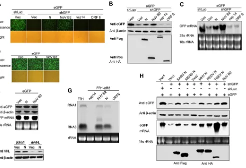
MERS-FIG 1Screening of potential VSR of SARS-CoV by reversal-of-silencing assays. 293T cells were cotransfected with plasmids encoding eGFP reporter (125 ng), eGFP-specific shRNA (shGFP) (1g), or luciferase-specific shRNA (shLuc) (1g) and viral protein of SARS-CoV (500 ng), respectively. (A to C) The expression level of eGFP was determined by Western blotting at 72 h posttransfection. Empty vector (Vec) and NoV B2 (B2) were used as a mock control and a positive control. The shLuc was used as an irrelevant silencing control.-Actin was used as a loading control. (D) The expression of SARS-CoV-encoded proteins as indicated was detected by Western blotting. (E) The relative eGFP reversion activity of different viral proteins in panels A to C was normalized by that of typical VSR NoV B2 control and is shown in a bar diagram.
on November 7, 2019 by guest
[image:3.585.93.489.63.409.2]CoV-Jeddah-human-1, (KF958702.1); Bat-CoV strain HKU9-1 BF_005I, NC_009021; MHV-A59,NC_001846; IBV strain Beaudette,NC_001451; TGEV strain PUR46-MAD,NC_002306; PEDV strain CV777,NC_003436; and H-CoV strain 229E,NC_002645.
Virus infection and real-time PCR.At 24 or 48 h posttransfection, Neuro-2a cells or L2 cells were infected with MHV strain A59 at a multi-plicity of infection (MOI) of 0.1. At 16 h postinfection, the supernatants were collected, and the virus titer was determined by plaque assay on L2 cells (48). For mRNA detection, total RNA was isolated from cells with TRIzol reagent. The RNA was reverse transcribed to first-strand cDNA using Moloney murine leukemia virus reverse transcriptase (Promega). The SYBR green master mix (Roche) was used for real-time PCR. Mouse GAPDH (glyceraldehyde-3-phosphate dehydrogenase) mRNA was used as an internal control. The primers used are shown in Table S3 in the supplemental material.
RESULTS
Identification of coronaviral N protein as a VSR in mammalian
cells.
To evaluate whether coronaviruses encode a VSR, we
screened the proteins encoded by SARS-CoV using the
rever-sal-of-silencing assay in HEK293T cells (
Fig. 1A
,
B
, and
C
).
NoV B2, a well-known VSR in mammalian cells (
23
), was used
as a positive control. The expression level of eGFP reporter
gene was tested by Western blotting and fluorescence
micros-copy, and the relative eGFP reversion activities of each protein
toward NoV B2 were calculated. As shown in
Fig. 1E
,
SARS-CoV N protein could efficiently revert the expression of
RNAi-silenced eGFP, as well as NoV B2. In contrast, the accessory
protein 7a, which was reported as a VSR of SARS-CoV (
32
),
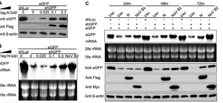
FIG 2N protein of coronaviruses represses shRNA-induced RNAi in mammalian cells. (A to C) HEK293T cells were cotransfected with plasmids encoding eGFP reporter (125 ng), shGFP, or shLuc (1g) and plasmids encoding viral proteins as indicated (300 ng). The expression of eGFP reporter was analyzed 72 h after cotransfection. (A) The intensity of eGFP was observed under fluorescence microscopy. (B) Cell lysates were harvested and analyzed by Western blotting. (C) Cellular total mRNAs were harvested and analyzed by Northern blotting. (D and E) HEK293T cells were cotransfected with plasmids encoding eGFP and SARS-CoV N protein or NoV B2, respectively. eGFP fluorescence, protein, and mRNA levels were determined by fluorescence microscopy (D) and Western and Northern blotting (E), respectively. (F) HEK293T cells were cotransfected with plasmids of mock vector pLko.1 (1g) or shRNA targeting endogenous gene VHL (shVHL) in the presence or absence of SARS-CoV N protein (300 ng). At 72 h posttransfection, cells were harvested and subjected to Western blotting to determine the endogenous VHL expression. (G) S2 Cells were transfected with 0.03g of pFR1 or 0.6g of pFRNA1-⌬B2 and with SARS-CoV N or FHV B2 as indicated above. At 48 h posttransfection, FHV RNA transcription was induced by incubation with CuSO4at 0.5 mM. At 24 h after induction, the cellular total mRNA was harvested for Northern blot analysis by a probe recognizing FHV RNA1 and RNA3. The band between RNA1 and RNA3 represents the mRNA transcribed from B2 expression plasmid. (H) HEK293T cells were cotransfected with plasmids encoding eGFP, shLuc, or shGFP and N proteins of different coronaviruses (Flag-tagged N proteins of SARS-CoV and MERS-CoV and HA-tagged N proteins of MHV, PEDV, and TGEV), respectively. The eGFP expression level and mRNA level were determined by Western blotting and Northern blotting. Empty vector (Vec, Vec1, or Vec2), nsp14, and ORF6 were used as negative controls, while NoV B2 was used as a positive control. The shLuc was irrelevant control shRNA.-Actin and rRNAs were used as loading controls for Western and Northern blotting, respectively. Cui et al.
on November 7, 2019 by guest
http://jvi.asm.org/
[image:4.585.47.540.64.400.2]showed extremely limited VSR activity in the screening assays.
We detected the protein expression level of some constructs,
including nsp7-16, N, 7a, ORF6, M, and B2, and the results
showed that most of these proteins were readily detectable but
at variable levels (
Fig. 1D
). Therefore, the VSR activity and its
strength observed in the initial screening were not conclusive
and had to be verified by further systematic experiments.
Be-cause SARS-CoV N showed relatively higher VSR activity in the
reversal-of-silencing assay, we continued to characterize
SARS-CoV N as a potential VSR.
To confirm the RNAi suppressor activity of SARS-CoV N
protein, we assayed the capability of N protein in suppressing
shRNA-induced eGFP silencing at protein and mRNA level.
The eGFP-specific shRNA (shGFP) caused a strong decrease of
eGFP expression (
Fig. 2A
and
B
) and transcription (
Fig. 2C
)
compared to the irrelevant shRNA that targets luciferase
(shLuc). The nsp14 with nonspecific RNA binding activity (
49
)
and ORF6 protein with IFN antagonistic activity (
50
) were
used as negative controls. Notably, transfection of the N
ex-pression plasmid remarkably resurrected the exex-pression of
RNAi-silenced eGFP at both protein and mRNA levels, as well
as B2, but ORF6 and nsp14 could not (
Fig. 2A
,
B
, and
C
),
indicating that N suppressed the shRNA-induced RNA
silenc-ing of eGFP. Importantly, N protein did not affect the
expres-sion efficiency of eGFP in the absence of shRNA (
Fig. 2D
and
E
), confirming that N protein inhibited the effects of
shGFP-mediated RNAi rather than promoted eGFP transcription or
translation. To exclude that the observed effects specifically
exists in the eGFP reporter system, the RNAi suppression
ac-tivity of N protein was determined in an endogenous RNAi
system (
Fig. 2F
). We demonstrated that the presence of N
pro-tein restored the endogenous VHL expression silenced by
VHL-specific shRNA (shVHL), whereas the VHL expression
level was not affected in mock vector (pLko.1)-transfected cells
(
Fig. 2F
). Since the RNAi pathway is conserved from plants to
animals, some VSRs of mammalian viruses are also functional
in insect cells such as influenza A virus NS1 and vaccinia virus
E3L (
22
). We further confirmed the VSR activity of SARS-CoV
N protein using FHV RNA1 replication system (
Fig. 2G
).
Transfection of pRNA1 led to the self-replication of FHV
RNA1 and the transcription of RNA3 in S2 cells. FHV B2 was
well known to inhibit RNA silencing induced by virus RNA
replication. B2-deficient mutant (pRNA1-
⌬
B2) failed to
accu-mulate FHV RNA1 and RNA3. This defect could be partially
rescued by cotransfection with plasmids either expressing
SARS-CoV N protein or FHV B2 (
Fig. 2G
). To explore whether
the RNAi inhibition activity of N protein is universal among
coronaviruses, the N proteins from alpha coronavirus (PEDV
and TGEV) and beta coronavirus (SARS-CoV, MERS-CoV,
and MHV) were tested. As shown in
Fig. 2H
, the indicated
coronavirus N proteins inhibited shRNA-induced RNAi to
var-ious degrees in the reversal-of-silencing system, wherein
MERS-CoV N showed notably low VSR activity.
To investigate whether the VSR activity is dependent on the
protein expression levels, the increasing amounts of SARS-CoV N
were tested using the reversal-of-silencing assay (
Fig. 3A
and
B
).
The reversal effect of eGFP silencing increased progressively at
both protein and mRNA levels, along with the gradual increase of
the transfected N-expressing plasmid (
Fig. 3A
and
B
). We also
detected eGFP expression at different time points, and the results
showed that the reversal of eGFP silencing could be observed as
early as 24 h posttransfection and was more effective at 48 and 72
h (
Fig. 3C
), indicating that the VSR activity was dependent on the
expression level of SARS-CoV N protein.
N protein inhibits Dicer-mediated siRNA generation and
siRNA-induced RNAi.
The shRNA-induced RNAi pathway
re-quires Dicer-mediated dsRNA cleavage into siRNA. To find out
whether N protein blocks the aforementioned step, small RNAs
harvested from HEK293T cells were subjected to Northern
blot-ting with DIG-labeled probes which recognized both shRNA and
siRNA (
Fig. 4A
and
B
). In the presence of SARS-CoV N protein, an
increase in the ratio of shRNA to 21-nt Dicer-processed siRNA
was detected. However, the increase was more obvious in the
pres-ence of NoV B2 (
Fig. 4A
and
B
). This phenomenon is consistent
with the eGFP expression and its mRNA level observed in the
reversal-of-silencing assays (
Fig. 2
). To further demonstrate
whether N protein could block RNAi induced by siRNA (the
post-Dicer product), 40 nM eGFP-specific siRNA (siGFP) was used to
FIG 3N protein represses shRNA-induced RNAi in a dose-dependent and time-dependent manner in mammalian cells. Increasing amounts of the plasmid expressing SARS-CoV N protein were transfected into HEK293T cells, as indicated in the upper panel in the reversal-of-silencing assay. At 72 h after transfection, Western blotting (A) and Northern blotting (B) were performed to determine the eGFP protein and mRNA levels, respectively. (C) Northern blotting (upper panel) and Western blotting (lower panel) were performed 24, 48, and 72 h posttransfection, respectively.
on November 7, 2019 by guest
[image:5.585.113.472.65.232.2]induce a remarkable reduction of eGFP expression (
Fig. 4C
). As
shown in
Fig. 4D
and
E
, the decrease in eGFP expression and its
mRNA level in HEK293T cells was partially inhibited in the
pres-ence of SARS-CoV N protein comparing with NoV B2. We
inves-tigated the ability of N protein to suppress RNA silencing in insect
cells. Consistent with the phenomenon observed in cells, the
pres-ence of SARS-CoV N protein efficiently abolished the
eGFP-spe-cific RNAi induced by dsRNA (dsRNA-GFP) while remarkably
inhibiting the siGFP-induced RNAi in
Drosophila
S2 cells (
Fig.
4F
). Thus, the N protein could inhibit the RNA silencing in
mam-malian and insect cells at both Dicer-processing and post-Dicer
stages with different efficiencies.
N protein directly binds to RNAs and prevents dsRNA from
RNase III-mediated cleavage
in vitro
.
A number of VSRs could
sequester RNA duplexes from Dicer cleavage and the
incorpora-tion of siRNA into RISC by dsRNA binding activity, such as NoV
B2, EBoV VP35, and influenza virus NS1 (
20
,
22
,
23
). To
investi-gate the mechanism of N protein as a VSR, we used a gel shift assay
after the incubation of recombinant GST-tagged SARS-CoV N
protein (GST-N) and WhNV B2 (GST-B2) with DIG-labeled
500-nt ssRNA (
Fig. 5A
), DIG-labeled 244-bp dsRNA (
Fig. 5B
),
and synthetic 5
=
-hexachlorofluorescein phosphoramidite
(HEX)-labeled 21-nt siRNA (
Fig. 5C
), which mimic viral ssRNA, cellular
pre-Dicer dsRNA, and post-Dicer siRNA, respectively. As shown
in
Fig. 5A
to
C
, the shifting amount of all labeled RNAs increased
as more SARS N protein was used in the reaction up to 15
M,
while the WhNV B2 could not bind ssRNA. Interestingly, the
mo-bility of labeled ssRNA (
Fig. 5A
) and dsRNA (
Fig. 5B
) decreased as
more SARS-COV N protein was used, except that of labeled
siRNA (
Fig. 5C
), suggesting that one molecule of long RNAs
(ss-FIG 4N protein inhibits the production of siRNA and RNAi in both mammalian and insect cells. (A) HEK293T cells were cotransfected with plasmid as indicated above, 72 h after transfection, and small RNAs were harvested from the cells and probed with DIG-labeled oligonucleotides that correspond to the target sites of siRNA produced from shGFP. The locations of bands corresponding to shRNA and siRNA are indicated with diagrams on the right side. Short exposures and long exposures are shown on the left and right, respectively. An ethidium bromide-stained gel of low-molecular-weight RNA is shown as a loading control. Neg., mock control transfected with shLuc. (B) The ratios of shRNA to siRNA in panel A were quantified based on the corresponding exposure signals and are shown as a bar diagram. (C) eGFP expression plasmid (200 ng) was cotransfected with multiple concentrations (5 to 40 nM) of synthetic eGFP-specific siRNA (siGFP) to confirm the effective siGFP concentration in HEK293T cells. At 72 h after transfection, cell lysates were harvested to determine the reduction in eGFP expression by Western blotting. (D and E) SARS-CoV N protein inhibits siRNA-induced RNAi. HEK293T cells were transfected with plasmids as indicated above. At 72 h after transfection, eGFP fluorescence (D), protein (E), and mRNA (F) were detected as described inFig. 2. (F) SARS-CoV N protein inhibits RNAi inDrosophilaS2 cells. eGFP-specific dsRNA (dsRNA-GFP) and siGFP were used to induce RNAi in S2 cells. The mRNA of eGFP was detected by Northern blotting. Empty vector (Vec), nsp14, and ORF6 were used as negative controls. NoV B2 and FHV B2 were used as positive controls. rp49 was used as a loading control.
Cui et al.
on November 7, 2019 by guest
http://jvi.asm.org/
[image:6.585.95.492.67.415.2]RNA and dsRNA) was bound by the multiple N proteins.
Subse-quently, we examined the possibility of SARS-COV N protein to
protect dsRNA from RNase III-mediated cleavage. RNase III is a
Dicer homolog and was widely used as the Dicer substitute
in vitro
as previously reported (
51
). As shown in
Fig. 5D
, in a gel shift
assay, 500-bp dsRNA formed a high-molecular-weight complex
with GST-N, as well as GST-B2, and therefore was protected from
RNase III digestion. However, without the protection of GST-N
or GST-B2, dsRNA was digested into 21-to 23-nt siRNA. These
data illustrated that N protein could directly bind dsRNA to
pre-vent the digestion of Dicer and partially sequester siRNA at
post-Dicer stages.
Positively charged residues Lys 258 and Lys 262 of SARS-CoV
N protein are critical for the RNAi repression activity.
Mutagen-esis and alanine scanning analyses were performed to identify the
critical domain or amino acid (aa) required for the VSR activity of
SARS-CoV N protein. The domain architecture of SARS-CoV N
protein is shown in
Fig. 6A
. Based on domain truncation analysis,
we found that the N-terminal (1 to 181 aa) and the C-terminal
(365 to 422 aa) of SARS-CoV N protein are not required for RNAi
inhibition activity (
Fig. 6B
), suggesting that the functional VSR
region might be located at the linkage region (Linker) and CTD of
SARS-CoV N protein (182 to 365 aa). The expression of truncated
proteins was evaluated (
Fig. 6C
). Previous studies revealed that
this region possesses an SR-rich domain (182 to 227 aa) and a
positively charged groove (248 to 280 aa) (
Fig. 6A
), which might
use positively charged residues arginine and lysine to mediate
electrostatic interaction with the phosphate backbone of RNAs
(
36
,
46
,
52
,
53
). Therefore, multiple sequence alignments of the
SR-rich domain (data not shown) and the positively charged
groove of the CTD of the coronavirus N protein (
Fig. 6A
) were
performed, and the single or double mutations of conserved
argi-nine and lysine were analyzed by the reversal-of-silencing assay in
HEK293T cells, as shown in
Fig. 6D
and
E
. Further mutational
analysis revealed that mutations of conserved Lys 258 and Lys262
in CTD of N protein (K258A and K262A) completely abolished its
ability to repress RNAi induced by shRNA in HEK293T cells (
Fig.
6F
). Taken together, these data show that the positively charged
residues Lys 258 and Lys 262 are critical for the RNAi repression
activity of SARS-CoV N protein, and these results support the
importance of the RNA-binding activity of protein N in RNAi
suppression.
SARS-CoV N protein and NoV B2 promote the replication of
coronavirus MHV.
To demonstrate the physiologic function of
coronavirus N protein as a VSR
in vivo
, we used coronavirus MHV
strain A59, which belongs to the same virus group as SARS-CoV,
as a model to investigate the influence of N protein and its mutants
on virus replication. We and others have reported that the
multi-ple functional N protein is essential for coronavirus, and the
ex-pression of N protein either in
cis
or in
trans
could facilitate the
viral replication (
40
,
54
,
55
). Therefore, SARS-CoV N protein, N
mutants and NoV B2 provided in
trans
were tested in the
in vivo
system. As shown in
Fig. 7A
to
D
, the MHV titer was upregulated
3- to 5-fold by SARS-CoV N protein in a dosage-dependent
man-ner in Neuro-2a cells (
Fig. 7A
), whereas the VSR inactive mutant
K258/262A failed to promote the replication of MHV (
Fig. 7B
). In
contrast, the VSR activity irrelevant mutant K267A (
Fig. 6E
)
ex-hibited promotion similar to that of the wild-type N protein
FIG 5N protein binds to RNAs and inhibits the Dicer-like RNase III cleavage reactionin vitro. Increasing amounts of purified GST-tagged SARS-CoV N protein (GST-N) from 0 to 15M were incubated with 0.2 pmol of 500-nt DIG-labeled ssRNA (A) or 244-bp DIG-labeled dsRNA (B) at 25°C for 30 min. Complexes were separated on 1.2% TBE–agarose gel and subjected to Northern blotting. The free ssRNA and dsRNA are indicated on the left side. (C) GST-N up to 15M was incubated with 0.2 pmol of 5=-HEX-labeled 21-nt siRNA as described in panels A and B. Complexes were applied to 4% native polyacrylamide gel, and the fluorescent signal was visualized by using a Typhoon 9200. The free siRNA is indicated on the left side. (D) DIG-labeled 500-bp dsRNA was incubated with purified proteins, as indicated above, at 25°C for 30 min before the processing of Dicer-like RNase III at 37°C for 30 min. The reaction products were separated on 1.2% TBE–agarose gel and subjected to Northern blotting. The free dsRNA is indicated on the left side, and the cleaved dsRNA is indicated on the right side. The shifted protein-RNA complexes are indicated by black arrows on the right side. The protein GST and GST-tagged WhNV B2 (GST-B2) were used as a mock control and a positive control.
on November 7, 2019 by guest
[image:7.585.136.449.66.287.2](
Fig. 7B
). Impressively, the typical VSR NoV B2 could also
facili-tate the replication of MHV (
Fig. 7B
), further suggesting that the
increase of MHV replication may be due to the general
suppres-sion of RNAi in host cells. The MHV titers were checked by the
plaque assays in L2 cells (
Fig. 7D
). These results were also
con-firmed by analyzing the synthesis of viral gene 7 mRNA (mRNA 7)
using reverse transcription-PCR (RT-PCR) in Neuro-2a cells (
Fig.
7C
), and the expression of corresponding proteins was evaluated
(
Fig. 7E
). As shown in
Fig. 7D
and
E
, the effect of SARS-CoV N
protein on the enhancement of MHV growth was also dose
de-pendent, a finding consistent with the previous observation (
Fig.
3A
and
B
). In the above-described assays, the steady-state protein
level of K258/262A mutant was similar to that of wild-type N
protein with the transfection dosage of 300 ng of DNA (
Fig. 7E
);
however, the mutant K258/262A could not promote MHV
growth, but 300 ng of wild-type N protein could still significantly
enhance virus replication (
Fig. 7A
,
D
, and
E
). Therefore, the
defi-ciency of K258/262A mutant in VSR activity was not due to
re-duced expression level.
Coronavirus N proteins have been reported to have IFN-

response inhibition activity in IFN induction systems (
41
,
50
,
56
),
suggesting that N might also promote MHV replication through
antagonizing IFN responses in host cells. However, previous
stud-ies on coronavirus infections showed that MHV induces a
mini-mal type I IFN response in several cell types, and the expression of
IFN-

is undetectable (
57–60
). When Neuro-2a cells were treated
with dsRNA analog poly(I·C), low level of IFN-

was produced,
but MHV infection did not induce expression of IFN-

(
Fig. 7F
).
We also analyzed the transcription of the
Ifn
-

mRNA and its
downstream gene
Isg56
mRNA that represent the expression level
of type I IFN. As shown in
Fig. 7G
and
H
, the mRNA levels of
Ifn
-

and
Isg56
are neither reduced compared to Nov B2 and vector
control nor significantly changed with N protein and its mutants,
which ruled out the possibility that the N protein increased MHV
production by antagonizing the IFN-

signaling pathway. These
results demonstrated that SARS-CoV N protein could promote
the virus replication independently of IFN signaling pathway and
that the critical residues Lys 258 and Lys 262 of SARS-CoV N
protein for VSR activity are involved in facilitating MHV
replica-tion by inhibiting the RNAi-mediated antiviral mechanism.
Moreover, the irrelevant viral protein NoV B2 could also promote
the replication of MHV, indicating that the MHV replication
might be sensitive to the RNAi-mediated antiviral response.
Knockdown of either Dicer1 or Ago2 facilitated the
replica-tion of MHV.
To further investigate the function of
RNAi-medi-ated antiviral response when the IFN-mediRNAi-medi-ated antiviral response
is highly repressed by coronaviruses, the Dicer1 and Ago2
tran-scripts in mouse Neuro-2a and L2 cells were knocked down by
FIG 6Conserved residues LysK258 and LysK262 of SARS-CoV N protein are critical for RNAi activity. (A) Schematic diagram of the domain architecture of the SARS-CoV N protein and multiple-sequence alignment of CTD spanning residues 248 to 281 of coronavirus N proteins. The conserved residues are indicated by solid black boxes. NTD, N-terminal domain; CTD, C-terminal domain; SR-rich, rich in serine and arginine; Linker, linkage region. (B and C) Mapping of critical residues of SARS-CoV N protein for VSR activity. The RNAi repression activity of Flag-tagged truncations of SARS-CoV N protein was analyzed by a reversal-of-silencing assay as described forFig. 2B. The expression of truncated SARS-CoV N proteins was detected by Western blotting. (D to F) The RNAi repression activity of Flag-tagged mutants of SARS-CoV N protein was analyzed by a reversal-of-silencing assay, as described forFig. 2. (F) The K257A, K258A, K262A, and R263A mutants were further analyzed based on the results from panel E.
Cui et al.
on November 7, 2019 by guest
http://jvi.asm.org/
[image:8.585.70.509.67.361.2]FIG 7SARS-CoV N protein promotes MHV replication when provided intrans. (A and B) Mouse Neuro-2a cells were transfected with plasmids as indicated and infected with MHV strain A59 at an MOI of 0.1 at 24 h posttransfection. At 16 h after infection, culture supernatants were collected and subjected to plaque assay on L2 cells to determine the MHV titers. The relative titer fold of MHV was quantified, as shown in bar diagrams. The actual virus titers are in indicated in panel A to show MHV replication efficiency. (C) Total RNAs were extracted from the transfected cells and subjected to RT-PCR using primers targeting the subgenomic RNA7 of MHV. The data were normalized to the abundance of endogenous mouse GAPDH mRNA. (D) Virus plaque formation was analyzed on L2 cells at a dilution of 10⫺6. (E) Protein expression levels in transfected cell lysates were detected by Western blotting with the indicated antibodies.-Actin was used as a loading control. (F) Neuro-2a cells were infected with MHV strain A59 at an MOI of 0.1 or transfected with 2g of poly(I·C). At 16 h postinfection or transfection, IFN-production was determined by enzyme-linked immunosorbent assay (ELISA). (G and H) Total RNAs were extracted from the transfected cells as described in panel B and subjected to RT-PCR to detectIfn-andIsg56mRNA. Error bars indicate the means and standard deviations for triplicate experiments. *,P⬍0.05; **,P⬍0.01; ns, not significant (unpaired Studentttest).
on November 7, 2019 by guest
[image:9.585.38.541.62.601.2]synthetic siRNAs (siDicer1 and siAgo2). As shown in
Fig. 8
, the
mRNA levels of
Dicer1
and
Ago2
were markedly reduced in cells
transfected with siDicer1-6 and siAgo2-4, respectively (
Fig. 8A
and
C
), whereas the
Ifn
-

mRNA was not significantly affected
(
Fig. 8B
and
D
). The RNAi-deficient Neuro-2a cells were still
competent in IFN production (
Fig. 9A
), indicating that RNAi
de-pletion did not nonspecifically abolish the IFN pathway. In
RNAi-deficient Neuro-2a cells, either in the presence or in the absence of
SARS-CoV N protein, the replications of MHV were increased to
the same level as in RNAi pathway healthy cells provided with
SARS N protein (
Fig. 9B
and
C
). Accordingly, there was no
reduc-tion in the mRNA level of
Isg56
observed in the presence of
SARS-CoV N (
Fig. 9D
), indicating that the increase of MHV replication
was affected by the attenuated RNAi pathway but not by
suppres-sion of type I IFN response. In fact, the mRNA level of
Isg56
in-creased to some extent, whereas the replication of MHV was still
promoted in Ago2-knockdown cells (
Fig. 9B
to
D
), strongly
sug-gesting that RNAi knockdown was involved in these cells.
More-over, the differences of MHV replication promotion between
wild-type SARS-CoV N protein, NoV B2, and mutant K285/262A
of SARS-COV N protein observed in Neuro-2a cells (
Fig. 7
) were
eliminated in RNAi knockdown cells by siDicer1 or siAgo2,
re-spectively (
Fig. 9E
,
F
,
H
, and
I
), whereas the expression of these
proteins did not result in different IFN responses (
Fig. 9G
and
J
).
The VSR activity was also analyzed in L2 cells that are highly IFN
sensitive. Both healthy and RNAi pathway-deficient L2 cells could
produce high levels of IFN-

(up to 10,000 pg/ml) when infected
by Sendai virus (SeV), but the production of IFN-

was still
un-detectable when infected by MHV (
Fig. 9K
). In RNAi-competent
L2 cells (treated with siGFP), N protein could significantly
in-crease MHV growth (
Fig. 9L
). Interestingly, when the RNA
path-way was depleted by siDicer1-6 or siAgo2-4, the increment of
MHV growth was kept in the absence of N or in the presence of
VSR-deficient mutant K258/262A (
Fig. 9L
). Together, the
obser-vations in L2 cells indicate that either suppression of RNAi by
SARS-CoV N or depletion of RNAi pathway by siRNAs could lead
to increment of MHV growth. These results were consistent with
that of Neuro-2a cells. Taken together, these results demonstrate
that SARS-CoV N protein is a novel VSR and the RNAi pathway
was involved in anticoronavirus response in mammalian cells.
DISCUSSION
Mammalian RNA viruses have evolved many mechanisms to
pro-tect their genomic RNAs and dsRNAs generated as replicative and
transcriptive intermediates (
30
,
31
,
59
,
61
) from host cell
recogni-tion. For example, they associate the replication-transcription
complex with the double-membrane vesicles to form a protective
microenvironment which prevents viral RNA from being detected
by host cell sensors (
62
,
63
), they encode several RNA processing
enzymes to undergo RNA modifications such as RNA capping to
mimic the host cell RNAs (
49
,
64
), and they adopt RNA binding
proteins to protect viral RNAs (
65
,
66
). However, virus-derived
small RNAs were still detectable in 41 human cell lines infected
with six different RNA viruses, respectively (
5
). Moreover, several
reports showed that virus-specific siRNAs accumulated in
in-fected mammalian cells, and recently the specific siRNAs of
en-cephalomyocarditis virus and Nodamura virus (NoV) were deep
sequenced (
6
,
7
). Therefore, in mammalian cells that possess a
functional RNAi pathway, the sequence-specific RNA
degrada-tion would still threaten the abundant viral mRNA in cytoplasm if
FIG 8Screen for siRNAs targeting mouse Dicer1 and Ago2. Mouse Neuro-2a cells were transfected with 40 nM siDicer1 (A and B) or siAgo2 (C and D). At 48 h posttransfection, total cellular mRNAs were extracted and subjected to RT-PCR to determine the mRNA levels of Dicer1 (A), Ago2 (C), and Ifn-(B and D). siGFP was used as a negative control. The data were normalized to the abundance of internal mouse GAPDH mRNA. Error bars indicate the means and standard deviations for triplicate experiments.
Cui et al.
on November 7, 2019 by guest
http://jvi.asm.org/
[image:10.585.142.449.64.332.2]FIG 9Knockdown of Dicer1 or Ago2 facilitated MHV replication. (A) Mouse Neuro-2a cells were transfected with siRNAs as indicated above and, at 48 h posttransfection, the cells were infected with MHV at an MOI of 0.1 or transfected with 2g of poly(I·C). At 16 h postinfection or posttransfection, IFN- production was determined by ELISA. (B to J) Mouse Neuro-2a cells were cotransfected with 40 nM siRNA and 500-ng protein expression plasmids as indicated and infected with MHV strain A59 at an MOI of 0.1 at 48 h posttransfection. At 16 h postinfection, the culture supernatants were collected and subjected to plaque assays on L2 cells to determine the MHV titers. (B, E, and H) The relative titer folds of MHV were quantified and are shown in the bar diagrams. Total RNAs were extracted from the transfected cells and subjected to RT-PCR to determine the indicated mRNA levels. (C, F, and I) MHV RNA7 level. (D, G, and J)Isg56mRNA level. (K) L2 cells were transfected with siRNAs as indicated above and, at 48 h posttransfection, the cells were infected with MHV or SeV. At 16 h postinfection, IFN-production was determined by ELISA. (L) L2 was transfected and infected similarly as described for Neuro-2a cells. The MHV titers were determined by plaque assay. Empty vector (Vec) and siGFP were used as the negative control. The data were normalized to the abundance of endogenous mouse GAPDH mRNA. Error bars indicate the means and standard deviations for triplicate experiments.
[image:11.585.40.537.30.622.2]without VSR during virus infection, especially for coronavirus, the
largest RNA virus as known. We present here evidence that
coro-naviruses also possess VSR, which was identified as N protein that
shares similar structural architecture and gRNA/subgenomic
RNA (sgRNA) binding activity to enhance the viral transcription,
replication, and assembly among coronaviruses (
37
,
38
,
40
,
65
,
66
). It is abundantly produced in infected cells to support its
mul-tiple functions, as well as VSR. In addition to the ssRNA binding
activity, we showed that N protein also possesses dsRNA and
siRNA binding activities. Moreover, SARS-CoV N protein could
bind more efficiently with long ssRNA and dsRNA than siRNAs
(
Fig. 5
), which may be resulted from cooperative binding of
mul-tiple N protein monomers. Coronaviruses possess the largest
sin-gle-stranded genomic RNA and abundant sgRNAs and dsRNA
intermediates (
29–31
,
61
). Therefore, we propose a hypothesis
that the coronavirus N protein might protect viral RNA from
RNAi-mediated gene silencing at three stages (
Fig. 10
): (i) binding
viral ssRNAs to prevent the positive- and negative-sense genomic
or subgenomic RNAs from formation of unnecessary
intramolec-ular and intermolecintramolec-ular dsRNA; (ii) shielding virus-derived
dsRNA from Dicer cleavage through dsRNA binding activity; and
(iii) binding to virus-derived siRNA to interfere with RISC
assembly.
In the present study, we showed that the VSR activity was
highly dependent on the protein expression levels (
Fig. 3A
and
B
;
Fig. 7D
and
E
). In the initial screening assays for putative VSRs
(
Fig. 1
), we did not evaluate the protein expression levels of
dif-ferent constructs, and therefore effects observed in the
reversal-of-RNA silencing could not be directly compared and may result
in either false-positive or false-negative outcomes. For example,
SARS-CoV 7a, previously reported as a VSR (
32
), did not show
apparent VSR activity in our assay system. Notably, the
MERS-CoV N protein expression level is higher than that of SARS-MERS-CoV N
protein, while the VSR activity of MERS-CoV N protein is less
than that of SARS-CoV N protein (
Fig. 2H
). These results suggest
that the N proteins of coronaviruses may have different intrinsic
activities in suppressing RNAi. We speculate that the low VSR
activity of MERS-CoV N might be compensated for by a second
VSR and one potential candidate could be the dsRNA-binding
IFN antagonist protein 4a (
67
).
Previous studies revealed that the three distinct and highly
conserved domains of N proteins (NTD, SRD, and CTD) could
bind with viral RNAs in different coronaviruses (
42
,
66
,
68
,
69
).
The NTD has been found to associate with a stem-loop structure
located at the 3
=
end of RNA genome, and the CTD was involved in
the specific binding of coronaviral packaging signal within
non-structural protein nsp15 region (
43
,
70
,
71
). The critical residues
Lys 258 and Lys 262 of SARS-CoV N protein for VSR identified
here are located in the positively charged groove of CTD (
Fig. 6
).
Moreover, Lys 258 was previously reported as a significant
deter-minant of SARS-CoV N protein CTD binding affinity toward
oli-gonucleotides (
36
). Here, we chose SARS-CoV N protein to
pro-mote MHV because SARS-CoV is closely related to MHV on one
side, while the CTD of SARS-CoV N protein cannot enhance the
specific selective packaging of MHV gRNA to facilitate the MHV
replication (
71
). Therefore, the fact that the VSR inactive mutant
K258/262A of SARS-CoV N protein led to the attenuation of
MHV replication might imply that the sufficient VSR activity is
important for coronavirus replication.
Although the IFN-mediated antiviral response is important for
mammalian cells, a lot of studies have indicated that MHV is a
poor inducer of type I IFN response (
59
). Moreover, MHV is
resistant to the pretreatment of IFN-
␣
/

and does not induce the
generation of IFN-

in various cell types, suggesting that it can
suppress IFN signaling pathway or its downstream gene effects at
multiple levels (
56
,
58
). In the present study, we showed that
MHV infection did not induce the production of IFN-

in both
mouse Neuro-2a and L2 cells (
Fig. 7F
and
9A
). In primary cells,
IFN is induced by MHV infection in plasmacytoid dendritic cells
and macrophages (
57
,
72
,
73
), but not in neurons, astrocytes, and
hepatocytes (
57
). Therefore, we used coronavirus MHV strain
A59 as a replication model, which might to some extent avoid the
impact of IFN response, to investigate the influence of N protein
and its mutants on virus replication. We demonstrated that
SARS-CoV N protein could promote MHV replication in both Neuro-2a
cells and L2 cells, the former being not very responsive to IFN
induction, whereas the latter were highly sensitive (
Fig. 7
and
9
).
We also tested the VSR activity of N protein in IFN-deficient
in-sect cells (
Fig. 2G
). The results indicated that the VSR activity of
SARS-CoV N protein is independent of either the cell type or the
IFN pathway. Moreover, the depletion of critical components
Di-cer1 or Ago2 of the RNAi pathway in Neuro-2a and L2 cells results
in a significant upregulation of MHV replication, which was not
dependent on the changes in IFN response (
Fig. 9
), further
strengthen the role of SARS-CoV N protein in IFN-independent
RNAi suppression.
In a study by Schelle et al. (
74
), the observed role of CoV N
protein in the transcomplementation was most likely related to
the virus recovery step before viral replication, whereas in our
experimental system, we are dealing with a wild-type virus that
does not require transcomplementation for virus recovery, and
thus the role of SARS-CoV N protein that we observed is its effect
FIG 10Model for the suppression of RNAi in mammalian cells by coronavi-rus N protein. After the entry and un coating of coronaviral virions, the single-stranded genomic RNA (gRNA) is protected by N proteins and serves as a template for the synthesis of negative-strand gRNA and a set of subgenomic RNA (sgRNA). The (⫺)gRNA and (⫺)sgRNA are replicated to generate full-length gRNA and a set of (⫹)sgRNA. The virus-derived dsRNA could be gen-erated during viral transcription and replication. sgRNA and gRNA sequences may also form an intramolecular hairpin structure. These viral dsRNAs may be recognized by Dicer, consequently triggering antiviral RNAi. Coronavirus N proteins may repress the RNAi at three different stages as indicated. Cui et al.
on November 7, 2019 by guest
http://jvi.asm.org/
[image:12.585.41.285.64.248.2]antiviral response in addition to the IFN-mediated innate
immu-nity in mammalian cells. These two important antiviral
mecha-nisms might work in turn in different situations and locations to
defend against virus infection, which would constantly provide a
positive selection toward the VSR and viral IFN antagonist during
the tachytelic evolution of viruses.
ACKNOWLEDGMENTS
We are grateful to Christopher S. Sullivan from the University of Texas, Stanley Perlman from the University of Iowa, Shaobo Xiao from Hua-zhong Agricultural University, Rong Ye from Fudan University, and Yan Zhou from Wuhan University for generously providing experimental ma-terials. We thank Ruidong Hao, Ruangang Pan, Zeng Cong, Xiaolu Lu, Yang Qiu, and Yujie Liu from Wuhan University for helpful discussions. This study was supported by the China 973 Basic Research Pro-gram (2013CB911101) and China NSFC grants (81130083, 81271817, 31170152, and 31221061).
REFERENCES
1.Carthew RW, Sontheimer EJ.2009. Origins and mechanisms of miRNAs and siRNAs. Cell136:642– 655.http://dx.doi.org/10.1016/j.cell.2009.01 .035.
2.Ding SW.2010. RNA-based antiviral immunity. Nat Rev Immunol10:
632– 644.http://dx.doi.org/10.1038/nri2824.
3.Umbach JL, Cullen BR. 2009. The role of RNAi and microRNAs in animal virus replication and antiviral immunity. Genes Dev23:1151– 1164.http://dx.doi.org/10.1101/gad.1793309.
4.Takeuchi O, Akira S.2009. Innate immunity to virus infection. Immunol Rev227:75– 86.http://dx.doi.org/10.1111/j.1600-065X.2008.00737.x. 5.Parameswaran P, Sklan E, Wilkins C, Burgon T, Samuel MA, Lu R,
Ansel KM, Heissmeyer V, Einav S, Jackson W, Doukas T, Paranjape S, Polacek C, FBdos Santos Jalili R, Babrzadeh F, Gharizadeh B, Grimm D, Kay M, Koike S, Sarnow P, Ronaghi M, Ding SW, Harris E, Chow M, Diamond MS, Kirkegaard K, Glenn JS, Fire AZ. 2010. Six RNA viruses and forty-one hosts: viral small RNAs and modulation of small RNA repertoires in vertebrate and invertebrate systems. PLoS Pathog
6:e1000764.http://dx.doi.org/10.1371/journal.ppat.1000764.
6.Li Y, Lu J, Han Y, Fan X, Ding SW.2013. RNA interference functions as an antiviral immunity mechanism in mammals. Science342:231–234. http://dx.doi.org/10.1126/science.1241911.
7.Maillard PV, Ciaudo C, Marchais A, Li Y, Jay F, Ding SW, Voinnet O.
2013. Antiviral RNA interference in mammalian cells. Science342:235– 238.http://dx.doi.org/10.1126/science.1241930.
8.Sagan SM, Sarnow P.2013. Molecular biology: RNAi, antiviral after all. Science342:207–208.http://dx.doi.org/10.1126/science.1245475. 9.Voinnet O.2005. Induction and suppression of RNA silencing: insights
from viral infections. Nat Rev Genet6:206 –220.http://dx.doi.org/10.1038 /nrg1555.
10. Bivalkar-Mehla S, Vakharia J, Mehla R, Abreha M, Kanwar JR, Tikoo A, Chauhan A.2011. Viral RNA silencing suppressors (RSS): novel strategy of viruses to ablate the host RNA interference (RNAi) defense system. Virus Res155:1–9.http://dx.doi.org/10.1016/j.virusres.2010.10.003.
Virology306:33– 41.http://dx.doi.org/10.1016/S0042-6822(02)00018-1. 16. Chao JA, Lee JH, Chapados BR, Debler EW, Schneemann A,
William-son JR.2005. Dual modes of RNA-silencing suppression byFlock House virusprotein B2. Nat Struct Mol Biol12:952–957.http://dx.doi.org/10 .1038/nsmb1005.
17. Li H, Li WX, Ding SW.2002. Induction and suppression of RNA silenc-ing by an animal virus. Science296:1319 –1321.http://dx.doi.org/10.1126 /science.1070948.
18. Qi N, Zhang L, Qiu Y, Wang Z, Si J, Liu Y, Xiang X, Xie J, Qin CF, Zhou X, Hu Y. 2012. Targeting of dicer-2 and RNA by a viral RNA silencing suppressor inDrosophilacells. J Virol86:5763–5773.http://dx .doi.org/10.1128/JVI.07229-11.
19. Qi N, Cai D, Qiu Y, Xie J, Wang Z, Si J, Zhang J, Zhou X, Hu Y.2011. RNA binding by a novel helical fold of b2 protein fromWuhan nodavirus
mediates the suppression of RNA interference and promotes b2 dimeriza-tion. J Virol85:9543–9554.http://dx.doi.org/10.1128/JVI.00785-11. 20. Haasnoot J, de Vries W, Geutjes EJ, Prins M, de Haan P, Berkhout B.
2007. The Ebola virus VP35 protein is a suppressor of RNA silencing. PLoS Pathog3:e86.http://dx.doi.org/10.1371/journal.ppat.0030086. 21. Bucher E, Hemmes H, de Haan P, Goldbach R, Prins M.2004. The
influenza A virus NS1 protein binds small interfering RNAs and sup-presses RNA silencing in plants. J Gen Virol85:983–991.http://dx.doi.org /10.1099/vir.0.19734-0.
22. Li WX, Li H, Lu R, Li F, Dus M, Atkinson P, Brydon EW, Johnson KL, Garcia-Sastre A, Ball LA, Palese P, Ding SW.2004. Interferon antagonist proteins of influenza and vaccinia viruses are suppressors of RNA silenc-ing. Proc Natl Acad Sci U S A101:1350 –1355.http://dx.doi.org/10.1073 /pnas.0308308100.
23. Sullivan CS, Ganem D.2005. A virus-encoded inhibitor that blocks RNA interference in mammalian cells. J Virol79:7371–7379.http://dx.doi.org /10.1128/JVI.79.12.7371-7379.2005.
24. Bennasser Y, Le SY, Benkirane M, Jeang KT.2005. Evidence that HIV-1 encodes an siRNA and a suppressor of RNA silencing. Immunity22:607– 619.http://dx.doi.org/10.1016/j.immuni.2005.03.010.
25. Chen W, Zhang Z, Chen J, Zhang J, Zhang J, Wu Y, Huang Y, Cai X, Huang A.2008. HCV core protein interacts with Dicer to antagonize RNA silencing. Virus Res133:250 –258.http://dx.doi.org/10.1016/j.virusres.2008 .01.011.
26. Basler CF, Wang X, Muhlberger E, Volchkov V, Paragas J, Klenk HD, Garcia-Sastre A, Palese P.2000. The Ebola virus VP35 protein functions as a type I IFN antagonist. Proc Natl Acad Sci U S A97:12289 –12294. http://dx.doi.org/10.1073/pnas.220398297.
27. Brand SR, Kobayashi R, Mathews MB.1997. The Tat protein of human immunodeficiency virus type 1 is a substrate and inhibitor of the interfer-on-induced, virally activated protein kinase, PKR. J Biol Chem272:8388 – 8395.http://dx.doi.org/10.1074/jbc.272.13.8388.
28. Geiss GK, Salvatore M, Tumpey TM, Carter VS, Wang X, Basler CF, Taubenberger JK, Bumgarner RE, Palese P, Katze MG, Garcia-Sastre A.
2002. Cellular transcriptional profiling in influenza A virus-infected lung epithelial cells: the role of the nonstructural NS1 protein in the evasion of the host innate defense and its potential contribution to pandemic influ-enza. Proc Natl Acad Sci U S A99:10736 –10741.http://dx.doi.org/10.1073 /pnas.112338099.
29. Perlman S, Netland J.2009. Coronaviruses post-SARS: update on
on November 7, 2019 by guest
cation and pathogenesis. Nat Rev Microbiol7:439 – 450.http://dx.doi.org /10.1038/nrmicro2147.
30. Knoops K, Kikkert M, Worm SH, Zevenhoven-Dobbe JC, van der Meer Y, Koster AJ, Mommaas AM, Snijder EJ.2008. SARS-coronavirus rep-lication is supported by a reticulovesicular network of modified endoplas-mic reticulum. PLoS Biol6:e226.http://dx.doi.org/10.1371/journal.pbio .0060226.
31. Weber F, Wagner V, Rasmussen SB, Hartmann R, Paludan SR.2006. Double-stranded RNA is produced by positive-strand RNA viruses and DNA viruses but not in detectable amounts by negative-strand RNA vi-ruses. J Virol 80:5059 –5064. http://dx.doi.org/10.1128/JVI.80.10.5059 -5064.2006.
32. Karjee S, Minhas A, Sood V, Ponia SS, Banerjea AC, Chow VT, Mukherjee SK, Lal SK.2010. The 7a accessory protein of severe acute respiratory syndrome coronavirus acts as an RNA silencing suppressor. J Virol84:10395–10401.http://dx.doi.org/10.1128/JVI.00748-10. 33. Yount B, Roberts RS, Sims AC, Deming D, Frieman MB, Sparks J,
Denison MR, Davis N, Baric RS.2005. Severe acute respiratory syn-drome coronavirus group-specific open reading frames encode nonessen-tial functions for replication in cell cultures and mice. J Virol79:14909 – 14922.http://dx.doi.org/10.1128/JVI.79.23.14909-14922.2005. 34. Tan YJ, Lim SG, Hong W. 2006. Understanding the accessory viral
proteins unique to the severe acute respiratory syndrome (SARS) corona-virus. Antivir Res72:78 – 88.http://dx.doi.org/10.1016/j.antiviral.2006.05 .010.
35. Tang TK, Wu MP, Chen ST, Hou MH, Hong MH, Pan FM, Yu HM, Chen JH, Yao CW, Wang AH.2005. Biochemical and immunological studies of nucleocapsid proteins of severe acute respiratory syndrome and 229E human coronaviruses. Proteomics5:925–937.http://dx.doi.org/10 .1002/pmic.200401204.
36. Takeda M, Chang CK, Ikeya T, Guntert P, Chang YH, Hsu YL, Huang TH, Kainosho M.2008. Solution structure of the c-terminal dimerization domain of SARS coronavirus nucleocapsid protein solved by the SAIL-NMR method. J Mol Biol380:608 – 622.http://dx.doi.org/10.1016/j.jmb .2007.11.093.
37. Baric RS, Nelson GW, Fleming JO, Deans RJ, Keck JG, Casteel N, Stohlman SA.1988. Interactions between coronavirus nucleocapsid pro-tein and viral RNAs: implications for viral transcription. J Virol62:4280 – 4287.
38. Grossoehme NE, Li L, Keane SC, Liu P, Dann CE, III, Leibowitz JL, Giedroc DP.2009. Coronavirus N protein N-terminal domain (NTD) specifically binds the transcriptional regulatory sequence (TRS) and melts TRS-cTRS RNA duplexes. J Mol Biol394:544 –557.http://dx.doi.org/10 .1016/j.jmb.2009.09.040.
39. Tylor S, Andonov A, Cutts T, Cao JX, Grudesky E, Van Domselaar G, Li XG, He RT.2009. The SR-rich motif in SARS-CoV nucleocapsid protein is important for virus replication. Can J Microbiol55:254 –260. http://dx.doi.org/10.1139/W08-139.
40. Almazan F, Galan C, Enjuanes L.2004. The nucleoprotein is required for efficient coronavirus genome replication. J Virol78:12683–12688.http: //dx.doi.org/10.1128/JVI.78.22.12683-12688.2004.
41. Lu X, Pan J, Tao J, Guo D. 2011. SARS-CoV nucleocapsid protein antagonizes IFN-beta response by targeting initial step of IFN-beta induc-tion pathway, and its C-terminal region is critical for the antagonism. Virus Genes42:37– 45.http://dx.doi.org/10.1007/s11262-010-0544-x. 42. Chang CK, Sue SC, Yu TH, Hsieh CM, Tsai CK, Chiang YC, Lee SJ,
Hsiao HH, Wu WJ, Chang WL, Lin CH, Huang TH.2006. Modular organization of SARS coronavirus nucleocapsid protein. J Biomed Sci13:
59 –72.http://dx.doi.org/10.1007/s11373-005-9035-9.
43. Huang Q, Yu L, Petros AM, Gunasekera A, Liu Z, Xu N, Hajduk P, Mack J, Fesik SW, Olejniczak ET. 2004. Structure of the N-terminal RNA-binding domain of the SARS CoV nucleocapsid protein. Biochem-istry43:6059 – 6063.http://dx.doi.org/10.1021/bi036155b.
44. Luo H, Ye F, Chen K, Shen X, Jiang H.2005. SR-rich motif plays a pivotal role in recombinant SARS coronavirus nucleocapsid protein multimerization. Biochemistry44:15351–15358.http://dx.doi.org/10 .1021/bi051122c.
45. Luo H, Chen J, Chen K, Shen X, Jiang H.2006. Carboxyl terminus of severe acute respiratory syndrome coronavirus nucleocapsid protein: self-association analysis and nucleic acid binding characterization. Biochem-istry45:11827–11835.http://dx.doi.org/10.1021/bi0609319.
46. Chen CY, Chang CK, Chang YW, Sue SC, Bai HI, Riang L, Hsiao CD, Huang TH.2007. Structure of the SARS coronavirus nucleocapsid
pro-tein RNA-binding dimerization domain suggests a mechanism for helical packaging of viral RNA. J Mol Biol368:1075–1086.http://dx.doi.org/10 .1016/j.jmb.2007.02.069.
47. Liu D, Wu A, Cui L, Hao R, Wang Y, He J, Guo D.2014. Hepatitis B virus polymerase suppresses NF-B signaling by inhibiting the activity of IKKs via interaction with Hsp90. PLoS One9:e91658.http://dx.doi.org /10.1371/journal.pone.0091658.
48. Chang GH, Luo BJ, Lu P, Lin L, Wu XY, Li J, Hu Y, Zhu QY.2011. Construction and genetic analysis of murine hepatitis virus strain A59 Nsp16 temperature-sensitive mutant and the revertant virus. Virol Sin
26:19 –29.http://dx.doi.org/10.1007/s12250-011-3145-x.
49. Chen Y, Su C, Ke M, Jin X, Xu L, Zhang Z, Wu A, Sun Y, Yang Z, Tien P, Ahola T, Liang Y, Liu X, Guo D.2011. Biochemical and structural insights into the mechanisms of SARS coronavirus RNA ribose 2= -O-methylation by nsp16/nsp10 protein complex. PLoS Pathog7:e1002294. http://dx.doi.org/10.1371/journal.ppat.1002294.
50. Kopecky-Bromberg SA, Martinez-Sobrido L, Frieman M, Baric RA, Palese P.
2007. Severe acute respiratory syndrome coronavirus open reading frame (ORF) 3b, ORF 6, and nucleocapsid proteins function as interferon antagonists. J Virol
81:548–557.http://dx.doi.org/10.1128/JVI.01782-06.
51. Ji X.2008. The mechanism of RNase III action: how dicer dices. Curr Top Microbiol Immunol320:99 –116.
52. Lingel A, Simon B, Izaurralde E, Sattler M.2005. The structure of the flock house virus B2 protein, a viral suppressor of RNA interference, shows a novel mode of double-stranded RNA recognition. EMBO Rep
6:1149 –1155.http://dx.doi.org/10.1038/sj.embor.7400583.
53. Chang CK, Hsu YL, Chang YH, Chao FA, Wu MC, Huang YS, Hu CK, Huang TH.2009. Multiple Nucleic acid binding sites and intrinsic disor-der of severe acute respiratory syndrome coronavirus nucleocapsid pro-tein: implications for ribonucleocapsid protein packaging. J Virol83:
2255–2264.http://dx.doi.org/10.1128/JVI.02001-08.
54. McBride R, van Zyl M, Fielding BC.2014. The coronavirus nucleocapsid is a multifunctional protein. Viruses6:2991–3018.http://dx.doi.org/10 .3390/v6082991.
55. Pan J, Peng X, Gao Y, Li Z, Lu X, Chen Y, Ishaq M, Liu D, Dediego ML, Enjuanes L, Guo D. 2008. Genome-wide analysis of protein-protein interactions and involvement of viral proteins in SARS-CoV replication. PLoS One3:e3299.http://dx.doi.org/10.1371/journal.pone.0003299. 56. Ye Y, Hauns K, Langland JO, Jacobs BL, Hogue BG.2007. Mouse
hepatitis coronavirus A59 nucleocapsid protein is a type I interferon an-tagonist. J Virol81:2554 –2563.http://dx.doi.org/10.1128/JVI.01634-06. 57. Roth-Cross JK, Bender SJ, Weiss SR.2008. Murine coronavirus mouse
hepatitis virus is recognized by MDA5 and induces type I interferon in brain macrophages/microglia. J Virol82:9829 –9838.http://dx.doi.org/10 .1128/JVI.01199-08.
58. Roth-Cross JK, Martinez-Sobrido L, Scott EP, Garcia-Sastre A, Weiss SR.2007. Inhibition of the alpha/beta interferon response by mouse hep-atitis virus at multiple levels. J Virol81:7189 –7199.http://dx.doi.org/10 .1128/JVI.00013-07.
59. Rose KM, Weiss SR.2009. Murine coronavirus cell type dependent in-teraction with the type I interferon response. Viruses1:689 –712.http://dx .doi.org/10.3390/v1030689.
60. Zhou H, Perlman S.2007. Mouse hepatitis virus does not induce beta interferon synthesis and does not inhibit its induction by double-stranded RNA. J Virol81:568 –574.http://dx.doi.org/10.1128/JVI.01512-06. 61. Versteeg GA, Bredenbeek PJ, van den Worm SH, Spaan WJ. 2007.
Group 2 coronaviruses prevent immediate-early interferon induction by protection of viral RNA from host cell recognition. Virology361:18 –26. http://dx.doi.org/10.1016/j.virol.2007.01.020.
62. Gosert R, Kanjanahaluethai A, Egger D, Bienz K, Baker SC.2002. RNA replication of mouse hepatitis virus takes place at double-membrane ves-icles. J Virol76:3697–3708.http://dx.doi.org/10.1128/JVI.76.8.3697-3708 .2002.
63. Snijder EJ, van der Meer Y, Zevenhoven-Dobbe J, Onderwater JJ, van der Meulen J, Koerten HK, Mommaa