Immunohistochemical Evaluation of Estrogen
Receptor (ER) and Progesterone Receptor (PR)
in Endometrial Carcinomas and Its Precursors
A DISSERTATION SUBMITTED IN PART FULFILMENT OF THE REGULATION
FOR THE AWARD OF THE DEGREE OF M.D. PATHOLOGY BRANCH III.
Immunohistochemical Evaluation of
Estrogen Receptor (ER) and
Progesterone Receptor (PR) in
Endometrial Carcinomas and Its
Precursors
A dissertation in part fulfillment of the rules and regulations for
the M.D. Branch III (Pathology) Degree Examination of the Tamil
CERTIFICATE
This is to certify that this dissertation titled “Immunohistochemical Evaluation of
Estrogen Receptor (ER) and Progesterone Receptor (PR) in Endometrial Carcinomas
and Its Precursors” is a bonafide work done in the department of General Pathology by
Dr. Rima S (Postgraduate Registrar), in part fulfillment of the rules and regulations for the M.D. Branch III (Pathology) Degree Examination of the Tamil Nadu Dr. M.G.R Medical University, to be held in May 2019.
Dr. Vivi M. Srivastava, MBBS, MD Professor and Head,
Department of General Pathology, Christian Medical College, Vellore
Dr. Anna Pulimood, MBBS, MD, Ph.D Principal,
CERTIFICATE
This is to certify that this dissertation titled “Immunohistochemical Evaluation of
Estrogen Receptor (ER) and Progesterone Receptor (PR) in Endometrial Carcinomas
and Its Precursors” is a bonafide work done under my supervision by Dr. Rima S, in part
fulfillment of the rules and regulations for the M.D. Branch III (Pathology) Degree Examination of the Tamil Nadu Dr. M.G.R Medical University, to be held in May 2019. The candidate has independently reviewed the literature, performed the data collection, analyzed the methodology and carried out the evaluation towards completion of the thesis.
Dr. Mayank Gupta, M.B.B.S., M.D., D.N.B Associate professor,
Department of General Pathology Christian Medical College,
CERTIFICATE
This is to certify that this dissertation titled “Immunohistochemical Evaluation of Estrogen
Receptor (ER) and Progesterone Receptor (PR) in Endometrial Carcinomas and Its
Precursors” is a bonafide work done by me, under the guidance of Dr. Mayank Gupta, in part
fulfillment of the rules and regulations for the M.D. Branch III (Pathology) Degree Examination of the Tamil Nadu Dr. M.G.R Medical University, to be held in May 2019. I have independently reviewed the literature, performed the data collection, analyzed the methodology and carried out the evaluation towards completion of the thesis.
Dr. Rima S,
Postgraduate Registrar,
This is to certify that this is the dissertation work entitled “Immunohistochemical evaluation of Estrogen receptor (ER) and Progesterone Receptor (PR) in Endometrial carcinomas and its precursors” of the candidate Rima. S with registration Number 201613355 for the award of Degree of MD Pathology in the Branch III. I personally verified the urkund.com website for the purpose of plagiarism Check. I found that the uploaded thesis file contains from introduction to conclusion pages and result shows three percent (3%) of plagiarism in the dissertation.
ACKNOWLEDGEMENT
It is said that the best gift a person can give the other is time. The following people have given me that gift for which I will always be indebted to them.
At the outset, I wish to express my sincere gratitude to my guide Dr. Mayank Gupta, M.B.B.S., M.D., DNB, Associate Professor, Department of General Pathology, Christian Medical College, Vellore, for his constant encouragement, constructive criticism, personal attention and guiding me throughout this study.
I am grateful to my co-guide Dr. Ramani Manoj Kumar, M.B.B.S., M.D., Professor, Department of General Pathology, Christian Medical College, Vellore, for her invaluable guidance and timely advice.
I am grateful to my co-guide Dr. Abraham Peedicayil, M.B.B.S., M.D., Professor, Department of Obstetrics & Gynecology, Christian Medical College, Vellore, for his invaluable guidance and timely advice.
I am highly indebted to Dr. Vivi M Srivastava, M.B.B.S., M.D., Professor and Head, Department of General Pathology, Christian Medical College, Vellore, for her invaluable and timely advice and constant support which have enabled me to complete this study.
I express my heartfelt thanks to all the teaching faculties of Department of General Pathology, Christian Medical College, Vellore, for their constant support and guidance during the study.
I would like to acknowledge my parents, husband & brother for their moral support and inspiration.
Thanks wouldn’t suffice for my Postgraduate Colleagues in the Department of General Pathology, Christian Medical College, Vellore, who were patiently by my side, sharing my difficulties. Their camaraderie has made this experience a memorable one.
I express my heartfelt thanks to the non-teaching staffs of Department of General Pathology, Christian Medical College, Vellore, for their assistance during the study.
I wish to place on record my gratitude to all those who at some time or another have given advice and help.
At the end I thank God the almighty for giving me an opportunity to do my destined work.
Abbreviations
ER - Estrogen Receptor PR - Progesterone Receptor DPEM - Disordered proliferation ET - Endometrial thickness
SEIC - Serous Endometrial Intraepithelial Carcinoma
Table of Contents
INTRODUCTION ... 1
AIMS AND OBJECTIVES ... 3
REVIEW OF LITERATURE ... 4
MATERIALS AND METHODS ... 36
RESULTS ... 42
DISCUSSION ... 65
CONCLUSION ... 76
LIMITATIONS ... 78
BIBLIOGRAPHY ... 79
1
INTRODUCTION:
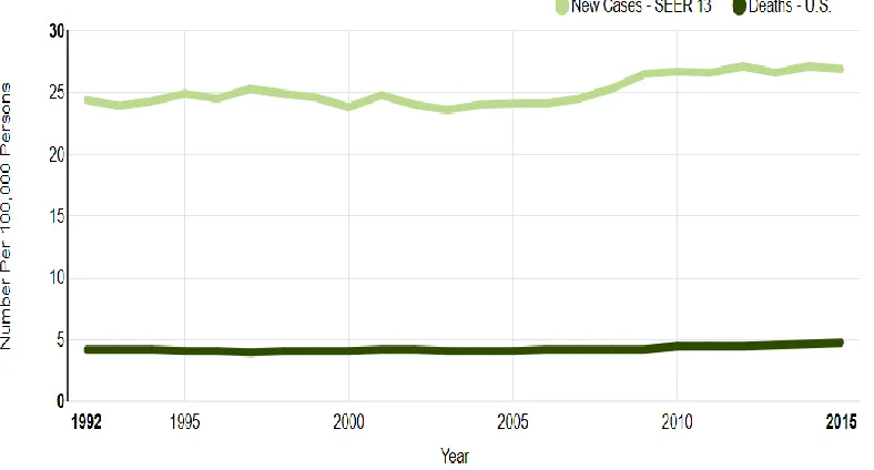
Endometrial carcinoma is the fourth most common malignancy among women and most common gynecologic malignancy in the developed countries (1). Its incidence in the developing countries is also increasing. The incidence is over 60,000 new cases per year in India. Approximately about 1,50,000 cases are diagnosed each year in the world (2). The death rate due to endometrial carcinoma is about 4.5 per 100,000 women per year (3). It is most commonly seen in age group of 45-74 years.
Most of the endometrial carcinomas (90%) are sporadic and the rest 10% are due to hereditary factors (4). Approximately 5% of the cancers diagnosed in women younger than 55 years are associated with hereditary causes (5). The clinical management of patients with endometrial cancer depends on various pathological parameters like stage of the tumour, grade of the tumour, presence of lymphovascular invasion, lymph node status and histological subtype of endometrial carcinomas (6). Around 20% of women who were diagnosed with low risk tumours will develop recurrent disease. (7)
2 clinical practice due to various reasons (9)
3
AIMS AND OBJECTIVES:
AIM:
1) To assess the expression of ER and PR in endometrial carcinomas and its precursors by immunohistochemistry.
OBJECTIVE:
1)To evaluate the immunohistochemical expression of ER and PR in endometrial biopsy samples of disordered proliferation, typical and atypical hyperplasia, and resection specimens of Type I and Type II carcinomas and carcinosarcoma.
4
REVIEW OF LITERATURE:
Epidemiology:
Endometrial carcinoma is the third most common female genital tract malignancy in females of south east Asia (12). Though, endometrial carcinoma is more common in developed countries, the annual incidence in developing countries is projected to increase because of change in life style and high incidence of obesity (13). They usually arise as a continuum of premalignant lesions such as disordered proliferation, typical hyperplasia and atypical hyperplasia. Endometrial carcinomas are broadly classified as Type I
[image:15.612.99.493.461.673.2](70-80%) and Type II (10-20%) carcinomas. Type I carcinomas follow estrogen dependent pathway, develop in a background of hyperplastic endometrium and are usually low grade. Type II carcinomas follow estrogen unrelated pathway, develop in an atrophic endometrium and are high grade. (2)
5 Histologically the endometrium is made of two layers (14)
1. Stratum basale 2. Stratum functionalis.
[image:16.612.83.525.296.504.2]The cyclical changes occurring in the endometrium are mostly controlled by estrogen and progesterone (15). The functionalis layer occupies almost one third of the endometrium and is responsible for proliferation and tissue degeneration. The stratum basalis lies beneath the stratum functionalis and has regenerative function. (16)
Figure 2: Phases of menstrual cycle (20)
6 Estrogen receptors:
Estrogen receptor is one of the important members of steroid hormone super family of nuclear receptors (18). It is an important sex hormone for the development and physiological functions of reproductive organs. Estrogen also plays a main role in metabolism, bone remodeling and functioning of the cardiovascular system (19). The biological activity of estrogen is mediated by its binding to the estrogen receptors. There are two types of estrogen receptor.
1. Estrogen receptor Alpha (ERα) 2. Estrogen receptor Beta (ERβ)
The estrogen receptor alpha (ERα) is located on the chromosome 6. The estrogen receptor beta (ERβ) is located on the chromosome 14 (20). The physiologic and biological activity of estrogen is mainly dependent on the distribution of the receptors in the target organs. Estrogen receptor alpha (ERα) is mainly seen in uterine parenchyma, stroma of prostate, Leydig cells, theca cells of ovary, liver and breast. On the other hand, ERβ is expressed mainly in prostatic epithelium, granulosa cells of ovary, bone marrow and brain (21).
The estrogen receptor has five domains with different functions. (22) 1.A/B Domain- Contains the activator function 1 (AF1) responsible for the transcriptional activity.
7 4. E/F receptor- Interaction site and has ligand dependent activator function 2
[image:18.612.133.478.138.430.2](AF2).
Figure 3: Mechanism of estrogen receptor signaling pathway (26)
The estrogen receptors remain in an inactive form, due to the presence of Heat Shock Protein (in the absence of hormones).
Role of estrogen receptors in the endometrium:
8 ERα is highest in the estrogen dominant proliferative phase of the menstrual cycle. Expression of both the estrogen receptors (ERα and ERβ) are high in the early proliferative endometrium, mid proliferative endometrium and early secretory endometrium. Expression of both the estrogen receptors subsequently decreases in the mid secretory endometrium and late secretory endometrium (24). In the late secretory phase, ERβ is expressed more than ERα in the endometrial stroma. This is due to the fact that the expression of both the estrogen receptors in the epithelium decreases as the cycle enters the secretory phase, but the stromal expression of ERβ is relatively maintained or increased (25).
ER expression in post-menopausal status:
The hormonal environment of pre and post-menopausal endometrium is entirely different. In postmenopausal women, the endometrium is subjected to low levels of estradiol and absent progesterone (26). The expression of ERβ in post-menopausal endometrium is weaker than that of ERα in both the epithelial and stromal elements. Zang et al proposed that the expression of ERβ increases in the
9 Progesterone hormone and its receptors:
Progesterone plays an important role in the female reproductive system. In combination with estrogen (estradiol), it helps in the implantation and maintenance of pregnancy (28). The progesterone exerts most of its actions in the following tissues.
1. Epithelial cells in the endometrium. 2. Stromal cells in the endometrium. 3. Stromal fibroblasts.
4. Smooth muscle cells in the myometrium. 5. Glandular epithelial cells in the cervix.
The actions of progesterone in the female reproductive tract are controlled and mediated by progesterone receptors. There are two types of receptors based on its location in the cell (29).
1. Nuclear Progesterone receptor- Ligand activated and involved in genomic actions. 2. Cytoplasmic Progesterone receptor- G- Protein coupled receptor and single trans
membrane receptors. These receptors are involved in non-genomic actions.
The progesterone receptor gene is located on the chromosome 11. There are two isoforms of progesterone receptors. The activities of progesterone are mediated by the combined effects of these isoforms.
10 The progesterone receptor is composed of four regions.
1. N-Terminal domain- Activation and Inhibitory functions. 2. DNA binding domain (DBD).
3. Hinge region
4. Ligand binding domain (LBD)
Figure 4: Structure of progesterone receptor (29)
CLASSIFICATION:
In 1983, Bokhman, first classified endometrial carcinoma based on clinico-pathological features and molecular background into two groups:
1. Type I
2. Type II
11 definite categorization cannot be made (4). Based on this classification a ‘dualistic model of endometrial carcinogenesis’ was proposed (4).
Dualistic model of endometrial carcinogenesis:
According to the ‘dualistic model of endometrial carcinogenesis’, it is speculated that endometrial carcinoma (Type I) develops following a continuum of precursor lesions ranging from disordered proliferation, typical and atypical hyperplasia and Type II carcinomas develop from endometrial intraepithelial neoplasia (30). Hence the exact diagnosis of precursor lesions in routine endometrial biopsies is very crucial in patient care. The precursor lesions are diagnosed in endometrial biopsies by histo-morphological features such as presence or absence of nuclear atypia, which has poor reproducibility and high inter-observer variability (14). Hence the use of immunohistochemical stains and mutational analysis can aid in the diagnosis of premalignant lesions and endometrial carcinoma in routine endometrial biopsies.
12 I.EPITHELIAL TUMOURS AND PRECURSORS:
Precursors:
Typical hyperplasia.
Atypical hyperplasia/Endometrioid intraepithelial neoplasia. Endometrioid carcinoma
Squamous differentiation Villoglandular
Secretory
Mucinous carcinoma
Serous endometrial intraepithelial carcinoma. Clear cell carcinoma
Neuroendocrine tumours
II.MESENCHYMAL TUMOURS Leiomyoma
Smooth muscle tumour of uncertain malignant potential Leiomyosarcoma
Endometrial stromal and related tumours. Miscellaneous
III.MIXED EPITHELIAL AND MESENCHYMAL TUMOURS Adenomyoma
13 Adenofibroma
Adenosarcoma Carcinosarcoma
IV.MISCELLANEOUS TUMOURS
V.LYMPHOID AND MYELOID TUMOURS VI.SECONDARY TUMOURS
Table 2: The WHO classification of tumours of uterine corpus
TYPE I ENDOMETRIAL CARCINOMA:
Type I endometrial carcinoma accounts for approximately 70-80% of total sporadic cases of endometrial carcinoma (4). Type I endometrial carcinoma is more common than Type II (2) and usually have indolent clinical behavior as compared to Type II tumours.
Risk factors: Estrogen excess:
14 (33). In post-menopausal women the baseline levels of circulating sex hormones is low. On treatment with estrogen only hormone replacement therapy (HRT) there is a two to three times increase in the risk of developing endometrial carcinoma. The use of combined hormone replacement therapy and oral contraceptive are associated with a protective effect (34). The other constitutional factors are early age of menarche and late age of menopause.
b) Nulliparity:
Nulliparity is associated with increased risk (up to three fold) due to chronic anovulatory cycles, which in turn result in unopposed estrogenic stimulation. Hence the development of endometrial carcinoma is more in married nulliparous women than for unmarried women (35).
c) Obesity:
Obesity is associated with approximately 40% of endometrial carcinomas (36). In obese individuals there is extra glandular estrone formation due to peripheral conversion of androgens in the adipose tissue as well as insulin resistance, which results in decreased production of sex hormone binding globulin in the liver and leads to excess circulating estrogen (37). A Body Mass Index (BMI) more than 25 kg/m2 increases the risk of developing endometrial carcinoma by two fold (38).
d) Life style factors:
15 fruits, reduced amounts of saturated fatty acids and moderate intake of wine is associated with decreased incidence of endometrial cancer (39).
e) Others:
Diabetes mellitus, Tamoxifen therapy, polymorphisms in the estrogen receptor are also associated with increased incidence of endometrial carcinoma (2).
Genetic factors:
A number of cancer causing genes have been identified in the development of endometrial carcinoma (Type I) such as PTEN, PIK3CA, PI3K, microsatellite instability and K-ras mutation. Among the various genetic alterations, PTEN (phosphatase and tensin homologue) gene is the most frequently mutated gene in Type I endometrial carcinoma(30-54%) (40).
16 Pathogenesis of Type I endometrial carcinomas:
Figure 5: Pathogenesis of Type I pathway (41)
17 hyperplasia was first introduced by Cullen in 1900 (43). This classification is based on cytologic and architectural abnormalities (2).
Typical hyperplasia
1. Simple hyperplasia without atypia 2. Complex hyperplasia without atypia. (adenomatous without atypia)
Atypical hyperplasia
1. Simple atypical hyperplasia 2. Complex atypical hyperplasia (adenomatous with atypia)
Table 4: WHO classification of precursor lesions of endometrium (1994)
These categories are descriptive in nature and the interpretation is subjective. These individual categories do not specify individual clinical management algorithms (44). Most of the studies, proposed that the risk of progression of hyperplasia to carcinoma depends more on the cytological atypia, than architectural atypia (45). According to Lacey et al, the risk of progression of hyperplasia without atypia to carcinoma was only 10%, on the other hand the risk of progression of hyperplasia with atypia to carcinoma was 40% (46). Following this, the WHO simplified the classification in 2014 as a two tier system.
1. Typical hyperplasia
18 Typical hyperplasia occurs secondary to unopposed estrogen stimulation (47).
The definition is “Exaggerated proliferation of glands of irregular size and shape, with an associated increase in the gland to stroma ratio compared with proliferative endometrium, but without significant cytological atypia” (48).
The development of the lesion is strongly associated with unopposed estrogenic stimulation, and hence the duration and dose of estrogen exposure affects the overall histological picture. According to Kurman et al, the risk of progression to carcinoma occurs in 1-3% of patients with hyperplasia without atypia (45). The term disordered proliferation is used when there is proliferation of glands with no cytological atypia, that exceeds that of proliferative endometrium but falls short of the crowding in hyperplasia (48).
Atypical hyperplasia/Endometrioid intraepithelial neoplasia:
Endogenous or exogenous hyperestrinism is the important risk factor for the development of atypical hyperplasia (49). Cytological atypia superimposed on endometrial hyperplasia defines atypical hyperplasia (48). The distinction between typical hyperplasia and atypical hyperplasia is based on cytological atypia (following nuclear features) (45,50).
19 These histological features are subjective with much intra-observer and Inter- observer variability. In approximately 25-40% of women, atypical hyperplasia coexist with carcinoma (51). Atypical hyperplasia shows most of the genetic changes that are seen in Type I endometrial carcinomas, such as microsatellite instability, PAX2 inactivation and PTEN mutation (52). The risk of progression of atypical hyperplasia to carcinoma is 25-33% during the first year of follow up (45,51,53)
Management of preneoplastic lesions:
20
Figure 6: Management of endometrial precursor lesions (14)
Type I endometrial carcinomas:
21 producing ovarian tumours are at risk for developing endometrial carcinoma (54). Patients with Lynch Syndrome and Cowden syndrome are also at an increased risk of developing endometrial carcinoma (55). The average age of diagnosis is around 63 years (56). The most common presenting symptom is post-menopausal bleeding. Postmenopausal bleeding is defined as uterine bleeding after permanent cessation of menstruation resulting from loss of ovarian follicular activity (57). The other symptoms include vaginal discharge and pelvic pain. In few cases, abnormal cells may be found in routine cervical cytology (PAP smear) (58). The initial imaging done in patients with abnormal uterine bleeding is pelvic ultrasonography. A cut off value >11mm endometrial thickness is associated with an increased risk of cancer by 6.7% (59).
Gross appearance:
Most of the endometrial tumours have an exophytic diffuse growth. A few may present as tan nodules in the endometrium. Hemorrhage and necrosis are common.
22 Histology:
Well differentiated endometrioid carcinoma is differentiated from atypical hyperplasia based on the following features (60).
1. An irregular infiltration of glands with desmoplastic response.
2. Confluent glandular and cribriform pattern in which glands are uninterrupted by the stroma.
3. Excessive papillary pattern(villoglandular)
4. Stromal replacement by masses of squamous epithelium.
Patterns in endometrioid carcinoma:
1. Endometrioid carcinoma with squamous differentiation. 2. Endometrioid carcinoma with secretory features.
3. Endometrioid carcinoma with villoglandular pattern. 4. Endometrioid carcinoma with sertoliform pattern 5. Endometrioid carcinoma with microglandular pattern.
23 The endometrioid carcinomas are graded by the WHO based on the architecture (62).
Grade 1 Less than 5% non-squamous or non-morular growth pattern. Grade 2 6%-50% non-squamous or non-morular growth pattern. Grade 3 More than 50% non-squamous or non-morular growth pattern.
Table 5: Grading of endometrioid carcinoma
Tx Primary tumour cannot be assessed.
T0 No evidence of primary tumour.
Tis Carcinoma in situ
T1 Tumour confined to the corpus uteri
T1a Tumour limited to endometrium or invading less than half of myometrium.
T1b Tumour invades one half or more of myometrium.
T2 Tumour invades cervical stroma, but does not extend beyond the uterus
T3 Local and/regional spread
T3a Tumour invades the serosa of the corpus uteri or adnexa(direct extension or metastasis)
T3b Vaginal or parametrial involvement(direct extension or metastasis) T4 Tumour invades bladder/bowel mucosa.
N1 Metastasis to pelvic or para-aortic lymph nodes.
N2 Metastasis to para-aortic lymph nodes with or without metastasis to pelvic lymph nodes
M1 Distant metastasis(exclude metastasis to vagina, pelvic serosa or adnexa)
24 Genetic profile of endometrial carcinoma:
Table 7: Molecular classification of endometrial carcinoma (31)
Prognosis of endometrioid carcinoma:
The prognosis of endometrioid carcinoma is dependent mainly on the following factors: FIGO stage, age of the patient, stage of the disease, histological grade, depth of myometrial involvement and the lymphovascular invasion. The risk of recurrence is mainly dependent in the depth of myometrial invasion. Tumour involving more than half of the myometrium (outer half) is significantly associated with diminished survival rate in patients (63). Survival rate of endometrial carcinoma:
25
Stage Percentage
IA IB IC
91% 88% 81% IIA
IIB
77% 67% IIIA
IIIB IIIC
60% 41% 32% IVA
IVB
20% 5%
26
Table 9: 2009 FIGO system for staging of carcinoma endometrium
Serous carcinoma:
27 ligation and past history of breast cancer are more often seen in cases of endometrial serous carcinomas (65).
Gross appearance:
These tumors are usually small and are associated with atrophic uterus. They may have an exophytic and papillary appearance. In few cases, a benign appearing polyp may be seen. Serous carcinoma and serous endometrial intraepithelial carcinoma are known to develop within a polyp (66).
28 Histogenesis of serous carcinoma:
29 The precursor lesions for serous carcinoma is termed as ‘serous endometrial intraepithelial carcinoma (SEIC). They frequently develop on a polyp or in atrophic endometrium (68). On microscopy, SEIC often show a mild papillary architecture lined by columnar cells with enlarged, hyper chromatic smudged nuclei. Few cells can display hobnail morphology. There is brisk mitotic activity among the atypical cells. These lesions are diffusely positive for p53 on immunohistochemistry.
Figure 10: Serous endometrial intraepithelial carcinoma in a polyp (68)
30
Figure 11: p53 staining in serous endometrial intraepithelial carcinoma (68)
Microscopy of uterine serous carcinoma:
31 Genetic profile of serous carcinoma:
The following are the most common somatic mutations in serous carcinoma of uterus:
Mutation Percentage
TP53 80-90%
PIK3CA 24-40%
FBXW7 20-30%
PPP2R1A 18-28%
Table 10: The mutations associated with serous carcinoma (71)
Prognosis:
Serous carcinoma patients should be carefully staged because they may have extra-uterine spread even in the absence or very minimal myometrial invasion. The involvement of extra uterine sites such as ovary, peritoneum and fallopian tubes can occur very early in the course of disease compared to other endometrial adenocarcinomas.
According to Abeler et al, the 5 year and 10 year survival rates(all stages of tumour) of serous carcinoma are 36% and 18% respectively (64). The prognostic factors for shorter survival are as follows:
1. Age more than 60 years. 2. Vascular invasion.
32 Carcinosarcoma:
Carcinosarcoma [Malignant Mullerian Mixed Tumors (MMMT)] comprise less than 5% of all the malignancies in the uterus (72). By definition, carcinosarcoma is a biphasic tumour composed of high grade carcinomatous and sarcomatous elements (48). The mean age of patients presenting with carcinosarcoma is about 70 years. The risk factors for carcinosarcoma are difficult to determine because of the low prevalence of the disease. Most of the studies proposed that patients with carcinosarcoma and endometrioid adenocarcinoma share the same risk factors.
Gross:
Carcinosarcomas are mostly polypoidal and fills the entire uterine cavity. In about half of the patients, the tumour protrudes through the cervix (73). The tumour has a fleshy, soft to firm tan cut surface.
33 Histogenesis:
MMMT likely represent carcinomas with a mesenchymal component as a result of metaplasia and tumour progression. The recent clinico-pathological, immunohistochemical and molecular findings suggest that these tumors arise from a single multipotent stem cell, by a process called ‘bidirectional differentiation’ (74).
Microscopy:
Carcinosarcomas are composed of a mixture of high grade epithelial and mesenchymal components. The epithelial component is composed mostly of endometrioid carcinoma, but other subtypes such as serous, mucinous, squamous and mesonephric carcinomas can also be seen (75). The mesenchymal component is composed of a high grade sarcoma. Heterologous differentiation such as chondrosarcoma, osteosarcoma and rhabdomyosarcoma is seen in around 50% of the cases (76).
Genetic profile:
Many genetic and molecular studies have confirmed that similar molecular pathways are involved in the pathogenesis of endometrioid carcinoma and carcinosarcoma. The phenotypic characteristic of carcinosarcoma is dependent on changes in Akt/β catenin pathway and repression of E- Cadherin (77).
Prognosis:
Carcinosarcomas are associated with poor outcome with a five year survival of 15%-30% in advanced stage disease (78). The prognosis is dependent on the following factors:
34 2. Extra uterine extension
3. Deep myometrial invasion.
4. Presence of serous or clear cell carcinoma as epithelial component. 5. Presence of heterologous sarcomatoid elements.
Treatment of endometrial carcinomas:
The main stay of treatment for endometrial carcinoma is surgery, followed by adjuvant therapy such as radiotherapy and chemotherapy. Few targeted therapies are also available.
The standard treatment for endometrioid carcinoma is surgery (Total hysterectomy and bilateral salpingo-oophorectomy). Post-operative progestin therapy is given for patients with no significant poor prognostic factors (79). Pelvic and para-aortic sampling should be done in patients with the following features (80):
1. More than 50% myometrial invasion 2. Grade 3 tumour.
3. Involvement of cervix.
4. Presence of extra uterine spread. 5. Palpably enlarged lymph nodes.
When treated with postoperative radiotherapy, the five year survival rate of women with positive lymph nodes is improved to 40%.
35 and para-aortic lymph node sampling. Because of the aggressive behavior of serous carcinoma, adjuvant therapy should always be considered. Few studies show that platinum based chemotherapy is useful in patients with high grade serous carcinoma (81).
Carcinosarcomas are treated by total hysterectomy and bilateral salpingo-oophorectomy with lymph node dissection, peritoneal and pelvic washings. The adjuvant therapies that was recently approved by the Gynecologic Oncology Group is the use of combination chemotherapy (cisplatin, ifosfamide and mesna) over abdominal radiotherapy (82).
Recent advances in endometrial cancer:
The understanding of molecular pathway in the pathogenesis of endometrial carcinoma help us to guide in the development of targeted therapies for its treatment. The following are the new targeted therapies available for the treatment of endometrial carcinoma (85):
36
MATERIALS AND METHODS:
This study was done in The Department of General Pathology. Cases diagnosed as disordered proliferation, typical and a typical hyperplasia on biopsy and resection specimens of endometrial adenocarcinoma, serous and carcinosarcoma during the time period of January 2014- December2016 are included in this study.
Inclusion criteria:
Cases diagnosed as disordered proliferative endometrium (DPEM), typical and
atypical hyperplasia in curettings and subsequently underwent hysterectomy. Cases which underwent hysterectomy for endometrial adenocarcinoma,
serous carcinoma and carcinosarcoma.
Exclusion criteria:
Cases of disordered proliferative endometrium (DPEM), typical and atypical
hyperplasia in curettings with no subsequent hysterectomy. Blocks handed over to the patients.
37 Methodology:
The clinical details of all the patients such as age, post-menopausal status, duration of post-menopausal status, clinical presentation, endometrial thickness, parity, history of exogenous hormone intake, family history of tumours, syndromic association, Body Mass Index (BMI) and comorbidities were retrieved from the Medical Records Department. Stage, grade, presence/ absence of lymphovascular invasion and lymph node metastasis if any were retrieved from the pathology work station.
Assessment of ER and PR on all the cases was done by immunohistochemistry. Precursor lesions:
The total number of cases diagnosed as disordered proliferation were 503. Out of which 30 cases had both curettings and resection specimens. Nine blocks were unavailable and hence 21 cases were included in this study. The total number of cases diagnosed as typical hyperplasia were 125. Out of which 32 cases had both resection and curettings. Three blocks were unavailable and hence 29 cases were included in this study. The total number of cases with atypical hyperplasia were 39. Fourteen blocks were unavailable and hence 25 cases were included in this study.
Endometrial carcinomas:
38
Preneoplastic lesions (2014 - 2016)
Figure 13: Schema figure for preneoplastic lesions
Typical hyperplasia
Disordered Proliferation Atypical hyperplasia 2014 – 176 cases
2015 – 179 cases
2016 – 148 cases 2014 – 65 cases 2015 – 32 cases 2016 – 28 cases
2014 – 17 cases 2015 – 17 cases 2016 – 5 cases
Only 30 cases had curettings
and Resection specimens Only 32 cases had curettings and Resection specimens 39 cases
Blocks not available - 5 cases & Outside blocks – 4 case
Blocks not available - 3 cases Blocks not available - 11 cases & Outside blocks – 3 case
Total cases included in our study - 21
Total cases included in
39
Carcinoma (2014 - 2016)
Figure 14: Schema figure for endometrial carcinoma
Study group Number of cases
Disordered proliferation (DPEM) 21
Typical hyperplasia 29
Atypial hyperplasia 25
Endometrioid carcinoma 20
Serous carcinoma 04
Carcinosarcoma 14
Table 11: Total number of cases in this study
Serous carcinoma
Endometrioid carcinoma Carcinosarcoma
2014 – 53 cases 2015 – 48 cases 2016 – 41 cases
Total 8 cases
Total 18 cases
Randomization done Blocks not available - 4 cases Blocks not available - 4 cases
Total cases included in
40 Immunohistochemical evaluation of ER and PR was performed according to the method described by Carcangiu ML et al., based on the percentage of stained cells and the intensity of nuclear stain.
Score Percentage of nuclei stained
1 0% to 25% of the nuclei
2 26% to 75% of nuclei
3 more than 76% of the nuclei
Table 12: Grading the percentage of stained cells
Score Intensity
1 absent or weak
2 Strong
3 very strong
Table 13: Grading the staining intensity of the cells
The sum of both parameters gave the immunohistochemical score.
41 Category I corresponded to a score of 2,
Category II corresponded to a score of 3 or 4, Category III corresponded to a score of 5 or 6.
42
RESULTS:
Clinical features:
Disordered proliferation:
In disordered proliferation, two out of twenty one patients (9%) were postmenopausal. The age distribution of disordered proliferation was 39-53 years. The most common clinical presentation in this group (85.71%) was perimenopausal bleeding. According to WHO, fourteen out of twenty cases (70%) had above normal body mass index (BMI). Nine out of twenty one cases (42%) were associated with a history of exogenous hormone intake. The average endometrial thickness in this group ranged from 0.5cm - 5.1cm. Three out of twenty one patients (14%) were associated with family history of other tumours.
Typical hyperplasia:
43 Atypical hyperplasia:
In atypical hyperplasia, thirteen out of twenty five patients (52%) were post-menopausal. The age distribution of atypical hyperplasia was 29-74 years. The most common clinical presentation in this group (52%) was postmenopausal bleeding. Seventeen out of twenty cases (85%) had above normal BMI. The BMI of five patients was not available. Ten out of twenty three cases (39%) were associated with a history of exogenous hormone intake. The average endometrial thickness in this group ranged from 0.5cm – 5.3cm. Two out of twenty five patients (12%) were associated with family history of other tumours.
Endometrial carcinoma:
In endometrial carcinoma, fifteen out of twenty patients (75%) were postmenopausal. The age distribution of endometrial carcinoma was 34-71 years. The most common clinical presentation in this group (75%) was postmenopausal bleeding. Ten out of nineteen cases (52%) had above normal BMI. The BMI of one patient was not available. Two out of twenty cases (10%) were associated with a history of exogenous hormone intake. The average endometrial thickness in this group ranged from 0.8cm – 7.9cm. Five out of twenty patients (25%) were associated with family history of other tumours.
Serous carcinoma:
44 Carcinosarcoma:
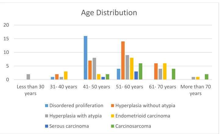
[image:55.612.127.488.337.557.2]In carcinosarcoma, thirteen out of fourteen patients were (92%) were postmenopausal. The age distribution of carcinosarcoma was 49-77 years. The most common clinical presentation in this group (93%) was postmenopausal bleeding. Ten out of fourteen cases (71%) had above normal BMI. The average endometrial thickness in this group ranged from 1.0cm – 7.8cm. Two out of fourteen cases (14%) were associated with family history of other tumours.
Figure 15: Age distribution of endometrial preneoplastic lesions and carcinomas
The mean age of presentation of preneoplastic lesions of endometrium was 51 years. In preneoplastic lesions, the frequency of disordered proliferation was a decade earlier than other lesions. In this study, atypical hyperplasia was randomly distributed among all age
0 5 10 15 20
Less than 30 years
31- 40 years 41- 50 years 51- 60 years 61- 70 years More than 70 years
Age Distribution
Disordered proliferation Hyperplasia without atypia
Hyperplasia with atypia Endometrioid carcinoma
45 groups. The most common age group for carcinomas was above 50 years. The mean age of carcinomas was 58 years. Endometrioid carcinoma was seen in a decade earlier than serous and carcinosarcoma. In summary, the preneoplastic lesions occurs at a relatively younger patients compared to carcinomas.
Figure 16: Postmenopausal status of endometrial preneoplastic lesions and carcinomas
This table suggests disordered proliferation was more frequently associated with pre and perimenopausal age group. On the other hand, carcinomas were more frequently associated with post-menopausal status.
0 5 10 15 20 25
Post Menopausal Status
46
Figure 17: Number of years after menopause
Presenting complaints:
The presenting complaints included post-menopausal bleeding, abnormal uterine bleeding, abdominal pain, and others including white discharge per vagina, primary infertility, itching and urinary retention. Three patients of atypical hyperplasia presented with primary infertility. Overall the most common clinical presentation was post-menopausal bleeding.
Clinical presentation Number of patients (n=113)
Post-menopausal bleeding 55
Abnormal uterine bleeding 46
Abdominal pain 7
Others 14
Table 14: Clinical presentation of preneoplastic lesions and endometrial carcinomas
0 2 4 6 8 10
Less than 5 years
6- 10 years 11- 15 years 16- 20 years 21-25 years 26-30 years
Years after Menopause
Disordered proliferation Hyperplasia without atypia
Hyperplasia with atypia Endometrioid carcinoma
47 Parity:
[image:58.612.126.488.278.499.2]Five out of thirty three patients with carcinoma were nulliparous. Seven out of seventy one patients with preneoplastic lesions were nulliparous. The parity status of nine patients was not known. Altogether, the incidence of carcinoma is 34.02 % and 41.67% in parous and nulliparous women respectively. Three out of fourteen carcinosarcoma patients were nulligravida (21%).
Figure 18: The incidence of carcinoma in nulliparous women
0.00% 5.00% 10.00% 15.00% 20.00% 25.00% 30.00% 35.00% 40.00% 45.00%
Parous Nulliparous
48 Body mass index:
[image:59.612.93.517.159.418.2]The body mass index (BMI) of the patients was classified according to the WHO.
Figure 19: Body mass index of preneoplastic lesions and carcinoma
In preneoplastic lesions, 1% were underweight, 17% were normal weight, 35% were overweight and 46% were obese. BMI of six patients was not available.
In carcinoma, 5% were underweight, 32% were normal weight, 30% were overweight and 32% were obese. BMI of one patient was not available.
Comorbidities:
The comorbidities associated with the study population were diabetes mellitus, hypertension, hypothyroidism and others. Others conditions included coronary heart disease, seizure, obstructive sleep apnoea and respiratory disorders.
0 5 10 15 20 25 30 35 40
Less than 18.5 18.5- 24.9 25- 25.9 30- 34.9 35- 39.9 More than 40
Body mass index
Disordered proliferation Hyperplasia without atypia Hyperplasia with atypia
49 Comorbidities Number of patients (n=113)
Diabetes 41
Hypertension 43
Hypothyroidism 12
Others 22
[image:60.612.123.488.285.499.2]Nil 42
Table 15: Comorbidities associated with preneoplastic lesions and carcinomas
Figure 20: Comorbidities associated with preneoplastic lesions and carcinomas
Diabetes and hypertension were the most common comorbidities associated with the study population. Most of the patients with preneoplastic lesions were without any comorbidities, probably as they presented at a younger age.
0 2 4 6 8 10 12 14
Diabetes Hypertension Hypothyroidism Nil Others
Comorbidities
Disordered proliferation Hyperplasia without atypia
Hyperplasia with atypia Endometrioid carcinoma
50 Association with exogenous hormones:
The incidence of carcinoma in patients who took hormonal therapy was 9.68% in our study population. The history of hormonal use was seen mostly among patients with preneoplastic lesions. Twenty eight out of seventy two patients (39%) with preneoplastic lesions had history of exogenous hormone intake. History of hormonal intake in three patients was not available.
History of other tumours:
In preneoplastic lesions, nine out of seventy five patients (12%) had family history of other tumours. In cases of carcinoma, seven out of thirty eight patients (19%) had family history of other tumours.
Endometrial thickness:
51 Parameter Endometrial preneoplastic lesions (n=75) Endometrial
carcinoma (n=38) P value*
Mean age 50.92+/- 9.05 58.11+/- 8.99 <0.001
Postmenopausal Yes No 35(46.67%) 40(53.33%) 32(84.21%)
6(15.79%) <0.001 Parity Gravida Nulligravida 64(90.14%) 7(9.86%) 33(86.84%) 5(13.16%) 0.600
History of tumour Yes
No
9(12.16%) 65(87.54%)
7(18.42%)
31(81.58%) 0.370
History of exogenous hormones Yes No 28(38.89%) 44(61.11%) 3(7.89%) 35(92.11%) 0.001 Presenting complaints PMB AUB Abdominal pain Others 31(41.33%) 39(52%) 3(4%) 5(6.67%) 24(63.16%) 7(18.42%) 4(10.53%) 9(23.68%) 0.028 0.001 0.174 0.009 Comorbidities
52 Diabetes mellitus Hypertension Nil Others 25(33.33%) 21(28%) 35(46.67%) 15(20%) 16(42.11%) 23(60.53%) 9(23.68%) 4(10.53%) 0.360 0.001 0.018 0.203 Endometrial
[image:63.612.124.488.454.668.2]thickness 13.27+/-9.65 26.13+/- 21.20 <0.001 *p value of less than 0.05 is considered as statistically significant.
Table 16: Comparison of clinical and radiological features between endometrial
preneoplastic lesions and carcinoma
Tumour size:
The mean tumour size was 4.84cm. The largest tumor in this study population was 11.5cm (endometrioid carcinoma, grade 3, Stage I). All the patients with carcinosarcomas had a tumour size of more than or equal to three.
Figure 21: Gross appearance of tumours
0 2 4 6 8 10 12 14 16
Polypoidal Diffuse fleshy Combined
Tumour - Gross appearance
53 Endometrioid carcinomas had polypoidal/proliferative friable growth in the endometrial cavity. Carcinosarcomas and serous carcinoma were bulky polypoidal masses.
Grading of carcinoma:
The endometrioid carcinomas were graded according to the WHO system of classification.
Grade No. of cases (n=20)
Well differentiated 6
Moderately differentiated 8
Poorly differentiated 6
Table 17: Grading of endometrioid carcinoma based on FIGO system
Depth of invasion:
54
Figure 22: Depth of myometrial invasion in endometrial carcinomas
Lymphovascular invasion:
Lymphovascular invasion was present in nine out of thirty eight cases of carcinomas (24%). In endometrioid carcinoma, three out of twenty patients (15%) had lymphovascular invasion. In carcinosarcoma, six out of fourteen patients (43%) had lymphovascular invasion. The most common lymph node group involved were left and right pelvic lymph nodes. None of the cases of serous carcinoma had lymphovascular invasion.
13
2
7 7
2
7
0 2 4 6 8 10 12 14
Endometrioid Carcinoma Serous carcinoma Carcino Sarcoma
Myometrial Invasion
55
Figure 23: Lymphovascular invasion in endometrial carcinoma
Stage of the tumour:
The tumour stage is based on TNM classification.
Stage Number of cases
1 30
III 7
[image:66.612.126.487.71.287.2]IV 1
Table 18: Distribution of cases according to the TNM staging
None of the cases in this study belonged to stage II. In endometrioid carcinoma,
seventeen out of twenty cases (85%) had stage I disease. In carcinosarcoma eleven out of fourteen cases (79%) had stage I disease. In serous carcinoma two cases each had stage I& stage III disease.
3 0 6 17 4 8 0 2 4 6 8 10 12 14 16 18
Endometrioid Carcinoma Serous carcinoma Carcino Sarcoma
Lymphovascular Invasion
56
Figure 24: Stage of tumour
Other associated lesions:
Fourteen out of thirty eight cases (37%) showed other associated benign lesions such as leiomyoma, adenomyosis and endometriosis. Two out of twenty (10%) of the endometrioid carcinoma were associated with endometriosis.
Associated lesions No. of cases (n=14)
Leiomyoma 8
Adenomyosis 4
[image:67.612.126.487.71.284.2]Endometriosis 2
Table 19: Lesions associated with endometrial carcinomas in resection specimen
17 2 1 2 2 0 11 3 0 0 2 4 6 8 10 12 14 16 18
Stage I Stage III Stage IV
Stage of tumour
57 Association with heterologous elements in carcinosarcoma:
[image:68.612.123.489.191.410.2]Six out of fourteen cases of carcinosarcoma (43%) were associated with heterologous elements. Out of six cases, four cases had cartilaginous differentiation and two cases had skeletal muscle differentiation.
Figure 25: Heterologous elements in carcinosarcoma
Immunohistochemical expression of ER and PR: ER:
Out of the 38 cases of carcinoma, 14 cases (37%) belonged to category I, 16 cases (42%) belonged to category II and 8 cases (21%) belonged to category III. All cases of endometrioid carcinomas belonged to category III.
Heterologous elements
58
Figure 26: ER expression in endometrial carcinoma
PR:
Among the 38 cases of tumour, 20 cases (53%) belonged to category I, 8 cases (21%) belonged to category II and 10 cases (26%) cases belonged to category III. All the cases of carcinosarcoma belonged to category I.
Figure 27: PR expression in endometrial hyperplasia
10% 50% 71.42% 55% 25% 28.58% 35% 25% 0
0% 10% 20% 30% 40% 50% 60% 70% 80%
Endometrioid carcinoma Serous carcinoma Carcinosarcoma
ER expression in carcinoma
Category III Catergory II Category 1
50% 75% 100.00% 50% 25% 0.00% 0% 0% 0%
0% 20% 40% 60% 80% 100% 120%
Endometrioid carcinoma Serous carcinoma Carcinosarcoma
PR expression in carcinoma
[image:69.612.124.489.449.668.2]59 Expression of ER and PR in precursor lesions:
ER:
[image:70.612.124.488.309.532.2]Out of the 75 cases, only one case (1%) belonged to category I, 6 cases (8%) belonged to category II and 68 cases (91%) belonged to category III. Out of the six cases in category II, five cases were of atypical hyperplasia. Out of the 5 cases, one case was diagnosed as complex hyperplasia with atypia in endometrial curettings and endometrioid carcinoma (grade 1) in resection specimen.
Figure 28: ER expression in preneoplastic lesions
PR:
Among the 75 cases, only one case (1%) belonged to category I, one case (1%) belonged to category II and remaining 73 cases belonged to category III (97%).
0.00% 0.00% 4.00%
0.00% 3.44%
20.00%
100.00% 96.56% 76.00%
0.00% 20.00% 40.00% 60.00% 80.00% 100.00% 120.00% Disordered proliferation
Hyperplasia without atypia Hyperplasia with atypia
ER expression in precursor lesions
60
Figure 29: PR expression in preneoplastic lesions
0.00% 0.00% 4.00%
0.00% 0.00% 4.00%
100.00% 100.00% 92.00%
0.00% 20.00% 40.00% 60.00% 80.00% 100.00% 120.00% Disordered proliferation
Hyperplasia without atypia Hyperplasia with atypia
PR expression in precursor lesions
61
Parameter
ER Category
ER p value*
1 2 3
Stage
1 9(64.29%) 13(81.25%) 8(100%)
0.173
3 – 4 5(35.71%) 3(18.75%) 0
Grade
Well differentiated 0 1(6.25%) 5(62.5%)
< 0.001
Moderately differentiated 1(7.14%) 7(43.75%) 2(25%)
Poorly differentiated 13(92.86%) 8(50%) 1(12.5%)
Lymphovascular Invasion
Yes 6(42.86%) 3(18.75%) 0
0.06
No 8(57.14%) 13(81.25%) 8(100%)
Diagnosis
Disordered Proliferation 0 0 21(27.63%)
< 0.001
Typical hyperplasia 0 1(4.55%) 28(36.84%)
Atypical hyperplasia 1(6.67%) 5(22.73%) 19(25%)
Endometrioid Carcinoma 2(13.33%) 11(50%) 7(9.21%)
Serous Carcinoma 2(13.33%) 1(4.55%) 1(1.32%)
Carcinosarcoma 10(66.67%) 4(18.18%) 0
Endometrial Thickness
Less than 5 mm 0 1(5.26%) 6(8.96%)
0.841
[image:72.612.56.560.70.634.2]More than 5 mm 12(100%) 18(94.74%) 61(91.04%)
62
Parameter
PR Category
PR p value
1 2 3
Stage
1 13(65%) 7(87.5%) 10(100%)
0.066
3 – 4 7(35%) 1(12.5%) 0
Grade
Well differentiated 0 2(25%) 4(40%)
< 0.001
Moderately differentiated 2(10%) 3(37.5%) 5(50%)
Poorly differentiated 18(90%) 3(37.5%) 1(10%)
Lymphovascular Invasion
Yes 8(40%) 1(12.5%) 0
0.025
No 12(60%) 7(87.5%) 10(100%)
Diagnosis
Disordered Proliferation 0 0 21(25.30%)
< 0.001
Typical hyperplasia 0 0 29(34.94%)
Atypial hyperplasia 1(4.76%) 1(11.11%) 23(27.71%)
Endometrioid Carcinoma 5(23.81%) 6(66.67%) 9(10.84%)
Serous Carcinoma 3(14.29%) 0 1(1.2%)
Carcinosarcoma 12(57.14%) 2(22.2%) 0
Endometrial Thickness
Less than 5 mm 0 1(11.11%) 6(8.33%)
0.409
[image:73.612.50.563.68.689.2]More than 5 mm 17(100%) 8(88.89%) 66(91.67%)
63 Comparison of ER and PR expression:
The ER and PR expression was compared with stage of the tumour, grade of the tumour, histological type, lymphovascular invasion and endometrial thickness.
Grade of the tumour:
62% tumours in category III (ER) were well differentiated and 93% of tumours in Category I (ER) were poorly differentiated. 40% of tumours in category III (PR) were well differentiated and 90% of tumours in Category I (PR) were poorly differentiated. The p value was <0.05 and is statistically significant.
Type of the lesion: ER expression:
All the cases of disordered proliferation (100%) belonged to Category III. In typical hyperplasia 97% cases belonged to Category III. In atypical hyperplasia nineteen out of twenty cases (76%) belonged to Category III. In endometrioid carcinoma, 10% cases belong to Category I, 55% cases belonged to Category II and 35% cases belonged to Category III. In serous carcinoma three out of four cases (75%) belonged to Category I. In carcinosarcoma, 71% of cases belonged to Category I and 29% cases belonged to Category II. The p value is <0.05 and is statistically significant.
PR Expression:
64 I, 30% cases belonged to Category II and 45% cases belonged to Category III. In serous carcinoma all the four cases belonged to Category I. In carcinosarcoma, 86% of cases belonged to Category I. The p value is <0.05 and is statistically significant.
Pathological correlation between curettings and resection specimens in preneoplastic lesions:
65
DISCUSSION:
Endometrial carcinoma is one of the most common gynecological malignancy in developed countries. However, the incidence of endometrial carcinoma is increasing in developing countries due to increasing incidence of life style diseases such as obesity and diabetes (86). Various studies have previously assessed the expression of different markers in endometrial carcinoma to predict the outcome and prognosis, one of which is expression of hormonal receptors, ER and PR. This study analyses the clinico-pathological features of preneoplastic lesions and carcinoma of the endometrium along with immunohistochemical expression of ER and PR in both of the groups and compares it with the previously published studies.
Clinico-pathological features: Age:
66 in the degree of abnormality of the lining epithelium. According to Masjeed et al and Kumari et al, the peak incidence of endometrial hyperplasia (preneoplastic lesions) was in the fifth decade and endometrial carcinomas was in the sixth decade. (87,88) which was similar to this study.
Postmenopausal status:
In this study, 84% cases of carcinoma were post-menopausal, compared to 47% cases of preneoplastic lesions. This was statistically significant (p<0.05). According to Hileeto et al, preneoplastic lesions were more prevalent (42%) in the perimenopausal age group(89), which was similar to this study. As a result Chavez et al, proposed the role of minimally invasive techniques like hysteroscopy and sonohysterogram in the evaluation of menstrual irregularities in younger women as one of the reasons for earlier detection of preneoplastic lesions. (90)
Presenting complaints:
The most common clinical presentation (63%) in cases of carcinoma was post-menopausal bleeding. The most common presentation (52%) in preneoplastic lesions was abnormal uterine bleeding. This was statistically significant (p<0.05). This was in concordance with the study by Masjeed et al in which the most common clinical presentation was postmenopausal bleeding (52%), followed by menorrhagia (44%). Risk factors of endometrial carcinoma:
67 carcinoma had a BMI>30 kg/m2 (91), which was similar to this study. This was mainly due to the fact that in post-menopausal females, the main site of conversion of androstenedione to estrone is the adipose tissue (39). The incidence of carcinoma was 34% and 42% in parous and nulliparous women respectively in this study. According to Poccobelli et al, ever having birth was associated with a 35% reduced risk of developing endometrial cancer (odds ratio: 0.65) (92). This was in concordance with our study. In this study 39% and 8% had history of exogenous hormone intake in preneoplastic lesions and carcinoma respectively. Higher intake of exogenous hormones in preneoplastic cases was due to the fact these cases presented with menstrual irregularities at a relatively younger age. This was statistically significant (p<0.05)
Endometrial thickness:
In this study, the mean endometrial thickness (ET) in cases with preneoplastic lesions was 13.27mm. The mean endometrial thickness in cases of carcinomas was 26.13mm. This was statistically significant (p<0.05). In this study, 90% of patients with endometrial carcinoma had ET> 5mm. Various studies in the past(Gull et al and Ferrizzi et al) assessed the usefulness of ET in predicting the risk of cancer development in post-menopausal females (93,94). Meta-analysis of 5892 symptomatic women showed than an ET greater than or equal to 5mm was seen in 95% of endometrial cancers (95). This was similar to the present study.
Immunohistochemical expression of ER and PR:
68 Combining category II and III in this study, ER was positive in 100% cases of disordered proliferation, 100% cases of typical hyperplasia, 96% of atypical hyperplasia. ER was positive in 90% of endometrioid carcinoma. PR was positive in 100% of disordered proliferation, 100% of typical hyperplasia, 96% of atypical hyperplasia. PR was positive in 75% of endometrioid carcinoma. None of the serous and carcinosarcoma cases were positive for ER and PR expression. This was statistically significant (p<0.05).
69
Diagnosis
ER expression Present
study Masjeed et al Orejuela et al Bozdogan et al Kumara et al Disordered
proliferation 100 % --- --- --- ---
Typical hyperplasia 100 % 92.85 % 100 % 96.1 % 100 %
Atypical hyperplasia 96 % 85.71 % --- --- --- Endometrioid
carcinoma 90 % 60.71 % 71.4 % 86.3 % 68 % Serous carcinoma 25 % --- ---- --- ----
[image:80.612.54.561.70.480.2]Carcinosarcoma 28 % --- ---- ---- ----
70 Diagnosis
PR expression
Present
study Masjeed et al
Orejuela et al
Bozdogan
et al Kumara et al Disordered
proliferation 100 % --- --- --- ---
Typical hyperplasia 100 % 90 % 94.7 % 100 % 100 %
Atypical hyperplasia 96 % 85.71 % --- --- ---
Endometrioid
carcinoma 75 % 64.28 % 94.7 % 90.9 % 76 %
Serous carcinoma 25 % --- ---- --- ----
[image:81.612.59.557.72.425.2]Carcinosarcoma 14 % --- ---- ---- ----
Table 23: Comparison of PR expression between present study and other studies
Grade of the tumour:
71 shows the expression of ER and PR in different grades of endometrioid carcinoma in the previous studies (95,96). Few of the recent studies done by Moriya et al, showed that the positive expression of PR correlates with the low grade of the tumour, low recurrence rate and higher survival in patients. The findings were similar to this study.
Grade
ER Expression
Present study Srijaipracharoen
et al Kounelis et al
Well differentiated 100 %
77 % 88 %
Moderately
differentiated 85 %
[image:82.612.90.527.250.473.2]Poorly differentiated 83 % 36.2 % 30.8 %
Table 24: Comparison of ER expression in grade of tumour between present study and
72 Grade
PR Expression
Our study Srijaipracharoen
et al Kounelis et al Well differentiated 100 %
82 % 85.2 %
Moderately
differentiated 85 %
Poorly differentiated 33.3 % 44.7 % 46.2 %
Table 25: Comparison of PR expression in grade of tumour between present study and
other studies
Stage of the tumour:
73 Stage ER Expression Present study p value Srijaipracharoen et
al p value
Kounelis et
al p value
Stage I 70 %
0.117
62.4 %
0.208
100%
0.0001 Stage II ----
Stage
[image:84.612.75.540.70.291.2]III & IV 37 % 47.8 % 40%
Table 26: Comparison of ER expression in stage of tumour between present study and
other studies Stage PR Expression Present study p value Srijaipracharoen et
al p value
Kounelis et
al p value
Stage I 56.66 %
0.045
69.4 %
0.122
100%
0.0006 Stage II ----
Stage
III & IV 12.5% 52.2 % 45%
Table 27: Comparison of PR expression in stage of tumour between present study and
[image:84.612.78.540.377.600.2]74 Lymphovascular invasion was seen in 9 out of 38 (24%) cases of carcinoma. In endometrioid carcinoma, 3 out of 20 (15%) cases had lymphovascular invasion. None of the cases of serous carcinoma had lymphovascular invasion. In carcinosarcoma, 6 out of 14 (42.85%) cases had lymphovascular invasion. The expression of PR in patients with lymphovascular invasion was statistically significant with a p value of <0.05.
Lymphovascular invasion
Our study Srijaipracharoen et al Maniketh et al
ER +ve ER -ve ER +ve ER -ve ER +ve ER -ve
Yes 33.33 % 66.67 % 45 % 55 % 83.3 16.7%
No 72.41 % 27.59 % 62.5 % 37.5 % 56.2% 43.8%
Table 28: Comparison of ER expression in relation to lymphovascular invasion between
75 Lymphovascular
invasion
Our study Srijaipracharoen et al Maniketh et al
PR +ve PR -ve PR +ve PR -ve PR +ve PR-ve
Yes 11.11 % 88.89 % 45 % 55 % 100% 0%
No 58.62 % 41.38 % 70.5 % 29.5 % 81.25% 18.75%
Table 29: Comparison of PR expression in relation to lymphovascular invasion between
present study and other studies
76
CONCLUSION:
Precursor lesions:
1. The mean age of preneoplastic lesions of endometrium is 50 years.
2. History of exogenous hormone use is more frequently associated with preneoplastic lesions.
Carcinomas:
1. The mean age of carcinoma is 58 years.
2. Endometrioid carcinoma, occurred a decade earlier than serous and carcinosarcoma.
3. Endometrial carcinomas are more frequently associated with post-menopausal status.
4. Nulliparity is associated with relatively increased risk of carcinoma. 5. The mean endometrial thickness in patients with carcinomas is 26.13mm. 6. Carcinomas are associated with mean endometrial thickness of >5mm.
ER and PR expression:
1. There was no significant difference in the expression of ER and PR in the preneoplastic lesions.
2. Strong expression of ER and PR is seen in well and moderately differentiated endometrioid carcinoma compared to poorly differentiated endometrioid carcinomas, which are mostly negative.
77 4. The difference in the immunohistochemical expression of ER and PR in endometrioid and serous carcinoma support the different pathogenesis of their development.
5. ER and PR expression decreases as the grade of the tumour increases. 6. PR expression decreases as the stage of the tumour increases.
78
LIMITATIONS:
1. Small sample size in individual groups.
2. The intensity of ER and PR expression is subjective to assess, especially between strong (2+) and very strong (3+).
FUTURE DIRECTIONS
:
79
BIBLIOGRAPHY:
References:
1. Buhtoiarova TN, Brenner CA, Singh M. Endometrial Carcinoma: Role of Current and Emerging Biomarkers in Resolving Persistent Clinical Dilemmas. Am J Clin Pathol. 2016 Jan;145(1):8–21.
2. Robert J.Kurman, Lora Hedrick Ellenson and Brigitte M. Ronett. Blaustein’s Pathology of the Female Genital Tract. Sixth. Springer;
3. corp.html [Internet]. [cited 2018 Jun 24]. Available from: https://seer.cancer.gov/statfacts/html/corp.html
4. Bansal N, Yendluri V, Wenham RM. The Molecular Biology of Endometrial Cancers and the Implications for Pathogenesis, Classification, and Targeted Therapies. Cancer Control. 2009 Jan;16(1):8–13.
5. Gruber SB, Thompson WD. A population-based study of endometrial cancer and familial risk in younger women. Cancer and Steroid Hormone Study Group. Cancer Epidemiol Biomark Amp Prev. 1996 Jun 1;5(6):411.
6. Bakkum-Gamez JN. Refining the Definition of Low-Risk Endometrial Cancer: Improving Value. Gynecol Oncol. 2016 May 1;141(2):189–90.
7. Werner HMJ, Salvesen HB. Current Status of Molecular Biomarkers in Endometrial Cancer. Curr Oncol Rep. 2014 Jul 27;16(9):403.
80 Early-stage Endometrial Cancer—Combined Analysis of the PORTEC Cohorts. Clin Cancer Res. 2016 Aug 15;22(16):4215.
9. Talhouk A, McConechy MK, Leung S, Li-Chang HH, Kwon JS, Melnyk N, et al. A clinically applicable molecular-based classification for endometrial cancers. Br J Cancer. 2015 Jun 30;113:299.
10. H Diep C, Daniel A, Mauro L, Knutson T, A Lange C. Progesterone action in breast, uterine, and ovarian cancers. Vol. 54. 2015.
11. Kreizman-Shefer H, Pricop J, Goldman S, Elmalah I, Shalev E. Distribution of estrogen and progesterone receptors isoforms in endometrial cancer. Diagn Pathol. 2014 Mar 31;9(1):77.
12. Kim JW, Kim SH, Kim YT, Kim DK. Clinicopathologic and Biological Parameters Predicting the Prognosis in Endometrial Cancer. Yonsei Med J. 2002 Dec;43(6):769–78. 13. Felix AS, Yang HP, Bell DW, Sherman ME. Epidemiology of Endometrial