Latency-Associated Transcript during Herpes Simplex Virus 1 Latency
Sariah J. Allen,aAntje Rhode-Kurnow,bKevin R. Mott,aXianzhi Jiang,cDale Carpenter,cJ. Ignacio Rodriguez-Barbosa,dClinton Jones,e Steven L. Wechsler,c,fCarl F. Ware,bHomayon Ghiasia
Center for Neurobiology and Vaccine Development, Department of Surgery, Cedars-Sinai Medical Center, Los Angeles, California, USAa
; Laboratory of Molecular Immunology, Infectious and Inflammatory Diseases Center, Sanford-Burnham Medical Research Institute, La Jolla, California, USAb
; Gavin Herbert Eye Institute, University of California, Irvine, School of Medicine, Irvine, California, USAc
; Immunobiology Laboratory, Institute of Biomedicine, University of Leon, Campus de Vegazana, Leon, Spaind
; School of Veterinary Medicine and Biomedical Sciences, Nebraska Center for Virology, University of Nebraska, Lincoln, Nebraska, USAe
; Department of Microbiology and Molecular Genetics, and Center for Virus Research, University of California, Irvine, Irvine, California, USAf
Herpesvirus entry mediator (HVEM) is one of several cell surface proteins herpes simplex virus (HSV) uses for
attachment/en-try. HVEM regulates cellular immune responses and can also increase cell survival. Interestingly, latency-associated transcript
(LAT), the only viral gene consistently expressed during neuronal latency, enhances latency and reactivation by promoting cell
survival and by helping the virus evade the host immune response. However, the mechanisms of these LAT activities are not well
understood. We show here for the first time that one mechanism by which LAT enhances latency and reactivation appears to be
by upregulating HVEM expression. HSV-1 latency/reactivation was significantly reduced in
Hvem
ⴚ/ⴚmice, indicating that
HVEM plays a significant role in HSV-1 latency/reactivation. Furthermore, LAT upregulated HVEM expression during latency
in
vivo
and also when expressed
in vitro
in the absence of other viral factors. This study suggests a mechanism whereby LAT
up-regulates HVEM expression potentially through binding of two LAT small noncoding RNAs to the HVEM promoter and that the
increased HVEM then leads to downregulation of immune responses in the latent microenvironment and increased survival of
latently infected cells. Thus, one of the mechanisms by which LAT enhances latency/reactivation appears to be through
increas-ing expression of HVEM.
T
he herpes simplex virus 1 (HSV-1) infects its human host
through multiple routes, stimulating strong immune
re-sponses that resolve the acute infection but prove unable to
pre-vent the virus from establishing latency in peripheral sensory
neu-rons or preventing reactivation from latency (
1–4
). The latent
phase of HSV infection is characterized by the presence of viral
genome without detectable infectious virus production except
during intermittent episodes of reactivation from latency (
2
,
5–7
).
During HSV-1 neuronal latency in mice, rabbits, and humans, the
only viral gene that is consistently expressed at high levels is the
latency-associated transcript (LAT) (
3
,
5
). The primary LAT RNA
is
⬃
8.3 kb in length. A very stable 2-kb intron is readily detected
during latency (
1
,
4
,
6
,
8
). LAT is important for wild-type (WT)
levels of spontaneous and induced reactivation from latency (
9
,
10
). The LAT region plays a role in blocking apoptosis in rabbits
(
11
) and mice (
12
). Antiapoptosis activity appears to be the critical
LAT function involved in enhancing the latency-reactivation cycle
because LAT-deficient [LAT(
⫺
)] virus can be restored to full
wild-type reactivation levels by substitution of different
antiapo-ptosis genes (i.e., baculovirus inhibitor of apoantiapo-ptosis protein gene
[cpIAP] or cellular FLICE-like inhibitory protein [FLIP]) (
13–
15
).
Experimental HSV-1 infection in mice and rabbits shows that
HSV-1 establishes a latent phase in sensory neurons (
2
,
5–7
).
Al-though spontaneous reactivation occurs in rabbits at levels similar
to those seen in humans, spontaneous reactivation in mice occurs
at extremely low rates (
16
). During latency, in addition to LAT,
some lytic cycle transcripts and viral proteins appear to be
ex-pressed at very low levels in ganglia of latently infected mice (
17
,
18
), suggesting that very low levels of reactivation and/or abortive
reactivation can occur in mice.
HSV-1 utilizes several routes of entry to initiate the infection of
cells including herpesvirus entry mediator (HVEM; TNFRSF14),
nectin-1, nectin-2, 3-O-sulfated heparan sulfate (3-OS-HS),
paired immunoglobulin-like type 2 receptor
␣
(PILR
␣
) (
19–21
),
nonmuscle myosin heavy chain IIA (NMHC-IIA) (
22
), and
my-elin-associated glycoprotein (MAG) (
23
). This apparent
redun-dancy of HSV-1 receptors may contribute to the ability of HSV-1
to infect many cell types (
19
,
21
,
24–28
). The virion envelope
glycoprotein D (gD) of HSV-1 is the primary viral protein that
engages the HVEM molecule (
25
,
26
,
29
).
HVEM is a member of the tumor necrosis factor (TNF)
recep-tor superfamily (TNFRSF) that regulates cellular immune
re-sponses, serving as a molecular switch between proinflammatory
and inhibitory signaling that aids in establishing homeostasis (
30
,
31
). HVEM is activated by binding the TNF-related ligands,
LIGHT (TNFSF14) and lymphotoxin-
␣
, which connect HVEM to
the larger TNF and lymphotoxin cytokine network (
30
). HVEM
also engages the immunoglobulin superfamily members CD160
and B and T lymphocyte attenuator (BTLA) (
32
,
33
). HVEM as a
ligand for BTLA activates tyrosine phosphatase SHP1 that
sup-presses antigen receptor signaling in T and B cells (
32
,
34
). BTLA
and HVEM are coexpressed in hematopoietic cells, forming a
complex in
cis
that restricts HVEM activation by its ligands in the
Received27 August 2013 Accepted25 November 2013
Published ahead of print4 December 2013
Address correspondence to Homayon Ghiasi, [email protected]. Copyright © 2014, American Society for Microbiology. All Rights Reserved. doi:10.1128/JVI.02467-13
on November 7, 2019 by guest
http://jvi.asm.org/
microenvironment (
34
). HVEM is broadly expressed in the
hema-topoietic compartment but is also expressed in epithelial cells in
many organs. For example, HVEM expressed in intestinal mucosa
cells limits the inflammatory action of T cells and innate effector
cells through activation of BTLA (
35
). HVEM activates NF-
B
survival programs that appear necessary for survival of long-term
memory T cells that arise from persistent inflammatory processes
(
36
). These observations define the HVEM pathway as a
commu-nication network formed between cells in the immune system and
tissues in the surrounding microenvironment to achieve
homeo-stasis.
The HSV-1 virion envelope gD forms a complex with HVEM
which mimics the BTLA-HVEM interaction (
37
), allowing the
virus to directly access NF-
B-dependent cell survival pathways
through HVEM, providing a strong selective pressure. However,
given the diversity in entry routes, the evolution of the gD-HVEM
interaction in the context of the acute phase of infection seems less
critical as a selective pressure, leading us to consider a role for
HVEM in viral latency and reactivation.
We report here that HSV-1 latency and reactivation from
la-tency are significantly impaired in mice deficient in the HVEM
gene. The experiments demonstrate that two small noncoding
RNAs (scnRNAs) in the LAT gene (
38
) induce HVEM expression
in trigeminal ganglia of latently infected mice. In addition, the
effect of LAT on latency is dramatically lost in mice deficient in
HVEM. Replacement of LAT with a viral ortholog of the cellular
inhibitor of apoptosis (cIAP) restores viral latency but not HVEM
expression. Moreover, the signature of immune T cells and
cyto-kines recruited into the trigeminal ganglia is selectively altered in
Hvem
⫺/⫺mice. These results indicate that LAT regulates viral
latency and reactivation at least in part by increasing HVEM
ex-pression, which in turn increases survival of cells harboring latent
virus and limits effector T cell activation. These results identify a
LAT-HVEM relationship as a novel mechanism that manipulates
homeostatic pathways involved in HSV-1 latency.
MATERIALS AND METHODS
Virus and mice.Plaque-purified HSV-1 strains, the wild-type McKrae expressing LAT [LAT(⫹)], dLAT2903 [LAT(⫺)], and other LAT(⫺) vi-ruses, were grown in rabbit skin (RS) cell monolayers in minimal essential medium (MEM) containing 5% fetal calf serum (FCS), as described pre-viously (9,39). Four different LAT(⫺) viruses, all derived from HSV-1 McKrae, were used: (i) dLAT2903 has both copies of the LAT promoter (one in each viral long repeat) and the first 1,667 nucleotides (nt) of the LAT transcript deleted (9); (ii) dLAT-gK3has LAT nt 76 to 1499 in both copies of LAT replaced by the open reading frame (ORF) encoding HSV-1 glycoprotein K (resulting in the virus containing three copies of gK [gK3]) (40); (iii) dLAT-CD80 contains the complete murine CD80 ORF in place of LAT nt 76 to 1499 in both copies of LAT; and (iv) dLAT-cpIAP contains the complete baculovirus inhibitor of apoptosis protein gene (cpIAP) ORF in place of LAT (15). C57BL/6 and C57BL/6-Hvem⫺/⫺mice (33) were used in this study. C57BL/6 mice were purchased from Jackson Lab-oratories, while the knockout mice were bred in-house. Animal research protocols were approved by the Institutional Animal Care and Use Com-mittees.
Ocular infection.Mice were infected via the ocular route with 2⫻105 PFU of virus suspended in 2l of tissue culture medium (supplemented with 5% serum). Viruses were administered as an eye drop without prior corneal scarification.
Titration of virus in tears of infected mice.Tear films were collected from both eyes of 10 mice per group on days 1 to 4 postinfection (p.i.) using a Dacron-tipped swab (41). Each swab was placed in 0.5 ml of tissue
culture medium and squeezed, and the amount of virus was determined by a standard plaque assay on RS cells.
In vitroexplant reactivation assay.Mice were sacrificed at 30 days p.i., and individual trigeminal ganglia (TG) were removed and cultured in tissue culture medium as we described previously (42). Aliquots of me-dium were removed from each culture daily for up to 10 days and plated on indicator cells (RS cells) to assay for the appearance of reactivated virus. As the medium from the explanted TG cultures was plated daily, the time at which reactivated virus first appeared in the explanted TG cultures could be determined.
C1300 and Neuro2A studies.C1300 cells stably expressing the LAT region from LAT nt⫺361 to⫹3225 were described previously (43). LAT-expressing C1300 cells and controls were grown in MEM supplemented with 10% fetal bovine serum (FBS) in the presence of 1g/ml puromycin. Control C1300 cells were grown without antibiotic. Neuro2A cells ex-pressing the LAT region from LAT nt⫺361 to⫹1499 were described previously (44) and grown as described above but with 1 mg/ml G418 antibiotic. These two LAT(⫹) stable cell lines were made using different cells, at different times, in two different labs, by two different people, and using different LAT(⫹) plasmids. Thus, the similar results seen here with both LAT(⫹) cell lines are extremely unlikely to be due to cloning artifacts or contamination.
sncRNA1 and sncRNA2 transfection.Construction of sncRNA1 and sncRNA2 in the plasmid vector pSilence was described previously (45). Neuro2A cells were transfected with plasmid DNA and Lipofectamine 2000 (Invitrogen, Carlsbad, CA) according to the manufacturer’s proto-col. Expression of HVEM mRNA was determined by quantitative real-time PCR (qRT-PCR) analysis of total cellular RNA. sncRNA1 and sncRNA2 expression levels were normalized to expression with cells transfected with empty pSilence vector. The experiment was repeated three times.
Immunostaining of TG.The trigeminal ganglia (TG) of naive and infected mice were removed at necropsy at 30 days postinfection (p.i.), embedded in optimal cutting temperature compound (OCT) (Tissue-Tek; Sakura, Torrance, CA) for cryosectioning, and stored at⫺80°C. Transverse sections were cut 15m thick and air dried for 15 min. Rep-resentative sections (spaced 50m apart) throughout the TG were fixed for 2 h in 4% paraformaldehyde at 4°C, followed by a 30-min incubation in Dako Serum-Free Protein Block. Rat anti-mouse HVEM clone 10F3 antibody (46) was incubated in protein block at 4°C overnight. After three rinses for 5 min each in 1⫻phosphate-buffered saline (PBS), slides were incubated for 1 h at 25°C with secondary antibody labeled with Alexa Fluor-488 (green) (Invitrogen, Carlsbad, CA). Slides were washed three times with PBS, air dried, and mounted with Prolong Gold with 4=,6= -diamidino-2-phenylindole (DAPI) mounting medium (Invitrogen). The fluorophores were imaged in separate channels with a Zeiss ApoTome-equipped Axio Imager Z1 (Carl Zeiss Microimaging). Images were then analyzed using ImageJ software, release 1.40g.
Immunostaining of cell cultures.Neuro2A cells expressing LAT or control cells were grown to confluence in two-chamber culture slides (BD Falcon, San Jose, CA). Culture slides were fixed for 10 min in ice-cold methanol, followed by 1 min in ice-cold acetone and finally blocked for 30 min in Dako Serum-Free Protein Block. Rat anti-mouse HVEM clone 10F3 antibody was incubated in protein block at 4°C overnight. After three rinses for 5 min each in phosphate-buffered saline (PBS), slides were incubated for 1 h at 25°C with Alexa Fluor-488 (Invitrogen, Carlsbad, CA). Slides were again washed three times with PBS, air dried, and mounted with Prolong Gold with DAPI mounting medium (Invitrogen). The fluorophores were imaged in separate channels with a Zeiss Apo-Tome-equipped Axio Imager Z1 (Carl Zeiss Microimaging). Images were then analyzed using ImageJ software, release 1.40g. Each experiment was repeated three times.
Flow cytometry.Neuro2A cells expressing LAT or control cells were grown to confluence, and the cells were harvested, washed, resuspended in fluorescence-activated cell sorting (FACS) buffer, and incubated for 15
on November 7, 2019 by guest
http://jvi.asm.org/
min at 4°C with purified 2.4G2 antibody (Fc block; BD Biosciences, San Diego, CA), followed by subsequent incubation with phycoerythrin (PE)-HVEM antibody (eBioscience, San Diego, CA) at 4°C for 1 h and then by fixation with BD Cytofix/Cytoperm solution for 20 min at 4°C. The cells were washed again and analyzed using FACScan instrumentation (Bec-ton, Dickinson). The experiment was performed in duplicate.
DNA extraction and PCR analysis for HSV-1 gB DNA.DNA was isolated from homogenized individual TG using a commercially available DNeasy Blood and Tissue Kit (Qiagen, Stanford, CA) according to the manufacturer’s instructions. PCR analyses was done using gB specific primers (forward, 5=-AACGCGACGCACATCAAG-3=; reverse, 5=-CTGG TACGCGATCAGAAAGC-3=; and probe, 5= -FAM-CAGCCGCAGTAC-TACC-3=, where FAM is 6-carboxyfluorescein). The amplicon length for this primer set is 72 bp. Relative copy numbers for the gB DNA were calculated using standard curves generated from the plasmid pAc-gB1. In all experiments glyceraldehyde-3-phosphate dehydrogenase (GAPDH) was used for normalization of transcripts.
RNA extraction, cDNA synthesis, and TaqMan RT-PCR.TG from individual mice were collected on day 3, 5, or 30 p.i., immersed in RNAl-ater RNA stabilization reagent, and stored at⫺80°C until processing. LAT-expressing C1300 cells and Neuro2A cells as well as their controls were grown to confluence in six-well plates. QIAzol RNA reagent (Qia-gen) and 1-bromo-2 chloropropane (BCP) were used to extract RNA from each well or individual TG. Total RNA extraction was carried out as we have described previously (40,47). Following RNA extraction, 1,000 ng of total RNA was reverse transcribed using random hexamer primers and murine leukemia virus (MuLV) reverse transcriptase from a High Capacity cDNA Reverse Transcription Kit (Applied Biosystems, Foster City, CA), in accordance with the manufacturer’s recommendations.
The differences in the mRNA expression levels of nectin-1, nectin-2, HVEM, PILR␣, 3-O-sulfated heparin sulfate, NMHC-IIA, BTLA, and LIGHT were evaluated using commercially available TaqMan Gene Ex-pression assays (Applied Biosystems, Foster City, CA) with optimized primers as described below. In all experiments GAPDH was used for nor-malization of transcripts.
Primer probe sets consisted of two unlabeled PCR primers and the FAM dye-labeled TaqMan minor groove binder (MGB) probe formulated into a single mixture. All cellular amplicons included an intron-exon junction to eliminate signal from genomic DNA contamination. The as-says used in this study were as follows: (i) HVEM, Mm00619239_m1 (amplicon size, 65 bp); (ii) nectin-1, ABI Mm00445392_m1 (amplicon size, 71 bp); (iii) nectin-2, ABI Mm00436144_m1 (amplicon size, 65 bp); (iv) PILR␣, ABI Mm00463324_m1 (amplicon size, 77 bp); (v) heparin sulfate-3-O-sulfotransferase, ABI Mm00479621_m1 (amplicon size, 65 bp); (vi) NMHC-IIA (Myh9), ABI Mm01197036_m1 (amplicon size, 61 bp); (vii) LIGHT, ABI Mm00444567_m1 (amplicon size, 68 bp); (viii) BTLA, ABI Mm00616981_m1 (amplicon size, 71 bp); and (ix) GAPDH, ABI assay Mm999999.15_G1 (amplicon length, 107 bp). Additionally, a custom-made primer and probe set was used for LAT as follows: forward primer, 5=-GGGTGGGCTCGTGTTACAG-3=; reverse primer, 5=-GGAC GGGTAAGTAACAGAGTCTCTA-3=; and probe, 5= -FAM-ACACCAGC-CCGTTCTTT-3=(amplicon length, 81 bp).
Quantitative real-time PCR (qRT-PCR) was performed using an ABI ViiA 7 Sequence Detection System (Applied Biosystems, Foster City, CA) in 384-well plates as we described previously (40,47). Real-time PCR was performed in triplicate for each tissue sample. The threshold cycle (CT) values, which represent the PCR cycles at which there is a noticeable in-crease in the reporter fluorescence above baseline, were determined using SDS, version 2.2 software.
Statistical analysis.Student’sttest and analysis of variance (ANOVA) were performed using the computer program Instat (GraphPad, San Di-ego, CA). Results were considered statistically significant at aPvalue of
⬍0.05.
RESULTS
HSV-1 receptors and latency.
To investigate the role of HVEM
during HSV-1 infection, we utilized a mouse model of viral
la-tency following acute ocular infection with HSV-1 strain McKrae.
This strain does not require corneal scarification for efficient
oc-ular infection. We examined mRNA levels of HSV-1 receptors in
wild-type (WT) C57BL/6 mice infected with wild-type HSV-1
strain McKrae [LAT(
⫹
)] or the McKrae-derived LAT(
⫺
) virus
dLAT2903 (
9
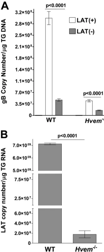
). Quantitative RT-PCR analysis of mRNA levels in
trigeminal ganglia (TG) at 30 days postinfection (p.i.), when
la-tency is well established, revealed that HVEM mRNA depended
on the presence of LAT (
Fig. 1A
) (
P
⬍
0.0001). In LAT(
⫹
)
virus-infected mice HVEM mRNA was increased over unvirus-infected mice,
while in LAT(
⫺
) virus-infected mice HVEM mRNA was
de-creased. There were no significant differences in the mRNA levels
of nectin-1, nectin-2, 3-O-sulfated heparan sulfate (3-OS-HS),
PILR
␣
, or NMHC-IIA in LAT(
⫹
) versus LAT(
⫺
) virus-infected
mice, with nectin-1, nectin-2, 3-OS-HS, and PILR
␣
levels
increas-ing relative to those in uninfected mice with both viruses while
NMHC-IIA decreased. In contrast to latent infection, LAT had no
statistically significant effect on HVEM mRNA levels during the
acute phase of infection (days 3 and 5 p.i.) although there was a
trend for increased HVEM mRNA with LAT(
⫹
) virus compared
to LAT(
⫺
) virus (
Fig. 1B
) (
P
⬎
0.05).
Immunohistochemical staining of HVEM in TG from mice
latently infected with LAT(
⫹
) and LAT(
⫺
) viruses revealed
dis-tinctive patterns of HVEM expression between LAT(
⫹
) (
Fig. 1C
,
left panels) and LAT(
⫺
) viruses (
Fig. 1C
, right panels). In LAT(
⫹
)
TG, HVEM staining localized broadly to large cells with dim
nu-clei consistent with neurons (
Fig. 1C
, 200
⫻
). In contrast, HVEM
staining in LAT(
⫺
) TG appeared more punctate and localized to
smaller cells (
Fig. 1C
, 200
⫻
). In the bottom panels of
Fig. 1C
(400
⫻
) the HVEM signal appears localized to neurons in LAT(
⫹
)
TG (bottom left panel), while this signal is considerably reduced
and/or absent in LAT(
⫺
) TG (bottom right panel). These data
suggest that LAT, or a LAT-induced cellular function, regulates
the level and pattern of HVEM expression in TG of HSV-1 latently
infected mice.
Viral latency and reactivation in HVEM-deficient mice.
The
impact of HVEM on the ability of LAT to increase the amount of
latency was investigated in HVEM-deficient (
Hvem
⫺/⫺) mice.
Replication levels of LAT(
⫹
) and LAT(
⫺
) HSV-1 strains in eyes
during the first 4 days of infection were similar to each other and
not significantly different between WT and
Hvem
⫺/⫺mice (
Fig.
2
). However, there was a trend toward decreased virus replication
in
Hvem
⫺/⫺mice, suggesting that there may be some effect of
HVEM on acute HSV-1 infection. This would be consistent with a
recent study in which in a corneal scarification model of ocular
HSV-1 infection, HVEM affected acute infection (
48
). The
rela-tive amount of latency on day 30 p.i. was determined by
quanti-tative PCR (qPCR) using primers from the gB region of the HSV-1
genome. Consistent with previous reports (
12
,
49
), there was
sig-nificantly more HSV-1 DNA in TG from WT mice latently
in-fected with LAT(
⫹
) virus than in those infected with LAT(
⫺
)
virus (
Fig. 3A
, WT) (
P
⬍
0.0001), which is characteristic of more
latency with LAT(
⫹
) than LAT(
⫺
) virus in WT mice. Strikingly,
Hvem
⫺/⫺mice infected with LAT(
⫹
) virus had significantly fewer
latent genomes than WT mice infected with LAT(
⫹
) virus (
Fig.
3A
) (
P
⬍
0.0001). In fact, the amount of latency in LAT(
⫹
)
on November 7, 2019 by guest
http://jvi.asm.org/
on November 7, 2019 by guest
http://jvi.asm.org/
infected
Hvem
⫺/⫺mice was similar to that in LAT(
⫺
)
virus-in-fected WT mice. Even less latency was detected in
Hvem
⫺/⫺mice
infected with LAT(
⫺
) virus than in WT mice infected with
LAT(
⫺
) virus (
Fig. 3A
) (
P
⬍
0.0001). Thus, HVEM appeared to
play a role in increasing the amount of latency in TG of mice
infected with both LAT(
⫹
) and LAT(
⫺
) viruses. As expected,
since LAT(
⫹
) virus produced less latency, as judged by the
num-ber of viral genomes in
Hvem
⫺/⫺mice compared to that of WT
mice, and since LAT levels during latency are related to the
amount of latency, LAT(
⫹
) latently infected
Hvem
⫺/⫺mice also
had less LAT than WT mice (
Fig. 3B
) (
P
⬍
0.0001). These results
suggest that HVEM and LAT both influence the amount of latency
that is established and/or maintained.
In contrast to the differences in the level of HVEM expression
between LAT(
⫹
) and LAT(
⫺
) viruses (
Fig. 1A
), mRNA levels of
LIGHT and BTLA were not significantly altered in WT mice
la-tently infected with LAT(
⫹
) virus versus LAT(
⫺
) dLAT2903 or
versus LAT(
⫺
) dLAT-gK
3virus (
Fig. 4A
and
B
). We have
previ-ously shown that HVEM expression is independent of BTLA or
LIGHT (
34
).
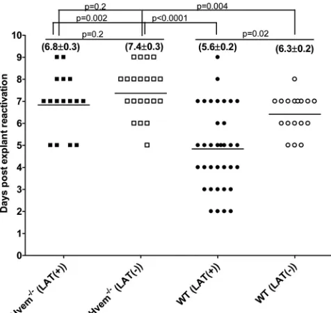
Although spontaneous reactivation from latency is too low to
study in mice, induced reactivation is routinely analyzed by
ex-planting individual TG into tissue culture medium and
monitor-FIG 1Effect of LAT on HVEM expression in TG of infected mice. (A) Effect of LAT on expression of HSV-1 receptors in latently infected mice. C57BL/6 mice were ocularly infected with HSV-1 strain McKrae [LAT(⫹)] or dLAT2903 [LAT(⫺)]; the TG from surviving mice were isolated individually on day 30 postinfection, and quantitative RT-PCR was performed using total RNA. Nectin-1, nectin-2, HVEM, PILR␣, NMHC-IIA, and 3-O-sulfated heparin sulfate (3-OS-HS) expression in naive mice was used to estimate the relative expression of each transcript in TG. GAPDH expression was used to normalize the relative expression of each transcript in TG of latently infected mice. Each bar represents the mean⫾standard error of the mean from 20 TG. (B) Expression of HVEM in TG of WT infected mice during primary infection. C57BL/6 mice were infected ocularly with McKrae [LAT(⫹)] or dLAT2903 [LAT(⫺)], and expression of HVEM in TG was determined on days 3 and 5 p.i. as described above. GAPDH expression was used to normalize the relative expression of each transcript in TG of latently infected mice. Each point represents the mean⫾standard error of the mean from 10 TG. (C) Upregulation of HVEM in TG of mice infected with LAT(⫹) virus. C57BL/6 mice were infected as described above. At 30 days p.i., TG from mice latently infected as indicated were isolated and stained with HVEM antibody as described in Materials and Methods. Nuclei are stained with DAPI (blue), and HVEM is stained in green. With LAT(⫹) virus infection, staining appears mostly at the surface of large cells (arrow), likely neurons. With LAT(⫺) virus infection, staining is mostly of small nonneuronal-like cells (arrow). Magnifications are indicated at the right of the panels.
FIG 2Virus titers in WT and HVEM⫺/⫺eyes during primary ocular infection. WT C57BL/6 and C57BL/6 HVEM⫺/⫺ mice were infected ocularly with LAT(⫹) or LAT(⫺) virus, and the amount of infectious HSV-1 in tear films was determined daily by standard plaque assays as described in Materials and Methods. For each time point, the virus titer (yaxis) represents the average of the titers from 20 eyes⫾standard error of the mean.
FIG 3Effect of LAT and HVEM on HSV-1 latency and reactivation in TG of latently infected mice. WT and HVEM⫺/⫺mice were ocularly infected with HSV-1 strain McKrae [LAT(⫹)] or dLAT2903 [LAT(⫺)] as described in the leg-end ofFig. 1. On day 30 p.i., TG were harvested from the latently infected surviving mice. Quantitative PCR and RT-PCR were performed on each individual mouse TG. In each experiment, an estimated relative copy number of gB or LAT was calculated using a standard curve generated from pGem-gB1 or pGEM-5317, re-spectively. Briefly, DNA template was serially diluted 10-fold such that 5l con-tained from 103to 1011copies of gB or LAT and then subjected to TaqMan PCR
with the same set of primers. By comparing the normalized threshold cycle of each sample to the threshold cycle of the standard, the copy number for each reaction product was determined. GAPDH expression was used to normalize the relative expression of gB DNA in the TG. Each bar represents the mean⫾standard error of the mean from 56 TG for WT mice and from 20 TG for HVEM⫺/⫺mice.
on November 7, 2019 by guest
http://jvi.asm.org/
[image:5.585.336.502.66.468.2] [image:5.585.59.268.68.258.2]ing the time required for production of infectious virus (
9
,
49–52
).
Consistent with previous studies, the time to reactivation in WT
mice was significantly shorter with LAT(
⫹
) virus than with
LAT(
⫺
) virus (5.6
⫾
0.2 days versus 6.3
⫾
0.2 days;
P
⫽
0.02)
(
Fig. 5
). The time to reactivation was significantly delayed in
Hvem
⫺/⫺mice [6.8
⫾
0.3 days with LAT(
⫹
) virus,
P
⫽
0.002;
7.4
⫾
0.3 days with LAT(
⫺
) virus,
P
⫽
0.004]. Although in
Hvem
⫺/⫺mice LAT(
⫹
) virus appeared to reactivate faster than
LAT(
⫺
) virus, this difference did not reach statistical significance
(
P
⫽
0.2). The alterations in latency and reactivation in
Hvem
⫺/⫺mice were largely independent of significant
immunopathogen-esis, as monitored by corneal scarring at day 30 p.i. or by mouse
survival (data not shown).
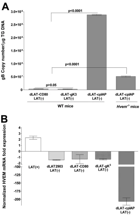
Mechanisms involved in LAT-HVEM regulation.
To define
the mechanism of LAT-HVEM regulation, we utilized
recombi-nant HSV-1 in which LAT is replaced with genes involved in cell
survival or immune modulation. Mice were infected with HSV-1
containing either the antiapoptosis gene from
Cydia pomonella
granulosis virus (dLAT-cpIAP) (
15
), the CD80 T cell activating
coreceptor (dLAT-CD80) (unpublished data), or, as a control, the
HSV-1 envelope glycoprotein gK (dLAT-gK
3) (
40
). The amount
of latency as judged by qPCR of viral DNA in mice latently
in-fected with dLAT-cpIAP was similar to that of wild-type HSV-1
(compare
Fig. 3A
and
6A
). This was expected since we previously
showed that this virus has a WT [LAT(
⫹
)] reactivation phenotype
(
15
). In contrast, dLAT-gK
3and dLAT-CD80 did not support
wild-type levels of latent virus (
Fig. 6A
) (
P
⬍
0.0001) and, like
LAT(
⫺
) virus, dLAT-gK
3and dLAT-CD80 did not upregulate
HVEM mRNA (
Fig. 6B
). In
Hvem
⫺/⫺mice dLAT-cpIAP had
re-duced latency, similar to LAT(
⫹
) virus in
Hvem
⫺/⫺mice
(com-pare
Fig. 3A
and
6A
). However, in contrast to LAT(
⫹
) virus,
dLAT-cpIAP did not upregulate HVEM mRNA levels in latently
FIG 4Effect of LAT on LIGHT and BTLA expression in TG of latently infected WT mice. WT C57BL/6 mice were ocularly infected with HSV-1 strain McKrae [LAT(⫹)], dLAT2903 [LAT(⫺)], or dLAT-gK3[LAT(⫺)]. TG were isolated
individually on day 30 postinfection, and quantitative RT-PCR was performed using total RNA. LIGHT and BTLA expression in naive WT mice was used to estimate the relative expression of each transcript in TG. GAPDH expression was used to normalize the relative expression of each transcript in TG of la-tently infected mice. Each point represents the mean⫾standard error of the mean from 8 TG.
FIG 5Effect of HVEM on kinetics of induced reactivation in explanted TG from latently infected mice. At 30 days postinfection individual TG were har-vested from HVEM⫺/⫺or WT mice. Each individual TG was incubated in tissue culture medium, and a 10-l aliquot was removed from each culture daily and used to infect RS cell monolayers for 10 days, as described in Mate-rials and Methods. The RS cells were monitored daily for the appearance of cytopathic effect for up to 5 days to determine the time of first appearance of reactivated virus from each TG. The results are plotted as the number of TG that reactivated daily. Numbers indicate the average time that the TG from each group first showed cytopathic effect⫾standard error of the mean. For each group, 20 TG from 10 mice were used.
on November 7, 2019 by guest
http://jvi.asm.org/
[image:6.585.301.539.66.291.2]infected WT mice. In fact, dLAT-cpIAP appeared to drastically
reduce HVEM mRNA (
Fig. 6B
). These results suggest that LAT
had a direct effect on HVEM mRNA levels, rather than the effects
on HVEM mRNA being the result of an increased latent viral load
in TG with LAT(
⫹
) compared to LAT(
⫺
) viruses.
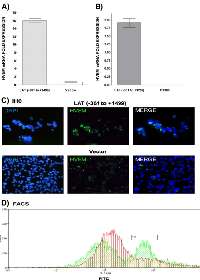
The increased HVEM mRNA levels in LAT(
⫹
) virus-infected
mice, but not those of other receptor mRNAs, prompted us to
investigate whether LAT could regulate HVEM expression in the
absence of other viral genes. HVEM mRNA levels were analyzed
by qRT-PCR in two neuroblastoma lines, C1300 and Neuro2A,
that stably express LAT (
43
,
44
). In both LAT(
⫹
) cell lines (
Fig. 7A
and
B
) HVEM mRNA expression was significantly upregulated
compared to cell lines containing the empty vector suggesting a
direct effect of LAT on HVEM gene expression. To estimate
rela-tive HVEM protein levels, the Neuro2A cells were stained with
mouse HVEM antibody. There appeared to be more
HVEM-pos-itive cells in the LAT(
⫹
) than in the LAT(
⫺
) cell line (
Fig. 7C
). In
addition, more high-intensity HVEM-positive cells were also
de-tected in the LAT(
⫹
) than in the LAT(
⫺
) cell line using flow
cytometry (
Fig. 7D
). Thus, LAT appeared to upregulate
expres-sion of HVEM in neuronal-derived C1300 and Neuro2A cells in
the absence of other viral genes.
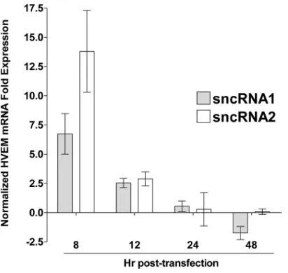
Previously, we showed that two small noncoding RNAs
(sncRNAs) (
38
) that do not appear to be miRNAs and that are
located within the region of LAT involved in the spontaneous
reactivation phenotype and the blocking of apoptosis (the first 1.5
kb of LAT) affect both viral infection and apoptosis (
45
). Neuro2A
cells were transfected with sncRNA1 or sncRNA2 as we described
previously (
45
) and harvested at 8, 12, 24, and 48 h
posttransfec-tion. HVEM expression in empty vector-transfected control cells
was used to normalize the relative expression of HVEM. Both
sncRNA1 and sncRNA2 transiently increased HVEM mRNA
ex-pression at 8 and 12 h posttransfection, with sncRNA2 having a
greater effect at 8 h than sncRNA1 (
Fig. 8
).
DISCUSSION
During HSV-1 latency, LAT is the only viral gene product
consis-tently detected in abundance in infected mice, rabbits, and
hu-mans (
1
,
3
,
5
,
6
,
10
,
53
). LAT is important for high, WT levels of
spontaneous (
9
) and induced (
10
) HSV-1 reactivation from
la-tency. The results presented here indicate that the HSV-1 LAT
gene targets HVEM in its capacity to help establish and maintain
viral latency. Our results using an HSV-1 mouse ocular infection
model indicate that LAT manipulates HVEM expression, which in
turn increases virus latency and enhances the latency-reactivation
cycle in the trigeminal ganglia. Moreover, HVEM appears
essen-tial to maintaining a normal immune signature in the TG,
suggest-ing its importance for host immunity dursuggest-ing latency. These results
indicate that LAT-HVEM forms a critical pathogen-host axis
con-tributing to viral latency.
Little is known regarding a role of HSV-1 entry receptors in
latency and reactivation and the role that LAT may play in this
process. In contrast to the other known entry routes for HSV-1
(
19–23
), HVEM mRNA levels significantly increased in a
LAT-dependent fashion in latently infected TG of normal mice. This
finding is surprising given the lesser role HVEM plays in viral
entry in mucosa, brain, and, as shown here, the ocular infection
route. The upregulation of HVEM by LAT(
⫹
) virus appeared to
be a result of LAT’s expression rather than an increase in viral load
in the TG during latency or a result of increased unapparent
spon-taneous reactivation with LAT(
⫹
) versus LAT(
⫺
) viruses. This
conclusion is based on several lines of reasoning. First, the
dLAT-cpIAP mutant virus, which establishes latency and reactivates in
the same way as LAT(
⫹
) virus (
15
), did not increase HVEM levels.
This result suggests that the upregulation of HVEM function is
unique and specific to LAT. Second, cell lines stably expressing
LAT had increased HVEM levels compared to control cell lines.
Third, in transient-transfection experiments, plasmids expressing
either of the two LAT sncRNAs (
38
,
45
) significantly upregulated
FIG 6Effect of recombinant viruses expressing foreign genes in place of LAT on latency and HVEM expression. (A) gB DNA. WT C57BL/6 and C57BL/6-HVEM⫺/⫺mice were ocularly infected with dLAT-cpIAP. As controls, some of the WT mice were similarly infected with dLAT-CD80 or dLAT-gK3. On day
30 postinfection, TG were harvested from the latently infected surviving mice, and quantitative PCR was performed on each individual mouse TG. In each experiment, an estimated relative copy number of gB was calculated using standard curves. GAPDH expression was used to normalize the relative expres-sion of gB DNA in the TG. Each point represents the mean⫾standard error of the mean from 10 TG. (B) HVEM mRNA. C57BL/6 mice were ocularly in-fected with the HSV-1 McKrae [LAT(⫹)] strain or the LAT(⫺) dLAT2903, dLAT-CD80, dLAT-gK3, or dLAT-cpIAP strain; the TG of surviving mice were
isolated individually on day 30 postinfection, and quantitative RT-PCR was performed using total RNA. HVEM expression in naive mouse TG was used to estimate the relative expression of HVEM transcript in TG of infected mice. GAPDH expression was used to normalize the relative expression of each transcript in TG of latently infected mice. Each point represents the mean⫾ standard error of the mean from 10 TG.
on November 7, 2019 by guest
http://jvi.asm.org/
[image:7.585.41.282.64.432.2]FIG 7Effect of LAT on HVEM expressionin vitro. (A and B) HVEM mRNA is upregulated in the presence of LATin vitro. C1300 (A) and Neuro2A (B) cells expressing LAT nt⫺361 to⫹3225 and⫺361 to⫹1499, respectively, were grown to confluence, and quantitative RT-PCR was performed using total RNA. HVEM expression in vector-only control cells was used to estimate the relative expression of HVEM mRNA. GAPDH expression was used to normalize the relative expression. Each bar represents the mean⫾standard error of the mean from three independent experiments. (C and D) HVEM protein is upregulated in the presence of LATin vitro. Neuro2A cells expressing LAT⫺361 to⫹1499 (top) or vector without HSV-1 LAT (bottom) were grown to confluence, stained with HVEM antibody, and subjected to immunohistochemistry (IHC) (C) or FACS (D) analyses as described in Materials and Methods. Nuclei are stained with DAPI (blue). HVEM is shown in green. FACS of Neuro2A cells expressing LAT or containing empty vector. Cells were stained and gated for HVEM, and results are shown as an overlay. Green represents LAT, and red represents an empty vector.
on November 7, 2019 by guest
http://jvi.asm.org/
[image:8.585.85.495.61.634.2]HVEM mRNA levels. Thus, LAT was able to upregulate HVEM
expression, independently of other viral factors.
To date, no LAT-encoded protein that regulates the
latency-reactivation cycle has been identified, suggesting that LAT
regu-lates the latency-reactivation cycle by exerting its effect as an RNA
molecule rather than by directing production of a protein. The
HSV-1 LAT locus includes several microRNAs, at least two of
which affect expression of a viral protein (
54
). However, these
microRNAs all map outside the first 1.5 kb of the primary 8.3-kb
LAT transcript, which is the region of LAT that we previously
demonstrated was both sufficient and required for LAT’s ability to
enhance the reactivation phenotype in mouse or rabbit models of
infection (
9
,
55
,
56
). Thus, these microRNAs are unlikely to be
involved in enhancing latency/reactivation in these animal
mod-els. In contrast, we identified two small noncoding RNAs
(sncRNAs) that are located within the first 1.5 kb of LAT (
38
,
45
).
These LAT sncRNAs do not appear to be microRNAs, based on
their sizes and their predicted structures. In this report we show
that following transient transfection, both of these sncRNAs can
independently upregulate expression of HVEM mRNA. In addition,
the RNAhybrid algorithm (
http://bibiserv.techfak.uni-bielefeld.de
/rnahybrid
) predicts interaction between the mouse HVEM
pro-moter and both of the LAT sncRNAs. The analysis suggests that
LAT sncRNA1 can interact with the HVEM promoter at position
493 in the forward direction while sncRNA2 can interact with the
HVEM promoter in the reverse direction at position 87. These
results suggest a direct impact of LAT RNA on HVEM expression.
Both LAT and HVEM directly contribute to cell survival within
their respective contexts. The LAT region plays a role in blocking
apoptosis of infected cells in rabbits (
11
) and mice (
12
) and in
human cells (
11
). The antiapoptosis activity appears to be a critical
function of LAT involved in enhancing the latency-reactivation
cycle because the LAT(
⫺
) virus can be restored to a full wild-type
reactivation phenotype by substitution of different prosurvival/
antiapoptosis genes (i.e., baculovirus inhibitor of apoptosis
tein gene [cpIAP] and FLIP [cellular FLICE-like inhibitory
pro-tein]) (
13
,
14
). HVEM activation by BTLA or LIGHT contributes
to survival of chronically stimulated effector T cells
in vivo
(
36
,
57
). Both LIGHT and BTLA induce HVEM to activate NF-
B
(RelA) transcription factors known to enhance survival of
acti-vated T cells (
34
,
58
). Furthermore, the LAT sncRNAs can
stimu-late NF-
B-dependent transcription in the presence of the RNA
sensor, RIG-I (
59
). HVEM, like its related tumor necrosis factor
receptor superfamily (TNFRSF) paralogs, utilizes TNF
receptor-associated factor 2 (TRAF2) and cellular IAPs as part of the
ubiq-uitin E3 ligases that regulate NF-
B activation pathways (
60–62
).
cpIAP, an ortholog of the cellular IAP E3 ligases (
63
), and cFLIP,
an NF-
B-regulated antiapoptosis gene (
64
), mimic the activated
HVEM signaling pathway. These results lead us to suggest that in
addition to upregulating HVEM expression, LAT also promotes
active HVEM signaling.
Our results indicate that HVEM signaling plays a significant
role in HSV-1 latency. We found that the level of latent viral
ge-nomes of LAT(
⫹
) virus in
Hvem
⫺/⫺mice compared to that of WT
mice was significantly reduced. Similarly, reactivation of latent
virus in TG explant cultures was also significantly reduced in
Hvem
⫺/⫺mice compared to levels in WT mice, demonstrating
that HVEM is a significant factor in increasing HSV-1 latency and
reactivation. However, differential replication and spread in the
eye and possibly the reactivation efficiencies may influence these
results. We found that, in contrast to increasing HVEM
expres-sion, LAT did not significantly alter LIGHT or BTLA mRNA
lev-els. This is consistent with the idea that LIGHT and BTLA
expres-sion occurs in immune cells in the microenvironment of the
latently infected cell and is therefore not affected by LAT
expres-sion in latently infected neurons.
We have previously shown that LAT functions as an immune
evasion gene (
49
,
65
), as an antiapoptosis gene (
11
), and as an
inhibitor of productive infection (
45
). All three of these LAT
func-tions would seemingly contribute to enhancing HSV-1 latency
and the HSV-1 reactivation phenotype. The results reported here
suggest that these important LAT functions contribute to LAT
increasing expression of HVEM in latently infected neurons. The
results presented here identify HVEM as an important target of
LAT that influences latency, reactivation, and survival of
gangli-on-resident T cells. We found that HVEM is upregulated by two
LAT sncRNAs and that in the absence of HVEM (i.e., in
Hvem
⫺/⫺mice), HSV-1 latency and reactivation significantly decreased.
This result suggests that increasing HVEM above a threshold level
by LAT leads to more efficient binding of HSV-1 gD to HVEM in
the latent microenvironment and therefore enhances HSV-1
la-tency and reactivation. HSV-1 targets the HVEM pathway by at
least two distinct mechanisms—at entry by direct interaction with
gD and in latency through LAT-dependent transcriptional
regu-lation—suggesting that HVEM is a critical node of selective
pres-sure in alphaherpesvirus evolution. This concept may apply to
other herpesviruses based on the observations that human
cyto-megalovirus encodes an HVEM-like ortholog (UL144) that
spe-cifically engages BTLA (
24
,
66
).
ACKNOWLEDGMENTS
S.J.A. was supported by T32 AI89553. S.L.W. was supported by NIH grant EY013191, The Discovery Eye Foundation, The Henry L. Guenther Founda-tion, and a Research to Prevent Blindness Challenge grant. C.J. was supported by a USDA grant, Agriculture and Food Research Initiative Competitive FIG 8Effect of LAT sncRNAs on HVEM expressionin vitro. Neuro2A cells
were transfected with sncRNA1 or sncRNA2, and expression of HVEM mRNA was determined as described above. HVEM expression in untransfected con-trol cells was used to normalize the relative expression of HVEM. GAPDH expression was used to normalize relative expression. Each bar represents the mean⫾standard error of the mean from three independent experiments.
on November 7, 2019 by guest
http://jvi.asm.org/
[image:9.585.60.266.64.259.2]Grants Program (09-01653), and the Nebraska Center for Virology (1P20RR15635). C.F.W. was supported by NIH grants R37AI033068 and AI048073. This study was fully supported by Public Health Service NIH grants EY14966, EY13615, EY15557, and AI093941, and by the Cedars-Sinai Medical Center to H.G.
REFERENCES
1.Wechsler SL, Nesburn AB, Watson R, Slanina S, Ghiasi H.1988. Fine mapping of the major latency-related RNA of herpes simplex virus type 1 in humans. J. Gen. Virol.69:3101–3106.http://dx.doi.org/10.1099/0022 -1317-69-12-3101.
2.Cook ML, Bastone VB, Stevens JG.1974. Evidence that neurons harbor latent herpes simplex virus. Infect. Immun.9:946 –951.
3.Stevens JG, Wagner EK, Devi-Rao GB, Cook ML, Feldman LT.1987. RNA complementary to a herpesvirus alpha gene mRNA is prominent in latently infected neurons. Science 235:1056 –1059.http://dx.doi.org/10 .1126/science.2434993.
4.Rock DL, Fraser NW. 1983. Detection of HSV-1 genome in central nervous system of latently infected mice. Nature302:523–525.http://dx .doi.org/10.1038/302523a0.
5.Rock DL, Nesburn AB, Ghiasi H, Ong J, Lewis TL, Lokensgard JR, Wechsler SL.1987. Detection of latency-related viral RNAs in trigeminal ganglia of rabbits latently infected with herpes simplex virus type 1. J. Virol.61:3820 –3826.
6.Wechsler SL, Nesburn AB, Watson R, Slanina SM, Ghiasi H.1988. Fine mapping of the latency-related gene of herpes simplex virus type 1: alter-native splicing produces distinct latency-related RNAs containing open reading frames. J. Virol.62:4051– 4058.
7.Jordan MC, Jordan GW, Stevens JG, Miller G.1984. Latent herpesvi-ruses of humans. Ann. Intern. Med.100:866 – 880.http://dx.doi.org/10 .7326/0003-4819-100-6-866.
8.Wagner EK, Flanagan WM, Devi-Rao G, Zhang YF, Hill JM, Anderson KP, Stevens JG.1988. The herpes simplex virus latency-associated tran-script is spliced during the latent phase of infection. J. Virol.62:4577– 4585.
9.Perng GC, Dunkel EC, Geary PA, Slanina SM, Ghiasi H, Kaiwar R, Nesburn AB, Wechsler SL.1994. The latency-associated transcript gene of herpes simplex virus type 1 (HSV-1) is required for efficient in vivo spontaneous reactivation of HSV-1 from latency. J. Virol.68:8045– 8055. 10. Hill JM, Sedarati F, Javier RT, Wagner EK, Stevens JG.1990. Herpes simplex virus latent phase transcription facilitates in vivo reactivation. Virology174:117–125.http://dx.doi.org/10.1016/0042-6822(90)90060-5. 11. Perng GC, Jones C, Ciacci-Zanella J, Stone M, Henderson G, Yukht A, Slanina SM, Hofman FM, Ghiasi H, Nesburn AB, Wechsler SL.2000. Virus-induced neuronal apoptosis blocked by the herpes simplex virus latency-associated transcript. Science287:1500 –1503. http://dx.doi.org /10.1126/science.287.5457.1500.
12.Branco FJ, Fraser NW. 2005. Herpes simplex virus type 1 latency-associated transcript expression protects trigeminal ganglion neurons from apoptosis. J. Virol.79:9019 –9025.http://dx.doi.org/10.1128/JVI.79 .14.9019-9025.2005.
13. Jin L, Perng GC, Carpenter D, Mott KR, Osorio N, Naito J, Brick DJ, Jones C, Wechsler SL.2007. Reactivation phenotype in rabbits of a herpes simplex virus type 1 mutant containing an unrelated antiapoptosis gene in place of latency-associated transcript. J. Neurovirol.13:78 – 84.http://dx .doi.org/10.1080/13550280601164333.
14. Jin L, Carpenter D, Moerdyk-Schauwecker M, Vanarsdall AL, Osorio N, Hsiang C, Jones C, Wechsler SL.2008. Cellular FLIP can substitute for the herpes simplex virus type 1 latency-associated transcript gene to sup-port a wild-type virus reactivation phenotype in mice. J. Neurovirol.14: 389 – 400.http://dx.doi.org/10.1080/13550280802216510.
15. Jin L, Perng GC, Mott KR, Osorio N, Naito J, Brick DJ, Carpenter D, Jones C, Wechsler SL.2005. A herpes simplex virus type 1 mutant expressing a baculovirus inhibitor of apoptosis gene in place of latency-associated tran-script has a wild-type reactivation phenotype in the mouse. J. Virol.79: 12286 –12295.http://dx.doi.org/10.1128/JVI.79.19.12286-12295.2005. 16. Willey DE, Trousdale MD, Nesburn AB.1984. Reactivation of murine
latent HSV infection by epinephrine iontophoresis. Invest. Ophthalmol. Vis. Sci.25:945–950.
17. Feldman LT, Ellison AR, Voytek CC, Yang L, Krause P, Margolis TP. 2002. Spontaneous molecular reactivation of herpes simplex virus type 1
latency in mice. Proc. Natl. Acad. Sci. U. S. A.99:978 –983.http://dx.doi .org/10.1073/pnas.022301899.
18. Kramer MF, Coen DM.1995. Quantification of transcripts from the ICP4 and thymidine kinase genes in mouse ganglia latently infected with herpes simplex virus. J. Virol.69:1389 –1399.
19. Satoh T, Arii J, Suenaga T, Wang J, Kogure A, Uehori J, Arase N, Shiratori I, Tanaka S, Kawaguchi Y, Spear PG, Lanier LL, Arase H. 2008. PILR␣is a herpes simplex virus-1 entry coreceptor that associates with glycoprotein B. Cell132:935–944.http://dx.doi.org/10.1016/j.cell .2008.01.043.
20. Spear PG, Eisenberg RJ, Cohen GH.2000. Three classes of cell surface receptors for alphaherpesvirus entry. Virology275:1– 8.http://dx.doi.org /10.1006/viro.2000.0529.
21. Taylor JM, Lin E, Susmarski N, Yoon M, Zago A, Ware CF, Pfeffer K, Miyoshi J, Takai Y, Spear PG. 2007. Alternative entry receptors for herpes simplex virus and their roles in disease. Cell Host Microbe2:19 –28. http://dx.doi.org/10.1016/j.chom.2007.06.005.
22. Arii J, Goto H, Suenaga T, Oyama M, Kozuka-Hata H, Imai T, Minowa A, Akashi H, Arase H, Kawaoka Y, Kawaguchi Y.2010. Non-muscle myosin IIA is a functional entry receptor for herpes simplex virus-1. Na-ture467:859 – 862.http://dx.doi.org/10.1038/nature09420.
23. Suenaga T, Satoh T, Somboonthum P, Kawaguchi Y, Mori Y, Arase H. 2010. Myelin-associated glycoprotein mediates membrane fusion and en-try of neurotropic herpesviruses. Proc. Natl. Acad. Sci. U. S. A.107:866 – 871.http://dx.doi.org/10.1073/pnas.0913351107.
24. Cheung TC, Humphreys IR, Potter KG, Norris PS, Shumway HM, Tran BR, Patterson G, Jean-Jacques R, Yoon M, Spear PG, Murphy KM, Lurain NS, Benedict CA, Ware CF.2005. Evolutionarily divergent her-pesviruses modulate T cell activation by targeting the herpesvirus entry mediator cosignaling pathway. Proc. Natl. Acad. Sci. U. S. A.102:13218 – 13223.http://dx.doi.org/10.1073/pnas.0506172102.
25. Yoon M, Zago A, Shukla D, Spear PG.2003. Mutations in the N termini of herpes simplex virus type 1 and 2 gDs alter functional interactions with the entry/fusion receptors HVEM, nectin-2, and 3-O-sulfated heparan sulfate but not with nectin-1. J. Virol.77:9221–9231.http://dx.doi.org/10 .1128/JVI.77.17.9221-9231.2003.
26. Montgomery RI, Warner MS, Lum BJ, Spear PG.1996. Herpes simplex virus-1 entry into cells mediated by a novel member of the TNF/NGF receptor family. Cell87:427– 436.http://dx.doi.org/10.1016/S0092-8674 (00)81363-X.
27. Shukla D, Liu J, Blaiklock P, Shworak NW, Bai X, Esko JD, Cohen GH, Eisenberg RJ, Rosenberg RD, Spear PG.1999. A novel role for 3-O-sulfated heparan sulfate in herpes simplex virus 1 entry. Cell99:13–22. http://dx.doi.org/10.1016/S0092-8674(00)80058-6.
28. O’Donnell CD, Kovacs M, Akhtar J, Valyi-Nagy T, Shukla D.2010. Expanding the role of 3-O sulfated heparan sulfate in herpes simplex virus type-1 entry. Virology 397:389 –398. http://dx.doi.org/10.1016/j.virol .2009.11.011.
29. Connolly SA, Landsburg DJ, Carfi A, Wiley DC, Cohen GH, Eisenberg RJ.2003. Structure-based mutagenesis of herpes simplex virus glycopro-tein D defines three critical regions at the gD-HveA/HVEM binding inter-face. J. Virol. 77:8127– 8140. http://dx.doi.org/10.1128/JVI.77.14.8127 -8140.2003.
30. Ware CF.2011. The TNF receptor super family in immune regulation. Immunol. Rev. 244:5– 8. http://dx.doi.org/10.1111/j.1600-065X.2011 .01065.x.
31. Murphy KM, Nelson CA, Sedy JR.2006. Balancing co-stimulation and inhibition with BTLA and HVEM. Nat. Rev. Immunol.6:671– 681.http: //dx.doi.org/10.1038/nri1917.
32. Sedy JR, Gavrieli M, Potter KG, Hurchla MA, Lindsley RC, Hildner K, Scheu S, Pfeffer K, Ware CF, Murphy TL, Murphy KM.2005. B and T lymphocyte attenuator regulates T cell activation through interaction with herpesvirus entry mediator. Nat. Immunol.6:90 –98.http://dx.doi.org/10 .1038/ni1144.
33. Cai G, Anumanthan A, Brown JA, Greenfield EA, Zhu B, Freeman GJ. 2008. CD160 inhibits activation of human CD4⫹T cells through interac-tion with herpesvirus entry mediator. Nat. Immunol.9:176 –185.http: //dx.doi.org/10.1038/ni1554.
34. Cheung TC, Oborne LM, Steinberg MW, Macauley MG, Fukuyama S, Sanjo H, D’Souza C, Norris PS, Pfeffer K, Murphy KM, Kronenberg M, Spear PG, Ware CF.2009. T cell intrinsic heterodimeric complexes be-tween HVEM and BTLA determine receptivity to the surrounding
on November 7, 2019 by guest
http://jvi.asm.org/
croenvironment. J. Immunol.183:7286 –7296.http://dx.doi.org/10.4049 /jimmunol.0902490.
35. Steinberg MW, Turovskaya O, Shaikh RB, Kim G, McCole DF, Pfeffer K, Murphy KM, Ware CF, Kronenberg M. 2008. A crucial role for HVEM and BTLA in preventing intestinal inflammation. J. Exp. Med. 205:1463–1476.http://dx.doi.org/10.1084/jem.20071160.
36. Soroosh P, Doherty TA, So T, Mehta AK, Khorram N, Norris PS, Scheu S, Pfeffer K, Ware C, Croft M.2011. Herpesvirus entry mediator (TNFRSF14) regulates the persistence of T helper memory cell populations. J. Exp. Med. 208:797– 809.http://dx.doi.org/10.1084/jem.20101562.
37. Compaan DM, Gonzalez LC, Tom I, Loyet KM, Eaton D, Hymowitz SG.2005. Attenuating lymphocyte activity: the crystal structure of the BTLA-HVEM complex. J. Biol. Chem.280:39553–39561. http://dx.doi .org/10.1074/jbc.M507629200.
38. Peng W, Vitvitskaia O, Carpenter D, Wechsler SL, Jones C. 2008. Identification of two small RNAs within the first 1.5-kb of the herpes simplex virus type 1-encoded latency-associated transcript. J. Neurovirol. 14:41–52.http://dx.doi.org/10.1080/13550280701793957.
39. Osorio Y, Ghiasi H.2003. Comparison of adjuvant efficacy of herpes simplex virus type 1 recombinant viruses expressing TH1 and TH2 cyto-kine genes. J. Virol.77:5774 –5783.http://dx.doi.org/10.1128/JVI.77.10 .5774-5783.2003.
40. Mott KR, Perng GC, Osorio Y, Kousoulas KG, Ghiasi H. 2007. A recombinant herpes simplex virus type 1 expressing two additional copies of gK is more pathogenic than wild-type virus in two different strains of mice. J. Virol.81:12962–12972. Epub 12007 Sep 12926.http://dx.doi.org /10.1128/JVI.01442-07.
41. Ghiasi H, Bahri S, Nesburn AB, Wechsler SL.1995. Protection against herpes simplex virus-induced eye disease after vaccination with seven in-dividually expressed herpes simplex virus 1 glycoproteins. Invest. Oph-thalmol. Vis. Sci.36:1352–1360.
42. Mott KR, Ghiasi H.2008. Role of dendritic cells in enhancement of herpes simplex virus type 1 latency and reactivation in vaccinated mice. Clin. Vaccine Immunol. 15:1859 –1867. http://dx.doi.org/10.1128/CVI .00318-08.
43. Carpenter D, Hsiang C, Brown DJ, Jin L, Osorio N, BenMohamed L, Jones C, Wechsler SL.2007. Stable cell lines expressing high levels of the herpes simplex virus type 1 LAT are refractory to caspase 3 activation and DNA laddering following cold shock induced apoptosis. Virology369:12– 18.http://dx.doi.org/10.1016/j.virol.2007.07.023.
44. Jiang X, Chentoufi AA, Hsiang C, Carpenter D, Osorio N, BenMohamed L, Fraser NW, Jones C, Wechsler SL.2011. The herpes simplex virus type 1 latency-associated transcript can protect neuron-derived C1300 and Neuro2A cells from granzyme B-induced apoptosis and CD8 T-cell killing. J. Virol.85:2325–2332.http://dx.doi.org/10.1128/JVI.01791-10.
45. Shen W, Sa e Silva M, Jaber T, Vitvitskaia O, Li S, Henderson G, Jones C.2009. Two small RNAs encoded within the first 1.5 kilobases of the herpes simplex virus type 1 latency-associated transcript can inhibit pro-ductive infection and cooperate to inhibit apoptosis. J. Virol.83:9131– 9139.http://dx.doi.org/10.1128/JVI.00871-09.
46. del Rio ML, Jones ND, Buhler L, Norris P, Shintani Y, Ware CF, Rodriguez-Barbosa JI.2012. Selective blockade of herpesvirus entry me-diator-B and T lymphocyte attenuator pathway ameliorates acute graft-versus-host reaction. J. Immunol.188:4885– 4896.http://dx.doi.org/10 .4049/jimmunol.1103698.
47. Mott KR, Osorio Y, Brown DJ, Morishige N, Wahlert A, Jester JV, Ghiasi H.2007. The corneas of naive mice contain both CD4⫹and CD8⫹T cells. Mol. Vis.13:1802–1812.http://www.molvis.org/molvis/v13/a201/. 48. Karaba AH, Kopp SJ, Longnecker R.2012. Herpesvirus entry mediator is
a serotype specific determinant of pathogenesis in ocular herpes. Proc. Natl. Acad. Sci. U. S. A.109:20649 –20654.http://dx.doi.org/10.1073/pnas .1216967109.
49. Allen SJ, Hamrah P, Gate DM, Mott KR, Mantopoulos D, Zheng L, Town T, Jones C, von Andrian UH, Freeman GJ, Sharpe AH, Benmo-hamed L, Ahmed R, Wechsler SL, Ghiasi H.2011. The role of LAT in increased CD8⫹T cell exhaustion in trigeminal ganglia of mice latently infected with herpes simplex virus type 1. J. Virol.85:4184 – 4197.http: //dx.doi.org/10.1128/JVI.02290-10.
50. Steiner I, Spivack JG, Deshmane SL, Ace CI, Preston CM, Fraser NW. 1990. A herpes simplex virus type 1 mutant containing a nontransinduc-ing Vmw65 protein establishes latent infection in vivo in the absence of viral replication and reactivates efficiently from explanted trigeminal gan-glia. J. Virol.64:1630 –1638.
51. Leib DA, Bogard CL, Kosz-Vnenchak M, Hicks KA, Coen DM, Knipe DM, Schaffer PA.1989. A deletion mutant of the latency-associated tran-script of herpes simplex virus type 1 reactivates from the latent state with reduced frequency. J. Virol.63:2893–2900.
52. Sawtell NM, Thompson RL.1992. Rapid in vivo reactivation of herpes simplex virus in latently infected murine ganglionic neurons after tran-sient hyperthermia. J. Virol.66:2150 –2156.
53. Stevens JG.1989. Human herpesviruses: a consideration of the latent state. Microbiol. Rev.53:318 –332.
54. Umbach JL, Kramer MF, Jurak I, Karnowski HW, Coen DM, Cullen BR.2008. MicroRNAs expressed by herpes simplex virus 1 during latent infection regulate viral mRNAs. Nature454:780 –783.http://dx.doi.org /10.1038/nature07103.
55. Perng GC, Esmaili D, Slanina SM, Yukht A, Ghiasi H, Osorio N, Mott KR, Maguen B, Jin L, Nesburn AB, Wechsler SL.2001. Three herpes simplex virus type 1 latency-associated transcript mutants with distinct and asymmet-ric effects on virulence in mice compared with rabbits. J. Virol.75:9018 –9028. http://dx.doi.org/10.1128/JVI.75.19.9018-9028.2001.
56. Perng GC, Ghiasi H, Slanina SM, Nesburn AB, Wechsler SL.1996. The spontaneous reactivation function of the herpes simplex virus type 1 LAT gene resides completely within the first 1.5 kilobases of the 8.3-kilobase primary transcript. J. Virol.70:976 –984.
57. Hurchla MA, Sedy JR, Murphy KM.2007. Unexpected role of B and T lymphocyte attenuator in sustaining cell survival during chronic allostimulation. J. Immunol.178:6073– 6082.http://www.jimmunol .org/content/178/10/6073.
58. Cheung TC, Steinberg MW, Oborne LM, Macauley MG, Fukuyama S, Sanjo H, D’Souza C, Norris PS, Pfeffer K, Murphy KM, Kronenberg M, Spear PG, Ware CF.2009. Unconventional ligand activation of herpes-virus entry mediator signals cell survival. Proc. Natl. Acad. Sci. U. S. A. 106:6244 – 6249.http://dx.doi.org/10.1073/pnas.0902115106.
59. da Silva LF, Jones C.2013. Small non-coding RNAs encoded within the herpes simplex virus type 1 latency associated transcript (LAT) cooperate with the retinoic acid inducible gene I (RIG-I) to induce beta-interferon promoter activity and promote cell survival. Virus Res.175:101–109.http: //dx.doi.org/10.1016/j.virusres.2013.04.005.
60. Beug ST, Cheung HH, LaCasse EC, Korneluk RG.2012. Modulation of immune signalling by inhibitors of apoptosis. Trends Immunol.33:535– 545.http://dx.doi.org/10.1016/j.it.2012.06.004.
61. Kenneth NS, Duckett CS.2012. IAP proteins: regulators of cell migration and development. Curr. Opin. Cell biol.24:871– 875.http://dx.doi.org/10 .1016/j.ceb.2012.11.004.
62. Varfolomeev E, Goncharov T, Maecker H, Zobel K, Komuves LG, De-shayes K, Vucic D.2012. Cellular inhibitors of apoptosis are global regulators of NF-B and MAPK activation by members of the TNF family of receptors. Sci. Signal.5:ra22.http://dx.doi.org/10.1126/scisignal.2001878.
63. O’Riordan MX, Bauler LD, Scott FL, Duckett CS.2008. Inhibitor of apoptosis proteins in eukaryotic evolution and development: a model of thematic conservation. Dev. Cell15:497–508.http://dx.doi.org/10.1016/j .devcel.2008.09.012.
64. Budd RC, Yeh WC, Tschopp J.2006. cFLIP regulation of lymphocyte activation and development. Nat. Rev. Immunol.6:196 –204.http://dx .doi.org/10.1038/nri1787.
65. Chentoufi AA, Kritzer E, Tran MV, Dasgupta G, Lim CH, Yu DC, Afifi RE, Jiang X, Carpenter D, Osorio N, Hsiang C, Nesburn AB, Wechsler SL, BenMohamed L.2011. The herpes simplex virus 1 latency-associated transcript promotes functional exhaustion of virus-specific CD8⫹T cells in latently infected trigeminal ganglia: a novel immune evasion mecha-nism. J. Virol.85:9127–9138.http://dx.doi.org/10.1128/JVI.00587-11. 66. Benedict CA, Butrovich KD, Lurain NS, Corbeil J, Rooney I, Schneider
P, Tschopp J, Ware CF.1999. Cutting edge: a novel viral TNF receptor superfamily member in virulent strains of human cytomegalovirus. J. Im-munol.162:6967– 6970.
on November 7, 2019 by guest
http://jvi.asm.org/