Acknowledgement
I was able to carry out and complete this project on time, only with the help, cooperation and goodwill of many people, to whom I will be forever indebted.
First and foremost, I would like to thank GOD for giving me the strength, knowledge, ability and oppourtunity to undertake this study and to complete it satisfactorily. I thank him for blessing me much more than I deserve.
I express my gratitude to our Chairman, Dr.Velayudhan Nair, for his untiring efforts in achieving the enviable standards in academics and patient care in our institution.
I wish to express my heartfelt thanks to our Director, Dr.Rema V. Nair, for her unrelenting support and encouragement without which this study would not have been completed. Her dedication and sincerity towards the welfare of the institution is admirable.
It is my privilege to record my deepest sense of gratitude to Dr.S.Saraswathy, Professor, Department of Obstetrics and Gynaecology, for her guidance in enabling me to undertake this study. Her profound knowledge and understanding of the subject has been a big source of inspiration.
I extend my sincere thanks to Dr.Sreelakshmi Ajay, Assistant Professor, Department of Obstetrics and Gynecology, Sree Mookambika Institute of Medical Science, for being a pillar of support.
I am grateful to Dr.Usha Sadasivan, Professor, Department of OBG,
Dr.Jesu Thankam, Professor, Department of OBG for their words of advice and infinite goodwill.
I am extremely thankful to Dr.Shwetha B.R., for her selfless help and timely tips which have been a guiding light throughout the journey.
I gratefully acknowledge the patients who co-operated to submit themselves for study, without whose co-operation, this work would not have been completed.
I would also like to thank my senior and junior post graduates and my co-pg for all the valuable advice and immense cooperation.
I render gratitude to My parents, Dr.P.Senthil kumar and Dr. P.N.Pushpalatha who have made invaluable sacrifices, and have inspired,
encouraged and blessed me to succeed in all my efforts. I wish to thank them for their everlasting love and all time support.
I would also like to thank my husband Dr.T.Sriram for his valuable help, suggestions and encouragement during this study.
I offer my regards to all of those who supported me during the completion of this thesis.
Urkund Analysis Result
Analysed Document: Plagerism 3-10.pdf (D30973629)
Submitted: 10/3/2017 10:56:00 AM
Submitted By: aiswarya0000@gmail.com
Significance: 2 %
Sources included in the report:
amandeep Antiplagiarism 159.docx (D30563466) PLAGIARISM FILE AMAN.docx (D30614454) THESIS 1.docx (D30657802)
Binder1.pdf (D17562850)
Instances where selected sources appear:
6
CONTENTS
Page No.
1 INTRODUCTION 1-2
2 SCIENTIFIC JUSTIFICATION OF THE STUDY 3
3 AIMS AND OBJECTIVES 4
4 REVIEW OF LITERATURE 5-47
5 MATERIAL AND METHODS 48-50
6 RESULTS 51-67
7 DISCUSSION 68-71
8 CONCLUSION 72
9 SUMMARY 73-75
10 REFERENCES i-ix
LIST OF TABLES
[image:10.612.94.528.96.664.2]Sl.No CONTENTS Page No
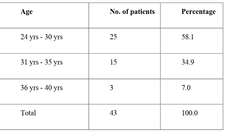
Table-1 Distribution of patients according to age 51 Table-2 Distribution of number of patients according to Socio
economic status
53
Table-3 Distribution of number of patients according to their years of marriage
54
Table-4 Distribution of number of patients according to Menstrual cycle
55
Table-5 Distribution of number of patients according to Dysmenorrhoea
56
Table-6 Distribution of number of patients according to foul smelling Vaginal discharge
57
Table-7 Distributionof number of patients according to the Treatment for Infertility
58
Table-8 Distribution of number of patients according to co- morbidities
60
Table-9 Distribution of number of patients according to Family H/O of Infertility
62
Table-10 Distribution of number of patients according to BMI 63 Table-11 Distribution of number of patients according to Pelvic
Examination
64
Table-12 Distribution of number of patients according to USG 65 Table-13 Distribution of number of patients according to
Hysteroscopy
67
LIST OF FIGURES
[image:11.612.91.525.113.713.2]Sl.No CONTENTS Page No.
Figure - 1 Anatomy of ovary 8
Figure - 2 Histology of ovary 12
Figure - 3 Histology of Graffian follicle 18
Figure - 4 histology of Corpus luteum 18
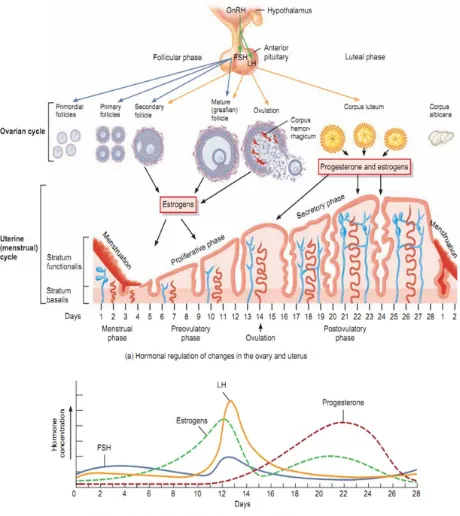
Figure - 5 Physiology of menstruation 22
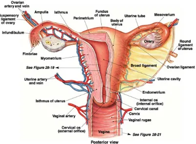
Figure - 6 Female reproductive system 26
Figure - 7 Distribution of percentage of patients according to age 52
Figure - 8 Distribution of percentage of patients according to socio economic status
53
Figure- 9 Distribution of percentage of patients according to their years of marriage
54
Figure- 10 Distribution of percentage of patients according to Menstrual cycle
55
Figure- 11 Distribution of percentage of patients according to Dysmenorrhoea
56
Figure- 12 Distribution of percentage of patients according to foul smelling Vaginal discharge
57
Figure- 13 Distribution of percentage of patients according to the Treatment for Infertility
59
Figure- 14 Distribution of percentageof patients according to co- morbidities
61
Figure-15 Distribution of percentage of patients according to Family H/O of Infertility
Figure- 16 Distribution of percentage of patients according to BMI
63
Figure- 17 Distribution of percentage of patients according to Pelvic Examination
64
Figure- 18 Distribution of percentage of patients according to USG
66
Figure- 19 Distribution of number of percentage according to Hysteroscopy
[image:12.612.89.528.74.293.2]1.INTRODUCTION:
Infertility is the inability of a couple to achieve pregnancy over an average
period of one year (in a woman under 35 years of age) or 6 months (in a woman
above 35 years of age) despite adequate, regular (3-4 times per
week),unprotected sexual intercourse . Infertility may also be referred to as the
inability to carry a pregnancy to the delivery of a live baby. Infertility can be
due to the woman, the man, or both; primary or secondary. In primary
infertility, the couples have never been able to conceive; while in secondary
infertility there is difficulty in conceiving after having conceived (either carried
the pregnancy to term or had a miscarriage).(1,3)
The World Health Organization (WHO) has defined infertility as a failure to
conceive over 12 months of exposure (which is a good practical guide to
management), and leaves a longer term residual incidence of infertility of 10–
15%.However, the chance to conceive is reduced almost twofold after the age
of 35 years.1) Epidemiological data suggest that approximately 80 million
people worldwide are infertile. WHO indicates the highest incidence in some
regions of Central Africa where the infertility rate may reach 50%, compared to
20% in the Eastern Mediterranean region, and 11% in the developed world. The
WHO estimates the overall prevalence of primary infertility in India to be
between 3.9 and 16.8 %.(1,2,6)
Infertility has a direct impact on both psychological well-being and social
status of women all over the world. A WHO task force revealed that tubal factor
demonstrable causes in 40% of cases.4A similar distribution was found in Asia,
Latin America, and the Middle East.In Africa, most of the women were infertile
due to tubal factor.(4,5,7) The risk factors for infertility can be classified into:
genital, endocrinal, developmental and general factors. Pelvic inflammatory
disease (PID) due to sexually transmitted diseases, unsafe abortion, or puerperal
infection is the main cause of tubal infertility mainly caused by chlamydial
infection.6Polycystic ovarian syndrome (PCOS) is the most commonest cause of
2. HYPOTHESIS AND JUSTIFICATION
According to review of literature, 80 million people worldwide are infertile
(10-15%) and about two-third of infertility cases are caused exclusively by female
factors.10 This study helps to identify and quantify the causes for female
infertility and to calculate the risk percentages, among the patients presenting
3.AIMS AND OBJECTIVES
1. To estimate the prevalence of causes of female infertility.
4.REVIEW OF LITERATURE
Anatomy, Physiology and Reproductive Endocrinology in the Female
Ovarian cyclicity upon which fertility depends is controlled by an elaborate
neuroendocrine feedback system that involves the hypothalamus, pituitaries and
ovaries.
The hypothalamus is a part of brain which is a small neural structure located at
the base of the brain above the optic chiasma and below the third ventricle.
Hypothalamus is divided into three zones- the periventricular, medial and lateral
zones, each of which is further subdivided. The hypothalamus interacts with
many regions of the brain through various pathways that form feedback loops.11
The major hormones secreted by hypothalamus are the pituitary releasing
factors :
• Gonadotrophin-releasing hormone(GnRH), that helps in the secretion of
follicle stimulating hormone(FSH) and luteinizing hormone(LH). Also called
luteinizing hormone-releasing hormone(LHRH), GnRH is a decapeptide
produced by neurons in the arcuate nucleus of the hypothalamus. GnRH is a
unique releasing hormone as it must be produced and secreted in a pulsatile
fashion maintain its function, and its pulsatile release influences the release of
both gonadotrophins. This pulsatility is necessary as GnRH has very short
half-life(2-4 minutes) as a result of rapid proteolytic cleavage. The frequency and
increase in the late follicular phase, and decrease in the luteal phase. These
variations in amplitude and frequency are directly responsible for varying
proportions of gonadotrophin secretion from the pituitary.12
• Corticotropin-releasing factor(CRF), that controls the release of
adrenocorticotropic hormone(ACTH).
• Growth hormone releasing hormone-releasing hormone(GHRH), which
controls the release of growth hormone(GH).
• Thyrotropin-releasing hormone(TRH), which regulates the release of
thyroid stimulating hormone(TSH).
The pituitary is divided into three lobes: anterior, intermediate and posterior.
Embryologically, the larger anterior pituitary(adenohypophysis, pars distalis) is
derived from Rathke’s pouch, an outpouching of the oral cavity. It is quite
different from the smaller posterior pituitary(neurohypophysis, infundibular
bulb), which is derived from a continuous extension of magnocellular neuronal
axons, which terminate in the infundibular bulb. The adult pituitary gland
weighs 0.5gm, measures 10 x 13 x 6mm and occupies a cavity in the sphenoid
bone, the sellaturcica.
The anterior pituitary is responsible for the production of FSH, LH, TSH,
ACTH, GH and prolactin. The gonadotrophin; FSH and LH, are produced by
the gonadotrophs, which are situated diffusely throughout the adenohypophysis
and are responsible for ovarian steroid secretion and follicle stimulation.
identical α subunits and differing β subunits. The synthesis of the β subunits is
the rate limiting step in gonadotrophin biosynthesis.11 Prolactin is a 198 amino
acid polypeptide secreted by the lactotrophs, and is the primary factor
responsible for the synthesis and secretion of breast milk
Endogenous opioids, three structurally similar families of naturally occurring
substances produced by the CNS, are the endorphins, encephalins and
dysmorphins. They play a vital role in the regulation of hypothalamic-pituitary
function, and inhibitsGnRH release.
Hypothalamic-pituitary-ovarian feedback mechanisms may be divided into four
stages:
• FSH and LH secretion during the early follicular phase in response to
low serum oestradiol(E2) and progesterone levels, after the preceding corpus
luteum cease to function (negative feedback).
• Reduced FSH secretion from midfollicular to late follicular phase in
response to rising E2 production in maturing follicles (negative feedback).
• Midcycle LH surge initiating ovulation and corpus luteum function, in
response to preovulatory E2 (positive feedback).
• Decreased FSH and LH secretion during the luteal phase in response to
Ovary
The ovaries are are unique organs in that their primary function is
gametogenesis, and all their endocrine activities (namely sex hormone
production) subserve this function. The ovarian cycle is designed to ensure that
mature female gametes (oocytes) are produced in good condition in an
appropriate number at ovulation so that they are available for fertilization by
[image:20.612.98.548.320.709.2]sperm, thus ensuring survival of the species.
Anatomy
The mature ovaries are paired organs, 4 x 3 x 1cm in size, situated in the
abdominal cavity weighing approximately 8 grams each. The ovaries are
suspended between the lateral pelvic wall and the uterus, laterally by the
infundibulopelvic ligament and the medially by uteroovarian ligament. Inferior
attachment of the hilar surface is to the broad ligament by the mesovarium and
through this run blood vessels and nerves. Each ovary consists of a large outer
cortex containing germ cells and small central medulla composed mainly of
stromal cells. A single layer of cuboidal epithelium covers each ovary, which is
continuous with the mesovarium. The ovary receives its blood supply from the
ovarian artery.
During each menstrual cycle, the ovaries produce a single dominant follicle
which is responsible for ovulation. After ovulation the dominant follicle
becomes corpus luteum, and secretes large amounts of progesterone during the
secretary phase of the menstrual cycle. Oestradiol and progesterone acts on the
endometrium of the uterus and prepare it for the implantation of embryo.14
Embryonic development:
At approximately 5 weeks of fetal life, primordial germ cells usually migrates
from the yolk sac to the genital ridges. There they undergo rapid mitotic
division and give rise to oogonia, which continue to divide and reach a
maximum of 6 to 7 million by the 20th week of fetal life. During the later half
that at birth, only one to two million follicles are present. This number further
decrease by 80 to 90% so that at puberty, only 300,000 follicles remain, of
which only 300 to 400 are destined to ovulate. The developing oogonia enter
meiotic prophase 1 at about 8 weeks of life, when they are known as primary
oocytes. Only the group of oocytes which enter meiosis will survive the wave
of atresia that sweeps the fetal ovary; these oocytes remain arrested in leptotene
stage of prophase till ovulation, when meiosis resumes.15
Follicular development:
The dynamic process of folliculogenesis happens mainly within the ovarian
cortex and it consists of four major developmental events:
i. recruitment of primordial follicle;
ii. development of preantral follicle;
iii. selection and growth of the preovulatory follicle;
iv . ovulation or death by follicular atresia.
Folliculogenesis is divided into 2 phases, the preantral
(gonadotrophin-independent ) and antral phase (gonadotrophin-dependent ). The preantral
phase occurs when the oocyte grows and differentiates which is controlled by
autocrine/paracrine mechanisms through locally produced growth factors . In
antral phase (graafian phase ) there will be increase in the size of the follicle
itself and is regulated by Follicular stimulating hormone and Leutinising
Chronology:
In each menstrual cycle, from the primordial follicle which has undergone
recruitment almost 1 year back develops in to a dominant follicle and ovulates.
The class 1 phase or preantral phase is divided into 3 major stages: the
primordial follicle, primary follicle, and secondary follicle stages. About 290
days or about 10 regular menstrual cycles required for the development of a
primordial to a full grown secondary follicle.16
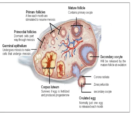
Primordial follicle:
Primordial follicles or oocytes are the essential reproductive cells of the ovary
as they, cause all menstrual cycles by giveing rise to all the dominant follicles.
The primordial follicle to primary follicle transition is otherwise known as
recruitment occurs when the arrested promodial follicle enters in to the group of
growing follicles. Histologically a primordial follicle consists of a small
primary oocyte (approximately 25µm in diameter) which has gort arrested in
the late diplotene stage of meiosis. It also contains a single layer of flattened or
squamous granulosa cells and a basal laminal layer. As all oocytes are formed
in intrauterine life, they are present in ovaries at birth. Hence the number of
eggs or primordial follicles in a women’s ovaries constitutes her ovarian
Figure 2 : Histology of ovary
In the fetus some primordial follicles gets recruited and soon after their
formation it grows. Primordial follicles gets recruited at a regular rate during
first 3 decades of a women’s life. On the other hand, when the Ovatian Reserve
reaches a significant number of around 25000 at about 40 years of age, the
propotion of loss of primordial follicles doubles. A decrease in fecundity
accompanies this increased loss of OR. The primary to secondary transition is
highly influenced by high concentration of Follicular stimulating hormone in
plasma.
The histological hallmarks of recruitment are transformation of cell from
squamous to cuboidal and the acquirement of mitotic potential in granulosa
primary mechanism that control recruitment consists of the granulosa cells and
the oocyte is a responding tissue to the primary activation event.5 Initial
recruitment is gonadotropin independent, but shortly thereafter FSH helps in
control of differentiation and growth, and subsequent steps are gonadotropin
dependent. Mullerian inhibiting substance (MIS) has been found to inhibit
recruitment.6Inspite of its clinical importance, we know nothing about how
recruitment is controlled in women.17
Primary follicle :
A primary follicle has more granulosa cells that are cuboidal in shape which
are placed in a single layer around a oocyte. The developmental events in
primary follicle consists of expression of FSH receptor, growth and oocyte
differentiation.The oocyte increases in width of about 25 µm to 120 µm during
the preantral period.
Occurrence of the gap junctions linking the oocyte and granulosa cells is an
important event in primary follicle development. Gap junctions are intercellular
channels composed of connexins (a type of protein) that connects adjacent cells
helps in the transport of various ions, metabolites and molecules like cAMP and
calcium.17
When the oocyte completes its growth during the preantral folliculogenesis, it
gets capacity to resume meiosis. Rarely the full grown oocytes resume meiosis
system. Eventhough the nature of this mechanism is unclear, it is evident that
cAMP is involved.
Secondary follicle :
Secondary follicle development is differentiated by the accumulation of
increased numbers of granulosa cells, a change from simple cuboidal cells to
stratified cells or pseudostratified columnar epithelial cells, and the acquisition
of the theca. The theca develops very early in folliculogenesis. 2 primary theca
cell layers starts appearing,
inner theca interna gives rise to theca interstitial cells.
outer theca externa that gives rise to smooth muscle cells.
It is accompanied by the formation of several small blood vessels transporting
nutrition and hormones in to the follicle and waste and secretory products out
from the developing follicle.
At the end of the folliculogenesis (preantral phase), a mature secondary follicle
contains five distinct structures: a fully grown oocyte bounded by a
zonapellucida, approximately nine layers of granulosa cells, a basal laminar
layer, a theca interna, a theca externa and capillaries within the theca tissue.18
Graafianfollicle :
A fully grown graffian follicle is consists of a cavity or antrum containing
follicular liquid called liquor folliculi. This liquor folliculi is the medium in
transported. Graafian follicle growth and development can be grossly divided
into four stages according to their size. Each dominant follicle has the potential
to complete transformation from the small (1-6mm), medium (7-11mm), large
(12-17mm), to the pre ovulatory state (18-23mm). An atretic follicle does not
develop beyond the small to medium stage (1-10mm).
OVULATORY FOLLICLE AND OVULATION
Increase in size of follicle occurs due an increase in amount of follicular fluid
with regression of the theca cells and as a result, the layer of granulose cells
becomes stretched and thin. The oocyte is attached to the granulosa cells by a
thin stalk of cell. The follicle now bulges from the surface of granulose cells by
a thin stalk of cells, a distinct stigma is formed with increased blood vessels in
the stigma. There is rupture at base of the stigma releasing gently the follicular
fluid and cumulus mass of cells enclosing the oocyte. A small amount of
bleeding usually occurs but it soon ceases as the vessels in the region of stigma
constrict. The rupture is infrequently accompanied with pain. This mid cycle
pain is known as Mittel schmerz occurs infrequently. Following the release of
ovum, residual part of follicle wall collapses in to the empty space and a clot is
formed. Unless fertilized, the ovum does not survive more than 24 hours. The
post ovulatory follicle is comprised of a fibrin core surrounded by several layers
of granulose cells enclosed within a fibrous theca capsule.
There is as yet no unanimous agreement as to the process of follicular rupture.
rupture. One theory proposes that muscular contraction in ovary contribute, and
the second theory proposes that enzymatic changes within the follicle cause
dissolution of the follicle wall and induces follicular rupture. In a 28 day
menstrual cycle , ovulation occurs around day 14. In irregular cycles it is the
follicular phase which varies, secretory or postovulatory phase remains
constant. Stress, emotional factors, violent physical exercise, behavioural and
endocrine factors can disturb or modify the timing of ovulation. The hormone
assays for detecting plasma or urinary estradiol levels can correctly detect the
time of ovulation. It occurs 24-34 hours after peak level of estradiol is reached.
The basal body temperature rises following ovulation due to increase in plasma
levels of progesterone.19
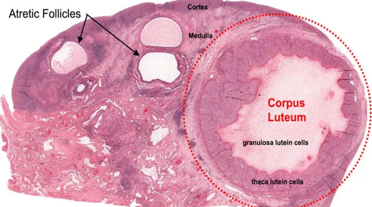
FORMATION OF CORPUS LUTEUM AND LUTEOLYSIS
The corpus luteum forms after ovulation with the fibrosis of the fibrin core and
some of the theca cells becoming incorporated within the developing corpus
luteum. The process of lutenization begins just before ovulation. There is an
increase in level of 17 alpha hydroxyl progesterone at midcycle soon after the
beginning of the LH surge. The corpus luteum has a limited life span. If there is
no implantation of the fertilized ovum, then it undergoes luteolysis.
Two types of cells are identified in the corpus luteum, theca and granulose
lutein cells. There may possibly be another cell type, the K cells. The formation
of luteal body encompasses several tages like proliferation, hyperaemia, and
droplets, and are shoewn to have granules containing lipofuscin, ehich gives the
characteristic yellow colour to the luteal body. The cells contain distinct golgi
complex, numerous vesicles, agranular endoplasmic reticulum and extracellular
canaliculi. These extra cellular canaliculi function as a system to transport
steroid from the cells to capillaries. On days 6-8 post ovulation, the blood
vessels reach the centre of the luteal mass and steroid secretion reaches peak
levels. The corpus luteum , by this time , reaches its stage of maximum
development. This stage is followed by declining steroid levels unless
blastocyst implantation occurs. The fully formed corpus luteum releases large
amount of 17-beta estradiol and progesterone. From about datys 7-8 post
ovulation , luteal regression is initiated. Degeneration of the cells occurs. The
cells undergo lipolysis, atropy and bloodflow declines. The corpus albicans is
formed which is composed of collagen fibres and few cells. LH is luteotrophic
Figure 3 : Histology of Graffian follicle
[image:30.612.97.480.482.695.2]ENDOMETRIAL CYCLE
The most obvious manifestation of a normal menstrual cycle is the presence of
regular menstrual periods. Every month the uterus prepares for a pregnancy by
generating a thick bed of secretory endometrium for the implantation. Due to
failure of fertilization of the oocytes or implantation, the menses starts. Hence,
menstruation is described as “weeping of a disappointed uterus for a baby”.
The endometrium is the superficial epithelium, which lies in the uterine
cavity. It has two principle components, the glandular epithelium and
supporting stromal cells. During the menstrual cycle the epithelium
differentiates to form three functional zones. The basalis spongiosum and
stratum compactum.21
The endometrial events can be divided into three phases.
1. Menstrual phase
2. Proliferative phase
3. Secretory phase
MENSTRUAL PHASE
The beginning of each endometrial cycle is characterized by complete shedding
of the spongiosum and stratum compactum layers (day 1) during menstruation
which lasts for 3 to 5 days. The fall in plasma progesterone and oestrogen levels
due to degeneration of corpus luteum leads to withdrawal of hormonal support
vasoconstriction of uterine blood vessels, which leads to decreased supply of of
oxygen and nutrients to endometrium. Disintegration starts in the entire lining
except the basalis layer that will regenerate the endometrium in the next cycle.
After the initial period of vasoconstriction, the endometrial arteriole dilates
resulting in hemorrhage through vascular capillary walls. The menstrual flow
consists of blood mixed with endometrial debris and mucus.
PROLIFERATIVE PHASE
Menstrual flow ceases and endometrium begins to thicken as it regenerates from
the basalis layer. The period of growth lasts for 10 days or so between cessation
of menstruation and occurrence of ovulation. The ovarian follicular phase
corresponds to menstrual proliferation phase of endometrial cycle. The uterine
changes during the menstrual cycle are caused by the changes in plasma
concentration of oestrogen and progesterone. During proliferative phase a rising
plasma oestrogen level will lead to reconstruction and growth of endometrium.
Both glandular and stromal component achieve proliferation which peaks at 8 to
10 days of cycle and corresponds to peak oestrogen level. During this phase, the
SECRETORY PHASE
Soon after ovulation the endometrium begins to secrete various substances and
the part of the menstrual cycle between ovulation and the onset of next
menstruation is called secretory phase. The circulatory progesterone which is
secreted by the corpus luteum after ovulation acts upon the oestrogen primed
endometrium to convert it to actively secreting tissue. Its glands become coiled
and filled with glycogen. The blood vessels become more numerous and
densely coiled. Various enzymes are secreted in the glands and connective
tissue. These changes are essential to make the endometrium a hospitable
Uterus :
The uterus is a fibro muscular organ usually divided into an upper corpus
or body and lower cervix. The size and configuration of the uterus undergoes
many changes throughout life, at birth the cervix and the uterus are
approximately the same size and during reproductive years the corpus grows to
2 to 3 times the size of the cervix. It measures approximately 7x5x3cm, with a
volume of 3 to 8 ml. After menopause it involutes and becomes atrophic. The
endometrial cavity is triangular in shape and represents the mucosal surface of
the corpus. The ultimate function of the uterus, to house and support the
conceptus, is dependent on the cyclic alterations in the endometrium. The
myometrium consists of interlacing smooth muscle fibres, and the serosa is the
outermost layer, formed by the peritoneum. The blood supply is by the uterine
artery, which anastomosis with the ovarian and vaginal arteries.23
CERVIX AND VAGINA
The cervix has lesser muscle cells compared to the myometrium. These are
dispressed in a ground substance containing glycosaminoglycans and collagen
fibrils. The collagen synthesized by the connective tissue is arranged in an
orderly fashion. Functions of the cervix are :
To help the passage of sperm towards the uterus
To prevent expulsion of fetus during pregnancy
At term, the cervix undergoes significant structural and functional changes
referred to as ‘ripening’ of the cervix. This involves changes in the structure og
the tissue matrix with the loss of collagen and increase in the
glycosaminoglycans (CAG) content which helps in loosening of collagen
fibrils. Estrogen and progesterone determine the nature of secretion from the
endocervical glands and the cervical ripening in preparation for labour.
The vagina consists of three layers: mucous, muscularis ans fibrous sheeth. The
mucosal layer shows several folds lined by stratified squamous epithelium. The
epithelium contains high amount of glycogen. Under estrogen dominance
glycogen synthesis is increased. The levels of circulating estrogen and
progesterone also influence the nature of vaginal fluid. When the estrogen level
in blood is high, the vaginal fluid becomes acidic due to the action of
Doderlein’s bacilli on the carbohydrates in the vaginal epithelium.24
Fallopian tube:
The oviduct forms an important part of the reproductive system and links the
uterine cornua to the ovaries. Its functions include ovum pickup, the mechanical
transport of gamates, provision of a physical environment for conception and
physiologic sustenance of the early conceptus.
The fallopian tube are paired hollow structures approximately 6 to 15 cm long
(average 11cm). their caudal ends anastomose with the uterus and their
formed by the two sheets of abdominal serosa, encloses the tube, and within it
run muscle fibres, supporting ligaments, nerves, blood vessels and lymphatics.
Depending on the anatomic location , the amount of circular smooth muscles of
the wall, the degree of folding and cellular composition of the mucosa, as well
as autonomic innervation, its possible to distinguish four distinct segments of
the tube. From cephalic to caudal these are fimbria, ampulla, isthmus and
interstitial parts of the tube. The tubal mucosa is ciliated columnar epithelium
with predominance of ciliated and secretory cells at the fimbrial end, which
decrease in number as the uterus is approached.
The fallopian tube is supplied by a rich anastomosing network of vessels fed
from both the uterine artery and the tubal branch of the ovarian artery, and the
innervation is also dual, from the uterovaginal and ovarian plexuses.
Oocyte capture occurs through the coordinated efforts of the tube and ovary.
Oocytes reside in the ampulla for about 72 hours, during which time
fertilization occurs. Through the exact in vivo nutritional and metabolic
conditions are not known, an iso osmotic balanced solution of amino acids,
pyruvate, lactate and albumin are required to support the pre embryo to
blastocyst stage. Ovum transport in the tube occurs through the integrated
activities of the muscular myosalpinx and cilia. The isthmus serves as the
transitional area between the tube and the uterus, and has both permissive and
FEMALE INFERTILITY
Failure to conceive within a year , when a couple is staying together and
cohabiting with an idea of having a pregnancy is labelled as infertility, it may be
primary when there has been no conception at all or secondary when a woman
has had prior conception and now fails to conceive.
Incidence: the incidence of fertility in any community varies between 8 - 15%,
though the exact estimation of the same is difficult.
Of the couples staying together and not using any contraception , approximately
60% conceive in 6 months, 75% in 9 months and 80% in 1 year. Another 5-10%
may conceive subsequent years.25
MAJOR CAUSES OF INFERTILITY
Several factors influence the process of conception, the failure of which can be
due to defects in any of these. For normal conception to occur, there must be
Normal spermatogenesis
Semen deposition in the vagina at the time of ovulation
Normal ovulation
Functionally normal fallopian tubes able to pick up the ovum
Normal uterine cavity for the implantation as well as the continuation of
pregnancy
Cervical secretion which are under cyclic hormonal control and are normal to
There should be no immunological incompatibility.
The male partner alone may be at fault in approximately one-third of the cases,
the female partner alone in another third and in the remaining thirdboth may be
at fault some of the defects may be remediable.26
NORMAL REPRODUCTIVE EFFICIENCY
Human reproduction is not efficient. In presumably fertile couples, a maximal
monthly fecundity rate of 20-30 % is demonstrated (5-7). Likewise, pregnancy
loss is common. This has been demonstrated as early as 1949, when Hertig and
Rock reported in their now classic studies in which early embryos were
recovered from 107 women who had intercourse at the estimated time of
ovulation, prior to undergoing hysterectomy (8). One-third of the embryos was
deemed to be abnormal; however, this was limited to the morphologic
assessment of the embryos. Forty to sixty % of conceptuses (as defined by a
positive HCG value) fail to achieve 12 weeks of gestation, with the majority of
losses occurring prior to the recognition of pregnancy by the mother (5-7).
Miscarriage rates appear to be affected most by the maternal age, with natural
conception cycles resulting in losses for 7-15% of women less than 30 years,
8-21 % for ages 30-34, 17-28 % for ages 35-39 and 34-52 % for ages 40 and
INDICATION FOR EVALUATION
Formal evaluation of infertility is generally indicated in women attempting
pregnancy who fail to conceive after a year or more of regular, unprotected
intercourse as 85% of couples will achieve pregnancy with out assistance within
one year of time. Earlier evaluation and treatment is indicated in women with
1. Age more than 35 years
2. H/O oligomenorrhea/ amenorrhea
3. Known case of uterine/tubal disease, endometriosis, or diminished
ovarian reserve
4. A partner who is known to be or suspected to be sub fertile.
The extent of evaluation should take into account the couple’s wishes, both the
partner’s age, the duration of infertility, and unique features of the medical
history and physical examination.
EVALUATION
• The couple must be considered as a single unit as each partner
contributes a share to the infertility potential of the couple . Initial consultation
with the infertile couple should include a complete medical and menstrual
history including a review of lifestyle and social habits, physical examination,
and pre conception counselling. The evaluation of female infertility assesses
the peritoneum. The initial screening evaluation of the male partner should
include, at a minimum, a reproductive history and two properly performed
semen analysis. A careful h/o history and physical evaluation of each partner
can direct further investigation. Evaluation of both partners should be initiated
simultaneously as it is imperative that infertility is approached as a couple
disorder. 27
HISTORY
Couples attending their first visit to the reproductive center are asked to
complete an extensive self assessment questionnaire prior to their visit. The
importance of this questionnaire is to ascertain relevant medical and surgical
history. In the female partner, the menstrual history, outcome of previous
pregnancies, length of infertility, sexually transmitted diseases, outcome of
previous surgeries, as well as prior infertility investigation and treatment are
critical issues. Specifically, it is necessary to carefully review any prior
treatment attempts, including evidence of ovulation with clomid, number of
follicles developed in response to gonadotropin therapy, amount of drug
utilized, peak estradiol levels, number of oocytes produced with ART, and
embryo progression.
It is important to provide some privacy during history taking to allow the patient
and the partner to disclose any relevant that she or he may be reluctant to
discuss in front of her or his partner. Such sensitive issues could include a
and/or elective terminations, and so on. The regularity, length, and frequency
of the women’s menstrual cycle, as well as associated symptoms such as pain
should be documented. A history of oligomenorrhea/amenorrhea and signs of
androgen excess may be suggestive of polycystic ovary syndrome. Complaints
of chronic pelvic pain, dysmenorrhoea or dyspariunia may suggest the presence
of endometriosis. Episodes of sexually transmitted disease, a history of prior
pelvic/abdominal surgery, or a ruptured appendix may indicate significant
pelvic adhesions tubal damage or obstruction. The history of previous abnormal
pap smears and any subsequent treatment should be elicited. Cervical factors
account for a very small percent (3-5%) of all infertility cases and may be
present in patients with a history of multiple D&C, cone biopsy, cryosurgery, or
LEEP. Prior surgical procedures of the cervix may also result in the decreased
cervical mucous production and cervical stenosis.
It is also important to obtain a detailed sexual history in terms
methods of contraception, the frequency and timing of intercourse, ability to
reach orgasm, and incidents of separation. The couple should be questioned
about the use of coital lubricants and postcoitaldouching because both are
potentially spermicidal.
Other relevant aspect of the medical history includes: current medications and
allergies; occupation and use of tobacco, alcohol, and other drugs; family
history of birth defects, premature ovarian defects, pre mature ovarian failure,
mental retardation or reproductive failure; symptoms of thyroid dysfunction,
Nutritional status, marked weight gain or loss, or intensive physical
activity may signal ovulation dysfunction. Often overlooked while eliciting a
history are the social and lifestyle factors that can negatively impact fertility.
These modifiable factors need to be addressed as part of thorough assessment. 25
PHYSICAL EXAMINATION
A through physical examination along with pelvic examination should be
undertaken and focused on the issues that could be relevant to fertility. Absence
of secondary sexual characteristics should be noted, and stages of pubertal
development could be relevant in some patients by using Tanner’s scale.
Scanty axillary and pubic hair may suggest testicular feminisation syndrome,
which is due to androgen resistance. Such patients are genetically males and
phenotypically females. The abscence of axillary and pubic hair accompanied
by anosmia should raise suspicion to Kallmann’s syndrome, which is due to
congenital absence of the GnRH. Increased facial or midline hair may be
indicative of androgen excess and highlight the possibility of polycystic
ovarian syndrome or other relevant adrenal or ovarian disorders.
It is important to realize that mild forms of late onset of adrenal
hyperplasia due to 21-beta- hydroxylase deficiency could be clinically
indistinguishable from PCOS. This is due to the fact that partial enzyme
deficiency is usually associated with normal cortisol and mineralocorticoid
levels unlike the classical infantile type, which is characterized by decreased
acanthosisnigricans is a sign of insulin resistance and may be associated with
PCOS, diabetes mellitus of thyroid dysfunction. Central obesity, buffalo neck,
and moon faces may suggest Cushing’s syndrome, which could be associated
with abdominal striae and other pathognomonic features. Turner’s syndrome is
characterized by short stature, webbed neck, shield chest, undeveloped breast
and cubitus valgus. The thyroid gland should be evaluated and palpated and
features of multinodulargoitre should be observed. Other features of thyroid
should be noted ( thin hair, myxedema, skin texture, bradycardia or tachycardia,
tremors, diarrhoea, weight loss, and exopholthalmos) . Both hypothyroidism or
hyperthyroidism are associated with infertility. The former when present tends
to be associated with some degree of hyperprolactinemia.
Breast examination for any evidence of galactorrhea should not be
missed during the general examination. However. 50 % of women reporting
galactorrhea have normal prolactin levels. Other signs of prolactinomas should
be assessed (headache, anorexia, vomiting, and visual defects. Body mass index
should be calculated if the patient is noted to be over or under weight. The BMI
calculated by dividing the weight in the kilograms with the height meter
squared, with the normal range falling between 18.5 and 24.9 kg/meter square.
The association between obesity and ovulatory dysfunction is well
documented. Women with increased BMI are encouraged to reduce weight,
which may help in the resumption of ovulation spontaneously and increase
should be encouraged to increase their weight to within the normal range prior
to ovulation induction to decrease the risk of a low birth weight baby. 27
The importance of pelvic examination can never be over emphasized and should
be performed at initial visit. Unfortunately, with increasing use of ultrasound,
many practitioners are reluctant in performing complete pelvic examination,
which should not be case. Congenital malformations of the vaginal tract should
be screened for such as double cervices and vaginal septum. Congenital absence
of the vagina (Mayer-Rokitansky-Kuster-Hauser syndrome) remains to be most
frequent cause of primary amenorrhea.
Speculum examination and thorough evaluation of the cervix should be
performed. Cervical smear and Chlamydia screening should be performed if
not already done. Chlamydia infection is central in the pathogenesis of pelvic
infection that may result in the development of tubal infertility, pelvic pain, and
ectopic pregnancy. Bimanual examination is helpful in determining the size,
shape and mobility of the uterus. The adnexa are examined for the presence of
ovarian endometriomas or cysts. Nodularity in the utero sacral ligaments on
rectovaginal examination suggests the presence of endometriosis.
In summery, as outlined in the ASRM 2006 Practice comittee opinion, the h/o
of the patient should include
1. Gravidity, parity index, pregnancy outcome, and associated complications
2. Age at menarche, characteristics of menstrual cycle and onset/severity of
3. Methods of contraception used and coital frequency
4. Duration of infertility and results of any previous evaluation and
treatment
5. Past surgery, its indication, and outcome, previous hospitalizations,
serious illness or injuries, pelvic inflammatory disease or exposure to
STD and unusual childhood diseases.
6. Any Previous abnormal pap smears and subsequent treatment in the past
7. Current medications and allergies
8. Occupation and use of tobacco, alcohol other drugs
9. Family h/o birth defects, mental retardation or infertility
10. Symptoms of thyroid disease, pelvic or abdominal pain, galactorrhea,
hirsutism, and dyspareunia.
The physical examination should note the patient’s weight and body mass
index and identify any
1. Thyroid enlargement, tenderness or nodule
2. Breast secretions and their character
3. Signs of excess androgen
4. Pelvic or abdominal tenderness, organ enlargement, or mass
6. Uterine size, shape, position and mobility
7. Adnexal mass or tenderness, and
8. cul-de-sac mass, tenderness or nodularity. 26
ASSESSMENT OF ANATOMICAL FACTORS
TUBAL FACTOR
The patency of fallopian tubes can be tested by several tests which are
carried out in the immediate post menstrual phase of the cycle. This timing
reduces (i) false positive test (ii) decreases the risk of accidental embolism and
endometriosis and (iii) avoids accidental disturbance of undiagnosed pregnancy.
Tubal insufflating carbon-dioxide gas through the cervix in to the uterine cavity
and tubes. The insufflation cannula is made air tight and is attached to a
kymograph. Gas is slowly passed at a rate of 60ml/min. as it flows, to begin
with there is a rise in pressure to about 100mm of Hg which falls to 70-80 mm
of Hg as the free flow of gas is established. A peristaltic activity is also
recorded on the kymograph. Patency is also judged by simultaneously
auscultating over the midinguinal point where a gurgling sound is heard. A rise
of pressure up to 200 mm of Hg indicates bilateral tubal block. The test is now
abandoned.
This test has a very high false negative and a very high false positive
test may help in establishing tubal patency by breaking flimsy adhesions due to
pressure of gas.28
HYSTEROSALPINGOGRAPHY
In this test, instead of gas a radioopaque dye is injected through the
canula under fluoroscopic control and X-ray films are taken. It helps in the
identification of uterine abnormalities and exact site of tubal block if any. False
negative rate is low, the block being missed in 5% of cases. Over diagnosis of
tubal block can be there in 10-20% of the cases.
HYSTEROSALPINGOGRAPHY
This is a new test which can be done instead of hystero-salpingography.
Normal saline is pushed in the uterine cavity either through a paediatric Foleys
catheter the bulb of which is inflatted at the cervical or through the Rubins
canul. Ultrasound is done to follow the fluid through the uterus and then
through the tubes in to the peritoneal cavity. Any uterine polyps or
malformation and tubal block can be diagnosed. There is no exposure to
radiation. If patient fails to conceive for 6 months, despite tubal patency on
HSG a laparoscopic evaluation is done.28
LAPAROSCOPY
It is done under general anaesthesia. After introducing a laparoscope,
chromotubation is done by injecting methylene blue through the
through the cervix. The dye can be seen through the laparoscope passing
through the tubes and coming out of the fimbrial ends if the tubes are patent.
Laparoscopy also helps in the diagnosis ofbicornuate or unicornuate uterus,
peritoneal adhesions, endometriosis, pelvic tuberculosis, tubal kinks, and
hydrosalpinx. The segment distal to the site of block and available tube length
can also be evaluated. Currently only one method of tube testing is done
depending on the availability of the facility present laparoscopy is gold stranded
as it can obviate false positive test seen due to spasm of the uterine cornua
during other tubal tests.
UTERINE FACTOR
Abnormalities of the uterus are uncommon causes for infertility. Failure
of conception may result from local pathology resulting in inadequate
preparation of the endometrium and defective implantation.
Local pathology may be tubercular endometritis, moderate to severe uterine
hypoplasia, myoma or asherman’s syndrome. A diagnosis of tubercular
endometritis is made on endo metrial biopsy.
Intra-uterine synechiae or submucous myomas can be diagnosed, by
hysterosonography. The hysteroscopy is preferred mode of investigation for
detecting intrauterine pathology and is fast becoming popular. It can offer
treatment at the same sitting.it is routinely combined with laparoscopy which
CERVICAL FACTOR
There may be an abnormal cervix, cervical infection or abnormal
cervical secretion. These can be diagnosed on inspection and easily rectified.
A post-coital test (Sim’s Hunyer’s): evaluates sperm penetration in vivo. The
test is done at the time of ovulation. The women is called 4-8 hours after sexual
intercourse, cervical mucus is collected from endocervical canal and examined
under microscope. The number of motile sperms per high field are counted. If
no sperms are identified , either coitus has not taken place or the husband is
azoospermic or the whole discharge has leaked out. Identification of only dead
sperm indicates cervical mucus hostility or infection. The couple needs to be
investigated further for infection and immunological factors. If the number of
live sperms is 15-20/high power field, it is an excellent or good test and
indicates good cervical function.
PERITONEAL FACTOR
Adhesions (peritubal, perifimbrial, periovarian and omental) altering
tubal function and ova pick up by the fimbriae and endometriosis are major
peritoneal problems. Diagnostic laparoscopy is the best rocedure for evaluation
IzatullaJumayev et al (2012) in the study of social correlates of female
infertility in Uzbekistan, concluded, that 65.0% of patients who had infertility
were diagnosed to have anovulation. Tubal blockage was the next major cause
for infertility in 23.3%, immunologic factors in 1.7%, and the remaining 10.0%
of women were suffering unexplained infertility.31
Adamson et al (2011) in the study of Prevalence& correlates of primary
infertility among young women in Mysore, India, reported the mean age of the
women was 25.9 yr which ranges from 16-30 year and the prevalence of
primary infertility was 12.6 per cent. HSV-2 seropositivity was the main factor
associated with primary infertility.32
Cornillie, et al (1995) found in their study that the preventable risk factors for
infertility were: PCOS: 18%, Endometriosis: 2.3%, genital infection: 10%,
menstrual cycle irregularity,13.7%, history of previous surgical operations:
5.2%.33
Friday EbhodagheOkonofua et al (2005) studied on the epidemiology of
infertility in Nigeria with special references comparing the role of genital tract
infections and sexual and reproductive risk factors, has concluded that there is a
strong correlation between episodes of reproductive tract infection and
infertility among women in Nigeria.34
In a study which was carried out in Aberdeen Fertility Centre from 1993-2006,
the factors that affects couples infertility including age of male and female
partner, year of first visit, diagnosis, duration and type of infertility were
primary infertility was seen in 51.4%. The mean female age was 31.2 (5.2 SD)
years. It was evident that there was a strong association between age of the
female and the cause of female infertility. Most of the women over 35 years of
age had unexplained infertility (26.6 versus 21.0%, p< 0.001). When Compared
with women under 30 years, the adjusted odds ratio (95% confidence intervals,
CI) of the following diagnoses in women over 35 were: unexplained infertility;
1.8 (1.4-2.2), ovulatory dysfunction; 0.3 (0.3-0.4) and tubal factor; 2.2 (1.7-2.7),
57.35
Moghadamet al.(2013), in Epidemiology of Female Infertility studied
Polycystic ovarian syndrome causes anovulation there by causing infertility in
young women and it comprises of about 70% of anovulation infertility, and
15.6% of primary infertilities.36
Wang et al. (2003) prospectively studied 518 newly married Chinese textile
workers (20–34 years of age) trying to conceive. They recorded various factors
like vaginal bleeding, sexual intercourse and collected daily first-morning urine
specimens for up to 1 year or until the women become pregnant. Survival
curves (Kaplan and Meier, 1958) were calculated for proportion of conceptions
over number of menstrual cycles. In their study population, ,50% of women
concieved in the first two cycles and .90% of women became pregnant in the
first six cycles. According to their study the monthly fecundity varied between
30 and 35%.32
Olooto ,WasiuEniola et al in, A review of Female Infertility; important
15 % of couples worldwide. In Africa, itsprevalence is mainly high in
sub-Sahara ranging from 20% to 60% of the couples. It is estimated that 50-80%
accounts for female factors and unexplained infertility and the male factor is
responsible for 20-50% of the cause of infertility in different parts of Nigeria.38
Sule, Erigbali and Eroum et al.,(2008) in their study on Prevalence of Female
Infertility in a Southwestern Nigerian Community, concluded; Cases of
infertility reported were caused by tubalfactor 39.5%, uterine factor30%,
ovarian factor13% and malefactor 6.5% . Less common causes werepelvic
inflammatory disease, 5.5%, cervical factor 3.0% and endometriosis 2.5%.37
SamihaMokhtar, Hassan Ali Hassan, NehadMahdy et al., (2006) studied Risk
Factors For Primary and Secondary Female Infertility in Alexandria: A Hospital
Based Case Control Study; results were The highest reported population
attributable risk percentages were: for primary infertility were as PCOS as
19.4% followed by irregular mensteruation as 15.9% then STI as 7.9% and
finally surgical operation as 4.6 %. The highest calculated PAR% in secondary
infertility were irregular mensis as 19.2% followed by 15.4% for PCOS then
14.1 % for STI and 6.5% for surgical operation.35
G.Sudha and K.S.N.Reddy (2013) in their study on causes of female infertility
concluded Maintaining a healthy lifestyle, getting regular checkups with the
doctor and maintenance of normal body weight can avoid fertility problems.
Fertility increases with identification and control of chronic diseases such as
MeraiyebuAjibola, Akintayo Christopher oloruntoba et al., in A study on effect
of prolactin hormone on female infertility in National Hospital Abuja, Nigeria
concluded that abnormal prolactin level has no effect in causing primary
infertility. It has also been studied that abnormal prolactin level was also not the
major cause for secondary infertility among the infertile female patients.40
M G R HULL,C M A GLAZENER et al., in Population study of causes,
treatment, and outcome of infertility stated that polycystic ovarian disease is
the only major unsolved problem. Tubal damage (except operative sterilisation)
is a major problem that occurs about 14% of cases and causesvery low chance
of successful pregnancy.41
Steinkeler et al., in Female Infertility: A Systematic Approach to Radiologic
Imaging and Diagnosis concluded that the genital causes of female infertility
include abnormalities in tubes, peritoneum, uterus, endometrium, cervix, and
ovary. A multimodality imaging approach may be useful for identifying the
cause of infertility and helps in the management.42
AbhaMaheshwar et al., in Effect of female age on the diagnostic categories of
infertility stated that age is the prime factor that is associated with the
prevalence of different causes of female infertility. Unexplained and tubal
factor infertility is common in women more than 35 years of age. The
increased incidence of secondary infertility were mainly due to tubal factor,
MARY ANNE ROSSING et al ., in OVARIAN TUMORS AMONG A
GROUP OF INFERTILE WOMEN concluded that the risk of ovarian tumors
are increased with prolonged use of clomiphene with ovulatory abnormalities.44
C.Augood et al., in Smoking and female infertility: a systematic review and
meta-analysis, stated that there is a significant rise in infertility among the
smoking women comprising of 60% increase in the risk of infertility
among cigarette smokers.45
Izumi Imaoka et al., in MRImaging of Disorders Associated with Female
Infertility: Use in Diagnosis, Treatment, and Management concluded that MR
imaging is a useful modality for routine work-ups in female with infertility.
Pituitary adenoma can be diagnosed very effectively when patients are
suspected to have disorder of the hypothalamic- pituitary-ovarian axis. The MR
imaging helps in assessment of the pelvic cavity in patients with infertility by
evaluation of the functioning uterus and ovaries, differentiation of mullerian
duct anomalies, and accurate noninvasive diagnosis of adenomyosis,
leiomyoma, and endometriosis.46
D.Meirow and J.G.Schenker et al., in The link between female infertility and
cancer: epidemiology and possible aetiologiesstated In many infertile patients,
various factors like hormonal status, height, weight , BMI measurements and
growth factors (composition and concentration) are considerably disturbed.
Epidemiological, clinical and experimental studies have shown the association
Willemsen et al. (1993), studied 12 female patients in whom a granulosa cell
tumour was discovered after ovulation induction treatment with clomiphene
citrate and/ or gonadotrophins, and has described about the prevelance of a
granulosa cell tumour in this population was 0.23%, which is much higher than
the incidence of granulose cell tumour in the female population as a whole.
These data has not proved the existence of a causal relationship between
ovulation induction treatments and the development of granulosa cell tumours.
The possibility of such an association may exists and to confirm it further
wide-ranging investigations by registered authorities should be done.48
Ron et al. (1987) evaluated 2672 women who were treated for infertility
(primary or secondary) between 1964 and 1974 with >31 600 patient years of
follow-up. In his study the major infertility cause were found to be as hormonal
(anovulation and menstrual irregularities) or other causes of infertility like male
factors, mechanical, unexplained). In the infertile patients group there was a
4.8-fold increased risk of endometrial carcinoma and a 10.3-fold increased risk
of endometrial carcinoma in infertile women with chronic anovulation.49
According to Rossing et al. (1994), the maximum risk of ovarian tumours
associated with ovulatory abnormalities was seen in PCOS patients.50
B. R. Moller et al ., in Serological evidence that chlamydiae and mycoplasmas
are involved in infertility of women stated that antibody to M. genitalium was
Conway et al,(1984) emphasize that pelvic inflammatory disease due to
chlamydia results in the tubal damage which increases the risk of ectopic
pregnancy and infertility.52
Tyagi et al., believe that local changes in the endometrium along with the
systemic effect are responsible for the menstrual abnormalities.53
Marcus et al., concluded that IVF-ET offers the only realistic treatment for
tuberculous infertility. Women with Tubercular endometritis and synechiae
formation preclude the use of IVF. In such case the couples are best suited for
adoption.54
Parikh et al., in Genital tuberculosis – a major pelvic factor that causes
infertility in Indian women stated that genital tuberculosis is a chronic
infectious disease which is one of the major etiologic factors that causes female
tubal infertility due to peritubal adhesions and tubal block, especially in the
India.55
Elizabeth A. Pritts et al., in Fibroids and infertility: an updated systematic
review of the evidence concluded Fertility outcomes are reduced in women who
had submucosal fibroids, and its removal had better outcome. Usually
Subserosal fibroids does not interfere with fertility outcomes, and its removal
does not improve the outcome. Intramural fibroids are known to decrease
fertility, but the results after its resection are unclear.56
Oliveira et al., stated that the anatomical distortion may reduce access to the
ostia, , whereas the large corneal fibroids or lesions results in impairment of
ovum retrieval by the tubes.57
Surreyet al.(2005) recently report IVF outcome in patients operated for
submucosal fibroids. The pregnancy rate in operated patients and in the control
group was 68 (69 /101) and 62% (900/1448), respectively (P-0.24). Overall,
even if the available evidence is still scanty, previous myomectomy does not
appear to negatively affect the chances of pregnancy in IVF cycles.58
Richards et al., stated that fibroids usually interfere with the migration of
sperms, ovum transport and implantation of embryo. The presence of
submucosal fibroids causes local inflammation which may result in a hostile
endometrial environment leading to the impairment of sperm transport and
embryo implantation.59
Somigliana et al ., in Fibroids and female reproduction: a critical analysis of the
evidence stated that in IVF cycles, the pregnancy rate is decreased in patients
with submucus or intramural fibroids but is not affected in the patients who
have undergone myomectomy. Available evidence also suggests that
submucosal, intramural and subserosal fibroids interfere with fertility in
5. MATERIALS AND METHODS:
a. Study design: Non-Randomized cross sectional study
b. Study setting: Obstetrics &gynaecologyoutpatientdepartment , pathology department, SreeMookambika Institute of Medical Science. Kulasekharam.
c. Duration of the study: One year
d.Study group: One group
e. Description of the group: women with history of infertility attending OG outpatient department, SreeMookambika Institute of Medical Science
Kulasekharam from 2014 to2015 taking in to consideration of inclusion and
exclusion criteria.
f. Sample size: 43
g.Scientific basis of sample size used in the study: 4pq/d2 = 42.85
p= 70%, q=(100-p), d=20% of p (Moghadamet al.(2013), in
Epidemiology of Female Infertility studied Polycystic ovary syndrome causes
infertility in young women and provides 70% of unovulation infertility, and
h. Inclusion criteria:
1. Women attending gynecology outpatient department with following criteria:
- age 20 to 40
- married for >1 year and living with husband
- not using any contraception
- Couples who fail to conceive following a previous pregnancy,
in the absence of contraception,breastfeeding or postpartum amenorrhoea for a
period of two years
i. Exclusion Criteria:
1. Those who are not willing to participate
2. Women on In Vitro Fertilization (IVF) treatment
j. Parameters to be studied:
i. Detailed history of the patient with general examination, height, weight,
speculum examination, basic blood investigations with TFT, Urine Routine and
USG abdomen and pelvis
k. Methods /technique/instrument/reagent/ kit used to measure the quantitative parameters along with their manufacturing source details:
Basic blood investigations with TFT, Urine Routine and USG abdomen and
l. Procedure in detail: Protocol will be submitted to Human Ethical Committee (HEC) for its approval. After getting approval from HEC, written
informed consent will be obtained from the patient before enrolling them into
study.
1. A detailed history to be taken ,Menstrual history, coital history, obstetric
history,past medical history, family history, personalhistory, surgical history.
2. A thorough general physical examination with reference to height, weight,
pulse, blood pressure, temperature, respiratory rate were noted followed by
Cardiovascular system, respiratory system, speculum examination, pelvic
examination.
3. Routine blood investigations, Blood sugar, TFT, USG abdomen and pelvis.
s. Statistical methods of analysis:
i) Significance level decided before starting of study: by calculating the statistical parameters like mean, standard deviation.
ii) Statistical tests to be used for data analysis: Chi-Square test/ ODDS ratio
iii) Software to be used for statistical analysis: All parameters will be entered in Microsoft Excel. Analysis will be done using SPSS software version 20.0