Copyrightq1996, American Society for Microbiology
DNA Immunization Confers Protection against Murine
Cytomegalovirus Infection
JUAN CARLOS GONZA´ LEZ ARMAS,1,2CHRISTOPHER S. MORELLO,3
LEE D. CRANMER,1,2ANDDEBORAH H. SPECTOR1,2*
Departments of Biology1and Pathology3and Center For Molecular Genetics,2
University of California, San Diego, La Jolla, California
Received 2 May 1996/Accepted 23 July 1996
The murine cytomegalovirus (MCMV) immediate-early gene 1 (IE1) encodes an 89-kDa phosphoprotein (pp89) which plays a key role in protecting BALB/c mice against the lethal effects of the MCMV infection. In this report, we have addressed the question of whether “naked DNA” vaccination with a eukaryotic expression vector (pcDNA-89) that contains the MCMV IE1 gene driven by a strong enhancer/promoter can confer protec-tion. BALB/c mice were immunized intradermally with pcDNA-89 or with the plasmid backbone pcDNAI/Amp (pcDNA) and then challenged 2 weeks later with either a lethal or a sublethal intraperitoneal dose of the K181 strain of MCMV. Variable results were obtained for the individual experiments in which mice received a lethal challenge. In four separate trials, an average of 63% of the mice immunized with pcDNA-89 survived, compared with 18% of the mice immunized with pcDNA. However, in two other trials there was no specific protection. The results of experiments in which mice were injected with a sublethal dose of MCMV were more consistent, and significant decreases in viral titer in the spleen and salivary glands of pcDNA-89-immunized mice were observed, relative to controls. At the time of peak viral replication, titers in the spleens of immunized mice were reduced 18- to >63-fold, while those in the salivary gland were reduced approximately 24- to 48-fold. Although DNA immunization elicited only a low level of seroconversion in these mice, by 7 weeks postimmunization the mice had generated a cytotoxic T-lymphocyte response against pp89. These results suggest that DNA vacci-nation with selected CMV genes may provide a safe and efficient means of immunizing against CMV disease.
Human cytomegalovirus (HCMV) is a ubiquitous pathogen that is responsible for congenital malformations in the new-born and severe disease in immunocompromised individuals (14). The serious problems associated with HCMV infection have made it imperative to understand the pathogenesis and immunology of this virus in order to develop strategies for its prevention and treatment. However, progress with the human virus has been slow primarily because the strict species speci-ficity of the virus has precluded studies with animal models. As an alternative, many in vivo studies have utilized murine CMV (MCMV) infection of mice, which greatly resembles its human counterpart with respect to acute infection, establishment of latency, and reactivation after immunosuppression (5, 14, 34). Since recovery from HCMV disease correlates with a cel-lular immune response rather than with the presence of CMV-neutralizing antibodies (22, 25), much of the effort with MCMV has been directed towards elucidating the basis of the cellular response. Adoptive transfer experiments showed that T lymphocytes limit virus replication and prevent histopathol-ogy (31). Moreover, in BALB/c mice, protection against lethal
MCMV disease could be attributed to CD81cytotoxic T
lym-phocytes (CTLs) specific for antigens expressed in the imme-diate-early (IE) phase of the viral replication cycle (28, 29). The dominant class of IE-specific CTLs in this case was found to be directed against a nonapeptide within the 89-kDa IE1 protein (9, 17–19, 30, 40). Immunization with recombinant vaccinia virus expressing this MCMV protein also was able to confer protection against lethal challenge with MCMV
through CD81cells (15, 40).
Current approaches to vaccination typically rely on virus-based vectors which are remarkably effective but have some clear disadvantages. In addition to the usual concerns regard-ing the safety of attenuated live virus vaccines, vectors such as vaccinia virus may be compromised by immune responses against the vector itself and preclude subsequent use of the vector with another immunogen (6). The direct inoculation of plasmid DNA (“naked DNA”) into animal tissues has become a new approach to vaccination which overcomes many of the problems associated with traditional immunization methods. For instance, the potential dangers of using an attenuated live vaccine are avoided since the highly purified plasmid DNA is free of infectious agents. In addition, because there is little or no immune response to plasmid DNA, the vector can be used for subsequent immunizations. Although this vaccination strat-egy is relatively new, it has already been shown to be successful in generating protective immunity against influenza virus (38), rabies virus (42), bovine herpesvirus 1 (7), lymphocytic chori-omeningitis virus (24, 43), and herpes simplex virus (4, 13, 20). An interesting recent report also provides evidence that im-munization with an expression library made from the DNA of Mycoplasma pulmonis can confer protection against challenge from the pathogen (3). In most cases, it has also been docu-mented that the generation of antibody or CTL responses was associated with the control of the infection.
Several methods of delivering plasmid DNA have been shown to be effective in generating an immune response (11, 27, 38). Initially, the intramuscular route of immunization was used since striated muscle cells can efficiently take up exog-enously added DNA and express high levels of the gene prod-uct (1, 41). However, it recently has been shown that intrad-ermal injection of DNA can also elicit both CTL and humoral responses that persist for long periods postinoculation (27). Although the transfection efficiency is lower, the success of this
* Corresponding author. Mailing address: Department of Biology 0357, University of California, San Diego, 9500 Gilman Dr., La Jolla, CA 92093-0357. Phone: (619) 534-9737. Fax: (619) 534-6083. Elec-tronic mail address: dspector@ucsd.edu.
7921
on November 9, 2019 by guest
http://jvi.asm.org/
we have used this model to examine the efficacy of a naked DNA vaccine using a eukaryotic vector expressing this protein under the direction of the strong HCMV IE enhancer/pro-moter sequences. Our studies demonstrate that intradermal inoculation of this recombinant vector limits viral replication in the spleen and salivary glands and confers protection against lethality when the mice are subsequently challenged with MCMV. Although the antibody response is variable, this vac-cination protocol does generate a specific CTL response. These results provide support for pursuing this approach in the development of a vaccine against CMV.
MATERIALS AND METHODS
Mice.For DNA immunization experiments, 8- to 10-week-old specific-patho-gen-free female BALB/c (H-2dhaplotype) mice were purchased from Harlan
Sprague Dawley, Inc. Mice were maintained in the Animal Facility of the Uni-versity of California, San Diego.
Cells and viruses.NIH 3T3 cells (ATCC CRL 1658) were grown in Dulbecco’s modified Eagle’s medium (DMEM) supplemented with heat-inactivated 10% calf serum (DMEM–10% CS). COS-7 cells (ATCC CRL 1651) were grown in DMEM supplemented with 10% heat-inactivated fetal calf serum (DMEM–10% FCS). All media contained, per milliliter, 0.29 mg ofL-glutamine, 200 U of
penicillin, 0.2 mg of streptomycin, 0.05 mg of gentamicin, and 1.5mg of ampho-tericin B. P815 mastocytoma cells (H-2dhaplotype; ATCC TIB 64) and
spleno-cytes from BALB/c mice were grown in RPMI 1640 medium (RPMI)–10% FCS with 0.29 mg ofL-glutamine, 200 U of penicillin, and 0.2 mg of streptomycin per
ml and 50mM 2-mercaptoethanol.
The K181 strain of MCMV was provided by Michael Oldstone and used in all experiments. Salivary gland stocks of virus were prepared following intraperito-neal inoculation of female BALB/c mice, as previously described (10). The same salivary gland stock was used in lethal challenge trials 1 through 5 (Table 1), both sublethal challenge trials (Fig. 4), and the multiple challenge dose experiment (Fig. 2). Another stock was used for lethal challenge trial 6 (Table 1). Tissue culture MCMV used in CTL assays was prepared by passaging the K181 strain in NIH 3T3 cells. A pp89-expressing recombinant vaccinia virus (designated vacc-89) was constructed by using the vaccinia virus strain WR as described elsewhere (40).
Plasmids.The plasmid pcDNA-89 was derived from a recombinant clone which contained an EcoRI fragment encoding the continuous open reading frame of the MCMV IE1 gene within the pSG62 vector (40). This EcoRI frag-ment encoding IE1 was gel purified and cloned into the EcoRI site in the poly-linker of the eukaryotic expression vector pcDNAI/Amp (designated pcDNA) from Invitrogen (San Diego, Calif.). This vector has the intron and polyadenyl-ation sequences from simian virus 40 and the enhancer/promoter sequence from the IE gene of HCMV for high-level expression of foreign genes (Fig. 1A). The correct orientation of the insert was confirmed by restriction enzyme analysis.
Enzymes were obtained from Bethesda Research Laboratories, Inc., and used as recommended by the manufacturer. DNA fragments were gel purified with GeneClean (Bio 101) according to the manufacturer’s recommendations. Plas-mid vectors were grown in transformed Escherichia coli DH5a(Bethesda Re-search Laboratories), in Luria-Bertani broth supplemented with ampicillin (100
mg/ml). Large-scale purification was conducted with a Qiagen maxiprep kit (Qiagen Inc., Chatsworth, Calif.) according to the manufacturer’s protocol. DNA was stored at 48C in 10 mM Tris-HCl–1 mM EDTA buffer, pH 8.0, and the endotoxin content was reduced by extraction with Triton X-114 (2) (Sigma Inc., St. Louis, Mo.) to 0.5 to 5 ng/mg of DNA, as determined by the Limulus amebocyte lysate assay (Associates of Cape Cod, Inc., Woods Hole, Mass.). The concentrations of the purified vectors were determined by measuring the optical density at 260 nm. Integrity of the multiple preparations of plasmid DNA as well as the absence of E. coli DNA or RNA contaminants were assessed by agarose gel electrophoresis. Before inoculation, DNA was resuspended in pyrogen-free normal saline.
Transient expression assays.Monkey COS-7 cell monolayers were transfected with plasmid DNAs by the DEAE-dextran technique previously described by Staprans et al. (35). Briefly, confluent monolayers in 75-cm2flasks were washed
twice with phosphate-buffered saline (PBS), pH 7.4, and then overlaid with 5 ml of plasmid DNA (1 to 10 mg) in DMEM (without FCS) containing 50 mM Tris-HCl, pH 7.4, and 400mg of DEAE-dextran (Sigma) per ml. After incubation
0.5-ml syringe with a 181/2-gauge needle. Two or six weeks after the last immu-nization, mice were challenged intraperitoneally with 43104
, 2.13105
(1.43 50% lethal dose [LD50]), 2.4310
5
(1.63LD50), 2.7310 5
(1.83LD50), or 33
105
(23LD50) PFU of MCMV in 0.5 ml of PBS, pH 7.4, per mouse. Survival was
assessed for 34 days.
Plaque assays.Spleen and salivary gland homogenates (10%, wt/vol) were prepared in DMEM–10% CS and 10% dimethyl sulfoxide and stored at2708C. The amount of infectious MCMV present in the organ homogenates was quan-titated by plaque assay on NIH 3T3 monolayers in 24-well dishes essentially as described by Mercer and Spector (21). Briefly, dilutions of the homogenates were adsorbed to the cells in 0.1 or 0.2 ml of DMEM–10% CS with occasional agitation. After a 2-h adsorption period, the inoculum was removed and replaced with 0.5 ml of DMEM–2% CS and 0.25% agarose. Plaques were counted 7 days later.
Seroconversion analysis.Blood was collected from the retro-orbital plexus 1 week after the last immunization. After overnight incubation at 48C, the clot was discarded, the tubes were spun for 2 min at 14,000 rpm to pellet the cells, and the supernatant was transferred to a clean Eppendorf tube and kept at2208C until use. To assess the serum for the presence of specific antibodies, Western blot analysis was used. An extract prepared from MCMV-infected NIH 3T3 cells at 48 h postinfection was subjected to sodium dodecyl sulfate-polyacrylamide gel electrophoresis (SDS-PAGE) and transferred to nitrocellulose. The blot was probed with sera from individual mice at a 1:25 dilution by using a Mini-PROTEAN II multiscreen apparatus from Bio-Rad, and the specific reactivity was detected by ECL according to the manufacturer’s recommendations.
CTL assay.To prepare the stimulator cells, syngeneic spleen cells from un-immunized mice were incubated in vitro for 3 days with 7mg of DEAE-dextran (Sigma) per ml and 25mg of lipopolysaccharide (Difco Laboratories, Detroit, Mich.) per ml, treated with 50mg of mitomycin (Boehringer Mannheim, India-napolis, Ind.) per ml, and then pulsed for 30 min at 378C with 200mg of the pp89 nonapeptide NH2-YPHFMPTNL-COOH per ml (30). Splenocytes from
immu-nized animals (effectors) were isolated 7 weeks after the last immunization and incubated for 5 days with the stimulator cells at a ratio of 73106
effectors to 3.53106
stimulators (effector/target [E/T] ratio, 2:1) in a total volume of 1.5 ml of RPMI–10% FCS in 24-well plates (Corning Costar Corp., Corning, N.Y.). Mouse recombinant interleukin-2 (Boehringer Mannheim) at 25 U/ml was then added, and 2 days later a standard 5-h51
Cr release assay was done in 96-well microtiter plates (Corning Costar Corp.). The P815 target cells were51
Cr labeled for 1 h at 378C while being pulsed with 5mg of the pp89 nonapeptide per ml. The target cells (104
per well) were then added to serial dilutions of the effector cells. Splenocytes from 8-week-old BALB/c mice, which were not immunized, served as a negative control for the effector cells. As a positive control, we used effector splenocytes harvested 3 weeks after mice were infected with 43104
PFU of tissue culture-passaged MCMV. Both positive-control and negative-control splenocytes were in vitro stimulated as described above. Results are expressed as percentages of specific51
Cr release, calculated as (experimental release2 spon-taneous release)/(maximum release2spontaneous release)3100, where max-imum release is calculated by lysis of the targets with 0.5% Triton X-100. Data shown represent the average of duplicate cultures.
RESULTS
pcDNA-89 immunization protects against a lethal MCMV challenge.For high-level expression of the MCMV IE1 pp89 gene product, we placed the cDNA for this protein into the eukaryotic expression vector pcDNAI/Amp (Invitrogen), which has the strong enhancer/promoter sequences from the IE gene of human CMV (Fig. 1A). To assess the expression of pp89 from this vector, the recombinant pcDNA-89 was transfected into COS cells. As a control, the vector without insert (de-signated pcDNA) was also used. Extracts were prepared at 48 h posttransfection and analyzed for the expression of the MCMV pp89 gene product by Western blot with hyperimmune serum from MCMV-infected mice. Protein extracts prepared from NIH 3T3 cells infected with MCMV or the pp89-express-ing vaccinia virus recombinant vacc-89 served as positive con-trols. Figure 1B shows that a specific 89-kDa gene product was expressed at high levels in the cells transfected with pcDNA-89
on November 9, 2019 by guest
http://jvi.asm.org/
but not in cells transfected with vector alone. This protein also comigrates with pp89 expressed in vacc-89- or MCMV-infect-ed, but not uninfectMCMV-infect-ed, NIH 3T3 cells.
To test the protective efficacy of vaccination with the pcDNA-89 recombinant, we performed a pilot study in which 10 8- to 10-week old female BALB/c mice were inoculated with either pcDNA-89 or pcDNA. Mice received three intradermal
inoculations of 50mg of recombinant DNA within 10 days and
then were challenged 2 weeks later with 3 3 105 PFU (23
LD50) of MCMV per mouse, administered intraperitoneally.
Four of the five mice immunized with pcDNA-89, but none of the mice immunized with pcDNA alone, were protected from the lethal effects of the MCMV infection (Table 1, trial 1). To confirm the protective effect of the vaccination, we repeated the immunization using a larger number of mice. In this second trial, 8 of 13 mice immunized with pcDNA-89 survived the lethal MCMV challenge, compared with 1 of 12 mice immu-nized with the control pcDNA. Thus, DNA immunization with the vector encoding pp89 afforded a 53% specific protection level. The difference in survival between the groups immunized
with pcDNA-89 and pcDNA is significant at the P,0.01 level
by the chi-square exact test.
The results from these two experiments were encouraging, but as can be seen in Table 1, the data generated in four additional trials with this protocol showed considerable ability in the specific protection conferred. Some of this vari-ability was due to errors inherent in determining the precise titer of the salivary gland-passaged viral stock. This is a prob-lem particularly when survival is the endpoint and the slope of the curve defining the relationship between viral titer and survival is steep. Thus, in trials 5 and 6, in which there was 0% specific protection, we could not determine whether the im-munization failed or whether a small underestimation of viral titer resulted in mice receiving a dose of MCMV that was sufficient to overwhelm any protection conferred. Of note, parallel vaccination trials in which BALB/c mice were immu-nized with a protective pp89-expressing vaccinia virus (vacc-89) and then challenged with the same MCMV stock as that used
in trials 5 and 6 showed that the putative 23LD50challenge
dose given in both types of vaccination experiments over-whelmed the vaccinia virus-immunized mice. To further ad-dress this problem, we performed an additional set of experi-ments with the remaining mice from trial 5, which had not been challenged with virus. At 6 weeks postimmunization, these mice were infected with different doses of MCMV from the same stock of virus as that used in trial 5. Figure 2 shows that the levels of specific protection obtained when the mice were
challenged with 2.13105, 2.43105, 2.73105, and 33105
PFU of MCMV were 30, 47, 7, and 20%, respectively. This
suggests that the 3 3105PFU challenge dose in trial 5 may
have overwhelmed the protective effect of the pcDNA-89 im-munization. Taken together, these results show that immuni-zation with pcDNA-89 can protect mice from the lethal effects of the MCMV infection, but the amount of virus being used in these survival experiments is close to the threshold at which the immune response is not sufficient for the mice to survive the infection.
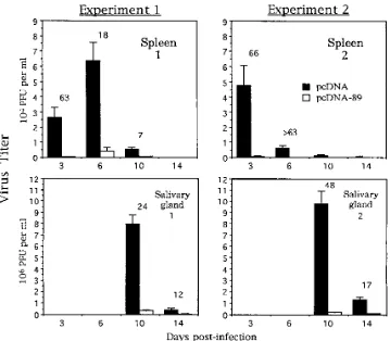
pcDNA-89 immunization reduces viral titers in the spleen and salivary glands.In view of the inherent variability in using survival as an endpoint, we investigated the effect of naked DNA immunization on viral replication when mice were in-jected intraperitoneally with a sublethal dose of virus. For these studies, we chose to examine viral titers in the spleen and salivary gland. BALB/c mice were immunized with pcDNA or
pcDNA-89 as described above and then challenged with 43
104PFU of MCMV. At 3, 6, 10, and 14 days postinfection, the
[image:3.612.70.301.74.240.2]spleens and salivary glands from 3 or 4 mice in each group were harvested, and the virus titer in these organs was assessed by plaque assay on NIH 3T3 cells. The results of two separate experiments are shown in Fig. 3. The progression of the infec-tion in mice immunized with pcDNA alone was identical to that seen in previous studies with mice that had received no immunizations (21). Peak viral replication occurred at days 3 to 6 in the spleen and at day 10 in the salivary gland. In
[image:3.612.318.557.602.703.2]FIG. 1. Recombinant eukaryotic vector pcDNA-89 and its expression. (A) pcDNA-89 is the recombinant eukaryotic expression vector used in our study. It contains the MCMV IE1 gene flanked by the HCMV IE promoter and simian virus 40 (SV40) sequences necessary for correct splicing and transcription ter-mination. (B) COS-7 cells were transfected with 10mg of pcDNA (as vector-alone control) or pcDNA-89 as described in Materials and Methods. Forty-eight hours later IE1 gene expression was assessed by Western blot analysis using a mouse MCMV hyperimmune serum and the ECL detection system. NIH 3T3 cells either were left uninfected or were infected with MCMV or vacc-89, the pp89-expressing recombinant vaccinia virus described in the text. MCMV and vacc-89 are positive controls representing the MCMV proteins expressed in NIH 3T3 cell extracts 48 and 75 h after infection, respectively.
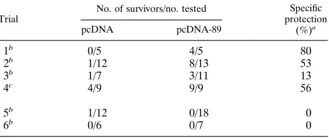
TABLE 1. Protection of BALB/c mice from lethal MCMV challenge by pcDNA-89 immunization
Trial
No. of survivors/no. tested Specific protection
(%)a
pcDNA pcDNA-89
1b 0/5 4/5 80
2b 1/12 8/13 53
3b 1/7 3/11 13
4c 4/9 9/9 56
5b 1/12 0/18 0
6b 0/6 0/7 0
aMean for trials 1 through 4, 45%. bChallenge, 33105PFU (23LD
50). cChallenge, 2.53105PFU.
on November 9, 2019 by guest
http://jvi.asm.org/
contrast, mice immunized with pcDNA-89 showed significant decreases in the amounts of virus present in both organs. At the time of peak viral replication, titers in the spleens of
im-munized mice were reduced 18- to.63-fold while those in the
salivary gland were 24- to 48-fold lower. For comparative pur-poses, a subgroup of the mice immunized as part of experiment
1 were also challenged with a lethal dose of virus (2.53 105
PFU). The results (trial 4 in Table 1) show that, in this group, all of the mice (a total of nine) immunized with pcDNA-89 survived the lethal MCMV infection, while only four of nine mice immunized with pcDNA alone survived. Taken together, these results demonstrate that immunization with pcDNA-89 limits viral replication in target organs and confers protection against the lethal effects of the virus.
pcDNA-89 immunization induces a low-level antibody re-sponse.To determine the serum antibody responses elicited by pcDNA-89 immunization, mice were immunized as described above, and sera were collected 1 week after the last immuni-zation. The sera from individual mice were then used to probe a Western blot of proteins isolated from MCMV-infected NIH 3T3 cells at 48 h postinfection. The results of a representative experiment are shown in Fig. 4 with sera obtained from the mice immunized in trial 4 (Table 1) and experiment 1 (Fig. 3). Of the eight sera from mice immunized with pcDNA-89 only one showed evidence of an antibody response against pp89. In other experiments, we also found that only a limited number of mice immunized with pcDNA-89 showed any evidence of se-roconversion (data not shown). As expected, antibodies to pp89 were not detected in any of the sera obtained from mice immunized with pcDNA alone. These results indicate that little if any humoral response is induced following immuniza-tion.
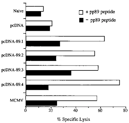
pcDNA-89 immunization induces a CTL response.In view of the lack of clear evidence of an immune response as as-sessed by seroconversion, we proceeded to determine whether pcDNA-89 immunization generated a CTL response using splenocytes from the same group of immunized mice which, as described above, were either subjected to a sublethal challenge of MCMV (experiment 1 in Fig. 3) or a lethal challenge (trial 4 in Table 1). At 7 weeks after the last immunization, spleno-cytes from one mouse immunized with pcDNA and from four mice immunized with pcDNA-89 were harvested and stimu-lated in vitro for 5 days with syngeneic spleen cells (stimula-tors) previously treated with mitomycin and pulsed with the
pp89 nonapeptide NH2-YPHFMPTNL-COOH. As a positive
[image:4.612.145.475.68.358.2]control, splenocytes were also harvested from an unimmunized mouse which had been infected 3 weeks previously with a sublethal dose of MCMV. Splenocytes from an uninfected and unimmunized naive mouse served as a negative control. After incubation for an additional 2 days with recombinant interleu-kin-2, the effector cells were harvested and tested in a conven-tional cytotoxicity test against P815 target cells or P815 target cells that had been pulsed with the nonapeptide. As shown in Fig. 5, a strong, pp89-specific CTL response was detected only in those mice immunized with pcDNA-89 or MCMV, with the percentage of specific lysis being 55 to 75% (E/T ratio, 10:1) or 57% (E/T ratio, 6:1), respectively. The percentage of specific lysis for MCMV-infected mice at an E/T ratio 18:1 was 75% (data not shown). In contrast, the specific lysis was only 21% (E/T ratio, 10:1) and 14% (E/T ratio, 6:1) with splenocytes isolated from the mouse immunized with pcDNA or from the naive mouse, respectively. These results show that pcDNA-89 can induce levels of pp89-specific CTLs which, when assayed
FIG. 2. Survival of pcDNA-89-immunized mice following challenge with different amounts of MCMV. BALB/c mice were immunized with pcDNA or pcDNA-89 as previously described. Six weeks later mice were challenged with different LD50s of MCMV, and survival was assessed for 34 days. These mice are from the group
challenged 2 weeks after the last immunization (trial 5 in Table 1).
on November 9, 2019 by guest
http://jvi.asm.org/
following in vitro stimulation, are comparable to those induced by the MCMV infection.
DISCUSSION
In this report, we show that intradermal injection of plasmid DNA expressing the MCMV 89-kDa IE1 phosphoprotein can immunize BALB/c mice against subsequent MCMV infection. Vaccinated mice showed a 13 to 80% specific protection (mean, 45%) against lethal challenge and decreases in spleen and salivary gland replication of as much as 66- and 48-fold, respectively, relative to mock-immunized controls. In our ini-tial characterization of the immune mechanisms underlying the protection of DNA-vaccinated mice, we found that pp89-spe-cific CTLs were elicited while detectable anti-pp89 antibody titers were only sporadically induced.
Although immunization with pcDNA-89 clearly provided protection to BALB/c mice from subsequent challenge of up to
23LD50of the highly virulent K181 strain of MCMV, the level
of protection in the different trials was quite variable. In more successful immunization trials, the level of protection was com-parable to that obtained following immunization with the pp89-expressing vaccinia virus-based vectors pp89-vacc (data not shown) or MCMV-ie1-vacc (15). However, in other trials there appeared to be little or no protection. This trial-to-trial variability in the level of protection against lethal challenge has commonly been seen with DNA immunizations against several viruses. Excluding the consistency achieved by gene gun
vac-cinations (11), reports often show maximal protection levels from DNA vaccination of 80 to 100% with the average of multiple trials being closer to 50% (27, 32, 38, 43). It has also been previously noted that even the survival rate of
pp89-FIG. 3. MCMV replication in spleen and salivary glands is inhibited in pcDNA-89-immunized mice. BALB/c mice were immunized with pcDNA or pcDNA-89 as previously described. Two weeks later, mice were challenged with a sublethal dose (43104
[image:5.612.129.488.67.381.2]PFU) of MCMV. At days 3, 6, 10, and 14, the spleens and salivary glands were harvested and virus titer was assessed by plaque assay using NIH 3T3 cells. For each time point, titer values presented are the geometric means of either three (experiment 1) or four (experiment 2) independent mouse organ titers, with error bars indicating the standard error of the geometric mean. Fold reductions of the organ titers in pcDNA-89-immunized mice relative to vector-alone-immunized controls are indicated above the bars. The mice used for experiment 2 were from the same group as those immunized in trial 4 (Table 1). The limit of sensitivity of the assay was 100 PFU per organ.
FIG. 4. Antibody response in pcDNA-89-immunized mice. BALB/c mice were immunized with pcDNA or pcDNA-89 as previously described and then bled 1 week after the last immunization. Seroconversion of three pcDNA-im-munized mice and eight pcDNA-89-impcDNA-im-munized mice was assessed by Western blot analysis using a Mini-PROTEAN II multiscreen apparatus. Protein extracts made from NIH 3T3 cells 48 h post-MCMV infection were subjected to SDS-PAGE and electrophoretic transfer to nitrocellulose. A 1:25 dilution of each serum was incubated separately on the nitrocellulose, and bound antibodies were detected by ECL. Mice used in this analysis were from the same group as those immunized in trial 4 (Table 1).
on November 9, 2019 by guest
http://jvi.asm.org/
expressing vaccinia virus-immunized mice showed consider-able variability (15). One source of variability in our experi-ments most likely stems from the heterogeneity of our salivary gland-derived MCMV stocks. Even among aliquots of a single preparation, virus may aggregate in the crude extracts, making difficult the delivery of a precise infectious-challenge dose. This problem is exemplified by the results of trial 5 (Table 1 and Fig. 2), in which there was no protection in the first chal-lenge group but 20% specific protection in a second group challenged with the same viral preparation. Furthermore, Fig. 2 demonstrates that there is a relatively narrow window in which the challenge dose is optimal for revealing protection against lethality. The difference in protection between trial 5 and the multiple challenge dose trials (Fig. 2) could also have been due to the difference in timing of the challenge, as the mice in Fig. 2 had an additional 4 weeks postimmunization to develop protective responses. Even when steps were taken to ensure the homogeneity of the viral stocks, other factors such as mouse-to-mouse variability, number and activation state of antigen-presenting cells proximal to the injection site, variability intrinsic to intradermal DNA and intraperitoneal virus injection, and overall immunogenicity of plasmid DNA-expressed pp89 may have confounded some of our lethal-challenge experiments.
Because of the difficulties in using survival as a sole means of measuring the efficacy of DNA immunization, we examined the effects of the vaccine on viral replication in spleen and salivary gland tissues after a sublethal challenge. The greatest effect of the pcDNA-89 immunization on replication was a 65-fold average reduction in spleen titers on day 3 postinfec-tion. At this time postinfection, the spleen in BALB/c mice is a major site of primary MCMV replication and titers are ap-proaching maximum levels (21). Natural killer activity is
thought to be important at this time (25), as CD81CTL
ac-tivity reaches a peak at approximately day 10 and is only barely
(21). At this time, day 10 postinfection, there was a 36-fold average reduction in salivary gland titer in mice immunized with pcDNA-89. Since clearance of MCMV virions from
sali-vary glands is mediated by CD41T lymphocytes (16), the titer
reduction in this tissue may be due to control of primary replication leading to fewer virions disseminating to the sali-vary glands. Plasmid immunization has been shown to prime
CD41T lymphocytes in mice (20), but we have not yet
deter-mined whether this specific T-lymphocyte subset was primed in our pcDNA-89-immunized animals.
As a qualitative measure of the generation of anti-pp89 antibodies produced in the immunized mice, we probed im-munoblots of MCMV-infected NIH 3T3 cells. Although these experiments are not precisely quantitative, they nevertheless indicated that only a low percentage of mice seroconverted following pcDNA-89 injections. Furthermore, analysis of sera collected throughout our trials showed various low levels of pp89 antibody, with animals producing the highest anti-body titers showing no greater incidence of survival (not shown). Accordingly, Jonjic´ and coworkers (15) showed that antisera produced in mice immunized with vaccinia virus ex-pressing pp89 conferred no protection against MCMV when adoptively transferred to unimmunized mice. Although the antibodies we detected are probably not effective in vivo for controlling the acute infection, their presence can serve as an indicator of the immune response. The generation of anti-pp89 antibodies confirms that, at least in some animals, the plasmid DNA was expressed and the resultant polypeptide was cor-rectly presented to B lymphocytes. We assessed the total anti-pp89 immunoglobulin in our assay since seroconversion was infrequent and anti-pp89 antibody titers were usually low when present, and thus we do not know which isotypes were raised by our plasmid immunization. However, others have found that immunoglobulin G is the predominant class induced by
this method (11, 23). A CD41helper T response is thought to
be necessary for immunoglobulin gene rearrangement and class switch in B lymphocytes and subsequently for the pro-duction of immunoglobulin G (12, 39). Any specific
stimula-tion of CD41cells by our vaccine, however, may be relevant
only if Th1 help results, since protection elicited by pp89-based
immunization is mediated by CD81T lymphocytes.
Protection of BALB/c mice from MCMV challenge by re-combinant vaccinia virus expressing pp89 has been shown to be
mediated primarily by CD81 T lymphocytes (15). A single
CTL epitope on pp89 was mapped to a nonapeptide region in the fourth exon of the IE1 gene, which could be recognized in vitro by pp89-specific CTLs and could also induce pp89-spe-cific CTLs in vivo (9). To determine if any pp89-spepp89-spe-cific CTLs were elicited from our plasmid injections, we measured the CTL activity of splenocytes from pcDNA-89-vaccinated mice using H-2 matched target cells presenting this nonapeptide. Our results show that mice immunized with pcDNA-89 mount a specific CTL response that is comparable to that generated in MCMV-immunized mice. These CTLs were specific to the same nonapeptide region of pp89 protective in recombinant vaccinia virus immunization, though it has not yet been deter-mined if CTLs specific to other epitopes of pp89 were gener-ated by plasmid DNA vaccination. In addition, the CTL
re-FIG. 5. CTL response in pcDNA-89 immunized mice. BALB/c mice were immunized with pcDNA or pcDNA-89 as previously described. Seven weeks after the last immunization, splenocytes were harvested and a standard 5-h51
Cr release assay was performed with P815 cells or cells pulsed with 5mg of the pp89 nonapeptide. As a negative control, we used splenocytes from naive, 8-week-old BALB/c mice. As a positive control, we used effector splenocytes harvested 3 weeks after the intraperitoneal challenge of mice with 43104
PFU of tissue culture-passaged MCMV. The E/T ratio for pcDNA- and pcDNA-89-immunized mice was 10:1, and for naive and MCMV-infected mice it was 6:1. Mice used in this analysis were from the same group as those immunized in trial 4 (Table 1).
on November 9, 2019 by guest
http://jvi.asm.org/
[image:6.612.74.283.69.268.2]sponses in vaccinated animals were assessed 5 weeks after the challenge date of the mice in trial 4 (Table 1) and organ titer experiment 2 (Fig. 3). Thus, lower CTL activities at the time of challenge compared with the time of CTL assay could explain the variable survival of pcDNA-89-immunized animals. The question of whether pcDNA-89 (or pp89) can raise a signifi-cant CTL response or confer protection in mice with other H-2 haplotypes has not yet been answered. Of interest, naked DNA immunization against the hepatitis B virus small surface anti-gen has been shown to raise a specific CTL response in an H-2b strain which could not be primed with soluble antigen or a recombinant vaccinia virus vector (33). Thus, this technique may be ideal for assessing the protective efficacy of MCMV antigens across different genetic backgrounds. However, the variable protection against lethal infection in our animals sug-gests that, although this antigen and delivery system produces a strong immune response, there are still unknown variables which influence the overall level.
Nucleic acid immunization using expression plasmids encod-ing viral surface glycoproteins or nucleocapsid antigens has been shown to induce both CTL and antibody responses and confer protection from DNA and RNA viruses. We have found that plasmid DNA expressing the nonstructural IE1 protein of MCMV can be used to protect BALB/c mice against lethal MCMV infection with nearly the efficiency of the pp89 vaccinia virus recombinant. It will be of interest to further delineate the specific immune cell types stimulated by plasmid DNA immu-nization and compare these immune responses with those pro-duced by vaccinia virus-based immunization and MCMV in-fection itself. Plasmid immunization conceivably could reveal other protective structural and nonstructural MCMV antigens which would otherwise be difficult to identify or labor-intensive to manipulate by other immunization strategies.
ACKNOWLEDGMENTS
We thank Laura Carter, Javier Herna´ndez, and Fernando Prados Mondejar for assistance with the CTL assays and Eyal Raz for helpful advice regarding the plasmid DNA immunization protocol. We also acknowledge members of the laboratory for critical reading of the manuscript.
Juan Carlos Gonza´lez Armas was the recipient of a fellowship from the Ministerio de Agricultura, Pesca y Alimentacio´n (INIA), of Spain. This work was supported by NIH grant AI20954 (D. H. Spector). Lee D. Cranmer was supported by a grant from the Life and Health Medical Research Fund and by NIH predoctoral training grant GM07198.
REFERENCES
1. Acsadi, G., G. Dickson, D. R. Love, A. Jani, F. S. Walsh, A. Gurusinghe, J. A.
Wolff, and K. E. Davies.1991. Human dystrophin expression in mdx mice after intramuscular injection of DNA constructs. Nature (London) 352:815– 818.
2. Aida, Y., and M. J. Pabst. 1990. Removal of endotoxin from protein solutions by phase separation using Triton X-114. J. Immunol. Methods 132:191–195. 3. Barry, M. A., W. C. Lai, and S. A. Johnston. 1995. Protection against mycoplasma infection using expression-library immunization. Nature (Lon-don) 377:632–635.
4. Bourne, N., L. R. Stanberry, D. I. Bernstein, and D. Lew. 1996. DNA immunization against experimental genital herpes simplex virus infection. J. Infect. Dis. 173:800–807.
5. Bruggeman, C. A. 1993. Cytomegalovirus and latency: an overview. Virchows Arch. B 64:325–333.
6. Cooney, E. L., A. C. Collier, P. D. Greenberg, R. W. Coombs, J. Zarling, D. E.
Arditti, M. C. Hoffman, S. L. Hu, and L. Corey.1991. Safety of and immu-nological response to a recombinant vaccinia virus vaccine expressing HIV envelope glycoprotein. Lancet 337:567–572.
7. Cox, G. J., T. J. Zamb, and L. A. Babiuk. 1993. Bovine herpesvirus 1: immune responses in mice and cattle injected with plasmid DNA. J. Virol.
67:5664–5667.
8. Cranmer, L. D., C. Clark, and D. H. Spector. 1994. Cloning, characteriza-tion, and expression of the murine cytomegalovirus homologue of the human
cytomegalovirus 28-kDa matrix phosphoprotein (UL99). Virology 205:417– 429.
9. Del Val, M., H. Volkmer, J. B. Rothbard, S. Jonjic´, M. Messerle, J.
Schicke-danz, M. J. Reddehase, and U. H. Koszinowski.1988. Molecular basis for cytolytic T-lymphocyte recognition of the murine cytomegalovirus immedi-ate-early protein pp89. J. Virol. 62:3965–3972.
10. Elliott, R., C. Clark, D. Jaquish, and D. H. Spector. 1991. Transcription analysis and sequence of the putative murine cytomegalovirus DNA poly-merase gene. Virology 185:169–186.
11. Fynan, E. F., R. G. Webster, D. H. Fuller, J. R. Haynes, J. C. Santoro, and
H. L. Robinson.1993. DNA vaccines: protective immunizations by paren-teral, mucosal, and gene-gun inoculations. Proc. Natl. Acad. Sci. USA 90: 11478–11482.
12. Gascan, H., J. F. Gauchat, M. G. Roncarolo, H. Yssel, H. Spits, and J. E. de
Vries.1991. Human B cell clones can be induced to proliferate and to switch to IgE and IgG4 synthesis by interleukin 4 and a signal provided by activated CD41T cell clones. J. Exp. Med. 173:747–750.
13. Ghiasi, H., S. Cai, S. Slanina, A. B. Nesburn, and S. L. Wechsler. 1995. Vaccination of mice with herpes simplex virus type 1 glycoprotein D DNA produces low levels of protection against lethal HSV-1 challenge. Antiviral Res. 28:147–157.
14. Ho, M. 1992. Cytomegalovirus biology and infection, 2nd ed. Plenum Pub-lishing Co., New York.
15. Jonjic´, S., M. del Val, G. M. Keil, M. J. Reddehase, and U. H. Koszinowski. 1988. A nonstructural viral protein expressed by a recombinant vaccinia virus protects against lethal cytomegalovirus infection. J. Virol. 62:1653–1658. 16. Jonjic´, S., I. Pavic´, P. Lucˇin, D. Rukavina, and U. H. Koszinowski. 1990.
Efficacious control of cytomegalovirus infection after long-term depletion of CD81T lymphocytes. J. Virol. 64:5457–5464.
17. Koszinowski, U. H., G. M. Keil, H. Schwarz, J. Schickedanz, and M. J.
Reddehase.1987. A nonstructural polypeptide encoded by immediate-early transcription unit 1 of murine cytomegalovirus is recognized by cytolytic T lymphocytes. J. Exp. Med. 166:289–294.
18. Koszinowski, U. H., M. J. Reddehase, G. M. Keil, and J. Schickedanz. 1987. Host immune response to cytomegalovirus: products of transfected viral immediate-early genes are recognized by cloned cytolytic T lymphocytes. J. Virol. 61:2054–2058.
19. Koszinowski, U. H., M. J. Reddehase, G. M. Keil, H. Volkmer, S. Jonjic, M.
Messerle, M. del Val, W. Mutter, K. Munch, and B. Buhler.1987. Molecular analysis of herpesviral gene products recognized by protective cytolytic T lymphocytes. Immunol. Lett. 16:185–192.
20. Manickan, E. R., J. D. Rouse, Z. Yu, W. S. Wire, and B. T. Rouse. 1995. Genetic immunization against herpes simplex virus. Protection is mediated by CD4 T lymphocytes. J. Immunol. 155:259–265.
21. Mercer, J. A., and D. H. Spector. 1986. Pathogenesis of acute murine cyto-megalovirus infection in resistant and susceptible strains of mice. J. Virol.
57:497–504.
22. Meyers, J. D. 1984. Cytomegalovirus infection following marrow transplan-tation: risk, treatment, and prevention. Birth Defects Orig. Artic. Ser. 20: 101–117.
23. Mor, G., D. M. Klinman, S. Shapiro, E. Hagiwara, M. Sedegah, J. A.
Norman, S. L. Hoffman, and A. D. Steinberg. 1995. Complexity of the cytokine and antibody response elicited by immunizing mice with Plasmo-dium yoelii circumsporozoite protein plasmid DNA. J. Immunol. 155:2039– 2046.
24. Pedroza Martins, L., L. L. Lau, M. S. Asano, and R. Ahmed. 1995. DNA vaccination against persistent viral infection. J. Virol. 69:2574–2582. 25. Quinnan, G., Jr., N. Kirmani, A. H. Rook, J. F. Manischewitz, L. Jackson, G.
Moreschi, G. W. Santos, R. Saral, and W. H. Burns.1982. Cytotoxic T cells in cytomegalovirus infection: HLA-restricted T-lymphocyte and non-T-lym-phocyte cytotoxic responses correlate with recovery from cytomegalovirus infection in bone-marrow-transplant recipients. N. Engl. J. Med. 307:7–13. 26. Quinnan, G. V., J. F. Manischewitz, and F. A. Ennis. 1978. Cytotoxic T lymphocyte response to murine cytomegalovirus infection. Nature (London)
273:541–543.
27. Raz, E., D. A. Carson, S. E. Parker, T. B. Parr, A. M. Abai, G. Aichinger,
S. H. Gromkowski, M. Singh, D. Lew, M. A. Yankauckas, S. M. Baird, and G. H. Rhodes.1994. Intradermal gene immunization: the possible role of DNA uptake in the induction of cellular immunity to viruses. Proc. Natl. Acad. Sci. USA 91:9519–9123.
28. Reddehase, M. J., and U. H. Koszinowski. 1984. Significance of herpesvirus immediate early gene expression in cellular immunity to cytomegalovirus infection. Nature (London) 312:369–371.
29. Reddehase, M. J., W. Mutter, K. Munch, H. J. Buhring, and U. H.
Koszi-nowski.1987. CD8-positive T lymphocytes specific for murine cytomegalo-virus immediate-early antigens mediate protective immunity. J. Virol. 61: 3102–3108.
30. Reddehase, M. J., J. B. Rothbard, and U. H. Koszinowski. 1989. A pen-tapeptide as minimal antigenic determinant for MHC class I-restricted T lymphocytes. Nature (London) 337:651–653.
31. Reddehase, M. J., F. Weiland, K. Mu¨nch, S. Jonjic, A. Lu¨ske, and U. H. Koszinowski.1985. Interstitial murine cytomegalovirus pneumonia after
on November 9, 2019 by guest
http://jvi.asm.org/
sequence requirements and trans-acting functions necessary for regulated expression of a human cytomegalovirus early gene. J. Virol. 62:3463–3473. 36. Steinman, R. M. 1991. The dendritic cell system and its role in
immunoge-nicity. Annu. Rev. Immunol. 9:271–296.
37. Towbin, H., T. Staehelin, and J. Gordon. 1979. Electrophoretic transfer of proteins from polyacrylamide gels to nitrocellulose sheets: procedure and some applications. Proc. Natl. Acad. Sci. USA 76:4350–4354.
38. Ulmer, J. B., J. J. Donnelly, S. E. Parker, G. H. Rhodes, P. L. Felgner, V. J.
Dwarki, S. H. Gromkowski, R. R. Deck, C. M. DeWitt, A. Friedman, L. A. Hawe, K. R. Leander, D. Martinez, H. C. Perry, J. W. Shiver, D. L.
Mont-41. Wolff, J. A., R. W. Malone, P. Williams, W. Chong, G. Acsadi, A. Jani, and
P. L. Felgner.1990. Direct gene transfer into mouse muscle in vivo. Science
247:1465–1468.
42. Xiang, Z. Q., S. Spitalnik, M. Tran, W. H. Wunner, J. Cheng, and H. C. Ertl. 1994. Vaccination with a plasmid vector carrying the rabies virus glycopro-tein gene induces protective immunity against rabies virus. Virology 199: 132–140.
43. Yokoyama, M., J. Zhang, and J. L. Whitton. 1995. DNA immunization confers protection against lethal lymphocytic choriomeningitis virus infec-tion. J. Virol. 69:2684–2688.