A DESCRIPTIVE STUDY OF LESIONS FOLLOWING THE LINES OF BLASCHKO
Dissertation Submitted in partial
fulfillment of the university regulations for
MD DEGREE IN
DERMATOLOGY, VENEREOLOGY AND LEPROSY
(BRANCH XII A)
APRIL 2012
THE TAMILNADU DR.M.G.R. MEDICAL UNIVERSITY
CERTIFICATE
This is to certify that this dissertation entitled ‘A DESCRIPTIVE STUDY OF LESIONS FOLLOWING THE LINES OF BLASCHKO’ submitted by Dr. SHEERJA BALI to The Tamil Nadu Dr.M.G.R. Medical University, Chennai is in partial fulfillment of the requirement for the award of M.D. [DERMATO VENEREO LEPROLOGY] and is a bonafide research work carried out by her under direct supervision and guidance.
Dr. A. S. Krishnaram. M.D., D.D., Professor and Head
Department of Dermatology Madurai Medical College & Government Rajaji Hospital Madurai.
Dr. D. Amal Raja. M.D., D.V., Professor and Head
Department of STD,
DECLARATION
I, Dr. SHEERJA BALI solemnly declare that I carried out this work on ‘A
DESCRIPTIVE STUDY OF LESIONS FOLLOWING THE LINES OF BLASCHKO’ at Department of Dermatology, Government Rajaji Hospital during the period of Oct 2009 – Sep 2011.
I also declared this bonafide work or a part of this work was not submitted by me or any other for any award, degree and diploma to any university, board either in India or abroad.
This is submitted to The Tamil Nadu Dr. M.G.R Medical University, Chennai in partial fulfillment of the rules and regulation for M.D., [D.V.L] Degree examination.
ACKNOWLEDGEMENT
Gratitude cannot be expressed through words. True, but unexpressed gratefulness weighs heavily on one’s heart. I may be permitted here to record valuable guidance, help, co- operation and encouragement from my teachers, colleagues and various other persons directly or indirectly involved in the preparation of this dissertation.
At the very beginning, I thank God almighty for bestowing upon me his blessings to help me bring this task to completion.
I would like to acknowledge my thanks and sincere gratitude to Prof. Dr. A. S. Krishnaram M.D., D.D., Professor and Head of the Department of Dermatology, Madurai Medical College, Madurai, for his valuable guidance and encouragement throughout the study and also during my postgraduate course. His clinical approach has been inspirational to me.
I express my thanks and deep sense of gratitude to Prof. Dr. S. Krishnan, M.D., D.D. for his valuable advice and encouragement. It is from him that I learnt the valuable lesson for life - humbleness.
preparation of this dissertation and also during my postgraduate course. I learnt to be empathetic towards patients and to be thorough in my work from her.
I would like to acknowledge my thanks to Prof. Dr. D. Amal Raja, M.D.,D.V., Head of the Department of Venereology, for his valuable guidance.
My heartful thanks to my teachers Dr. K. Senthil kumar, Dr. R. Kothandaraman, Dr. Sathesh, Dr. Balaji Adityan, Assistant professors
for their timely advice and patience during the preparation of this dissertation.
I would also like to acknowledge my thanks to Dr. M. S. Adityan, Dr. M. Senthil Kumar, Dr. Ayamperumal, Assistant professors of Department of STD, for their constant support during the period of my study.
I am extremely thankful to the Dean, Principal and Medical Superintendent for permitting me to use the hospital materials for this study.
I owe my thanks to my fellow postgraduate colleagues for their constant help and constructive criticism.
TABLE OF CONTENTS
S. NO PARTICULARS PAGE NO.
1 INTRODUCTION 1
2 AIM OF THE STUDY 3
3 REVIEW OF LITERATURE 4
4 MATERIALS AND METHODS 38
5 OBSERVATIONS AND RESULTS 40
6 DISCUSSION 53
7 SUMMARY & CONCLUSION 73 ANNEXURES
- BIBLIOGRAPHY - PHOTOGRAPHS - PROFORMA - MASTER CHART
INTRODUCTION
The concept of Blaschko’s lines was first put forth by Alfred Blaschko, a private practioner in dermatology, when he presented his findings on the distribution patterns of linear skin at the German Dermatological Society meeting in Breslau in 1901.1 In his original description he referred to Blaschko’s lines as ‘a system of lines on the human skin which the linear nevi and dermatosis follow.’1 The pattern of Blaschko’s lines does not correlate with the distribution of cutaneous nerves, blood vessels, or lymphatics and is distinct from pigmentary demarcation lines . Two mechanisms have been proposed to explain Blaschko’s lines: mosaicism and chimerism.
A number of congenital and acquired conditions follow these special lines. Almost all epidermal nevi follow Blaschko’s lines. Lesions in various X-linked disorder’s like incontinentia pigmenti , Goltz syndrome and chromosomal disorders like hypomelanosis of Ito also respect these lines. This pattern followed by these congenital diseases helps in their diagnosis not only in the newborn and infant, but also in adults.
AIM OF THE STUDY
1) To study and describe the patterns of Blaschko’s lines encountered in various dermatoses.
REVIEW OF LITERATURE
DEFINITION-
The lines of Blaschko represent a pattern assumed by many different nevoid and acquired skin diseases on the human skin and mucosae.2 These lines are characteristic of mosaic conditions of the epidermis and probably represent the routes of ectodermal cell migration from the neural crest. These were first described by Alfred Blaschko ( 1858- 1922) , a German Dermatologist in 1901.1These are also termed as cutaneous lines of embryogenesis.3These lines do not correspond to other patterns such as Langer’s lines of cleavage,4Voigt’s lines ( borders between areas of innervations by peripheral cutaneous nerves,5 embryonic clefts,6 pigmentary demarcation lines,7or the lines of lymphatic drainage or blood supply.8 These do not correspond to nervous structures.8
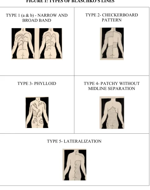
TYPES- (Figure 1)
Four main types of Blaschko’s lines have been described9: TYPE 1a-NARROW BANDS
TYPE 1b- BROAD BANDS
TYP FIG
E 1 (a & b BROA
TYPE
3-GURE 1: T
b) - NARRO AD BAND - PHYLLO TY TYPES OF OW AND D OID
YPE 5- LA
F BLASC T T ATERALIZ HKO’S L
TYPE 2- C P
[image:12.612.69.548.102.706.2]TYPE 3- PHYLLOID PATTERN- Multiple leaf-like or oblong macules reminiscent of the floral ornaments of art nouveau style.11Clinical example is novel neuro- cutaneous syndrome in the form of phylloid hypomelanosis.12
TYPE 4- PATCHY PATTERN WITHOUT MIDLINE SEPARATION
A fifth type, LATERALIZATION13 has been described which is classically seen in CHILD syndrome wherein a clear cut midline demarcation is seen.14
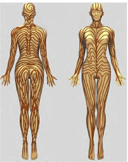
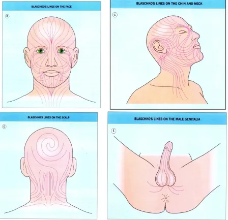
DISTRIBUTION PATTERN – (Figures 2 & 3)
These assume a V-shape over the upper spine, an S-shape on the abdomen, and an inverted U-shape from the breast area onto the upper arm. There are perpendicular lines down the front and back of the lower extremities.1The lines of Blaschko are less well defined on the head and neck.15Happle et al added lines to the posterior scalp,16,17 while Bolognia et al further delineated the lines on the lateral aspect of the face and neck.15Brown and Gorlin mentioned vertical striations on the lips, linear midline lesions on the hard and soft palate, and linear unilateral and / or midline bands on the tongue in patients with epidermal nevi.18
FIGURE 3: DISTRIBUTION PATTERN OF BLASCHKO’S – HEAD, NECK & GENITALS
chondrodysplasia punctata.23Witkop also described alternating vertical band of opaque white and translucent (normal- appearing) enamel on the central incisor of women heterozygous for X- linked hypo - maturation amelogenesis imperfecta.20
BASIS OF DISTRIBUTION PATTERN OF THE LINES OF BLASCHKO
One exp is that t but is embryo Figure 4: Howeve lines.24 lines.8A the stag more w longer t lateraliz The pa affected planation g transversal interfered .13 (Figure
Proposed exp er no singl Interesting Assuming th ge of devel widely disp the lines of zation & or attern of c d.26 (Table
given for th l proliferat
with the 4)
planation of th le theory a gly, cutan
hat the pat lopment at persed and f migration rganogene cutaneous I) he fountain tion of pre
longitudi
he fountain-li as yet clea neous mosa
ttern is det t which mo
more intim n.26Its timin
sis will als mosaicism
n-like patte ecursor cel inal growt
ike pattern of arly elucid aicism do termined b osaicism a mately mi ng in relat so influenc m also va
ern of Blas lls starts fr th and in
f Blaschko’s l dates the lo
es not alw by cell mig arises. The xed the m ion to the ce the patte aries acco
schko’s lin from the pr
creasing f
ines on the ba ocalization
ways follo gration, it w
earlier the mosaic clon processes ern.26
ording to
nes on the b rimitive st flexion of
ack.
TABLE I- EXPECTED PATTERN OF CUTANEOUS MOSAICISM ACCORDING TO CELL TYPE CELL AFFECTED EMBRYONIC MOVEMENT EXPECTED PATTERN MELANOBLAST Single cell migration Checker board, bands or
phylloid KERATINOCYTE Directional proliferation
following surface forces
Blaschko - bands
Sometimes, a severe linear lesion is superimposed on a background of milder generalized disease. Happle attributes this to mosaic loss of heterozygosity, and terms it Type 2 mosaicism. Thus, an individual who has inherited an autosomal dominant disorder suffers a postzygotic mutation affecting the other allele (‘second hit’) resulting in a linear area where the genetic burden is doubled. In fact, many disorders show dense or confluent linear lesions even in the absence of background lesions, for example, Darier’s disease, psoriasis and lichen planus. Type 2 mosiacism has now been proven for Hailey- Hailey disease.27
SKIN CONDITIONS FOLLOWING THE LINES OF BLASCHKO- (Table II and Figure 5) TABLE II- MOSIAC SKIN CONDITIONS CLASSIFIED ACCORDING TO THE NATURE OF THE GENERALIZED CONDITION
X- LINKED DOMINANT
LETHAL IN MALES Incontinentia pigmenti Goltz syndrome
Conradi—Hunermann—Happle syndrome (chondrodysplasia punctata) CHILD syndrome (congenital
hemidysplasia with ichthyosiform nevus and limb defects)
MIDAS syndrome
Oral-facial-digital syndrome type I X-LINKED RECESSIVE Hypohidrotic ectodermal dysplasia
Menkes disease (carrier females)
X-Iinked reticulate pigmentary disorder [ cutaneous amyloidosis]
Ectodermal dysplasia, hypohidrotic, with immune deficiency (carrier females)
AUTOSOMAL DOMINANT
SINGLE GENE DISORDERS Linear bullous ichthyosiform erythroderma Palmoplantar verrucous nevus
Nevus comedonicus Linear Darier’s disease Linear Halley—Halley disease Linear porokeratosis
Linear basal cell nevi
Segmental neurofibromatosis type 1 Linear angiofibromas
Segmental leiomyoma POSSIBLE AUTOSOMAL
DOMINANT SINGLE GENE DISORDERS Nevoid telangiectasia Linear syringomas Linear trichoepitheliomas Linear eccrine spiradenomas MULTIFACTORIAL INFLAMMATORY
DISORDERS WITH AUTOSOMAL DOMINANT OR POLYGENIC
INHERITANCE
Linear lichen nitidus Lichen striatus ‘Adult blaschkitis’ Linear morphea
Moulin atrophoderma linearis Segmental vitiligo
PRESUMED AUTOSOMAL DOMINANT LETHAL DISORDER RESCUED BY MOSAICISM ( Never Seen In A Generalized Form)
Linear epidermal /sebaceous nevus Epidermal nevus syndromes
PEODDN (porokeratotic eccrine ostial and dermal duct nevus)
Proteus syndrome
Nevus lipomatosis superficialis
Encephalocraniocutaneous lipomatosis Oculocerebrocutaneous syndrome ILVEN (inflammatory linear verrucous
epidermal nevus)
McCune—Albright syndrome Zosteriform lentiginous nevus Localized vascular anomalies Linear fibromatosis
CHROMOSOMAL Hypomelanosis of Ito Nevus depigmentosus
LWNH (linear whorled nevoid hypermelanosis)
A) X-LINKED CONDITIONS FOLLOWING BLASCHKO’S LINES
In women heterozygous for X-linked disorders, mosaicism due to random inactivation (lyonization) may produce skin lesions following Blaschko’s lines. Lyonization occurs synchronously in all cells at about the 1000- cell stage, so the two clones are intimately mixed from the beginning. Therefore, Blaschko’s lines in X- linked disorders are typically narrow and numerous. An exception is CHILD syndrome, which characteristically shows large unilateral blocks of abnormal skin; possibly because the mosaicism is due to later somatic mutation on the X chromosome rather than lyonization.
In males, only X-linked recessive conditions are compatible with survival (e.g. hypohidrotic ectodermal dysplasia). X- linked dominant conditions are usually lethal in males in utero itself (e.g. incontinentia pigmenti and Goltz syndrome). This lethal phenotype can only be rescued by mosaicism.
Some genes on the X chromosome do not undergo random inactivation.29 For example, the gene on the short arm of the X chromosome that encodes steroid sulfatase escapes inactivation. This explains why the epidermis of female carriers of X- linked recessive icthyosis lacks a mosaic pattern of scaling.15
I) INCONTINENTIA PIGMENTI (Synonyms-Bloch- Sulzberger syndrome; Bloch- Siemens syndrome)26
Incontinentia pigmenti (IP) is a rare X-linked dominant genodermatosis, first described by Bloch and Sulzberger in 1920’s.30Nearly all the patients (97%) are females. The name refers to the pathologic finding of pigmentary incontinence, especially in the third stage of the disease. The linear skin lesions reflect mosaicism secondary to X inactivation.
by linear warty streaks, often overlying the distribution of stage I lesions. This stage occurs in approximately 70% of the patients.32 The pigmentary or the third stage peaks between the twelfth and the twenty-sixth week of life and is characterized brown or grey streaks and swirls. The pigmentation is characteristic of the syndrome, and the bizarre ‘Chinese letter pattern’ is diagnostic.33
These streaks follow the lines of Blaschko and have a predilection for the trunk. In about 14% of patients, the third stage is said to occur without evidence of prior stages.32 The hyperpigmentation fades in weeks to years and usually does not persist into adulthood. The fourth stage is characterized by hypopigmented atrophic lesions that occur most commonly on the legs in a linear or reticulated pattern.34In an adult, the hypopigmented streaks may be the only manifestation of IP. Additional manifestations of IP include linear absence of hair and sweat glands, nail dystrophy and nail tumors, asymmetric breast development, supernumerary nipples, missing and conical teeth, microphthalmia, retinal vascular anomalies, cataracts, mental retardation, seizures, spastic hemi/ di/ tetraplegia, optic atrophy, skull anomalies, scoliosis and pulmonary hypertension.
frequent (20% and 30%, respectively) but rarely severe (8% and 7.5%, respectively).35
II) OTHER DISORDERS
Some other X- linked disorders following Blaschko’s lines include Conradi- Hunermann- Happle syndrome, Goltz Syndrome, Congenital Hemidysplasia with Icthyosiform Erythroderma and limb defects ( CHILD Syndrome), Menkes’ syndrome (female carriers), X- linked hypohidrotic ectodermal dysplasia (HED-female carriers), MIDAS syndrome, Oral- facial – digital syndromes (OFDS), X- Linked Reticulate Pigmentary Disorder- ( Partington Cutaneous Amyloidosis), ectodermal dysplasia, hypohidrotic, with immune deficiency.26
B) AUTOSOMAL DOMINANT CONDITIONS FOLLOWING BLASCHKO’S LINES
Mosaicism for autosomal disorders arises during gametogensis (half- chromatid mutation) or after fertilization (somatic mutation). As expected for a denovo mutation, there is generally no antecedent family history; rare exceptions have been attributed to inheritance of an unstable permutation.26
I) VERRUCOUS EPIDERMAL NEVUS
Verrucous epidermal nevi are congenital, non- inflammatory cutaneous hamartomas composed of keratinocytes. Their prevalence in adults is probably 0.1–0.5%, and they occur equally in males and females. 36 They are divided into epidermolytic and non-epidermolytic types based on histopathology and pathogenesis. The epidermolytic verrucous epidermal nevi represents a clone of cells expressing a mutation in one of the BIE genes KRT1 and KRT10 involved in
the pathogenesis of the autosomal dominant bullous ichthyosiform erythroderma (BIE). Recently, FGFR3 mutations associated with severe achondroplasia has been found in non- epidermolytic verrucous epidermal nevi, hence may represent an autosomal dominant lethal disorder rescued by mosaicism.26
developmental anomalies may occur in association with verrucous epidermal nevi of the non- epidermolytic type. They include localized anomalies such as megalopinna and aplasia cutis of the scalp, and syndromes such as epidermal nevus syndrome etc.36
II) ACNE NEVUS AND NEVUS COMEDONICUS SYNDROME
Nevus comedonicus was first described in 1895.38This rare nevus is usually present at birth, but becomes prominent at puberty, when the sebo- follicular element is stimulated by hormones.26
Munro observed that the acneiform lesions of nevus comedonicus and the localized skeletal malformations of nevus comedonicus syndrome are generalized in the autosomal dominant disorder Apert Syndrome. This syndrome is due to mutations on FGFR2 and thus comedo nevus is probably due to mosaicism for FGFR2 mutation.39This may present as several lesions in a linear, unilateral or, more rarely, bilateral distribution.
III) LINEAR DARIER’S DISEASE
morphologically and histologically, linear Darier disease is identical to the generalized form.40
Starink and Woerdeman reported seven cases showing unilateral, linear, or zosteriform patterns, without other findings of DD and suggested the name acantholytic dyskeratotic epidermal nevus.41
IV) OTHER DISORDERS
Other autosomal dominant conditions following Blaschko’s lines include Hailey- Hailey disease, linear porokeratosis, linear basal cell nevus, segmental Neurofibromatosis –type I and linear angiofibromas in Tuberous sclerosis.26
V) OTHER LINEAR BENIGN TUMORS
Syringomas, trichoepitheliomas and eccrine spiradenomas are heritable ectodermal tumors and can occasionally occur in a linear distribution.26
C) LINEAR INFLAMMATORY DISORDERS
linear form of these disorders may therefore reflect mosaicism for a ‘susceptibility’ mutation.26 This loss of heterozygosity may occur from a mutation, deletion, or DNA recombination and lead to the formation of a keratinocyte clone that is more susceptible to development of the skin disease. This concept was first introduced in 1991 to describe linear psoriasis and, since then, has been applied to segmental forms of atopic dermatitis, lichen planus, erythema multiforme, pemphigus vulgaris, vitiligo, granuloma annulare and so on.42,43 Segmental disease may be superimposed on non-segmental lesions, with the segmental lesions usually being more difficult to treat.44
I) LICHEN STRIATUS
( Synonyms- Linear Lichenoid Dermatosis)
Lichen striatus is an asymptomatic, uncommon, self- limited, linear dermatosis of unknown etiology that generally affects children. First described by Balzer and Mercier in 1898, and they termed it ‘lichenoid trophoneurosis’. Forty years later, Senear & Caro proposed the name ‘lichen striatus’.45
Lichen striatus is seen primarily in children between the ages of 4 months and 15 years. In 1988, Burton et al. wrote in their Textbook of Dermatology that LS may
rarely be seen in adults.46 The median age of onset is 2 to 3 years and the vast
females are affected two or three times as frequently as males and that the eruption of LS usually involutes within 1year.48In certain other studies, reported male: female ratio has varied from 1:1.6 to 1:2.49
Although the distribution of lichen striatus along the Blaschko’s lines points to somatic mosaicism, neither the genes involved nor the triggering factors are known.
Environmental agents, particularly viruses, have been implicated, given the predominance of the disorder in young children and its seasonal variation (it appears to be more common in summer and spring). However, to date, a viral association has not been proven via serologic testing or cultures.
In theory, during early fetal development, an aberrant clone(s) of epidermal cells produced by somatic mutation migrated out along the lines of Blaschko. Exposure to an infectious agent (e.g. virus, BCG vaccine) or other precipitant could then break previous tolerance to the aberrant clone inducing a novel membrane antigen.45
Lichen striatus may represent a manifestation of an atopic diathesis. A retrospective analysis by Patrizi A et al of 115 children with lichen striatus
revealed an association with atopic dermatitis in 70 of the cases.47In another series,
There are scattered reports of lichen striatus occurring at the sites of injury (e.g.
the periphery of a burn scar49 rather than along the Blaschko’s lines. Lastly, axial
distribution has been reported in some cases.51
The eruption consists of a continuous or interrupted band composed of discrete or
clustered pink, skin- colored or tan papules that are flat-topped, smooth or scaly,
range in diameter from 2 to 4mm.Infrequently, vesicles may be present.
Hypopigmentation may be prominent, especially in dark- skinned persons.
Typically, there is a single, unilateral streak on an extremity along the Blaschko’s
lines; occasionally, there is a bilateral distribution pattern and /or multiple parallel
bands.45In two-third of patients, an extremity is involved, in particular the arm; in
the remainder, the trunk or face is involved.52, 53 When lesions extend to the ends of
the digits, nail involvement may range from fraying, splitting, onycholysis to total
nail loss.54, 55
The eruption usually appears suddenly, develops fully over days to weeks, and
after several months to a year or more, undergoes spontaneous resolution, leaving
post-inflammatory hypopigmentation.45
II) LINEAR LICHEN PLANUS
Scattered linear lesions often occur in patients with lichen planus (LP) and are a
result of scratching and the Koebner phenomenon.15Less commonly, unilateral
lesions seen along the Blaschko’s lines.56Some authors consider linear LP as an
intermediate entity between LP and lichen striatus. This form has also been
referred as zosteriform but the distribution pattern of LP is not dermatomal (with
the rare exception of the koebnerization of LP into the site of a previous herpes
zoster infection). Linear LP accounts for less than 0.2 percent of all patients with
LP,44 except in Japan, where up to 10% of reported cases are linear.57 Because of
the polygenic nature of LP, family members may have the condition in its non
-segmental form. LP occurs in up to ten percent of first-degree relatives of affected
patients.44
The linear variant of LP can be persistent,58 but occasionally may resolve with post
inflammatory hyperpigmentation.59Usual sites involved are the extremities.60
Isolated linear lesions are more common in childhood.61Linear LP lesions are
usually only a few centimeters in length, but long, narrow linear lesions extending
along the whole length of a limb may occur. Such cases may overlap with
epidermal nevi, and the term lichenoid epidermal nevus has been introduced by
Brownstein et al.61
If the LP lesions extend to the end of a digit, the nail is often affected. Linear
III) SEGMENTAL (LINEAR) VITILIGO
Vitiligo is a multifactorial disorder that occasionally occurs in a linear distribution.
The lesions tend to be broad bands, patches or blocks, corresponding more to
dermatomes than Blaschko’s lines, perhaps in keeping with a neuronal
pathogenesis. This idea is consistent with mosaicism, as the neuronal abnormality
could be mosaic, or alternatively, there could be a clonal susceptibility of
melanocytes to neuronal or other influences.
Compared with symmetric vitiligo, the linear type is earlier in onset, less likely to
spread to other areas of the body, and less frequently associated with other
autoimmune diseases.26
IV) LINEAR MORPHEA
Linear morphea occurs as a linear band, usually with a single unilateral lesion. The
lower extremities are most often involved, followed, in frequency of occurrence,
by the upper extremities, frontal area of the head, and anterior thorax.63The
female-to-male ratio is 4:1. Linear scleroderma tends to affect children and adolescents.
Whether linear morphea follows Blaschko’s lines is controversial.64 Jackson first
described it,2 but later observed that although linear morphea was thought to follow
Blaschko’s lines it was probably dermatomal.65 In a detailed review, Bolognia et al
Subsequently, only few papers described this finding.66Three patients with fronto-
parietal scleroderma showing multiple lesions have been described. The lesions
were in two different lines that seemed to belong to Blaschko's lines.67
However, in many cases of linear morphea, it is not clear whether the distribution
is segmental, dermatomal or following Blaschko’slines.15
V) LINEAR LICHEN SCLEROSUS ET ATROPHICUS
Lichen sclerosus et atrophicus (LSA) is an inflammatory dermatosis of an unclear
pathogenesis.LSA was first described by Hallopeau in 1887.68Darier described the
characteristic histological findings in 1892.69It primarily affects the vulvar,
perineal and perianal skin of prepubertal, perimenopausal and postmenopausal
women.
It is sometimes distributed following Blaschko lines.70 The first case report of a
linear LSA was described in 1995 by Izumi and Tajima.71 Thereafter, a handful of
cases of linear LSA have been reported, among which some developed in a pattern
corresponding to the lines of Blaschko. Kim and Lee 72have summarized 6 cases of
linear LSA along the Blaschko's lines, and this occurred on the trunk, limbs or
face. Out of the 3 reported cases of linear LSA that appeared on the face, one case
showed facial lesion following the Blaschko's line without any oral mucosal lesion,
In the first stages it presents as interfollicular, pearly, polygonal papules, which
merge to form atrophic, sclerotic plaques. In more advanced stages, follicular
hyperkeratosis and telangiectasias are seen.75 Histologically, LSA has a
characteristic pattern.
VI) LINEAR LICHEN PLANUS PIGMENTOSUS
Lichen planus pigmentosus clinically differs from the classic lichen planus by
exhibiting dark brown macules and/or papules, mottled or reticulated
hyperpigmentation and a longer clinical course without scalp, nail, or mucosal
involvement. It is most common in sun-exposed areas such as the face and neck
and the flexural folds, including axillary, inguinal, and submammary regions.76
Some authors observed a striking predominance of lesions in intertriginous
locations, among which axillae are the most common.77 The most common pattern
of pigmentation is diffuse, whereas less common patterns include linear, reticular,
and perifollicular patterns.77
There are few reports in the literature of unilateral linear LPP. All except one of
these cases were females with absence of involvement of mucosa and nails. Their
ages varied from 16- 60 years. The sites involved were predominantly the lower
extremities and the neck in one patient. One case of bilateral linear LPP in a male
patients were asymptomatic and all the lesions in them were along the Blaschko’s
lines alone.78
VII) ‘ADULT BLASCHKITIS’-
(Synonyms- Blaschko linear acquired inflammatory skin eruption (BLAISE); Blaschkitis, or, Idiopathic dermatitis along the lines of Blaschko) 79
It was first described in 1990 by Grosshans and Marot in Bordex, France.80
It presents as pruritic papules and vesicles along multiple lines of Blaschko,
particularly on the trunk. It follows a relapsing course, with individual episodes
resolving spontaneously within days. This disorder may represent an adult
counterpart of lichen striatus, but the histology is more eczematous and less
lichenoid.81
Some authors are trying to find a difference between adult blaschkitis and lichen
striatus, but the discussion remains controversial. In trying to unite these two
entities, the term ‘Blaschko linear acquired inflammatory skin eruption’ (BLAISE)
has been created.
VIII) NEVOID PSORIASIS
Psoriasis occasionally occurs in a ‘nevoid’ form,82 possibly reflecting mosaicism
for a gene responsible for psoriasis. Linear psoriasis coexisting with generalized
psoriasis might co-exist with or develop as an isomorphic phenomenon 83-86 over a
pre-existing inflammatory linear verrucous epidermal nevus (ILVEN). Linear
psoriasis is extremely rare with many early cases having been subsequently
reclassified as ILVEN.
Onset is usually during childhood, but some otherwise typical cases have presented
at birth. In a study of 419 cases of childhood psoriasis from north India by Kumar
B et al, infantile psoriasis (below 1 year of age) accounted for 3.5% of patients, the
youngest being a four day old child, but none had lesions at birth.87 The lesions are
clinically indistinguishable from ordinary psoriasis other than in their distribution,
and are histologically identical. Linear psoriasis can be associated with nail pits
and psoriatic arthritis. This condition responds to anti-psoriatic therapy such as
ultraviolet radiation or dithranol. Linear psoriasis can distinguished from ILVEN
by its minimal pruritus and therapeutic responsiveness.
IX) LINEAR LICHEN NITIDUS
Lichen nitidus (LN) is a disorder of unknown etiology. Most cases occur in
children or young adults. Typical LN papules are minute, pinpoint to pinhead
sized, and have a flat or dome-shaped, shiny surface. They usually remain discrete,
the sites of predilection are the forearms, penis, abdomen, chest and buttocks. The
palms or soles can be involved in form of confluent hyperkeratosis.88
Mucous membrane lesions occur occasionally. Krook 89 described a generalized
case as having mucosal lesions mainly on the hard palate and maxillary alveolar
margins, which consisted of fairly closely grouped, greyish yellow, round, sharply
demarcated, discrete papules up to 1 mm in diameter. Linear LN has been
described by Pringent F et al, but is exceptionally rare.90
X) OTHER LINEAR INFLAMMATORY DISORDERS
Several other inflammatory disorders have been reported in a linear distribution,
suggestive of mosaicism for a susceptibility mutation. They include lupus
erythematosus, fixed eruption, atrophoderma of Pasini and Pierini (linear
atrophoderma of Moulin), chronic lichenoid GVHD, mycosis fungoides and
mucinosis.26
D) LETHAL DISORDERS RESCUED BY MOSAICISM
For some linear disorders no generalized counterparts can be recognized. These
cases probably represent mosaicism for a mutation that would be incompatible
with life if it involved all cells. Such conditions affect both men and women and
represent clonal loss of heterozygosity in a phenotypically normal carrier of a
lethal recessive mutation.
I) NON- EPIDERMOLYTIC VERRUCOUS EPIDERMAL NEVUS- (see verrucous epidermal nevus)
II) EPIDERMAL NEVUS SYNDROME
(Synonyms- Sebaceous nevus syndrome, Schimmelpenning’s syndrome, Feuerstein–Mims
syndrome, Organoid nevus syndrome, Jadassohn’s nevus phakomatosis)
Epidermal nevus syndrome (ENS) describes the association of sebaceous and/or
verrucous nevi with other developmental defects, particularly of the central
nervous system (CNS), eye and skeleton. This was first reported by Feuerstein and
Mims92 and Schimmelpenning 93 and named by Solomon in 1975.94 Happle in 1995
suggested that no one epidermal nevus syndrome exists, but rather includes at least
six separate disorders, namely: Schimmelpenning’s syndrome, Proteus syndrome,
comedo nevus syndrome, CHILD syndrome, Becker’s nevus syndrome and
phakomatosis pigmentokeratotica.95 The term ‘epidermal nevus syndrome’ as used
here does not include syndromes where the nevus is not a sebaceous or verrucous
ENS occurs in a sporadic fashion, probably reflecting genetic mosaicism for an
autosomal dominant mutation which would be lethal if not ‘rescued’ by
mosaicism.
Other skin manifestations include hypo – or hyperpigmentation, café-au- lait
macules, hamangiomas, aplasia cutis congenita and melanocytic nevi.26
Significant developmental anomalies occur in approximately1.7% of all neonates 96
and 10% of children with epidermal nevi, the risk correlating poorly with number
and extent of lesions. The most common systemic abnormalities are neurological
seen in 50% of these patients.97 Neurological abnormalities are much more
frequent in patients who have sebaceous nevi on the head and neck,97 but the
location of the skin lesions does not provide a reliable prediction of the laterality of
intracranial brain anomalies 98 which include hemi- megaloencephaly, seizures,
hemiparesis and retardation.
Some 35–70% of patients have ocular abnormalities, the commonest of which is
involvement of the eyelid or conjunctiva by the epidermal nevus, sometimes
causing trichiasis or interfering with lid closure. Other ocular problems have
included colobomas of the eyelid, iris and retina, choristomas, optic nerve
hypoplasia, retinal dysplasia, cortical blindness, microphthalmia, macrophthalmia,
Skeleton abnormalities include asymmetry, abnormal skull shape, kyphoscoliosis
and limb hypertrophy.26
Other reported associations include cardiac and genitourinary anomalies.26
Endocrine disease, including inappropriate antidiuretic hormone (ADH) secretion
and precocious puberty.100
Benign and malignant transformation may occur in these patients, exactly as it may
when such nevi exist without associated abnormalities. A relatively high incidence
of systemic malignancies has been found in these patients, often at a very early
age.101
III) INFLAMMATORY LINEAR VERRUCOUS EPIDERMAL NEVUS
(ILVEN) (Synonym- Dermatitic epidermal nevus)
Altman and Mehregan first coined the phrase “inflammatory linear verrucous
epidermal nevus” to describe a subset of epidermal nevi that were erythematous ,
inflamed, and pruritic. These nevi follow Blaschko’s lines.15
ILVEN is probably due to mosaicism for a dominant mutation, as yet unidentified,
which would be lethal if it affected all cells and is ‘rescued’ by mosaicism.102
ILVEN is usually sporadic, but there have been reports of familial cases.103
Although the lesions may be present at birth, the majority of ILVEN appear during
years of age.104About 25% extend beyond their initial presentation, usually over a
few months, but in one case extended over 9 years.105
They are characterized by pruritus, which may be intense. The lesions are linear,
most commonly on a limb, and comprise eczematous or psoriasiform papules.
There is a slight preference for the left side.104 Occasionally, ILVEN is bilateral
and widespread. Nail dystrophy may occur when the nail fold is affected.106
Unlike the non- inflammatory epidermal nevi, the ILVEN is not associated with
the neurologic defects. Rarely, there are ipsilateral skeletal abnormalities, usually
reduction deformities, suggesting that possibly ILVEN is a forme-fruste of CHILD
syndrome. An alternative acronym is PEN/ PENCIL (psoriasiform epidermal
nevus + congenital ipsilateral limb defects).107
ILVEN can be distinguished from true nevoid psoriasis by pruritus and lack of
response to anti-psoriatic treatments.108
IV) NEVUS SEBACEOUS
Nevus Sebaceous was first described by Jadasshon in 1895.These are present in
approximately 0.3% of newborns and appear as a waxy to verrucous plaque.109
Although usually congenital, they are occasionally first reported later in life.110 The
sex incidence is equal.36 Typically, there is a yellow to orange hue that reflects
and neck, 36 they can occur on the extremities as well as the trunk. The distribution
of sebaceous nevi is along the lines of Blaschko, but this may be difficult to
appreciate on the scalp, face or neck.15They can extend on to the oral mucosa.111
V) OTHER DISORDERS
Some other probably lethal diseases that can involve the Blaschko’s lines and are
rescued by mosaicism include porokeratotic eccrine ostial and dermal duct nevus
(PEODDN), encephalocraniocutaneous lipomatosis, oculocerebro- cutaneous
syndrome, Mc-Cune – Albright syndrome, zosteriform speckled lentiginous nevus,
linear fibromatosis and localized vascular malformations.26
E) CHROMOSOMAL MOSAICISM
I) HYPOMELANOSIS OF ITO
(Synonyms- Linear Nevoid Hypopigmentation, Pigmentary Mosaicism, Incontinentia pigmenti
achromians -historical)
The term hypomelanosis of Ito (HI) is a description rather than a diagnosis. The
phenotype of this multisystem disorder is highly variable, except in the skin, where
it always presents as hypopigmentation following Blaschko’s lines.26
HI was first described by Japanese dermatologist Minor Ito in 1952. 112 HI is the
sclerosis.113 It is diagnosed in 1 per 8000-10000 unselected patients in general
paediatric outpatient clinic and 1 out of every 790 in a paediatric dermatology
clinic.114 Unlike incontinentia pigmenti, HI affects both sexes. It probably occurs in
all races, but is easier to see in darkly pigmented skin.26
There have been a few reports of familial HI, but the majority of cases are
sporadic.115 Chromosomal mosaicism can be identified in the blood in about a third
of patients. The mosaic karyotype anomalies reported include a variety of defects
of chromosome structure and number and can affect autosomes or X
chromosomes.26 However, no consensus exists about the identity of the HI gene.
In contrast to the variable systemic manifestations, the consistency of the skin
appearance is remarkable. Clinically, lesions appear at birth or infancy as
asymmetric, whorled or streaked lesions in a “marble cake pattern” along the lines
of Blaschko, occurring on any part of the body.116 The hypopigmented streaks can
be unilateral or bilateral. Less often the distribution is patchy sparing the palms and
soles.117 Lesions usually appear by 1 year of age in 77% of patients and an initial
increase in extent of involvement can be followed by a gradual repigmentation. 118
A Wood’s lamp enhances the pattern. When the scalp is involved, streaks of
hypopigmented hairs are observed. 119 There is also evidence for somatic
mosaicism in the eye in which striated and mottled hypopigmentation of the fundus
Congenital abnormalities, mental retardation, and seizures are the most commonly
associated conditions, as reported in the medical literature. Cerebral malformations
may occur and visual impairment may be cortical in nature.121 Glomerulocystic
kidney disease has been reported.122 Other anomalies include cleft palate, hemi
hypertrophy of limbs, hand and/or foot abnormalities, nail abnormalities,
hypotonia, teeth abnormalities, hair anomalies, face and/or skull anomalies. The
associated anomaly rate is approximately 30%.26
II) NEVUS DEPIGMENTOSUS
(Synonym- Nevus achromicus)
This is a localized area of depigmented skin, first described by Lesser in 1884. 123
It occurs in 1 in 50 – 75 individuals.26The name is a bit of a misnomer as the areas
of leukoderma are actually hypomelanotic not amelanotic.
This circumscribed area of hypopigmentation is congenital, but may not be
apparent at birth. There are three clinical variants- isolated, segmental and
systematized.124The commonest variant is the single, isolated circumscribed,
rounded lesion. Segmental and systematized forms are very rare, and may resemble
hypomelanosis of Ito. The systematized form often consist of broad whorls and
streaks that follow Blaschko’s lines.125,126 Block-like areas of hypopigmentation
centimeters in diameter and have irregular but well- defined borders . Hairs within
the hypopigmented macules are usually depigmented.
The cutaneous findings in nevus depigmentosus are identical to those in HI, but are
fixed and usually more limited in distribution; and more importantly
extracutaneous associations are lacking.26 However, neurological abnormalities
such as seizures have been rarely reported.129
III) PHYLLOID HYPOMELANOSIS 26
This is a newly recognized syndrome occurring in patients with mosaic trisomy 13.
The hypomelanosis consists of: (1) round or oval lesions; (2) large asymmetric
areas reminiscent of the leaves of a begonia; (3) and pear shaped areas or oblong
macules. These patients also have associated CNS defects.
IV) LINEAR AND WHORLED NAEVOID HYPERMELANOSIS (LWNH)
This disorder was first described by different groups of authors in 1970s as a
unique pigmentary condition. The features and nomenclature were clarified by
Kalter and colleagues in 1988.130They characterized LWNH as follows: (1) onset
within a few weeks of birth and then progressing for 1-2 years before stabilizing ;
(2) linear and whorled nevoid hyperpigmentation following Blaschko’s lines
without preceding bullae or verrucae; (3) hyperpigmented areas with increased
incontinence of pigment; (4) sporadic male and female incidence; (5) sparing of
mucous membranes, eyes, palms and soles; and (6) possible associations with
congenital anomalies.
The exact pathogenesis is not known .Somatic mosaicism that develops during
embryogenesis appears to be the underlying etiology.26
Patients can have swirls and streaks that follow Blaschko’s lines as well as
reticulated pigmentation. The trunk, extremities, neck, face and genitalia are the
typical sites affected.131,132,133The pigmentation tends to persist indefinitely.26
Associated systemic abnormalities include atrial septal defect, dextrocardia,
deafness and neurological and musculoskeletal defects.15
Moulin et al 134 described five patients with linear atrophic streaks that were
unilateral and followed Blaschko’s lines. Onset occurred between 6 and 20 years
and the pigmentation was caused by increased epidermal melanosis as in LNWH.
Whether this disorder represents a tardive form of LNWH or is a separate entity
MATERIALS AND METHODS
This study was conducted in the Out Patient Department of Government Rajaji Hospital, Madurai during the period October 2009 to September 2011(24 months).
INCLUSION CRITERIA All consenting patients with -
1) Lesions related to Blaschko’s lines whose diagnosis could be confirmed on clinical basis alone.
2) Lesions related to Blaschko’s lines that were confirmed by histopathology where clinically there was a diagnostic dilemma.
EXCLUSION CRITERIA
1) Patients not willing to give consent.
2) Patients with lesions related to Blaschko’s lines, but where the diagnosis could not be confirmed on clinical basis and histological and other relevant investigations could not be performed due to various reasons.
The parameters studied were the age of onset, sex, side of involvement, site, type of Blaschko pattern followed, symptoms, extension/ regression of the lesions and direction of extension of the lesions, associations, family history and birth history. No literature clearly defining the width of narrow and broad – band types of Blaschko’s lines could be found. Hence, in this study we have arbitrarily categorized Blaschko’s lines in the type 1 pattern with width less than 3 centimeters to be narrow – band and those with a width more than 3 centimeters to be broad- band .
OBSERVATIONS AND RESULTS
In this study, 218 cases with lesions following Blaschko’s lines were encountered
in the outpatient department of Dermatology, Government Rajaji hospital, Madurai
during the period of October 2009 – September 2011. The following observations
were made.
I) INCIDENCE:
The overall incidence of lesions following the lines of Blaschko was found to be
2.1 per 1000 cases with dermatological complaints. The incidence in males was 2
per 1000 and in females was 3 per 1000 dermatology patients.
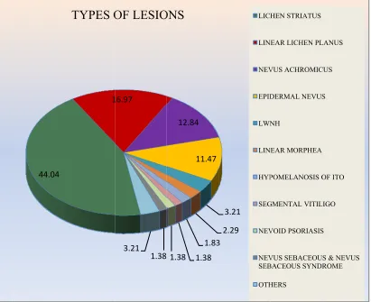
II) TYPE OF LESIONS:
Of the 218 patients, 96 (44.04%) cases had lichen striatus followed by linear LP in
37 (16.97%) cases, nevus achromicus with 28 (12.84%) cases, epidermal nevus
with 25 (11.4%) cases. There were 7 ( 3.21%) cases of LWNH, 5 (2.29%) cases of
linear morphea, 4 (1.83%)cases of HI, 3 (1.38%)cases each of linear vitiligo and
nevoid psoriasis; and 2 (0.92%) cases of nevus sebaceous and one(0.46%) case of
nevus sebaceous syndrome. Other diseases included Darier’s disease, IP, LSA,
lichen planus pigmentosus, lichen nitidus, dyschromatosis universalis & soft
F Others- Inc Lichen Pla FIGURE 6 continentia Pig anus Pigmentos 44.04
6: DISTR
gmenti, Darier’ sus, Soft Fibrom
1
TYPES O
RIBUTION
’s Disease, Dys ma
16.97
1.38 3.21
OF LESIO
N OF TYP
schromatosis U 12.84 11.47 1.83 1.38 1.38 ONS
PES OF LE
Universalis Her 3.21 2.29 3 LIC LIN NEV EPID LWN LIN HYP SEG NEV NEV SEB OTH ESIONS reditaria, LSA, HEN STRIATUS
NEAR LICHEN PL
VUS ACHROMIC DERMAL NEVU NH NEAR MORPHEA POMELANOSIS O GMENTAL VITIL VOID PSORIASIS VUS SEBACEOU BACEOUS SYND HERS `
, Lichen Nitidu
LANUS CUS S A OF ITO LIGO S
US & NEVUS ROME
III) SEX RATIO
In our study 93 cases were males and 125 cases were male, the overall male:
[image:52.612.69.516.194.508.2]female ratio being 0.75: 1. (Table III)
TABLE III- SEX DISTRIBUTION
DISEASE MALE FEMALE M:F TOTAL
Lichen Striatus 39 57 0.7:1 96
Linear Lichen Planus 9 28 0.3:1 37
Nevus Achromicus 17 11 1.5:1 28
Epidermal Nevus 15 10 1.5:1 25
LWNH 4 3 1.3:1 7
Linear Morphea 2 3 0.7:1 5
Hypomelanosis Of Ito 2 2 1:1 4
Segmental Vitiligo 1 2 1:2 3
Nevoid Psoriasis 1 2 1:2 3
Nevus Sebaceous & Nevus Sebaceous
Syndrome 3 0 - 3
Others 0 7 - 7
Total 93 125 0.75:1 218
OTHERS- Darier’s Disease, Incontinentia pigmenti, Dyschromatosis universalis hereditaria, Lichen nitidus, LSA, Lichen Planus Pigmentosus, Soft fibroma
On considering the individual diseases, male predominance was seen in nevus
achromicus, epidermal nevus and LWNH. Equal sex ratio was found in
hypomelanosis of Ito. Female predominance was noted in lichen striatus, linear LP,
The patients in the other diseases like Darier’s disease, IP, LSA, Lichen planus
pigmentosus, lichen nitidus, dyschromatosis universalis and soft fibroma were all
females. (Figure 7)
IV) AGE AT ONSET – DISTRIBUTION :
The combined (male + female) age at onset in various diseases is shown. TABLE IV- AGE AT ONSET DISTRIBUTION
LS-lichen striatus, LP- LS-lichen planus, NA- nevus achromicus, EN- epidermal nevus, LM- linear morphea, HI- hypomelanosis of Ito, LV- linear vitiligo, NP- nevoid psoriasis, NS- nevus sebaceous & nevus sebaceous syndrome, LPP- Lichen Planus Pigmentosus, LN- Lichen nitidus, S Fibroma- soft fibroma, DUH-dyschromatosis universalis hereditaria, DD – Darier’s disease.
AGE Δ 0-12m 1-5y 6-10y 11-20y 21-30y 31-40y 41-50y
>50y NR TOTAL
LS 4 52 23 16 1 - - - - 96
LP - 3 2 9 5 7 5 6 - 37
NA 26 - - 1 1 - - - - 28
EN 21 - 1 1 - - - - 1 25
LWNH 5 - 1 - - - 1 7
LM - - 1 3 1 - - - - 5
HI 2 - 2 - - - 4
LV - 2 1 - - - 3
NP 3 - - - 3
NS 2 - - - 1 - - - - 3
DD - - - 1 - 1
IP 1 - - - 1
LSA - - - 1 - - - 1
LPP - - - 1 - - - 1
LN - - 1 - - - 1
S Fibroma - - - 1 - - - 1
DUH - 1 - - - 1
LS- Lichen Hypomelan Incontinenti Pigmentosu 0 10 20 30 40 50 60 70 80 90 100 F
striatus, LP- line osis Of Ito, LV-ia Pigmenti, Dar us,Soft Fibroma
57
[image:54.612.78.538.89.481.2]28
FIGURE 7
ear lichen planus Linear Vitiligo, rier’s Disease, D
11
10
7: SEX- W
s, NA- nevus ach NP-Nevoid Pso Dyschromatosis U
0
3
WISE DIST
hromicus, EN- E riasis, NS-Nevu Universalis Here
3 2
TRIBUTI
Epidermal Nevu us Sebaceous & N
editaria, LSA, Li
2 2
ION
s,LM- Linear M Nevus Sebaceou ichen Nitidus, Li
0 7
Morphea,HI- us Syndrome, Ot
The maximum number of cases {65 (29%)} occurred in the first year, more so in
the first 1month with 56 (25.7%) cases , representative of epidermal nevi and
pigmentary disorders which included nevus achromicus, LWNH and
Hypomelanosis of Ito. The second most common age group was 1-5 years with 58
(25.8%) cases predominantly constituted by lichen striatus. (Figure 8)
AGE AT ONSET DISTRIBUTION- MALES AND FEMALES
The age at onset distribution in males and females separately is similar to that of
the total distribution. However in males, the maximum number of cases occurred
in the age group 0-12 months with 36 (38.7%) cases, followed by 25(26.9%) cases
in 1-5 years, 13(14%) cases in the ages 11-20 years and 10 (10.7%) cases in the
ages 6-10 years. The number of cases with age at onset above 20 years was
9(9.7%).
In females, the maximum number of cases {33 (26.4%)} occurred in the age-
group of 1-5 years, closely followed by 30(24%) cases in the first year. However,
as compared to the age distribution in males, the number of cases above 21 years
was 22 (17-6%). Twenty – two (17.6%) cases developed lesions between 6- 10
years and 18 (14.4%) cases developed lesions in the ages 11-20 years. (Figure 9)
FI LS- Lichen Hypomelan Incontinenti Pigmentosu 0 10 20 30 40 50 60 70 1 IGURE 8:
n striatus, LP- osis Of Ito, LV-ia Pigmenti, D us, Soft Fibroma
1-12m 1-5y
: DISTRIB
linear lichen p -Linear Vitiligo, Darier’s Disease , NR- not recall
6-10y 11
BUTION O
planus, NA- ne , NP-Nevoid Pso
, Dyschromatos ed
-20y 21-30y
OF AGE A
evus achromicu oriasis, NS-Nevu sis Universalis
31-40y 41-5
AT ONSE
s, EN- Epiderm us Sebaceous &
Hereditaria, L
50y >50y
ET- TOTA
mal Nevus,LM-& Nevus Sebaceo LSA, Lichen N
NR O N N L H L L E N L L AL
- Linear Morph ous Syndrome, O
FIGURE 9 -COMPARATIVE DISTRIBUTION OF AGE OF ONSET IN MALES, FEMALES AND COMBINED
0 10 20 30 40 50 60 70
1‐12M 1‐5Y 6‐10Y 11‐10Y 21‐30Y 31‐40Y 41‐50Y >50Y NR
V) SIDE- WISE DISTRIBUTION
Overall, the right side and left sides each were involved in approximately 45% of
the patients and both the sides were simultaneously involved in the remaining 10 %
cases. On scrutinizing the individual diseases, significant right side predominance
was observed in linear LP and left side predominance in nevus achromicus. Lichen
striatus, linear vitiligo and nevoid psoriasis involved the right side preferentially
while epidermal nevus, LWNH and nevus sebaceous preferentially involved the
[image:58.612.68.525.354.617.2]left side. Linear morphea showed equal occurrence on left and right side. (Table V)
TABLE V- SIDE OF INVOLVEMENT
DISEASE RIGHT LEFT BOTH TOTAL
Lichen Striatus 49 45 2 96
Linear Lichen Planus 21 13 3 37
Nevus Achromicus 5 16 7 28
Epidermal Nevus 10 13 2 25
LWNH 1 3 3 7
Linear Morphea 2 2 1 5
Hypomelanosis Of Ito 2 - 2 4
Linear Vitiligo 2 1 - 3
Nevoid Psoriasis 2 1 - 3
Nevus Sebaceous & Nevus Sebaceous
Syndrome - 3 - 3
Others 4 2 1 7
TOTAL 98 99 21 218
VI) DISTRIBUTION OF TYPE OF BLASCHKO’S LINES
Overall, the most common type of Blaschko pattern observed was the narrow band
type, followed by a combination of the narrow and broad- band types of
Blaschko’s lines, broad –band type alone and the phylloid pattern. Lateralization
was observed in two patients with nevus achromicus and a combination of broad-
[image:59.612.40.539.292.622.2]band and phylloid in pattern in one patient. (Table VI and Figure 10)
TABLE VI- TYPE OF BLASCHKO’S LINES FOLLOWED
DISEASE NB BB NB & BB PHYLLOID PHYLLOID
WITH BB LATERAL- IZATION
Lichen Striatus 87 2 7 - -
Linear Lichen Planus 33 2 2 - -
Nevus Achromicus 9 3 5 8 1 2
Epidermal Nevus 19 2 4 - -
LWNH 2 2 2 1 -
Linear Morphea - 5 - - -
Hypomelanosis Of Ito 3 1 - - -
Segmental Vitiligo - - - 3 -
Nevoid Psoriasis 3 - - - -
Nevus Sebaceous & Nevus
Sebaceous Syndrome 2 1 - - -
Others 6 1 - - -
Total 164 19 20 12 1 2
OTHERS- Darier’s Disease, Incontinentia pigmenti, Dyschromatosis universalis hereditaria, Lichen nitidus, LSA, Lichen Planus Pigmentosus, Soft fibroma
FIGUR
Narrow B
RE 10: DI
Band, BB- Bro
164
ISTRIBUT
oad Band
TION OF
12
1 2
TYPES O
20 2
OF BLASC
19
CHKO’S L
NB BB NB & BB PHYLLOID PHYLLOID LATERALIZ
LINES
VII) SITE DISTRIBUTION- This is represented and discussed according to the
individual diseases.
a) SITES OF INVOLVEMENT - LICHEN STRIATUS
Multiple sites were involved in 64.6 % patients. The single most common site
involved was the thigh in this study. (Table VII and Figure 11)
TABLE VII- SITE – WISE DISTRIBUTION- LICHEN STRIATUS
b) SITES OF INVOLVEMENT- LINEAR LICHEN PLANUS
Multiple sites were involved in 78 % of the patients (including cases with
coexisting oral lesions), the predominant site being the leg. Coexisting oral
mucosal involvement was seen in 7 (19%) patients of which one was male and six
were female patients. (Table VIII and Figure 12)
SITE TOTAL
Head & neck 7
Trunk 12
Upper limb 16
Lower limb 33
Multiple sites 28
FI
H & N - 0 5 10 15 20 25 30 35 GURE 11
Head & Ne H&N
7
: SITE- W
eck TRUN 12 SI WISE DIST NK 2 ITE DISTRIBU TRIBUTI UPPER LIMB 16 UTION-LICHE
ON – LIC
FIGURE 12: SITE – WISE DISTRIBUTION IN LINEAR LICHEN PLANUS
H & N - Head & Neck
1 1
8
18
9
0 2 4 6 8 10 12 14 16 18 20
H & N TRUNK UPPER LIMB LOWER LIMB MULTIPLE
SITES
TABLE VIII- SITE- WISE DISTRIBUTION- LINEAR LICHEN PLANUS
SITE TOTAL
Head & neck 1
Trunk 1
Upper limb 8
Lower limb 18
Multiple sites 9
Total 37
c) SITES OF INVOLVMENT- NEVUS ACHROMICUS
In this study, 24 (86%) out of 28 of patients had involvement of multiple sites, the
most common site being the trunk. (Table IX and Figure 13)
TABLE IX- SITE - WISE DISTRIBUTION - NEVUS ACHROMICUS
SITE TOTAL
Head & neck -
Trunk 4
Upper limb 3
Lower limb 3
Multiple sites 18
Total 28
d) SITES OF INVOLVEMENT- EPIDERMAL NEVUS
In our study, the most common site involved in epidermal nevi patients was the
head and neck region, followed by the involvement of multiple sites. One patient
H & N - Head & Neck
FIGURE 14: SITE- WISE DISTRIBUTION IN EPIDERMAL NEVUS
H & N - Head & Neck
0
4 3 3
18 0 2 4 6 8 10 12 14 16 18 20
H & N TRUNK UPPER LIMB LOWER LIMB MULTIPLE SITES
SITES OF INVOLVEMENT-NEVUS ACHROMICUS
11 4 1 0 9 0 2 4 6 8 10 12
H & N TRUNK UPPER LIMB LOWER LIMB MULTIPLE SITES
TABLE X- SITE - WISE DISTRIBUTION - EPIDERMAL NEVUS
SITE TOTAL
Head & neck 11
Trunk 4
Upper limb 1
Lower limb -
Multiple sites 9
Total 25
e) SITES OF INVOLVEMENT- OTHER DISEASES
The sites involved in the other diseases are shown in Table XI. In general, in most patients multiple sites were involved. LWNH preferentially involved the lower limbs and linear morphea preferentially involved the upper limbs.
TABLE XI- SITE - WISE DISTRIBUTION - OTHER DISEASES
SITE
Δ HEAD& NECK
TRUNK UPPER
LIMB LOWER LIMB MULTIPLE SITES TOTAL
LWNH - - 1 4 2 7
LM - - 3 2 - 5
HI 1 - - - 3 4
LV - - - 1 2 3
NP - - 2 1 - 3
NS 3 - - - - 3
DD - - - - 1 1
IP - - - - 1 1
LSA - - - 1 - 1
LPP - - 1 - - 1
LN - - 1 - - 1
S FIB 1 - - - - 1
DUH - - - - 1 1
TOTAL 5 - 8 9 10 32
VIII) SYMPTOM - WISE DISTRIBUTION
Overall, most of the patients in this study were asymptomatic. However, linear
lichen planus was symptomatic in 86%. Thirty percent of patients with lichen
striatus experienced pruritus. (Table XII and Figure 15)
TABLE XII- SYMPTOM- WISE DISTRIBUTION
DISEASE ASYMPTOMATIC SYMPTOMATIC
M F M F
Lichen Striatus 30 36 9 20
Linear Lichen Planus 1 4 8 24
Nevus Achromicus 17 11 - -
Epidermal Nevus 14 9 - 2
LWNH 4 3 - -
Linear Morphea 1 3 1 -
Hypomelanosis Of Ito 2 2 - -
Linear Vitiligo - 2 1 -
Nevoid Psoriasis - - 1 2
Nevus Sebaceous & Nevus Sebaceous Syndrome
2 - 1 -
Others - 5 - 2
Total 71 75 21 49
OTHERS- Darier’s disease, lichen planus pigmentosus, Soft fibroma, Dyschromatosis universalis hereditaria, Incontinentia pigmenti, Lichen nitidus, LSA
FIGUR LS- Lichen Hypomelan Incontinenti Pigmentosu 0 10 20 30 40 50 60 70 80 90 100
RE 15: SYM
striatus, LP- line osis of Ito, LV-L ia pigmenti, Dar us, Soft Fibroma
MPTOM-ear lichen planus Linear Vitiligo, N rier’s Disease, D
- WISE DI
s, NA- nevus ach NP-Nevoid Psor yschromatosis U
ISTRIBUT
hromicus, EN- E riasis, NS-Nevus Universalis Here
TION
Epidermal Nevu s Sebaceous & N editaria, LSA, Li
s,LM- Linear M Nevus Sebaceous
chen Nitidus, Li
SY A
[image:68.612.76.545.150.463.2]IX) EVOLUTION OF LESIONS- (Table XIII)
TABLE XIII- EVOLUTION OF LESIONS
Disease Extending Static Not recalled Initial
extension, now static
Total
Lichen striatus 73 1 18 4 96
Linear lichen planus 30 1 4 2 37
Nevus achromicus 7 17 1 3 28
Epidermal nevus 6 13 3 3* 25
LWNH 2 4 - 1* 7
Linear morphea 3 - 1 1 5
Hypomelanosis of ito 1 2 1 - 4
Linear vitiligo 3 - - - 3
Nevoid psoriasis 3 - - - 3
Nevus sebaceous & nevus
sebaceous syndrome - 2 - 1 3
Others 3 4 - - 7
Total 131 44 13 7 218
Others- Incontinentia pigmenti, Darier’s disease, Lichen planus pigmentosus, soft fibroma, dyschromatosis universalis hereditaria, lichen nitidus, LSA
(*) regression of lesions
In this study, most acquired lesions were still extending at the time of
presentation,these included lichen striatus, linear lichen planus, linear morphea,
linear vitiligo. The congenital disorders like epidermal nevus and nevus
achromicus were static in nature, exception being nevoid psoriasis which was
X) DIRECTION OF EXTENSION
In lichen striatus, the predominant direction of extension was distal to proximal
over the limbs and outward over the trunk and head and neck region. In Linear LP,
most lesions extended in the distal to proximal direction over the limbs and
outward direction over the trunk and head & neck region. In nevus achromicus, the
predominant direction of extension was outward over the trunk. Predominant
direction of extension in epidermal nevus was proximal to distal over the limbs &
outward over the trunk and head & neck regions. (Figure 16)
XI) ASSOCIATIONS
Diabetes mellitus was recorded in 3 patients and hypertension in one patient with
linear lichen planus. Our patient with Darier’s disease was both diabetic and
hypertensive. One patient with lichen striatus had coexistent pityriasis rosea. One
patient with linear LP had coexistent lesions of morphea not following the
Blaschko’s lines and another patient with linear LP had epilepsy with Parkinson’s
disease. Three patients with nevus achromicus had CNS abnormalities while one
Others- LW Nevus Seb LSA, Lich (*)- Lesion 0 10 20 30 40 50 60 70 80 90 100 FIGUR
WNH, Linear M baceous Syndro
en Nitidus, Lic
ns On Trunk W
RE 16: DI
Morphea, Hypo ome, Incontinen chen Planus Pig
With/ Without L
IRECTIO
omelanosis Of ntia Pigmenti, gmentosus,Sof
Limb Lesions,
N OF EXT
Ito, Linear Vit Darier’s Disea ft Fibroma
PD- proximal
TENSION
tiligo, Nevoid P ase, Dyschrom
to distal,
DP-N OF LES
Psoriasis, Nevu atosis Univers
distal to proxim
history of temporal association with tetanus vaccination and pregnancy. One of the
three patients with nevoid psoriasis had congenital valvular heart disease. Our
patient with soft fibroma was a known case of pemphigus vulgaris.
XII) BIRTH HISTORY
In this study, 170 (78%) patients were born out of non- consanguineous marriage
while 30 (14%) patients were born out of third degree consanguineous marriage
and 18 (8%) patients were born out of second degree consanguineous marriage.
XIII) FAMILY HISTORY
Only 2 patients reported with medical disorders in the family. The older female
sibling of the patient with incontinentia pigmenti died on 5th postnatal day due to
unknown causes. She did not have lesions of IP according to the history. The
daughter of one patient with epidermal nevus was diagnosed to have
DISCUSSION
The lines of Blaschko represent a pattern assumed by many different nevoid and
acquired skin diseases on the human skin and mucosae. Various congenital and
acquired dermatoses follow the Blaschko’s lines. There are five patterns of
Blaschko’s lines which include the narrow and broad – band pattern, checker board
pattern, phylloid pattern, patchy pattern without midline separation and the
lateralization pattern.
DISCUSSION ON EPIDEMIOLOGY OF BLASCHKO’S LINES
A) INCIDENCE
Out of a total 1, 03, 536 cases visiting the Dermatology outpatients during the
study period, the cases with lesions following Blaschko’s lines were 218 in total.
Thus the incidence rate of lesions following the lines of Blaschko was 2.1 per 1000
cases.
B) CASE DISTRIBUTION
The most common disease with lesions along the Blaschko’s lines was lichen
striatus, followed by linear LP, nevus achromicus and epidermal nevus. Other
diseases with few cases included LWNH, linear morphea, hypomelanosis of Ito,
Few rare conditions in which lesions are known to exist along the Blaschko’s lines
encountered were Darier’s disease, incontinentia pigmenti, LSA, linear lichen
nitidus and lichen planus pigmentosus . On