Evaluation of autonomic dysfunction in
patients with hepatic cirrhosis
Dissertation submitted in partial fulfilment
of the requirements for the degree of
D.M., (MEDICAL GASTROENTEROLOGY) - BRANCH IV
CERTIFICATE
This is to certify that the dissertation titled “Evaluation of Autonomic
Dysfunction in Patients with Hepatic Cirrhosis” is a genuine work done by
Dr.E.Kandasamy alias Kumar, Post Graduate in Medical Gastroenterology under
my supervision between April 2006 to March 2007 and is being submitted for the
partial fulfillment of the requirement for the awarding of D.M. (Medical
Gastroenterology) degree by the Tamilnadu Dr. M.G.R Medical University,
Chennai.
Dean
Prof. S. Barnaba Durairaj, MD., DM
Madras Medical College, Professor and Head,
Chennai – 600 003
Department of Medical Gastroenterology
Madras Medical College,
Chennai – 600 003
ACKNOWLEDGEMENT
I am greatly indebted to Dr.Kalavathy Ponnirivan B.Sc, M.D., Former
Dean, Madras Medical college and Government General Hospital, Chennai
for permitting me to carry out this study.
I am extremely thankful to my guide Dr. S. Barnaba Durairaj, M.D. D.M.,
Professor and Head, Department of Medical Gastroenterology for his fatherly
guidance and support throughout study period.
I am extremely thankful to Prof. Mythili Baskaran Director, Institute of
Physiology and experimental medicine and Dean In charge for permitting to
utilize the research laboratory facilities and for her guidance.
I thank Prof. P. Padmanaban, M.D., D.M., Additional Professor for his
valuable comments which facilitated my study enormously. My sincere thanks are
also due to my assistant professors of the department Dr. P. Ganesh,
Dr. K.Narayanaswamy, Dr. K. Premkumar for their valuable suggestions and
support.
I thank my colleagues Dr. P. Karthikeyan, Dr. G. Ramkumar, Dr. G. Rema
Krishna Kumar, Dr. Antony Joe and Dr. Mahadevan for their cooperation,
enthusiasm.
CONTENTS
CHAPTER TITLE Page No
1. INTRODUCTION 1
2. BACKGROUND 2
3. AIM OF STUDY 37
4. MATERIALS & METHODS 38
5. OBSERVATION 47
6. DISCUSSION 55
7. CONCLUSION 61
8. SUMMARY 61
9. APPENDIX 64
Evaluation of Autonomic Dysfunction in Patients with Hepatic
Cirrhosis
1. Introduction:
Cirrhosis of the liver leads to a number of complications, some of which may eventually provefatal. For more than a century,chronic alcoholics have been known to have peripheral neuropathy.It has been observed that alcoholics with liver damage have higher frequency of neuropathy than those without it. Thereare reports of association of chronic liver disease with autonomicneuropathy However; conflicting reports have also appearedcausing much confusion. Patients with cirrhosis and portal hypertension develop hyper dynamic circulation, with increased blood volume and cardiac output, and with a reduced peripheral vascular resistance. This disorder has been related to portal hypertension–induced arterial vasodilatation in the peripheral and splanchnic beds, but other factors may contribute, such as abnormally high levels of circulating vasodilators and false neurotransmitters.1
the continuous recordings of heart rate and arterial pressure reflect the activity of the efferent arc of the autonomic nervous sys tem that is modulating cardiovascular function. In particular, sympathetic and vagal outflows to the heart and circulation generate two main oscillatory rhythms: (1) a short-term rhythm, present in heart period and arterial pressure variabilities and defined as the low-frequency (LF) component, which is considered a marker of the sympathetic activity; and (2) a long-term rhythm, only occurring in heart period variability, related to respiration, and known as the high frequency (HF) component, which is thought to be a marker of the vagal modulation. These oscillatory components of heart rate and arterial pressure signals can be extracted from the variability signals through spectral analysis techniques.
2. Back ground :
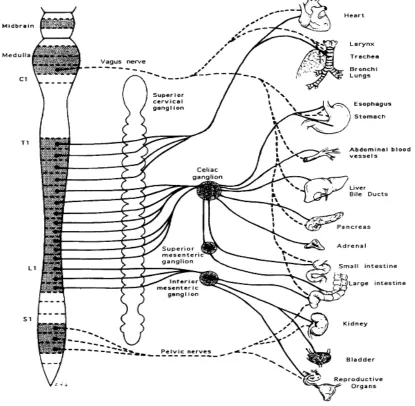
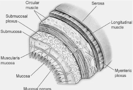
Neural regulation of the gastrointestinal tract
increases intracellular cyclic adenosine monophosphate (cAMP) levels and acetylcholine increases intracellular calcium in the target cell.6
Endocrine, paracrine, and neural transmitters existing within the lamina propria modulate effects on the gut immune system. Lymphocytes, macrophages, mast cells, neutrophils, and eosinophils are potential targets for endocrine and neural transmitters and participate in the inflammatory cascade. Moreover, inflammatory mediators can act directly on enteric nerves. Serotonin released from endocrine cells is involved in intestinal anaphylaxis and stimulates vagal afferent fibers that possess the 5-hydroxytryptamine 3 5-HT3) receptor.
Chemical messengers of the gastrointestinal tract
The enteric nervous system, through intrinsic and extrinsic neural circuits, controls gastrointestinal function. This control is mediated by a variety of chemical messengers, including motor and sensory pathways of the sympathetic and parasympathetic nervous systems. The parasympathetic preganglionic input is provided by cholinergic neurons and elicits excitatory effects on gastrointestinal motility via nicotinic and muscarinic receptors, whereas the sympathetic input occurs by postganglionic adrenergic neurons.
Acetylcholine
the mediator of postsynaptic membrane depolarization following acetylcholine receptor binding. Muscarinic receptors belong to the heptahelical GPCR family. There are five known muscarinic cholinergic receptors (M1 to M5). Muscarinic receptors can be further classified based on receptor signal transduction, with M1, M3, and M5 stimulating adenylate cyclase and M2 and M4 inhibiting this enzyme.
Acetylcholine is degraded by the enzyme acetylcholinesterase, and the products may be recycled through high-affinity transporters on the nerve terminal
Catecholamines
The primary catecholamine neurotransmitters of the enteric nervous system include norepinephrine and dopamine. Norepinephrine is synthesized from tyrosine and released from postganglionic sympathetic nerve terminals that innervate enteric ganglia and blood vessels. Tyrosine is converted to dopa by tyrosine hydroxylase. Dopa is initially converted into dopamine by dopa decarboxylase and packaged into secretory granules. Norepinephrine is formed from dopamine by the action of dopamine β -hydroxylase within the secretory granule. After an appropriate stimulus, norepinephrine-containing secretory granules are released from nerve terminals and bind to adrenergic receptors.
Norepinephrine signaling is terminated by intracellular monoamine oxidase or by rapid reuptake by an amine transporter. The actions of adrenergic receptor stimulation regulate smooth muscle contraction, intestinal blood flow, and gastrointestinal secretion.8
Dopamine is an important mediator of gastrointestinal secretion, absorption, and motility and is the predominant catecholamine neurotransmitter of the central and peripheral nervous systems. In the central nervous system, dopamine regulates food intake, emotions, and endocrine responses, and peripherally, it controls hormone secretion, vascular tone, and gastrointestinal motility.9 Characterization of dopamine in the gastrointestinal tract has been challenging for several reasons. First, dopamine can produce inhibitory and excitatory effects on gastrointestinal motility. Generally, the excitatory response, which is mediated by presynaptic receptors, occurs at a lower agonist concentration than the inhibitory effect that is mediated by postsynaptic receptors. Second, localization of dopamine receptors has been hampered by identification of dopamine receptors in locations that appear to be species specific. Finally, studies of dopamine in gastrointestinal tract motility have often used pharmacologic amounts of this agonist. Therefore, interpretation of results has been confounded by the ability of dopamine to activate adrenergic receptors at high doses.
exposed to agonists and antagonists. After release from the nerve terminal, dopamine is cleared from the synaptic cleft by a specific dopamine transporter.
Serotonin
Serotonin has long been known to play a role in gastrointestinal neurotransmission. The gastrointestinal tract contains more than 95% of the total body serotonin, and serotonin is important in a variety of processes, including nausea, emesis, epithelial secretion, and bowel motility. Serotonin is synthesized from tryptophan, an essential AA, and is converted to its active form in nerve terminals. Serotonin is inactivated in the synaptic cleft by reuptake via a serotonin-specific transporter. Most plasma serotonin is derived from the gut, where it is found in mucosal enterochromaffin cells and the enteric nervous system. Serotonin mediates its effects by binding to a specific receptor. There are seven different serotonin receptor subtypes found on enteric neurons, enterochromaffin cells, and gastrointestinal smooth muscle (5-HT1 to 5-HT7). Through these receptors serotonin regulates intestinal secretion, absorption, and motility.
Characterization of specific serotonin receptor subtypes has led to the development of selective agonists and antagonists for the treatment of gastrointestinal disorders such as irritable bowel syndrome and chronic constipation and diarrhea. For example, 5-HT3 receptor antagonists may be useful in diarrhea-predominant irritable bowel syndrome, and the 5-HT4 receptor agonist has prokinetic effects and may be useful in constipation or other motility disorders.
Histamine
In the gastrointestinal tract, histamine is best known for its central role in regulating gastric acid secretion and intestinal motility. Histamine is produced by enterochromaffin-like cells of the stomach and intestine as well as enteric nerves. Histamine is synthesized from l-histidine by histidine decarboxylase and activates three GPCR subtypes. H1 receptors are found on smooth muscle and vascular endothelial cells and result in activation of phospholipase C (PLC). As such, the H1 receptor mediates many of the allergic responses induced by histamine. H2 receptors are present on gastric parietal cells, smooth muscle, and cardiac myocytes. H2 receptor binding stimulates Gs and activates adenylate cyclase. H3 receptors are present in the central nervous system and gastrointestinal tract endochromaffin cells. These receptors signal through Gi and inhibit adenylate cyclase. Histamine can also interact with theN-methyl-d-aspartate (NMDA)
Unlike other neurotransmitters, there is no known trans porter responsible for termination of histamine's action. However, histamine is metabolized to telemethylhistamine by histamine N-methyltransferase, and is then degraded to telemethylimidazoleacetic acid by
monoamine oxidase B and an aldehyde dehydrogenase.
Nitric Oxide
Although smooth muscle physiologists have long known of an "endothelial-derived relaxing factor" responsible for vasodilation, it took many years for the chemical nature of this substance to be identified as nitric oxide (NO). NO is a unique chemical messenger produced with citrulline from l-arginine and oxygen by the enzyme nitric
oxide synthase (NOS). Three types of NOS are known. Types I and III are also know as
endothelial NOS and neuronal NOS and are constitutively active. Small changes in NOS
activity can occur through elevations in intracellular calcium. The inducible form of NOS (type II) is apparent only when cells become activated by specific cytokines and inflammation. This form of NOS is capable of producing large amounts of NO and is calcium independent. NOS is often colocalized with VIP and PACAP in neurons of the enteric nervous system. 10
physiology includes stimulation of epithelial secretion, vasodilation, and mucosal defense.
Adenosine
Adenosine is an endogenous nucleoside that acts through any of four GPCR subtypes. Adenosine causes relaxation of intestinal smooth muscle and stimulates intestinal secretion. Adenosine can also cause peripheral vasodilation and activation of nociceptors that participate in pain neural pathways.
Cytokines
marrow support after bone marrow transplantation. Chemokines initiate and propagate inflammation and are of two groups: CXC (α chemokines) and CC (β chemokines). Other cytokines, such as transforming growth factor (TGF)-β and platelet-derived growth factor (PDGF), have proliferative effects.
Normal Liver Blood Flow
Hepatic blood flow is normally about 1500 mL/minute, representing 15% to 20% of cardiac output. One third of this flow and 30% to 60% of the oxygen consumed by the liver are provided by the hepatic artery. Approximately two thirds of the hepatic blood supply is provided by portal venous blood.7,11 The high-pressure, well-oxygenated arterial blood mixes completely with the low-pressure, low-oxygen-containing, nutrient-rich portal venous blood within the hepatic sinusoids. After perfusing the sinusoids, blood flows, sequentially, into the hepatic venules, hepatic veins, and inferior vena cava. A fraction of the plasma entering the space of Disse is drained into lymphatic vessels.
maintenance of a low pressure in the hepatic sinusoids is critical to the maintenance of normal rates of transudation of sinusoidal fluid into the space of Disse.
Another feature that is unique to the hepatic circulation is the close interrelationship between blood flow in the portal vein and that in the hepatic artery. When portal blood flow increases, hepatic arterial flow decreases; when portal flow decreases, hepatic arterial flow increases. This phenomenon has been termed the hepatic arterial buffer
response and is an adenosine-mediated vascular reflex that ensures the maintenance of a
Figure 3. Hemodynamic principles in portal hypertension
Hyperdynamic circulation is a common and long-recognized feature of patients with advanced cirrhosis, consisting of elevated cardiac rate and output and reduced peripheral vascular resistance, so that arterial pressure is tendentially or frankly reduced. The clinical importance of this disorder was shown by subsequent studies, showing that the alterations of systemic hemodynamics, renal function, and vasoactive systems are prognostic indicators even more accurate than the tests exploring liver function. The setting of the hyperdynamic circulatory syndrome is the pathogenetic background of complications such as renal sodium and water retention and hepatorenal syndrome.Splanchnic blood pooling, opening of portal-systemic collaterals, and arterial vasodilatation, as well as a compensatory increase in blood volume, are the causative events of the hyperdynamic circulatory syndrome. The pathogenesis of arterial vasodilatation is still debated. It has been proposed that an overproduction of a variety of vasorelaxant agents, such as histamine, adenosine, gut-derived peptides and endothelial cell-derived vasodilators, and bile acid retention reduce the responsiveness of the vascular bed to endogenous vasoconstrictor stimuli.
Earlier studies showed that cross-perfusion between portal hypertensive and normal animals produces arteriolar vasodilation in the latter, lending support to the hypothesis that a transferable humoral vasodilator is present in the blood in portal hypertension. Much attention has since focused on putative vasoactive mediators responsible for the arteriolar vasodilation in splanchnic organs that underlies the increase in portal venous inflow. Investigators have postulated that endogenous vasodilators normally present in portal blood and cleared by the liver may escape hepatic removal either as a result of portosystemic shunting via portosystemic collaterals or as a result of impaired hepatocellular metabolism. A further possibility is that liver disease and portal hypertension lead to an increase in the production of certain vasodilators within either the hepatic or the splanchnic vascular beds. These vasodilators then reach high concentrations in the systemic circulation, thereby leading to systemic and splanchnic arterial vasodilation.
pharmacologic doses of somatostatin or its synthetic analog octreotide, which decreases glucagon release, produces vasoconstriction of both the splanchnic and the systemic circulation. Because somatostatin also inhibits the release of several other peptide vasodilators, such as substance P, vasoactive intestinal peptide (VIP), and calcitonin gene-related peptide (CGRP), it is conceivable that the effects of somatostatin on the circulation in portal hypertension may be mediated by other peptides in addition to, or apart from, glucagon. Also, somatostatin may exert a direct vasoconstrictive effect on vascular smooth muscle. Therefore, understanding of the role of glucagon as a mediator of systemic vasodilation in portal hypertension remains inconclusive, but on the basis of available data, hyperglucagonemia may account for approximately 30% to 40% of the splanchnic vasodilation of chronic portal hypertension.
Vasoactive factors produced by the vascular endothelium have attracted considerable attention with respect to a potential role in the pathogenesis of portal hypertension. There is increasing evidence for the involvement of nitric oxide (NO) and prostacyclin in the pathogenesis of the circulatory abnormalities in portal hypertension.
NO is a powerful endogenous vasodilator that is generated in several tissues by a constitutive vascular endothelial NO synthase (eNOS) and an inducible NO synthase (iNOS) from the amino acid l-arginine. NO is produced constitutively by eNOS and by
resistance and hence increased splanchnic arterial flow associated with portal hypertension.
Tumor necrosis factor-α (TNF-α) mediates the effects of endotoxin, a potent stimulant of NOS. A dramatic amelioration in the hyperdynamic circulation and the increased portal pressure has been described in portal hypertensive rats treated with antibody to TNF-α.
Administration of specific NO antagonists to animals with portal hypertension induces splanchnic and systemic vasoconstriction, thereby attenuating the hyperdynamic circulation. In addition, inhibition of NO synthesis, at least partially, corrects the blunted vascular responsiveness to vasoconstrictors that is characteristic of portal hypertension. The finding that patients with cirrhosis have increased serum and urinary concentrations of nitrite and nitrate (end-products of NO oxidation) also supports a role for NO in the genesis of the circulatory disturbances of portal hypertension. However, NO inhibition attenuates but does not normalize the hyperkinetic state of portal hypertension. Also, in one study chronic NO inhibition delayed but did not prevent the development of splanchnic vasodilation in experimental animals. These and other data suggest that other factors in addition to NO are involved in the vasodilatory phenomena associated with the hyperdynamic circulation of portal hypertension.
degree of portal pressure elevation in these patients. In one study inhibition of prostaglandin biosynthesis by indomethacin reduced the hyperdynamic circulation and portal pressure in patients who had cirrhosis and portal hypertension.
A variety of other circulating vasodilators have been evaluated, including bile acids, histamine, adenosine, and substance P without convincing evidence to date that they contribute to the systemic hyperdynamic state of portal hypertension.
The hyperdynamic circulation begins in the portal venous bed as a consequence of portal hypertension due to the increased resistance to flow from altered hepatic vascular morphology of chronic liver disease. Dilatation of the portal vein is associated with increased blood flow, as well as the opening up or formation of veno-venous shunts and splenomegaly. At the same time, portal hypertension leads to subclinical sodium retention resulting in expansion of all body fluid compartments, including the systemic and central blood volumes.
early liver transplantation in patients with advanced liver disease and autonomic dysfunction
Alcohol related neuropathy
The clinical symptoms of alcoholic peripheral neuropathy were described more than 200 years ago. The descriptions by Lettsom (1787) and Jackson (1822) have led to the recognition and association of peripheral nerve disease with excessive ethanol use. Several terms connote alcohol neuropathy, including neuritic beriberi, neuropathic beriberi, and alcoholic neuritis. In patients with alcoholic neuropathy, nutritional deficiency goes hand in hand with alcohol abuse.
The similarity between beriberi and alcoholic neuropathy had long been noted, but Shattuck in 1928 was the first to seriously discuss the relationship. He suggested that "polyneuritis of chronic alcoholism" was caused chiefly by failure to take or assimilate food containing a sufficient quantity of vitamin Bcomlex and might properly be regarded as true beriberi. However, this theory may be only partly true.
thiamine-mediated enzyme activity (transketolase) has been established, though this has not been conclusively established in the case of peripheral neuropathy.
In their comparison of alcoholics and nonalcoholic control subjects, Behse and Buchthal concluded that nutritional deficiencies alone did not produce the neuropathy. Monforte et al concluded that alcohol appears to be toxic to autonomic and peripheral nerves in a dose-dependent manner, on the basis of heart rate, blood pressure, and electrophysiologic examination. In a study of macaque monkeys, Hallet et al failed to produce clinical and electrophysiologic signs of neuropathy in monkeys that were given a certain amount of alcohol for 3-5 years.
Studies in rats also failed to demonstrate a direct toxic effect of alcohol on the peripheral nerves. Most studies of peripheral neuropathy in humans and animals implicate nutritional deficiency as an etiology as opposed to the direct toxic effect of alcohol.13,14
Frequency:
Internationally: Depending on criteria and patient selection, incidence of peripheral neuropathy ranging from 10-50% has been reported. These studies included alcoholics hospitalized for other reasons or for detoxification.
Mortality/Morbidity: Johnson and Robinson studied the mortality rate of alcoholics with autonomic dysfunction.
• Their findings suggested that evidence of vagal neuropathy in long-term alcoholics is associated with a significantly higher mortality rate than in the general population (a reported 88% survival rate at 7 years in alcoholics with autonomic dysfunction as compared to 94% in the general population).
• Deaths due to cardiovascular disease are a major factor.
• Many deaths were attributed to strokes, since heavy alcohol consumption is a significant risk factor for stroke.
Sex:
• A high incidence of alcoholic polyneuropathy has been observed in women.
History: Clinical manifestations of alcoholic neuropathy can be summarized as slowly progressive (over months) abnormalities in sensory, motor, autonomic, and gait function. Patients might ignore early symptoms, and seek help only when significant complications develop. Symptoms are often indistinguishable from other forms of sensory motor axonal neuropathy.
lancinating. Symptoms start typically distally, to progress slowly to involve proximally (dying-back neuropathy). When symptoms extend to involve above the ankle level, the fingertips often get similarly involved, giving rise to the well-known stocking and glove pattern of sensory involvement. Paresthesia might become unpleasant, even painful.15
• Motor manifestations include distal weakness and muscle wasting.
• When proprioception becomes involved, sensory ataxia will occur giving rise to gait difficulty, independent of alcoholic cerebellar degeneration.
• Autonomic disturbances are seen less commonly than other neuropathic conditions (eg, diabetes).
o Dysphagia and dysphonia are prominent secondary to degeneration of the vagus nerve. Other parasympathetic abnormalities include depressed reflex heart rate responses, abnormal pupillary function, sexual impotence, and sleep apnea.
o Sympathetic dysfunction is rare but if present can produce orthostatic hypotension and hypothermia.
Physical: Examination shows distal sensory loss in lower extremities. In severe cases, the hands may be involved. In addition to distal atrophy and weakness, deep tendon reflexes usually are decreased or absent. Stasis dermatitis, glossiness, and thinness of skin of the lower legs are common findings. Hyperesthesia and hyperalgesia may be seen along with hyperpathia. Excessive sweating of the soles and dorsal aspects of the feet and of the palms and fingers is a common manifestation of alcoholic neuropathy and is indicative of involvement of the peripheral (postganglionic) sympathetic nerve fibers. Occurrence of trophic ulcers is rare.
Causes:
• Variants
o Rare cases of acute or subacute alcoholic peripheral neuropathy have been
described. They mimic Guillain-Barré syndrome, although biopsy and electrodiagnostic studies had revealed an axonal neuropathy, with normal CSF parameters. A causal but an unproven association with ethanol is present.16
Lab Studies:
• The diagnosis is based on accurate history of prolonged and excessive alcohol intake, clinical signs and symptoms, and electrophysiologic testing. Behse and Buchtal suggested that a minimum of 100 mL of ethyl alcohol (3 L of beer or 300 mL of spirits) per day for 3 years will precipitate the neuropathy17.
Other Tests:
• Electrophysiologic findings primarily reveal evidence of primary axonal sensory motor polyneuropathy.
o Sensory conduction studies may be abnormal even before the advent of clinical symptoms.
Sural nerve sensory action potentials (SNAP) are reduced slightly to moderately in conduction velocity and SNAP amplitudes also are reduced.
As the condition worsens, the sensory potentials may become unobtainable. The median, radial, and ulnar nerves show the same response as the disease progresses.
distal latency. The upper extremity nerves follow the same pattern as time progresses.18
o The tibial H reflex latency is prolonged and becomes unobtainable if the condition continues to progress. The F waves are obtained more easily but reveal slight to moderate prolongation of latency.
• Needle electromyography (EMG) examination of the distal muscles of the lower extremities shows active denervation as well as chronic changes in the form of re-innervation patterns.
o Spontaneous activity (positive sharp waves and fibrillation) is seen in the tibialis anterior and gastrocnemius.
o The motor unit action potentials are reduced in recruitment pattern, with high-amplitude, long-duration, and polyphasic motor units.
• Avaria Mde et al (2004) have demonstrated that prenatal alcohol exposure is associated with abnormalities in nerve electrical properties and that the pattern is different from that seen in adults, showing conduction slowing and decrease in proximal and distal amplitude. Inference can be made by demonstrating other abnormalities of alcohol abuse, particularly abnormal liver function test results and red cell macrocytosis. Thiamine levels are not consistently reduced, but the thiamine-mediated enzyme transketolase estimation is often abnormal.19
• Patients have an increased risk of compression neuropathy, and electrodiagnostic findings can be complicated by superimposed mononeuropathies that are present. Recent methods of demonstrating small-diameter fiber neuropathy, such as quantitative sensory testing and intraepidermal nerve fiber density, have been applied but need to be applied in large scale.
• Sural nerve biopsy often shows evidence of generalized distal axonal loss affecting both large and small fibers but without distinctive pathologic features.
• Autonomic testing of parasympathetic and sympathetic reflexes is often abnormal (25% in one study), including analysis of heart rate variability, Valsalva maneuver, handgrip, tilt table, and standing maneuvers . The pattern of abnormalities often resembles the changes in diabetes and other causes of autonomic failure.17
Histologic Findings: Pathologic findings of the peripheral nerve in alcoholic neuropathy generally are agreed to consist of axonal degeneration with secondary segmental demyelination.
Prognosis:
• The prognosis of alcoholic neuropathy generally is good, as reported by Hillbom and Wennberg in their series of 10 patients.
o Provided that alcohol intake is discontinued and other causes of neuropathy (eg, malignancy, diabetes, nerve trauma) are carefully excluded, clinical and electrophysiologic examinations returned to normal or near normal. This is independent of age.
o Prognosis is generally better in patients who are healthy and well nourished. Recovery is presumed to be due to regeneration and collateral sprouting of damaged axons.
Hepatic disease–related neuropathy
Hepatic disease–related neuropathies, as with primary biliary cirrhosis (PBC), can be associated with autonomic dysfunction in 48% of patients. The cause of autonomic dysfunction in hepatic disease remains unclear, but it may be associated with toxic metabolite accumulation or related immune-mediated mechanisms. It may be reversible following liver transplantation. Maheshwari et al (2004) hypothesized that patients with autonomic neuropathies are more likely to develop hepatic encephalopathy due to a decreased intestinal transit time.20 Although this group's study did not show an independent effect of autonomic dysfunction on hepatic encephalopathy, their findings did demonstrate that patients with autonomic neuropathies were more likely to develop new-onset hepatic encephalopathy. In general, patients present with symptoms of both sympathetic and parasympathetic dysfunction, with or without symptoms of somatic nervous system dysfunction 21 Some symptoms, such as those of orthostatic intolerance, are common in autonomic neuropathies, whereas other symptoms, such as complete anhidrosis, are rare as a primary manifestation.
Ocular - Blurring then graying of vision, blacking out, tunnel vision, sensitivity to light, difficulty with focusing, reduced lacrimation, loss of pupillary size over time (which is often correlated with loss of visual symptoms)
Cardiovascular - Orthostatic onset of palpitations, nausea, tremulousness, presyncope with light-headedness, visual blurring, tinnitus, and even chest pain and shortness of breath Orthostatic hypotension may follow and is often associated with postprandial state, alcohol, exercise, or temperature-induced exacerbation of hypotension. Supine hypertension and a loss of diurnal variation in blood pressure may occur later.23
Micturition and defecation may induce presyncope. With worsening symptoms, episodes of syncope with complete loss of consciousness after standing may occur. In the most severe of autonomic neuropathies, orthostatic tolerance loss with inability to stand because of immediate syncope may occur. Episodes of palpitations, angina, dyspnea, and syncope may relate to cardiac arrhythmias as well.
Gastrointestinal - Constipation, episodic diarrhea, early satiety, increased gastric motility, dysphagia, bowel atony, bowel incontinence, gastroparesis in diabetes mellitus (which may cause food stasis and subsequent vomiting, hyposalivation, and altered sense of taste24
compensatory hyperhidrosis, gustatorysweating (Watkins, 1987) (Hyperpyrexia may occur in severe anhidrosis.)
Temperature regulation - Hypothermia (from loss of shivering and inability to vasoconstrict to prevent heat loss) and hyperpyrexia (may be of concern to patients with anhidrosis who are exposed to high temperatures) Feet - Burning feet most commonly observed in small-fiber sensory neuropathy (itching of feet may precede burning), pruritus, dysesthesia, allodynia, hyperalgesia, nocturnal exacerbation of symptoms, dry skin, loss of distal leg hair, brittle nails, pallor, and cold feet
Techniques of physical examination
symptoms. Pulse should be measured concurrently to examine for loss of compensatory tachycardia and the presence of excessive tachycardia response in the case of POTS.25
• Measurement of blood pressure during isometric exercise (sustained hand grip). The patient squeezes a handgrip dynamometer with one hand to maximum capability. Following this, grip is then reduced to 25-30% of maximum pressure for approximately 5 minutes. The normal response for diastolic blood pressure is an increase of >16 mm Hg in the opposite arm.. Measurement of postprandial blood pressures. An abnormal result would be to measure a drop in systolic blood pressure of >20 mm Hg approximately 15-20 minutes after a meal. Measurement of multiple daily blood pressures to examine for diurnal fluctuation. A difference of <15 mm Hg with either systolic or diastolic blood pressure between daytime (awake) values and nighttime (sleeping) values could indicate presence of autonomic dysfunction26.
• Measurement of heart rate and blood pressure during a cold pressor test with
hand immersed in ice cold water for at least 1 minute. The contralateral arm blood pressure is measured, with a drop of >10 mm Hg in diastolic blood pressure considered to be normal.
• Examination for skin shriveling in response to holding the hand in water for a prolonged time.
• Examination of the palms, soles, and axillae for sweat.
• Examination of pupillary responses to light and accommodation.
• Examination for presence of Horner syndrome with light palpation of both sides
of the face to determine unilateral anhydrosis, assessment of pupillary size to determine miosis, and assessment for ptosis. .
• Of note, ptosis in Horner syndrome is due to a sympathetic defect to Mueller
muscle, which is found in both superior and inferior eyelids; therefore, Horner syndrome can produce a ptosis of both upper and lower eyelids.
3. Aim of the Study
The study was conducted with following specific objectives in mind.
1. To investigate autonomic Dysfunctions in patients with cirrhosis of varied etiology (in both alcoholics and non-alcoholics)
2. To analyze characteristicsof patients who develop autonomic dysfunction
4. Materials and Methods
:Study Design : Case control study
The study was conducted in Government General Hospital, Chennai, during the period of April 2006 to March 2007.and protocol of the study was submitted to the ethical committee of the hospital and the approval was obtained .
The study was carried out on 40 patients with Cirrhosis (20 alcoholics - 80g of alcohol per day for 10 year and 20 non-alcoholics) and 40 age and sex matched controls in the Department of Medical Gastroenterology, Madras Medical College, Chennai.
The diagnosis of Cirrhosis was made, on history, clinical examination, laboratory parameters, ultrasonographic findings, and the presence of oesophageal varices. The controls were healthy volunteerswith no history of alcohol consumption and normal clinical and biochemical parameters. A detailed clinical history with special reference to symptoms of autonomic disturbance was taken from each subject and a thorough physical examination including neurological assessmentwas carried out.
protein, albumin, SAAG, amylase, cell count and cytology, viral markers like HBsAg, AntiHCV Ab, serum Cerulopalsmin in selected cases, Chest X ray, USG abdomen, Doppler study of portal venous system (in selected cases), and UGI endoscopy.
Amount of ethanol intake, frequency and type were noted and so also about other drug usage. All the patients and the controls were subjected to a battery of five standard autonomic function tests as detailedbelow.
Statistical analysis was carried out using SPSS windows 11.5 version.
INCLUSION CRITERIA
1. Symptoms and signs of parenchymal liver disease in the form of jaundice, swelling of legs and abdomen, unexplained asthenia, fever, anorexia, altered sleep pattern, bleeding tendency, spider nevi , palmar erythema, dupytren’s contracture, gynacomastia and astrexis.
EXCLUSION CRITERIA
1. Patients diagnosed as acute viral hepatitis
2. Liver secondaries with known or unknown primaries. 3. Obstructive jaundice as evidenced by ultrasound, or ERCP
4. Cases diagnosed to have Budd-Chiari syndrome, VOD, EHPVO or non cirrhotic portal hypertension.
5. Cases where EHPVO or NCPF could not ruled out with certain after exhaustive testing
6. Subjects who were known diabetes mellitus, ischemic heart disease and other medicalconditions, and drugs that causes autonomic disturbance were excluded fromthe study.
Tests reflecting sympathetic damage
Blood pressure response to standing
Blood pressure response to sustained hand grip
The blood pressure of the patient was taken three times before the manoeuvre. A modified sphygmomanometer was used for sustainedhandgrip manoeuvre. The patient was asked to grip the inflatable rubber bag and apply maximum voluntary pressure possible. A reading from the attached mercury manometer was taken during maximum voluntary contraction.
Thereafter, the patient was asked to maintain 30% of maximum voluntary contraction for as long as possible up to five minutes. Blood pressure was measuredat one minute intervals during the handgrip. The result wasexpressed as the difference between the highest diastolic bloodpressure during the handgrip exercise and the mean of the threediastolic blood pressure readings before the handgrip began.
Tests reflecting cardiac parasympathetic damage
Valsalva ratio = longest R-R interval after the manoeuvre ÷shortest R-R interval during the manoeuvre.The mean of the three Valsalva ratios was taken as the finalvalue.
Heart rate (R-R interval) variation during deep breathing
The subject was asked to breathe deeply at six breaths/min (five seconds "in" and five seconds "out") for one minute. An ECGwas recorded throughout the period of deep breathing and onsetof each inspiration and expiration was marked on ECG paper. The maximum and minimum R-R intervals during each breathingcycle were measured with a ruler and converted to beats/min. The results of the test were expressed as the mean of the differencebetween maximum and minimum heart rates for the six measured cycles in beats/min.
Immediate heart rate response to standing
abnormal; with early parasympathetic damage, if results of one of the three tests of parasympatheticwas abnormal; with definite parasympathetic damage,if two or more of the three tests of parasympathetic functionwere abnormal; and with combined damage, if one or both the tests of the sympathetic function were abnormal in addition to parasympathetic damage. For the purpose of the above mentioned classification the borderline tests were interpreted as normal.
A scoring system like the one suggested by Bellavere et al was also
utilized to assess the extent of autonomic nervous damage28 For each test "0" score was given for normal, "1" for borderline,and "2" for an abnormal value. By adding the score of each ofthe five standard tests of autonomic function, total autonomicfunction score was determined for every subject.
Picture :1 . Instrument to assess heart rate and blood pressure response
Picture 3: Blood pressure response to sustained Hand Grip
5. Observations:
The study group includes 40 patients with Cirrhosis (20 alcoholics and 20 non-alcoholics) and 40 age and sex matchedcontrols. Male:female ratio was 3:1. Twenty three (57.5%) were below 40 years of age. Common autonomic symptoms observed were dizziness while standing( 75%), pain in extremities(7.5%), palpitation(12.5%), and constipation (5%). Nine (22.5%) had family history of jaundice. Of which 3 had HBsAg positive, 2 were diagnosed to have Wilson disease and remaining 4 did not have any identifiable etiologies.
Eleven (27%) patients had recent UGI bleed.Upper GI endoscopy showed Grade I (17), Grade II (15), Grade III (5) and 3 had no esophageal varices.
Eighty percent (32) of patients with Cirrhosis were foundto have evidence of autonomic Dysfunction. Of these, six (15%)patients had early parasympathetic damage, ten (25%) had definite parasympathetic damage, and sixteen (40%) had combined (that is, both parasympathetic and sympathetic) damage.
liver disease belonging to Child class B and 13 (92.85%) of the 14 patientsbelonging to Child class C had autonomic dysfunction..
Table.1 : Child Turcotte Pugh score
Parameters Child A Child B Child C
Ascites none slight Moderate/severe
Encephalopathy none slight /moderate Moderate/severe
Bilirubin mg/dl <2.0 2-3 >3.0
Albumin g/dl >3.5 2.8-3.5 <2.8
[image:53.612.86.581.432.679.2]Prothrombin time 1-3 4-6 >6.0
Table.2: Interpretation of autonomic function tests as normal, borderline, or abnormal depending on the value of the parameter measured
Test Predominant Autonomicfunction
tested
Normal Border line Abnormal
Valsalva ratio parasympathatic >1.21 1.11-1.20 <1.10 Deep breathing
(Max-Min heart beat /min) parasympathetic >15 11-14 <10
Heart response to standing(30:15 ratio)
parasympathetic >1.04 1.01-1.03 <1.00
BP response to standing (fall in blood pressure in mm Hg)
Sympathetic <10 11-29 >30
BP response to sustained hand Grip
(increasein diastolic pressure in mm of Hg)
Table.3 Distribution of autonomic dysfunction according to Child class
Group Child class A
(n=2)
Child class B (n=24)
Child class C (n=14)
Early parasympathetic damage 1 4 1
Definite parasympathetic damage 0 6 4
Combined damage 0 8 8
[image:54.612.87.526.462.604.2]Total 1 18 13
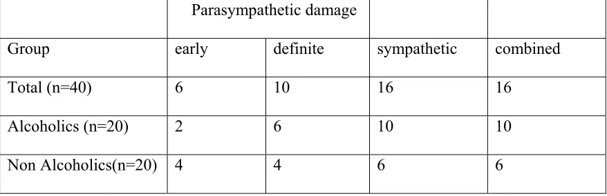
Table.4 Distribution of autonomic dysfunction in alcoholics and non alcoholics
Parasympathetic damage
Group early definite sympathetic combined
Total (n=40) 6 10 16 16
Alcoholics (n=20) 2 6 10 10
Graph:1 Distribution of Child Class in the study population
5%
60% 35%
Child A -2
Child B-24
Child C-14
Graph:2 Distribution of autonomic dysfunction according to Child class
1 0
1 4
6 18
1 4
13
0 2 4 6 8 10 12 14 16 18
Child class A (n=2)
Child class B (n=24)
Child class C (n=14)
Early parasympathetic damage
Definite parasympathetic damage
Combined damage
Graph:3 Distribution of autonomic dysfunction in alcoholics and non alcoholics
early PS Definite PS Sympathetic Combined
0 2 4 6 8 10 12 14 16
Alcoholic Non Alcoholic Total
Graph:4. Distribution of autonomic score in patients according to their Child class
0.45 0.5
3.95
6.53
0 1 2 3 4 5 6 7
ANS score
Graph:5. Distribution of autonomic score in alcoholics and non alcoholics
0.45
3.65 5.7
0 1 2 3 4 5 6
ANS score
Controls Nonalcoholic Alcoholic
Graph 6. Comparison of Valsalva ratio and Heart rate response to Standing in alcoholic and non alcoholic Groups
0 0.2 0.4 0.6 0.8 1 1.2 1.4 1.6 1.8 2
Alcoholic Non-alcoholic
Graph 7: Comparison of Heart rate response to deep breathing ,BP response to standing and BP response to sustained hand grip in alcoholic and non alcoholic Groups
Graph 8: Comparison of Valsalva ratio ,Heart rate response to deep breathing, Heart rate response to standing in Child A, B. and C groups
0 0.5 1 1.5 2 2.5 3 3.5 4 4.5 5
A B C
Valsalva Ratio Deep breathing Heart Rate
5 7 9 11 13 15 17
Alcoholic Non-alcoholic
6. Discussion:
Autonomic nervous dysfunction is a known complication of diabetes28 and alcohol abuse. Autonomic damage is expected in somepatients with alcohol related cirrhosis since autonomic dysfunction, especially of vagal origin, is seen in chronic alcoholics29. Evidence for vagal neuropathy in alcoholic cirrhosis is wellestablished by various studies30 However, in non-alcoholiccirrhosis there are conflicting reports.31
\ In our study, 32 of the 40 chronic liver disease patients (80%) were found
to havean abnormal result in one or more autonomic function tests.However, Barter and Tanner in their study of 30 subjects reportedevidence of parasympathetic damage in 16% and of combined parasympatheticand sympathetic neuropathy in an additional 20%..The lowerfrequency of autonomic dysfunction in their study could be dueto the fact that they included only 14 subjects with alcoholic liver disease while the rest had an alcohol dependence problemonly.
Szalay et al in their evaluation of 121 patients withchronic alcoholism—33
without liver disease, 33 with fattyliver, 33 with cirrhosis, 10 with biliary cirrhosis, and 12with cirrhosis of another origin—found autonomic reflexdamage in all.They observed significantly more damage in those with liver disease. Hendrickse and Triger reported cardiovascular autonomic dysfunction with predominantly parasympathetic abnormality in 35% of the patients with chronic liver disease.33
Hendrickse et al in another study reported vagal neuropathyin 45% of
Gentile et al foundautonomic dysfunction in 60% (71% in the alcoholic
group and 57% in the non-alcoholic group) of the 113 cirrhotics studied.35Like in the present study, alteration of parasympathetic functionwas significantly more frequent than that of sympathetic function.Dillon et al also detected abnormal cardiovascular reflexes
in 60% of 70 cirrhotics.36 Their study group included as manyas 42 patients belonging to Child class A and only 15 patientsin class C.
Gonzalez-Reimers et al have remarked in a study that in alcoholics,
autonomic and peripheral neuropathy are dependent on each otherand there is only weak correlation between liver function andboth autonomic and peripheral neuropathy. In the present study no apparent relationship was noticed between autonomic and peripheral neuropathy, and autonomic damage was observed to be more commonthan peripheral neuropathy. The main object of the present studywas to examine autonomic dysfunction and so, while careful systematicclinical examination was done in all the subjects, it is probable that more sensitive electrophysiological measurement would have yielded a much higher proportion of peripheral nerve abnormalities.
Maheshwari -Thuluvath et al in their study remarked that Autonomic
that patients with Autonomic dysfunction are more likely to develop hepatic encephalopathy (HE) due to prolonged intestinal transit time. In their study, they examined the incidence of new-onset HE in patients with and without AN. Seventy-two patients (Child A/B/C = 35/31/6) without evidence of HE at the time of autonomic function testing (AFT) were followed for 39.5 +/- 27.3 months. The end point of the study was the development of new onset HE.
Patients were followed until death or liver transplantation. Of the 72 patients, 42 (58%) patients did not develop HE (group A) while 30 (42%) developed HE (group B) during the follow-up. Both groups had similar baseline demographics. AN was more common in group B (27/30; 90%) compared to group A (28/42; 67%) (P = 0.02). Kaplan-Meier analysis showed a trend toward a higher incidence of HE in patients with Autonomic dysfunction . Mortality was higher in group B (12/30; 40%) compared to group A (8/42; 19%) (P = 0.04). Patients with Autonomic dysfunction were more likely to develop new onset HE. Although an independent effect of Autonomic dysfunction on HE was not established in that study, they speculated that delayed intestinal transit secondary to Autonomic dysfunction may explain the higher incidence of HE in patients with AN.
scores for alcoholics and and non-alcoholics were 5.70 and 3.65 respectively . These findings are similar to the observations of most other studies,37,38 which reported increasingfrequency of autonomic dysfunction with increasing severityof liver damage.
Hendrickse and Triger reported a strong correlationbetween the abnormal tests and Child-Pugh score (p<0.0001). . In their study, they found autonomic dysfunction in 69% of Childclass B and C patients and 23% in class A patients (p<0.0001).On the contrary, Gonzalez-Reimer et al in their study of 33alcoholics, 20 of them cirrhotics,
found a weak correlation between liver function and both autonomic and peripheral neuropathy.
neuropathy were significantly older and tended to havelower serum albumin than those with normal cardiovascular test.41,42
In our study, heart rate response to standing was the most frequently(22 out of 40 patients) abnormal test in test group. Barterand Tanner in their study noted the heart rate response to standingas the most sensitive test with high specificity. Thuluvathand Triger in their study reported the heart rate response todeep breathing as the most sensitive test. However, it is noteworthythat this test depends on the cooperation of the subject andis, thus not as reproducible as the heart rate response to standing..
Gentile
et al remarked that deep breathing test and handgriptests are the
most influenced by the compliance of the patient.. In the study, they found the deep breathing test and lying tostanding (heart rate response) tests to be most altered andthe most sensitive and specific tests respectively. . In the presentstudy, seven patients had abnormal heart rate response to deep breathing, twelve had abnormal blood pressure response to sustainedhandgrip, and eleven patients had an abnormal Valsalva ratio.
7. Conclusion
:This study shows that autonomic dysfunction is commonin patients with cirrhosis and it was comparable frequency bothin alcoholics and non-alcoholics. It increases in severity with increase in extent of liver damage, suggesting that liverdamage contributes to the neurological dysfunction
8. Summary:
It has been reported that the severity of autonomic dysfunction is proportional to the severity of cirrhosis, and its presence is an indicator of poor prognosis.
This study was conducted to investigate autonomic Dysfunctions in patients with cirrhosis of varied etiology, and to determinethe relationship between severity of liver damage and extentof autonomic function impairment.
• The present study included 40 patients with Cirrhosis and 40 age and sex
matched controls.
• All the patients and the controls were subjected to a battery of five standard
autonomic function tests. (Valsalva ratio, HR response to deep breathing, Heart rate response to standing, BP response to standing & BP response to sustained hand Grip)
damage, ten (25%) had definiteparasympathetic damage, and sixteen (40%) had combined (both parasympathetic and sympathetic) damage.
• Eighteen (90%) ofthe alcoholics and fourteen (70%) of the non-alcoholics had autonomicdysfunction.
• Autonomic dysfunctionwas significantly abnormal in advanced liver disease comparedwith early liver damage.
• Light headedness on standingwas the most common symptom of autonomic dysfunction which was more frequently observed in cirrhotics compared with controls (p=0.001).
• One patient (50%) in Child class A group, eighteen (75%) in Child class B and 13
(92.85%) in Child class C had autonomic dysfunction.
• Therewas no significant association between symptoms ofautonomic dysfunction
and objective evidence of autonomic damage
• None of our patients was found to have sympathetic dysfunctionalone. • Parasympathetic damage was always found in association with an evidence of
sympathetic damage.
• The mean (SD) total autonomic function scores in patients with cirrhosis and
controls were 4.57(1.84) and 0.45 (0.57) respectively and it was statistically significant (p<0.01).
9. Annexure –I
Proforma
Date: GE no: 1. Name :
2. Age/sex :
3. Address : 4. History
Abdominal distension Abdominal pain UGI bleeding Jaundice
Altered sensorium Fever
Pedal edema
Symptoms of autonomic dysfunction : 1. Dizziness while Standing
2. Altered sweating
5. Constipation 6. Nausea, vomiting 7. Difficulty in swallowing 8. Feeling full after eating little 9. Eye irritation/decreased lacrimation 10.Urinary frequency/ hesitancy
5. Drug history :
6. Family History :
7. Habits :
Alcohol abuse
8. BMI :
9. General examination :
10. Abdominal examination:
CTP score :
USG abdomen :
UGI Endoscopy :
Autonomic function tests
12.Basal Recordings
Heart Rate
Blood Pressure 13.Orthostatic standing Test
HR (30/15) B.P. (supine) B.P. Standing 14.Deep Breathing
HR
15.Valsalva Maneuver
HR
10. References:
1. DelValle J, Yamada T: The gut as an endocrine organ. Annu Rev Med 41:447–455, 1990.
2. Gershon MD, Erde SM: The nervous system of the gut. Gastroenterology 80:1571– 1594, 1981
3. .Larsson LI, Goltermann N, de Magistris L, et al: Somatostatin cell processes as pathways forparacrine secretion. Science 205:1393–1395, 1979.
4. Dockray GJ: Physiology of enteric neuropeptides. In Johnson LR (ed): Physiology of the Gastrointestinal Tract. New York, Raven Press, 000:129–167, 1994.
5. Murthy KS, Grider JR, Jin JG, et al: Interplay of VIP and nitric oxide in the regulation of neuromuscular activity in the gut. Arch Int Pharmacodyn Ther 329:27–38, 1995.
6. Makhlouf GM: Neural and hormonal regulation of function in the gut [published erratum appears in Hosp Pract (Off Ed) 25(5):15, 1990]. Hosp Pract (Off Ed) 25:79–87, 90–95, 98, 1990.
7. Furness JB, Clerc N: Responses of afferent neurons to the contents of the digestive tract, and their relation to endocrine and immune responses. Prog Brain Res 122:159– 172, 2000.
8.Willems JL, Buylaert WA, Lefebvre RA, et al: Neuronal dopamine receptors on autonomic ganglia and sympathetic nerves and dopamine receptors in the gastrointestinal system. Pharmacol Rev 37:165–216, 1985.
10.Lowenstein CJ, Dinerman JL, Snyder SH: Nitric oxide: A physiologic messenger. Ann Intern Med 120:227–237, 1994.
11.Huet P-M, Pomier-Layrargues G, Villeneuve J-P, et al: Intrahepatic circulation in liver disease. Semin Liver Dis 6:277, 1986
12.Hendrickse MT, Triger DR. Autonomic dysfunction in chronic liver disease.
Clin Auton Res 1993;3:227–31.
13.Barter F, Tanner AR. Autonomic neuropathy in alcoholic population. Postgrad Med J 1987;63:1033–6.
14.Bharucha AE, Camilleri M, Low PA, Zinsmeister AR: Autonomic dysfunction in gastrointestinal motility disorders. Gut 1993 Mar; 34(3): 397-401
15.Emond D, Lebel M: Orthostatic hypotension and Holmes-Adie syndrome. Usefulness of the Valsalva ratio in the evaluation of baroreceptor dysfunction. J Hum Hypertens 2002 Sep; 16(9): 661-2.
16.Gibbons CH, Freeman R: Autonomic neuropathy and coeliac disease. J Neurol Neurosurg Psychiatry 2005 Apr; 76(4): 579-81.
17.Butchal, Crawford TO, Hauer P, et al: Small-fiber sensory neuropathies: clinical course and neuropathology of idiopathic cases. Ann Neurol 1998 Jul; 44(1): 47-59. .
19.Avaria Mde et al Kudat H, Akkaya V, Sozen AB, et al: Heart rate variability in diabetes patients. J Int Med Res 2006 May-Jun; 34(3): 291-6
20..Maheshwari A, Thomas A, Thuluvath PJ: Patients with autonomic neuropathy are more likely to develop hepatic encephalopathy. Dig Dis Sci 2004 Oct; 49(10): 1584-8
21.Low PA, Novak V, Spies JM, et al: Cerebrovascular regulation in the postural orthostatic tachycardia syndrome (POTS). Am J Med Sci 1999 Feb; 317(2): 124-33.
22.Low PA, Caskey PE, Tuck RR, et al: Quantitative sudomotor axon reflex test in normal and neuropathic subjects. Ann Neurol 1983 Nov; 14(5): 573-80.
23.Low PA, Dyck PJ, Lambert EH, et al: Acute panautonomic neuropathy. Ann Neurol 1983 Apr; 13(4): 412-7.
24.Low PA: Clinical autonomic disorders: evaluation and management. 2nd ed. New York: Lippincott Raven; 1997.
25.Low PA, Vernino S, Suarez G: Autonomic dysfunction in peripheral nerve disease. Muscle Nerve 2003 Jun; 27(6): 646-61.
26.Lyu RK, Tang LM, Wu YR, Chen ST: Cardiovascular autonomic function and sympathetic skin response in chronic inflammatory demyelinating polyradiculoneuropathy. Muscle Nerve 2002 Nov; 26(5): 669-72.
28.Bellavere F, Bosello G, Fedele D, et al. Diagosis and management of diabetic
autonomic neuropathy [letter]. BMJ 1983;287:61.
29.Szalay F, Oravecz L, Kadar E, et al. Autonomic neuropathy in chronic liver disease.
Gastroenterology 1990;50:187–9.
30.Thuluvath PJ, Triger DR. Autonomic neuropathy and chronic liver disease. Q J Med
1989;72:737–47.
31.Gonzalez-Reimers E, Alonso-Socas M, Santolaria-Fernandez F, et al. Autonomic and
peripheral neuropathy in chronic alcoholic liver disease. Drug Alcohol Depend
1991;27:219–22.
32.Khosla SN, Sanyal S, Nand N. Autonomic function tests and clinical significance of dysautonomia in chronic liver disease. J Assoc Physicians India 1991;39:924–6.
33.Hendrickse MT, Triger DR. Autonomic dysfunction and hepatic function in chronic liver disease. Gut 1990;31:A1164.
34.Hendrickse MT, Thuluvath PJ, Triger DR. Natural history of autonomic neuropathy in chronic liver disease. Lancet 1992;339:1462–4
35.Gentile S, Marmo R, Peduto A, et al. Autonomic neuropathy in liver cirrhosis:
relationship with alcoholic aetiology and severity of disease. Ital J Gastroenterol
36.Dillon JF, Plevris JN, Nolan J, et al. Autonomic function in cirrhosis assessed by
cardiovascular reflex tests and 24-hour heart rate variability. Am J Gastroenterol
1994;89:1544–7.
37.Ewing DJ, Campbell IW, Clarke BF. Assessment of cardiovascular effects in diabetic autonomic neuropathy and prognostic implications. Ann Intern Med 1980;92:308–11.
38.Novak DJ, Victor M. The vagus and sympathetic nerves in alcoholic polyneuropathy.
Arch Neurol 1974;30:273–84.
39.Johnson RH, Robinson BJ. Mortality in alcoholics with autonomic neuropathy. J
Neurol Neurosurg Psychiatry 1988;51:476–80.
40.Duncan G, Johnson RH, Lambie DJ, et al. Evidence of vagal neuropathy in chronic
alcoholics. Lancet 1980;ii:1053–7.
41.Decaux G, Cauchie P, Soupart A, et al. Role of vagal neuropathy in the
hyponatraemia of alcoholic cirrhosis. BMJ 1986;293:1534–6.
42.Lenz K, Hortnagl, H, Magometschnigg, D, et al. Functions of the autonomic nervous
system in patients with hepatic encephalopathy. Hepatology 1985;5:831.
Master Chart Control Group
S.No Age sex AN symptoms Drug history Family History Total Valsalva ratio Score Deep breath score HR Score BP 1 score BP 2 score
1 40 2 0 2 0 1 1.22 0 16 0 1.05 0 16 1 16 0
2 30 1 0 2 0 1 1.24 0 16 0 1.06 0 12 0 18 0
3 35 1 0 2 0 1 1.24 0 18 0 1.08 0 14 1 20 0
4 46 2 4 2 0 1 1.22 0 18 0 1.06 0 14 1 20 0
5 32 1 0 2 1 1 1.28 0 16 0 1.07 0 12 0 18 0
6 55 1 4 2 0 1 1.26 0 17 0 1.06 0 14 1 18 0
7 46 1 1 2 0 0 1.23 0 17 0 1.06 0 6 0 20 0
8 15 1 1 2 1 0 1.24 0 16 0 1.06 0 6 0 18 0
9 27 2 4 2 0 0 1.22 0 18 0 1.08 0 8 0 18 1
10 42 2 4 2 0 0 1.23 0 16 0 1.07 0 6 0 18 1
11 37 1 1 2 1 0 1.24 0 17 0 1.09 0 6 0 16 0
12 30 1 1 2 1 0 1.26 0 16 0 1.08 0 8 0 16 1
13 40 1 1 2 1 0 1 22 0 18 0 1 06 0 6 0 16 0
13 40 1 1 2 1 0 1.22 0 18 0 1.06 0 6 0 16 0
14 25 2 1 2 0 0 1.25 0 18 0 1.06 0 8 0 18 1
15 37 1 0 2 1 0 1.22 0 18 0 1.08 0 6 1 18 0
16 40 1 5 2 1 0 1.26 0 16 0 1.08 0 6 0 16 0
17 36 1 0 2 1 0 1.26 0 16 0 1.06 0 4 0 20 1
18 45 2 0 2 1 0 1.26 0 18 0 1.08 0 8 0 20 0
19 34 1 0 2 1 0 1.22 0 16 0 1.06 0 6 0 18 0
20 45 2 0 2 1 1 1.28 0 12 1 1.06 0 4 0 14 0
21 40 1 1 2 0 1 1.26 0 14 1 1.06 0 6 0 12 1
22 47 1 1 2 1 1 1.12 1 12 1 1.08 0 6 0 12 0
23 34 1 1 2 1 1 1.18 1 12 1 1.08 0 6 0 16 1
24 45 1 1 2 0 1 1.24 0 14 1 1.06 0 8 0 12 1
25 41 1 1 2 1 0 1.22 0 16 0 1.06 0 8 1 12 0
26 26 1 1 2 1 0 1.26 0 16 0 1.07 0 6 0 12 1
27 35 2 3 2 0 1 1.24 0 14 1 1.08 0 8 0 12 0
28 22 2 6 2 0 1 1.22 0 16 0 1.06 0 12 1 12 1
S.No Age sex AN symptoms Drug history Family History Total Valsalva ratio Score Deep breath score HR Score BP 1 score BP 2 score
30 35 1 0 2 0 1 1.24 0 18 0 1.06 0 12 1 12 1
31 42 1 0 2 0 0 1.26 0 16 0 1.06 0 8 0 18 0
32 41 2 0 2 1 0 1.24 0 16 0 1.07 0 6 0 18 0
33 18 1 0 2 1 0 1.22 0 16 0 1.09 0 6 0 20 0
34 24 2 0 2 0 0 1.22 0 18 0 1.06 0 6 0 20 0
35 34 1 5 2 0 1 1.24 0 16 0 1.04 1 8 0 18 0
36 46 1 6 2 1 1 1.14 1 16 0 1.06 0 6 0 20 0
37 40 2 0 2 1 0 1.22 0 18 0 1.08 0 8 0 18 0
38 38 1 0 2 1 0 1.23 0 16 0 1.02 1 6 0 20 0
39 20 2 0 2 1 0 1.22 0 16 0 1.06 0 8 0 18 0
40 27 2 0 2 0 0 1.24 0 18 0 1.06 0 6 0 16 0
Male -1 1 Dizziness while Stand1 yes 1 jaundice >1 21= 0 >15 = 0 >1 04 = 0 <10 = 0 >16 = 0
Male -1 1. Dizziness while Stand1.yes 1.jaundice >1.21= 0 >15 = 0 >1.04 = 0 <10 = 0 >16 = 0
Female -2 2. Altered sweating 2.no 2.diabetes 1.11–1.20 = 1 11–14 = 1 1.01–1.03 = 1 11–29 = 1 11–15 = 1
3. Pain in extremities 3.HT <1.10 = 2 <10 = 2 <1.00 = 2 >30 = 2 <10 = 2
4. Palpitation
5. Constipation Valsalva ratio Deep breathing Heart rate BP 1 BP 2
6. Nausea, vomiting test (max-min heart response to response response to
7. Difficulty in swallowing rate beats/min) standing (30:15 ratio) to standing sustained hand grip
AN symptoms Drug history Family History Total Valsalva ratio Score Deep breath score HR Score BP 1 score BP 2 score 1 0 2 0 1 1.24 0 1 8 0 1.06 0 1 2 1 12 1 1 0 2 0 0 1.26 0 1 6 0 1.06 0 8 0 1 8 0 2 0 2 1 0 1.24 0 1 6 0 1.07 0 6 0 1 8 0 1 0 2 1 0 1.22 0 1 6 0 1.09 0 6 0 2 0 0 2 0 2 0 0 1.22 0 1 8 0 1.06 0 6 0 2 0 0 1 5 2 0 1 1.24 0 1 6 0 1.04 1 8 0 1 8 0 1 6 2 1 1 1.14 1 1 6 0 1.06 0 6 0 2 0 0 2 0 2 1 0 1.22 0 1 8 0 1.08 0 8 0 1 8 0 1 0 2 1 0 1.23 0 1 6 0 1.02 1 6 0 2 0 0 2 0 2 1 0 1.22 0 1 6 0 1.06 0 8 0 1 8 0 2 0 2 0 0 1.24 0 1 8 0 1.06 0 6 0 1 6 0 1 Dizziness while Stand 1 yes 1 jaundice >1 21= 0 > 1 5=0 >1 04 = 0 < 1 0=0 > 1 6=0 1 . Dizziness while Stand 1 .yes 1 .jaundice >1 .21= 0 >15 = 0 >1 .04 = 0 <10 = 0 >16 = 0
2. Altered sweating
2.no
2.diabetes
1.11–1.20 = 1
11–14 = 1
1.01–1.03 = 1
11–29 = 1
11–15 = 1
3. Pain in extremities
3.HT
<1.10 = 2
<10 = 2
<1.00 = 2
>30 = 2
<10 = 2
4. Palpitation 5. Constipation
Valsalva ratio
Deep breathing
Heart rate
BP 1
BP 2
6. Nausea, vomiting
test (max-min heart
response to
response
response to
7. Difficulty in swallowing
rate beats/min)
standing (30:15 ratio)
to standing
sustained hand grip