Copyright © 1998, American Society for Microbiology
Cloning and Expression of a Human T-Lymphotropic Virus
Type 1 Protein with Reverse Transcriptase Activity
S. MICHELE OWEN,1RENU B. LAL,1ANDRICHARD A. IKEDA2*
HIV and Retrovirus Diseases Branch, National Center for Infectious Diseases, Centers for Disease
Control and Prevention, Atlanta, Georgia 30333,1and School of Chemistry and Biochemistry,
Georgia Institute of Technology, Atlanta, Georgia 30332-04002
Received 30 October 1997/Accepted 16 February 1998
Unlike most other characterized retroviruses, there is little published information on the biochemical properties of human T-lymphotropic virus type 1 (HTLV-1) reverse transcriptase (RT). Specifically, no reports of a cloned functional RT enzyme have been published. Since the RT enzyme is an essential component of the virus, our objective was to clone, express, and purify a functional RT enzyme from HTLV-1. Our approach was to clone and express a protein of approximately 60 to 65 kDa that we hypothesized would correspond to the RT region encoded by the pol reading frame. The predicted region encoding the RT enzyme comprised nucleotides 2617 to 4312 of the HTLV-1 MT-2 isolate. A putative RT gene was obtained by PCR and was ligated into various prokaryotic expression vectors. A novel cloning approach allowed us to generate a stable clone in the pro-karyotic expression vector pGEX-4T-1 and produce a recombinant protein of approximately 60 to 65 kDa. The partially purified protein displays RT activity in both amplification RT (AMP-RT) assays and traditional RT assays. This is the first report of a cloned protein from HTLV-1 which displays RT activity and is the first step in the characterization of HTLV-1 RT.
Human T-lymphotropic virus type 1 (HTLV-1) is an onco-virus in the family Retroviridae (25). HTLV-1 was first isolated in the early 1980s from patients with adult T-cell leukemia/ lymphoma (28), and the virus has been subsequently shown to be clinically associated with adult T-cell leukemia/lymphoma (36), HTLV-1-associated myelopathy/tropical spastic parapa-resis (11), and a number of other chronic diseases (i.e., uveitis, arthritis, and infective dermatitis) (9, 18). Infections with HTLV-1 are endemic in Melanesia, Japan, the Caribbean, and sub-Saharan Africa and among U.S. risk groups such as intra-venous drug users and prostitutes (3, 9, 18).
HTLV-1 has been cloned, and a number of different isolates have been sequenced (5, 12, 30). The genome of HTLV-1 is approximately 9 kb in length and is flanked by long terminal repeats. Like other human retroviruses, HTLV-1 has three large open reading frames which encode the Gag (48 kDa), Pol (99 kDa), and Env (54 kDa) proteins, and a number of spliced, open reading frames that encode regulatory proteins (e.g., Rex and Tax, etc.) (3, 21).
All characterized onco- and lentiviruses share another com-mon feature. That feature is that none of the pol open reading frames begin with an initiator codon (ATG). The translation of
pol genes occurs through either amber codon suppression, as in
the case of murine leukemia virus, or, more commonly, through ribosomal frameshifts, as has been described for all other characterized onco- and lentiviruses (4). For example, in human immunodeficiency virus type 1 (HIV-1) and HIV-2 one ribosomal frameshift is required for the translation of the pol-encoded enzymes. In contrast, two ribosomal frame shifts, one in the gag-pro overlap and one in the pro-pol overlap, are required to synthesize the pol-encoded replication enzymes of HTLV-I (26).
Although HTLV-I and HIV-1 are structurally related
(hu-man retroviruses), their differences are notable. HIV-1 repli-cates quickly, mutates rapidly, exhibits great genetic diversity, produces large amounts of infectious virus, and causes AIDS in almost all infected individuals (8, 23). Furthermore, reverse transcriptase (RT) activity is easily detected in the sera of HIV-1-infected individuals (13), and HIV-1 RT is well char-acterized (15, 16, 33). In contrast, HTLV-1 replicates slowly, mutates very slowly, exhibits little genetic diversity, produces little or no cell-free infectious virus (transmission appears to require the exchange of infected cells), and causes a variety of diseases including T-cell leukemia, HTLV-1-associated my-elopathy/tropical spastic paraparesis in only a minority of the infected individuals (6, 7, 9, 10, 35). Unlike HIV-1-infected individuals, RT activity is almost nonexistent in the sera of HTLV-1-infected individuals (13, 22) and HTLV-1 RT has never been fully characterized. A better understanding of the HTLV RT may prove useful in understanding the differences in mutation rates and pathogenicities observed between HIV-1 and HTLV-1.
A potential HTLV-1 open reading frame that could encode RT (5, 12, 30) has been identified in the sequence of HTLV-1, but only two published reports specifically address the enzyme. In 1981, Gallo and coworkers (29) reported the purification of RT from HTLV-1 virions. The purified enzyme was shown to have Mg21-dependent RT activity, RNase H activity, and a
mass of 95,000 Da. In 1986, Johnson et al. (17) published a computer analysis of various retroviral pol coding sequences. This analysis showed that amino acids 33 to 279 of the pre-dicted HTLV-1 Pol protein aligned to known RT sequences, amino acids 465 to 599 aligned to ribonuclease H sequences, and amino acids 600 to 896 aligned to integrase sequences. These results along with the observed sites for ribosomal frameshifting (26) suggest that the HTLV-1 RT should be about 600 amino acids in length (an equivalent mass of about 65 kDa) and comparable in size to HIV-1 RT. Recently, the integrase of the closely related retrovirus HTLV-2 was identi-fied by Balakrishnan et al. (2). These investigators cloned a 920-bp fragment from the 39end of HTLV-2 pol, expressed a * Corresponding author. Mailing address: School of Chemistry and
Biochemistry, Georgia Institute of Technology, Atlanta, GA 30332-0400. Phone: (404) 894-4037. Fax: (404) 894-7452. E-mail: rick.ikeda @chemistry.gatech.edu.
5279
on November 9, 2019 by guest
http://jvi.asm.org/
protein corresponding to the last 300 amino acids of HTLV-2 Pol (encoded by nucleotides 4290 to 5186 of the G12 isolate of HTLV-2), and demonstrated that the purified recombinant protein exhibited integration and disintegration activities (27). Their results indicate that HTLV-2 pol encodes an integrase at its 39end. Alignment of the HTLV-1 and HTLV-2 pol coding regions by using CLUSTAL software (34) further supports our prediction of the region encoding RT (Fig. 1). Comparison of the nine complete HTLV-1 pol coding regions in GenBank demonstrates a low level of diversity in this coding region of HTLV-1. A comparison of the MT-2 and K30 amino acid sequences is shown in Fig. 1. The remaining seven isolates had similar differences in their amino acid sequences with only eight of the amino acid differences observed being nonhomolo-gous (data not shown).
In this report, we describe the cloning of the predicted HTLV-1 RT by using a novel approach that allows the
pro-duction and purification of a protein with RT activity. Our strategy for construction of an HTLV-1 RT expression clone was simple and direct. PCR was used to simultaneously amplify the predicted RT-coding sequences (nucleotides 2617 to 4312 of HTLV-1 MT2) (30), remove the pro-pol frameshift, add an appropriate stop codon, and append restriction sites to the ends of the RT gene. The amplified RT gene could then be ligated into a modern expression vector to finish the construc-tion of the HTLV-1 RT expression clone. When this was done with a variety of expression vectors (pET20B, pET22B, pET30A [32], and pGEX4T-1) the only products that could be recovered were recircularized vector or clones containing de-letion mutants of the predicted HTLV-1 RT gene. One expla-nation for these results is that HTLV-1 RT is toxic to
Esche-richia coli. Since a translatable RT gene could not be cloned
our solution was to clone an RT gene that begins with a stop codon. This approach was taken to prevent recombinant tein production due to incomplete suppression of the tac pro-moter or utilization of a cryptic propro-moter upstream of the cloning site. We postulated that the presence of the stop codon would increase the stability of the expression construct by allowing tight control over protein expression.
The 1.7-kb fragment of HTLV DNA that we postulated would encode the RT enzyme was amplified from 5 ng of the HTLV-1 infectious clone pK30 (37). The PCR mixture con-tained 10 mM Tris-HCl (pH 8.3); 2.5 mM MgCl2; 50 mM KCl; 200 mM (each) dATP, dTTP, dGTP, and dCTP; 100 ng each of MO40 and MO31; and 2.5 U of Taq Polymerase. (Perkin-Elmer Cetus). The cycling conditions consisted of an initial denaturation at 94°C for 2 min, followed by 35 cycles of 94°C for 1 min, 55°C for 1 min, and 72°C for 1.5 min. The primers that were designed for the PCR amplification of HTLV-1 RT are shown in Table 1. Primer MO40 anneals to nucleotides 2619 to 2632 of HTLV-1 (MT-2, accession no. J02029) and contains nucleotides which add a stop codon upstream (in a 59 direction) of the Asn181 codon and add an additional C that removes the frameshift between Asn181 and Pro33. Primer MO40 also adds an EcoRI restriction site that allows the 59end of HTLV-1 RT to be fused to the 39 end of a glutathione
S-transferase (GST) leader in the expression vector
pGEX-4T-1 (31). Primer MO31 anneals to nucleotides 4298 to 4313 of HTLV-1 (MT-2) and adds a stop codon and a SalI restriction site to the amplified product. The PCR generated a 1.7-kb fragment, which was gel purified, cut with EcoRI and SalI, and ligated between the EcoRI and SalI sites of pGEX-4T-1. Highly competent E. coli STBL2 (Gibco-BRL, Gaithersburg, Md.) was transformed with the ligation mixture (according to the manufacturer’s suggested protocol) and plated on Luria-Bertani agar plates containing 50mg of carbenicillin (Sigma Chemical, St. Louis, Mo.)/ml. Plasmid DNA was isolated from 10 representative colonies, and eight plasmids were found to contain an intact copy of the 1.7-kb RT DNA. The RT DNA of two of the clones was sequenced with T7 Sequenase, and no mutations were discovered. This confirmed the construction of pRT100, a stable clone of HTLV-1 RT (Fig. 2).
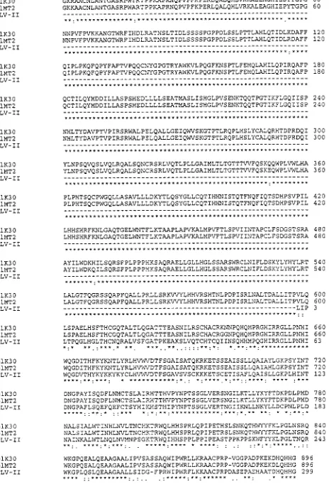
[image:2.612.60.292.79.420.2]FIG. 1. HTLV-1 RT is predicted to extend from Pro33 to Val598. The first amino acid of Pol that is translated is Pro33. Pro33 is the first possible pol-encoded amino acid of HTLV-RT and is indicated by the exclamation point. The location of the C-terminal end of HTLV-1 RT was approximated by aligning the HTLV-1 Pol protein to HTLV-2 integrase. CLUSTAL (14, 34) was used to align the predicted protein sequences encoded by two different HTLV-1 pol open reading frames (K-30, accession no. L03561, and MT-2, accession no. J02029) and HTLV-2 integrase (G12, accession no. M10060). Residue numbering of the Pol protein begins from the first amino acid of the protein encoded by the pol gene of HTLV-1. The HTLV-2 integrase was defined by Balakrishnan et al. to be last the 300 amino acids (amino acids 651 to 950) of the HTLV-2 Pol protein. Residue numbering of HTLV-2 integrase begins at the first amino acid in the integrase. In the aligned sequences, identical amino acids are denoted by aster-isks and similar amino acids are identified by periods.
TABLE 1. PCR primers for amplification of HTLV-1 RT DNAa
Primer Sequence
MO40 59ACTCGAATTCTAGATGAACCCAGAACGCCTCCA 39 MO31 59ATCGAGTCGACTTAGACAGGGGTGATTAGT 39
aThe bases in italics represent the restriction site sequences added to the amplified product. The underlined bases mark the inserted stop codons. The bases in boldface indicate the sequences that anneal to the RT DNA in the infectious clone pK30.
on November 9, 2019 by guest
http://jvi.asm.org/
The expression plasmid pRT100 would appear to be unable to produce HTLV-1 RT since a stop codon is fused to the beginning of the RT gene; however, stop codons can be by-passed in strains of E. coli that harbor suppressor tRNAs (19, 20, 24). As discussed below, the plasmid pRT100 produces GST-RT, a fusion protein, in E. coli strains that contain an amber suppressor.
The isolation of GST-RT fusion protein was begun by trans-forming an amber suppressor strain (amber-Gln E. coli) with pRT100. Transformation reactions were carried out as recom-mended by the manufacturer (Promega, Madison, Wis.). To control for potential endogenous RT activity present in E. coli, the suppressor strain was also transformed with the parental pGEX-4T-1 and carried through the subsequent experiments described for pRT100. A single colony from a transformation plate was used to inoculate a 400-ml culture of LB broth containing 50mg of carbenicillin/ml. The culture was grown at 37°C in a shaking incubator at 250 rpm to an optical density at 500 nm of 0.8. The tac promoter upstream of the GST-coding region was then induced with 1 mM isopropyl thiogalactoside. After induction, the culture was incubated for an additional 2 h, and the cells were harvested by centrifugation at 7,8003
g for 10 min at 4°C. The cells were resuspended in 20 ml of
phosphate-buffered saline (137 mM NaCl, 2.7 mM KCl, 4.3 mM Na2HPO4, 1.4 mM KH2PO4 [pH 7.4]) containing 1% Triton X-100, incubated with gentle mixing for 30 min at 4°C, and sonicated on ice. The sonicated sample was cleared by centrifugation at 12,0003g for 10 min at 4°C, and the
super-natant fraction was collected. The supersuper-natant fractions were loaded on a 200-ml glutathione-Sepharose column, and the column was washed with 6 ml of phosphate-buffered saline. The column was eluted with 600ml of 50 mM Tris-HCl, pH 8.0, containing 10 mM reduced glutathione. The elution fraction was then reduced to;50ml in a Centricon 30 concentrator (Millipore). Various volumes of the eluted fraction were used
in the previously described AMP-RT assay (13) to screen for RT activity. The AMP-RT assay like conventional RT assays measures the ability of RT to produce a DNA copy of a heterologous RNA template. The AMP-RT reaction buffer was composed of 50 mM Tris-Cl (pH 8.3), 50 mM KCl, 5 mM MgCl2, 0.4 mM (each) deoxynucleoside triphosphate, 1 mM EGTA, 0.06% Nonidet P-40, 2 mM dithiothreitol, and 100 ng of encephalomyocarditis virus RNA template. To increase sen-sitivity, the AMP-RT assay uses a detection system whereby the reverse-transcribed cDNA (encephalomyocarditis virus cDNA) is detected by PCR and Southern hybridization (13). Figure 3A shows the results of an AMP-RT assay with 1ml of the partially purified material isolated from the amber-Gln E.
coli strain transformed with pRT100.
[image:3.612.49.287.70.232.2]To determine if other suppressor strains would allow for
[image:3.612.353.494.71.381.2]FIG. 2. Plasmid pRT100. The plasmid pRT100 is pGEX-4T-1 (Pharmacia) with nucleotides 2617 to 4312 of HTLV-2 MT-2 (Seiki et al. [30]) inserted in the multiple cloning site and fused in-frame to the GST of the pGEX vector; however, the sequence of the HTLV-1 DNA has been slightly modified to facilitate cloning and expression of HTLV-1 RT. The modifications include (i) addition of an in-frame amber stop codon at the 59end (nucleotide 2617) of the cloned HTLV-1 RT sequences, (ii) removal of the pro-pol frameshift by insertion of a C between HTLV-1 nucleotides 2618 and 2619 (this generates an RT gene that encodes a protein beginning with Asn181 to Pro33), and (iii) addition of a stop codon to the 39end (nucleotide 4312) of the last codon (encoding Val598) of the cloned HTLV-1 RT gene. The amber stop codon preceding HTLV-1 RT separates the RT from GST and prevents translation of the RT; however, a GST-RT fusion protein can be produced by suppression of the amber stop codon.
FIG. 3. Lysates of suppressor E. coli containing pRT100 exhibit RT activity. The cDNA band that is produced by reverse transcription and PCR is detected by Southern hybridization. The bands seen in this figure indicate RT activity. (A) Autoradiograph of an AMP RT reaction of partially purified lysates from the amber-Gln E. coli strain transformed with pRT100 and the parental plasmid pGEX4T-1. Lane 1, size standards; lane 2, 1ml of lysate from parental pGEX 4t-1-transformed bacteria; lane 3, 1ml of lysate from pRT100-transformed bac-teria, lane 4, positive control (0.1 U of purified AMV RT). (B) Autoradiographs of AMP-RT assays of partially purified lysates from four suppressor strains. Top: lane 1, molecular mass markers; lane 2, 10ml of a lysate of E. coli containing the parental plasmid pGEX-4T-1; lane 3, 1ml of a lysate of amber-Gln E. coli containing pRT100 (sample from panel A); lane 4, 10ml of a lysate of amber-Gln E. coli containing pRT100; lane 5, 10ml of a lysate of amber-Tyr E. coli con-taining pRT100; lane 6, 10ml of a lysate of amber-Leu E. coli containing pRT100; lane 7, 10ml of a lysate of amber-Lys E. coli containing pRT100; lane 8, 0.1 U of purified AMV RT. Bottom: lane 1, molecular mass marker; lane 2, 1 ml of a lysate of amber-Gln E. coli containing pRT100; lane 3, 1ml of a lysate of amber-Tyr E. coli containing pRT100; lane 4, 1ml of a lysate of amber-Leu E. coli containing pRT100; lane 5, 1ml of a lysate of amber-Lys E. coli containing pRT100; lane 6, 0.1 U of purified AMV RT.
on November 9, 2019 by guest
http://jvi.asm.org/
better expression of the recombinant protein three additional suppressor strains (amber-Thr E. coli, amber-Leu E. coli, and amber-Lys E. coli; Promega) were transformed and screened for the presence of recombinant RT protein as described above. The results of the AMP-RT assay for the partially purified protein fractions from the three additional suppressor strains as well as the original amber-Gln E. coli are shown in Fig. 3B. These results demonstrate that RT activity is present in all of the fusion protein preparations, except the amber-Lys
E. coli lysate, and that the RT is active even when fused to
GST.
To determine the activity of the recombinant protein with-out the GST fusion tag, thrombin (10 U) was added to the remaining concentrated elution fractions, and the fractions were incubated at 25°C for 4 h. RT activity in proteolyzed elution fractions (equivalent amounts of protein as prior to cleavage) was analyzed by an AMP-RT assay (13) and by a traditional RT assay with a reaction mixture (15, 33) contain-ing 50 mM Tris (pH 7.8), 75 mM KCl, 2 mM dithiothreitol, 5 mM MgCl2, 5-mg/ml poly(A), 20-mg/ml oligo(dT) (12- to 18-mer), 0.05% Nonidet P-40, and 20-mCi/ml [a-32P][TTP]. Fig-ure 4 shows the results of the AMP-RT reactions and Fig. 5 shows the results of the traditional RT reactions. Both assays indicate that all four suppressor strain fractions contain RT activity. A 10-ml aliquot of the protein purified from the am-ber-Gln E. coli and cleaved from the GST tag was subjected to sodium dodecyl sulfate-polyacrylamide gel electrophoresis (SDS-PAGE) to determine the ratio of GST to RT. As can be seen from Fig. 6 the ratio of GST to RT is very high. It is
[image:4.612.311.547.68.303.2]estimated that the RT portion of the fraction is approximately 1% of the total protein preparation. A similar analysis was done with the protein fractions from the other suppressor strains, and similar results were obtained with the amber-Tyr strain. The amber-Lys and amber-Leu strains produced less FIG. 4. AMP-RT assays of partially purified HTLV-1 RT. A 1.8% agarose
gel containing AMP-RT PCR products from partially purified HTLV-1 RT produced from pRT100 in amber-Gln, amber-Tyr, amber-Leu, and amber-Lys strains of E. coli was stained with ethidium bromide. For each lane shown in the top and bottom panels 10ml of PCR product was loaded onto the gel. The ;320-bp bands seen in lanes 3, 5, and 7 (top) and lanes 2 and 4 (bottom) are the cDNAs that are produced by the amplification of a reverse transcription product. The bands indicate RT activity. (Top) Lane 1, 100-bp ladder; lane 2, negative control; lane 3, 1ml of partially purified HTLV-1 RT from amber-Gln E. coli; lane 4, 10ml of partially purified HTLV-1 RT from amber-Gln E. coli; lane 5, 1 ml of partially purified HTLV-1 RT from amber-Tyr E. coli; lane 6, 10ml of partially purified HTLV-1 RT from amber-Tyr E. coli; lane 7, 1ml of partially purified HTLV-1 RT from amber-Leu E. coli; lane 8, 10ml of partially purified HTLV-1 RT from amber-Leu E. coli. (Bottom) Lane 1, 100-bp ladder; lane 2, 1 ml of partially purified HTLV-1 RT from amber-Lys E. coli; lane 3, 10ml of partially purified HTLV-1 RT from amber-Lys E. coli; lane 4, positive control (1 U of AMV RT).
FIG. 5. Assay of recombinant HTLV-1 RT activity using poly(A)-oligo(dT) template-primer with 5 mM MgCl2. Protein preparations from four different
suppressor strains containing pRT100 or the parental pGEX 4T-1 were assayed in standard RT assays. The bars represent counts per minute of incorporated [32P]TTP from 5ml of purified sample which was cleaved from the GST fusion
tag.
FIG. 6. SDS-PAGE analysis of a partially purified sample of HTLV-1 RT. A Coomassie-stained SDS–12% polyacrylamide gel of HTLV-1 RT following cleavage of the GST fusion tag with thrombin. A total of 10ml of sample was loaded onto the gel. GST is readily apparent on the gel at approximately 26 kDa. A second faint band indicated by the arrow is visible at about 60 to 65 kDa; we predict that this is HTLV-1 RT. Lane 1, molecular weight markers; lane 2, protein purified from amber-Gln E. coli containing pRT100.
on November 9, 2019 by guest
http://jvi.asm.org/
[image:4.612.355.501.460.664.2]GST and RT (data not shown). This large excess of GST in comparison to RT was expected and is consistent with the production of GST-RT by amber suppression since suppres-sion is never 100% complete (19).
The results from the AMP-RT assay suggest that cleavage of the GST tag increases the activity of the recombinant RT. This conclusion is based on a qualitative comparison of the results from AMP-RT assays with equivalent amounts of protein preparations before and after cleavage of the GST tag. Prior to cleavage, no activity was detected in the amber-Lys E. coli lysate, whereas following cleavage, RT activity is clearly present (compare Fig. 3B with Fig. 4). Additionally, Southern hybridization was required to detect the PCR product of the AMP-RT reactions prior to cleavage; however, PCR products are easily visible on an ethidium bromide-stained agarose gel following cleavage of the GST fusion partner. Prior to cleavage of GST we were not able to detect RT activity in any of the protein fractions in a traditional RT assay (data not shown). The results from the AMP-RT assay also suggest that the GST fusion protein or some other component of the protein frac-tion may inhibit or decrease the activity of the RT protein. This conclusion is based on the fact that increasing the quantity of lysate (cleaved or uncleaved) actually inhibits the RT reaction (Fig. 3 and 4). To confirm the observation that increased vol-umes of the cleaved protein preparation can actually inhibit RT activity, various quantities of the cleaved protein fraction from the amber-Gln strain were added to control RT reactions containing purified avian myeloblastosis virus (AMV)-RT (Promega). The results of these experiments showed that 10ml of the cleaved protein fraction can decrease the counts per minute obtained for purified AMV-RT (.1 U) from 328,920 to 97,484 cpm. Similar results were observed for the AMP-RT assay, with a decreased signal observed in the AMP-RT assay of AMV-RT with the addition of 10ml of the cleaved RT from the amber-Gln strain (data not shown). These observations are highly suggestive that either large quantities of GST protein or some other component of the protein fractions such as the reduced glutathione can decrease the activity observed in the RT assays.
Since the earlier report by Rho et al. (29) demonstrated a template-primer preference of poly(C)-oligo(dG) at higher concentrations of MgCl2, we conducted a preliminary experi-ment using cleaved, partially purified protein to determine if our recombinant protein exhibited a similar preference. The same standard RT buffer described above was used with the following changes: poly(C)-oligo(dG) was substituted for the poly(A)-oligo(dT) and [a-32P]dGTP was used in place of [a-32P]dTTP. Additionally, two different concentrations of MgCl2(5 and 30 mM) were tested. The results of this exper-iment are shown in Fig. 7. As can be seen from Fig. 7, 5ml of the cleaved protein purified from the amber-Gln suppressor strain had no clear preference for the poly(C)-oligo(dG) tem-plate-primer over the poly(A)-oligo(dT) temtem-plate-primer at MgCl2 concentrations of 5 mM (529 versus 665 cpm for poly(A) and poly(C), respectively). However, there appeared to be a slight increase in activity on the poly(C) template compared to that on the poly(A) template when the MgCl2was increased to 30 mM (664 versus 300 cpm). The reported counts per minute are the average counts per minute for duplicate 5-ml samples. It is difficult to make a direct comparison be-tween our RT template preference data and those of Rho et al. For example, in their study they compared the two different template-primer combinations at different MgCl2 concentra-tions, i.e., poly(A)-oligo(dT) at 1 mM MgCl2 and poly(C)-oligo(dG) at 30 mM MgCl2. In our experiment the two tem-plates were compared at the same MgCl2concentrations. The
presence of large quantities of the GST fusion partner and the reduced glutathione found in our protein fractions also makes a direct comparison of our data to those of Rho et al. difficult. It appears, however, that our recombinant protein, like the native enzyme purified from virions, has increased activity on a poly(C) template at 30 mM MgCl2. Further analysis is under way to determine the true optimal conditions for our recom-binant protein.
This is the first report of a cloned protein from HTLV-1 which displays RT activity. Our approach was to clone and express a protein of approximately 60 to 65 kDa that we hy-pothesized to correspond to the RT region encoded by the pol reading frame. This prediction was based on three observa-tions: alignment of pol coding regions from various retrovi-ruses (17), determination of the putative ribosomal frameshift-ing sites by Nam et al. (26), and CLUSTAL (34) sequence alignment of the HTLV-1 and HTLV-2 Pol regions using the recently published amino acids known to constitute the HTLV-2 integrase (14, 34). A novel cloning approach allowed us to generate a stable clone of this region in the prokaryotic expression vector pGEX-4T-1 and produce a recombinant pro-tein of approximately 60 to 65 kDa that displays RT activity in both AMP-RT and traditional RT assays.
Experiments to optimize expression and purification are cur-rently in progress. Additional analysis of the biochemical prop-erties of the purified protein such as optimal salt concentration and pH and template preferences are also in progress.
The characterization of HTLV RT may lead to a better understanding of the low mutation rate observed for HTLV-1. Furthermore, biochemical characterization of this protein may better determine the role of RT in the pathogenicity of HTLV-1. Additionally, the novel cloning approach described
FIG. 7. Comparison of poly(A)-oligo(dT) and poly(C)-oligo(dG) template-primer combinations with 5 and 30 mM MgCl2. Bars represent average counts
per minute of incorporated [32P]TTP or [32P]GTP in duplicate 5-ml samples.
on November 9, 2019 by guest
http://jvi.asm.org/
in this study may prove useful for the production of other proteins which have been difficult to express in E. coli.
We gratefully acknowledge Thomas Kindt from the National Insti-tutes of Health for providing HTLV-1 infectious clone pK30 and Bill Critchfield from the CDC for providing helpful comments.
REFERENCES
1. Ausubel, F. M., R. Brent, R. E. Kingston, D. D. Moore, J. A. Smith, J. G. Seidman, and K. Struhl.1988. Current Protocols in Molecular Biology, p. 10.0.1–10.16.10. Wiley, Chichester, United Kingdom.
2. Balakrishnan, M., D. Zastrow, and C. B. Jonsson. 1996. Catalytic activities of human T-cell leukemia virus type II integrase. Virology 219:77–86. 3. Carrington, C. V. F., and T. F. Schulz. 1996. Virology of HTLV-I infection,
p. 111–139. In P. Hollsberg and D. A. Hafler (ed.), Human T-cell lympho-tropic virus. Wiley, Chichester, United Kingdom.
4. Coffin, J. M., 1990. Retroviridae and their replication, p. 1437–1489 In B. N. Fields, D. M. Knipe, and P. M. Howley (ed.), Fields virology, 2nd ed. Lippencott Raven Press, Philadelphia, Pa.
5. Dekaban, G. A., E. E. King, D. Waters, and G. P. A. Rice. 1992. Nucleotide sequence analysis of an HTLV-I strain from a Chilean patient with HAM/ TSP. AIDS Res. Hum. Retroviruses 8:1201–1207.
6. Ewald, P. W. 1994. Evolution of mutation rate and virulence among human retroviruses. Phil. Trans. R. Soc. Lond. B Biol. Sci. 346:333–343. 7. Fan, N., J. Gavalchin, B. Paul, K. H. Wells, M. J. Lane, and B. J. Poiesz.
1992. Infection of peripheral blood mononuclear cells and cell lines by cell-free human T-cell lymphoma/leukemia virus type I. J. Clin. Microbiol. 30:905–910.
8. Fenyo, E. M. 1993. Viral pathogenesis and heterogeneity, p. 31–47. In H. C. Neu, J. A. Levy, and R. A. Weiss (ed.), Frontiers of infectious diseases: focus on HIV. Churchill Livingstone, Edinburgh, United Kingdom.
9. Gessain, A. 1996. Epidemiology of HTLV-I and associated diseases, p. 31– 64. In P. Hollsberg and D. A. Hafler (ed.), Human T-cell lymphotropic virus. Wiley, Chichester, United Kingdom.
10. Gessain, A., and G. DeThe. 1994. Molecular epidemiology of HTLV-I/II, p. 139–161. In K. Takatsuki (ed.), Adult T-cell leukemia. Oxford University Press, Oxford, United Kingdom.
11. Gessain, A., J. C. Vernant, L. Maurs, F. Barin, O. Gout, A. Calender, and G. DeThe.1985. Antibodies to human T-lymphotropic virus type-I in patients with tropical spastic paraparesis. Lancet ii:407–410.
12. Gessain, A., E. Boeri, R. Yanagihara, R. C. Gallo, and G. Franchini. 1993. Complete nucleotide sequence of a highly divergent human T-cell leukemia (lymphotropic) virus type I (HTLV-I) variant from Melanesia: genetic and phylogenetic relationship to HTLV-I strains from other geographic regions. J. Virol. 67:1015–1023.
13. Heneine, W., S. Yamamoto, W. M. Switzer, T. J. Spira, and T. M. Folks. 1995. Detection of reverse transcriptase by a highly sensitive assay in sera from persons infected with human immunodeficiency virus type I. J. Infect. Dis. 171:1210–1216.
14. Higgins, D. G., A. J. Bleasby, and R. Fuchs. 1992. CLUSTAL V: improved software for multiple sequence alignment. Comput. Appl. Biosci. 8:189–191. 15. Huber, H. E., J. M. McCoy, J. S. Seehra, and C. C. Richardson. 1989. Human immunodeficiency virus 1 reverse transcriptase. J. Biol. Chem. 264:4669– 4678.
16. Hubner, A., M. Kruhoffer, F. Grosse, and G. Krauss. 1992. Fidelity of human immunodeficiency virus type I reverse transcriptase in copying natural RNA. J. Mol. Biol. 223:595–600.
17. Johnson, M. S., M. A. McClure, D.-F. Feng, J. Gray, and R. F. Doolittle. 1986. Computer analysis of retroviral pol genes: assignment of enzymatic functions to specific sequences and homologies with nonviral enzymes. Proc. Natl. Acad. Sci. USA 83:7648–7652.
18. Kaplan, J. E., and R. F. Khabbaz. 1993. The epidemiology of human
T-lymphotropic virus types 1 and 2. Rev. Med. Virol. 3:137–148.
19. Kleina, L. G., J. M. Masson, J. Normanly, J. Abelson, and J. H. Miller. 1990. Construction of Escherichia coli amber suppressor tRNA genes. II. Synthesis of additional tRNA genes and improvement of suppressor efficiency. J. Mol. Biol. 212:705–717.
20. Kleina, L. G., and J. H. Miller. 1990. Genetic studies of the lac repressor. XIII. Extensive amino acid replacements generated by the use of natural and synthetic nonsense suppressors. J. Mol. Biol. 212:295–318.
21. Koralnik, I. J. 1996. Structure of HTLV-I, p. 65–78. In P. Hollsberg and D. A. Hafler (ed.), Human T-cell lymphotropic virus. Wiley, Chichester, United Kingdom.
22. Lal, R. B., and W. Heneine. 1996. Testing of human T-lymphotropic virus types I and II: serological, virological and molecular detection, p. 167–195. In P. Hollsberg and D. A. Hafler (ed.), Human T-cell lymphotropic virus. Wiley, Chichester, United Kingdom.
23. Luciw, P. A. 1990. Human immunodeficiency viruses and their replication, p. 1881–1952. In B. N. Fields, D. M. Knipe, and P. M. Howley (ed.), Fields virology, 2nd ed. Lippencott Raven Press, Philadelphia, Pa.
24. Miller, J. H., L. G. Kleina, J. M. Masson, J. Normanly, and J. Abelson. 1989. Protein engineering with synthetic Escherichia coli amber suppressor genes. Genome 31:905–908.
25. Murphy, R. A., and D. W. Kingsbury. 1990. Virus taxonomy, p. 26–27. In B. N. Fields, D. M. Knipe, P. M. Howley (ed.), Fields virology, 2nd ed. Lippencott Raven Press, Philadelphia, Pa.
26. Nam, S. H., T. D. Copeland, M. Hatanaka, and S. Oroszlan. 1993. Charac-terization of ribosomal frameshifting for expression of pol gene products of human T-cell leukemia virus type I. J. Virol. 67:196–203.
27. Pardi, D., W. M. Switzer, K. G. Hadlock, J. E. Kaplan, R. B. Lal, and T. M. Folks.1993. Complete nucleotide sequence of an Amerindian human T-cell lymphotropic virus type II (HTLV-II) isolate: identification of a variant HTLV-II subtype b from a Guaymi Indian. J. Virol. 67:4659–4664. 28. Poiesz, B. J., F. W. Ruscetti, A. F. Gazdar, P. A. Bunn, J. D. Minna, and R. C.
Gallo.1980. Detection and isolation of type C retrovirus particles from fresh and cultured lymphocytes of a patient with cutaneous T-cell lymphoma. Proc. Natl. Acad. Sci. USA 77:7415–7419.
29. Rho, H. M., B. Poiesz, F. W. Ruscetti, and R. C. Gallo. 1981. Characteriza-tion of the reverse transcriptase from a new retrovirus (HTLV) produced by a human cutaneous T-cell lymphoma cell line. Virology 112:355–360. 30. Seiki, M., S. Hattori, Y. Hirayama, and M. Yoshida. 1983. Human adult
T-cell leukemia virus: complete nucleotide sequence of the provirus genome integrated in leukemia cell DNA. Proc. Natl. Acad. Sci. USA 80:3618–3622. 31. Smith, D. B., and K. S. Johnson. 1988. Single step purification of polypep-tides expressed in Escherichia coli as fusions with glutathione S-transferase. Gene 67:31–40.
32. Studier, F. W., A. H. Rosenberg, J. J. Dunn, and J. W. Dubendorff. 1990. Use of T7 RNA polymerase to direct expression of cloned genes. Methods En-zymol. 185:60–89.
33. Thimmig, R. L., and C. S. McHenry. 1993. Human immunodeficiency virus reverse transcriptase. J. Biol. Chem. 268:16528–16536.
34. Thompson, J. D., D. G. Higgins, and T. J. Gibson. 1994. Clustal V: improving the sensitivity of progressive multiple sequence alignment through sequence weighting, position specific gap penalties and weight matrix choice. Nucleic Acids Res. 22:4673–4680.
35. Wattel, E., J.-P. Vartanian, C. Pannetier, and S. Wain-Hobson. 1995. Clonal expansion of human T-cell leukemia virus type 1-infected cells in asymptom-atic and symptomasymptom-atic carriers without malignancy. J. Virol. 69:2863–2868. 36. Yoshida, M., M. Seiki, K. Yamaguchi, and K. Takatsuki. 1984. Monoclonal
integration of human T-cell leukemia provirus in all primary tumors of adult T-cell leukemia suggests causative role of human T-cell leukemia virus in the disease. Proc. Natl. Acad. Sci. USA 81:2534–2537.
37. Zhao, T. M., M. A. Robinson, F. S. Bowers, and T. J. Kindt. 1995. Charac-terization of an infectious molecular clone of human T-cell leukemia virus type I. J. Virol. 69:2024–2030.