COMPARATIVE ANALYSIS OF TETANUS
ANTITOXIN TITERS OF SERA FROM
IMMUNIZED GUINEA PIGS DETERMINED BY
TOXIN NEUTRALIZATION TEST AND
ENZYME-LINKED IMMUNOSORBENT ASSAY
A Dissertation Submitted to
THE TAMILNADU DR. M.G.R. MEDICAL UNIVERSITY, CHENNAI
In Partial fulfillment of the requirement for the Award of the Degree of
MASTER OF PHARMACY
(PHARMACEUTICAL BIO-TECHNOLOGY)
Submitted by
Reg. No: 26073821
Under the guidance of
Mr. T. SARAVANAN , M. Pharm
Assistant Professor, Department of Pharmaceutical Bio-technology,
NANDHA COLLEGE OF PHARMACY, PERUNDURAI ROAD,
Introduction
1
INTRODUCTION
The toxin neutralization(TN) test in mice is a highly sensitive, reproducible
and accurate technique for measuring tetanus antitoxin in sera. However ,the test is
cumbersome, requiring large number of laboratory animals active and stable tetanus
toxin. Well trained staff and relatively large volumes of sera. Therefore, several in
vitro serological tests for titration of tetanus antitoxin have been developed.
Although in vitro tests such as indirect haemagglutination,9,12 several version
of Enzyme linked immunosorbent assay (ELISA)3,4,5,29,32,33 and Toxin binding
inhibition test13 have been widely used in many laboratories. None of these has been
universally accepted as a substitute for the TN test.
ELISA has been widely recent years for titration of tetanus diphtheria
antitoxins in human and animals sera due to simplicity, ease of automation,
availability of stable reagents and objective interpretation. Different groups assign
antibody concentrations by different methods including titers, international or
antitoxin units (IU or AU)3,4,5,27,32,33 and weight based units µg / ml.30
In most reports, ELISA results have not been directly compared with TN in
mice results making a comparison of ELISA results from various laboratories
extremely difficult. In the present study tetanus antibody levels in sera of immunized
mice and guinea pigs were measured by ELISA at different intervals after
Introduction
2
HISTORY OF TETANUS
Tetanus still causes almost 180,000 neonatal deaths and 10,000 to 30,000
maternal deaths globally every year in developing countries, mostly in areas where
access to clean deliveries and other health services is limited.
In 1989, the World Health Assembly called for the elimination of
neonatal-tetanus, defined by WHO as less than one case of neonatal-tetanus per 1000 live
births in every district of every countries.22
In response, the maternal and neonatal tetanus (MNT) elimination initiative
was launched in January 1999, with a goal to eliminate the disease globally by 2005.
A strategy document was published by UNICEF/WHO/UNFPA in 2000, in which the
agreed goal of MNT elimination would be achieved by reaching all child-bearing age
women at high risk for tetanus with tetanus toxoid vaccine by 2005.34
About 30% of people with tetanus die from the infection. The risk of deaths is
greatest in those over age 50. Neonatal-tetanus is a form of tetanus that occurs in
newborn infants. Neonatal-tetanus is common in some developing countries. It causes
more than 270,000 deaths worldwide per year .28
1.1. TETANUS
Tetanus is caused by highly potent neurotoxin “tetanospasmin” produced by
anaerobic condition of growth of Clostridium tetani. This bacterium is resistant to
heat and disinfectantion. The end spores of clostridium tetani are commonly found in
hospital environments, in soil and dust, and in the feces of many farm animals and
Introduction
3
Clostridium tetani has 0.3 to 0.5µm in width and 2 to 2.5 µm in length.
Clostridium tetani is a strict anaerobic and usually cultivated in McIntosh and fildes’
jar at optimum temperature of 37° C, the range of growth being 14º C to 48° C. it
requires 3 to 4 days for good growth.25
The spore formation commences within 2 days at 37° C. The spores can
withstand boiling for 40 to 60 minutes. When the moist heat is applied, the spores are
killed at 105ºC at 25 minutes and 120°C in 20 minutes. They are killed by 5% phenol
and watery solution of iodine or hydrogen peroxide kill the spores in few hours.
1.2. PATHOGENESIS:
The clostridium tetani produces two types of toxins
Tetanospasmin
Tetanolysin
Tetanospasmin:
The clostridium tetani usually enters the body through open wound. The
anaerobic conditions allow germination of spores and production of toxins. The toxin
(tetanospasmin) binds in central nerve system. Toxin (tetanospasmin) interferes with
neurotransmitter (Gamma-amino butyric acid) release to block inhibitory impulses.
This leads to unopposed muscle contraction and spasm.
Tetanolysin:
It is responsible for haemolysis seen on blood agar plates. It is active only in
the reduced form that is under anaerobic condition and therefore termed
Introduction
4
1.3. TYPES OF TETANUS:
Local tetanus:
This is a rare and mild form of the disease. Local tetanus is characterized by
persistent contraction of muscle in the same anatomic area as the injury, and may
persist for several weeks before gradually subsiding.
Cephalic tetanus:
It is a particular form of generalized tetanus, occurring when the tetanus spores
enter through the middle ear, following middle ear infection or a head injury.
Generalized tetanus:
It accounts for about 80% cases worldwide. After a period of general malaise
Trismus (also knows as lockjaw) develops; this is characterized by spasm of the facial
muscles. Stiffness of the neck, difficulty in swallowing, autonomic dysfunction is
seen with the temperature rising between 2º C to 4° C above normal, elevated blood
pressure, and rapid heart beat.
Neonatal-tetanus:
Neonatal tetanus is a form of tetanus that occurs in newborn infants, most often
through the use of the unsterilized cutting instruments on the umbilical (unhealed)
stump. These babies usually have no temporary immunity passed from mother
Introduction
5
1.4. INCUBATION PERIOD:
The incubation period varies from 3-21 days with an average of eight days. The
further the injury site from central nervous system, the longer incubation period. The
shorter the incubation period, the higher risk of death (vaccine information.org,
2003).
1.5. TETANUS TOXOID:
Tetanus toxoid consists of a formaldehyde treated toxin. Used in EPI for
immunizing pregnant mother. There are two types of toxoid available- adsorbed
(aluminum salt precipitated) and fluid toxoids.
The adsorbed toxoid is preferred, because the antitoxin response reaches
higher titers and long lasting. Tetanus toxoid is available as a single antigen
preparation, or combined with diphtheria as paediatric DT or adult Td and with both
diphtheria toxoid and acellular pertussis vaccine as DPT .
Children younger than 7 years of age should receive either DtaP or pediatric
DT. Persons 7 years of age or older should receive the adult formulation.
1.6. VACCINATION SCHEDULE AND USE:
DOSE AGE INTERVALS
Primary-1 2-months - - - -
Primary-2 4-months 4-Weeks
Primary-3 6-months 4-Weeks
Introduction
6
Children WHO Receive DT:
The number of doses of DT needed to complete the series depends on the
child’s age at the first dose. If first dose given at > 12 months of age 4 doses are
needed. If first dose given at > 12 months 3 doses are needed.
Routine DtaP Schedule children < 7 years of age:
Booster Doses:
4-6 years, before entering school,
11-12 years of age, if 5 years since last dose,
Every 10 years thereafter.
Routine DT Schedule Unvaccinated Persons > 7 years of age:
Dose Interval
Primary-1 - - - -
Primary-2 4 weeks
Primary-3 6-12 weeks
DRUG DESCRIPTION
Tetanus Toxoid Adsorbed , for intramuscular use, is a sterile suspension of
alum-precipitated (aluminum potassium sulfate) toxoid in an isotonic sodium chloride
solution containing sodium phosphate buffer to control pH. The vaccine, after
Introduction
7
PREPARATION OF TETANUS TOXOID
Clostridium tetani culture is grown in a peptone-based medium and detoxified
with formaldehyde. The detoxified material is then purified by serial ammonium
sulfate fractionation, followed by sterile filtration, and the toxoid is adsorbed to
aluminum potassium sulfate (alum). The adsorbed toxoid is diluted with physiological
saline solution (0.85%) and thiomersal (a mercury derivative) is added to a final
concentration of 1:10,000.
Each 0.5 mL dose is formulated to contain 5 Lf (flocculation units) of tetanus
toxoid and not more than 0.25 mg of aluminum. The residual formaldehyde content,
by assay, is less than 0.02%. The tetanus toxoid induces at least 2 units of antitoxin
per mL in the guinea pig potency test.
INDICATIONS
Tetanus Toxoid Adsorbed vaccine is indicated for active immunization of
children 7 years of age or older, and adults, against tetanus, wherever combined
antigen preparations are not indicated.
This vaccine should not be used for immunizing children below 7 years of age. In children below 7 years of age, either Diphtheria and Tetanus Toxoids and Acellular Pertussis Vaccine Adsorbed (DTaP) – Tripedia®, or Diphtheria and Tetanus
Toxoids and Pertussis Vaccine Adsorbed USP (For Pediatric Use) (DTP) is
recommended. If a contraindication to pertussis immunization exists, the
recommended vaccine is Diphtheria and Tetanus Toxoids Adsorbed (For Pediatric
Introduction
8
For the prevention of neonatal tetanus in infants born of unvaccinated
pregnant women, see Pregnancy section.
This vaccine is NOT to be used for the treatment of tetanus infection. Only for
prevention of tetanus.
As with any vaccine, vaccination with Tetanus Toxoid Adsorbed may not
protect 100% of susceptible individuals.
If passive immunization is required, Tetanus Immune Globulin (Human)
(TIG) should be used .
DOSAGE AND ADMINISTRATION
Parenteral drug products should be inspected visually for extraneous
particulate matter and/or discoloration prior to administration whenever solution and
container permit. If these conditions exist, the vaccine should not be administered.
SHAKE VIAL WELL before withdrawing each dose. Discard vial of vaccine if it cannot be resuspended.
Inject intramuscularly in the area of the vastus lateralis (mid-thigh laterally) or
deltoid. The vaccine should not be injected into the gluteal area or areas where there
may be a major nerve trunk.
The following guidelines are derived from the Advisory Committee on
Immunization Practices (ACIP).
Primary Immunization for Persons 7 Years of Age and Older
Introduction
9
should be given intramuscularly; the second dose of 0.5 mL is given 4 to 8 weeks
after the first dose; and the third dose of 0.5 mL is given 6 to 12 months after the
second dose.
Children who remain incompletely immunized after their seventh birthday
should be counted as having prior exposure to tetanus and diphtheria toxoids (e.g., a
child who previously received two doses of DTaP or DTP needs only one dose of
Tetanus Toxoid Adsorbed vaccine to complete the primary series for tetanus.
Interruption of the recommended schedule with a delay between doses does
not interfere with the final immunity achieved with Tetanus Toxoid Adsorbed
vaccine. There is no need to start the series over again, regardless of the time elapsed
between doses.
Routine Booster Injections
To maintain adequate protection, a booster dose of 0.5 mL of Td (For Adult Use)
vaccine or Tetanus Toxoid Adsorbed vaccine every 10 years thereafter is
recommended.
Booster Injection After Injury
A thorough attempt must be made to determine whether a patient has
completed primary immunization. Patients with unknown or uncertain previous
immunization histories should be considered to have no previous tetanus toxoid doses.
Persons who had military service since 1941 can be considered to have received at
least one dose. Although most people in the military since 1941 may have completed
a primary series of tetanus toxoid, this cannot be assumed for each individual. Patients
Introduction
10
immunization at the time of wound cleaning and debridement.
Available evidence indicates that complete primary vaccination with tetanus
toxoid provides long-lasting protection 10 years for most recipients. Consequently,
after complete primary tetanus vaccination, boosters, even for wound management,
need to be given only every 10 years when wounds are minor and uncontaminated.
For other wounds, a booster is appropriate if the patient has not received tetanus
toxoid within the preceding five years. Persons who have received at least two doses
of tetanus toxoid develop antitoxin antibodies.
Tetanus and Diphtheria Toxoids Adsorbed For Adult Use (Td) is the preferred
vaccine for active tetanus immunization in wound management of patients 7 years
of age. Because a large proportion of adults are susceptible to diphtheria, this vaccine
enhances diphtheria protection. Thus, by taking advantage of acute health-care visits,
such as for wound management, some patients can be protected who otherwise would
remain susceptible. For inadequately vaccinated patients of all ages, completion of
primary vaccination at the time of discharge or at follow-up visits should be ensured.
If passive immunization for tetanus is needed, TIG (Human) is the product of
choice. It provides longer protection than antitoxin of animal origin and causes few
adverse reactions. The currently recommended prophylactic dose of TIG (Human) for
wounds of average severity is 250 units intramuscularly. When tetanus toxoid and
TIG (Human) are given concurrently, separate syringes and separate sites should be
used. The ACIP recommends the use of only adsorbed toxoid in this situation.
Introduction
11
NEUTRALISATION TESTS
Virus neutralization tests: Neutralization of virus by their antibodies can be
demonstrated in various systems. Neutralization of bacteriophages can be
demonstrated by the plaque inhibition test. When bacteriophages are selected in
appropriate dilution on lawn cultures of susceptible bacteria, plaques of lysis are
produced. Specific antiphage serum inhibits plaque formulation. Neutralization of
animals, eggs and tissue culture.
Toxin neutralization :
Bacterial exotoxins are good antigens and induce the formation of neutralizing
antibodies (antitoxins) which are important clinically, in protection against and
recovery from diseases such as diphtheria and tetanus. The toxicity of endotoxins is
not neutralized by anti sera.
Toxin neutralization can be tested in vivo or in vitro. Neutralization tests in
animals consist of injecting toxin – antitoxin mixtures and estimating the least amount
of antitoxin that prevents death or disease in the animals. With the diphtheria toxin,
which in small doses cause a cutaneous reaction, neutralization tests can be done on
rabbit skin. The Schick test is based on the ability of circulating antitoxin to neutralise
the diphtheria toxin given intradermally, and indicates immunity or susceptibility to
the disease. Toxin neutralization in vitro depends on the inhibition of some
demonstrable toxin effect. An example is the antistreptolysin O test, in which
antitoxin present in patient sera neutralizes the hemolytic activity of the streptococcal
Introduction
12
ENZYME – LINKED IMMUNOSORBENT ASSAY
History
Prior to the development of the EIA/ELISA, the only option for conducting an
immunoassay was radioimmunoassay, a technique using radioactively-labeled
antigens or antibodies. In radioimmunoassay, the radioactivity provides the signal
which indicates whether a specific antigen or antibody is present in the sample.
Radioimmunoassay was first described in a paper by Rosalyn Sussman Yalow and
Solomon Berson published in 1960.
Enzyme-Linked ImmunoSorbent Assay, also called ELISA, Enzyme ImmunoAssay or EIA, is a biochemical technique used mainly in immunology to detect the presence of an antibody or an antigen in a sample. The ELISA has been
used as a diagnostic tool in medicine and plant pathology, as well as a quality control
check in various industries. In simple terms, in ELISA an unknown amount of antigen
is affixed to a surface, and then a specific antibody is washed over the surface so that
it can bind to the antigen. This antibody is linked to an enzyme, and in the final step a
substance is added that the enzyme can convert to some detectable signal. Thus in the
case of fluorescence ELISA, when light of the appropriate wavelength is shone upon
the sample, any antigen/antibody complexes will fluoresce so that the amount of
antigen in the sample can be inferred through the magnitude of the fluorescence.
Performing an ELISA involves at least one antibody with specificity for a
particular antigen. The sample with an unknown amount of antigen is immobilized on
Introduction
13
adsorption to the surface) or specifically (via capture by another antibody specific to
the same antigen, in a "sandwich" ELISA). After the antigen is immobilized the
detection antibody is added, forming a complex with the antigen. The detection
antibody can be covalently linked to an enzyme, or can itself be detected by a
secondary antibody which is linked to an enzyme through bioconjugation. Between
each step the plate is typically washed with a mild detergent solution to remove any
proteins or antibodies that are not specifically bound. After the final wash step the
plate is developed by adding an enzymatic substrate to produce a visible signal, which
indicates the quantity of antigen in the sample. Older ELISAs utilize chromogenic
substrates, though newer assays employ fluorogenic substrates enabling much higher
sensitivity.
TYPES
INDIRECT ELISA
Primary Anti body Secondary Anti body Substrate
Primary Anti body
Introduction
14
The steps of the general, "indirect," ELISA for determining serum antibody
concentrations are:
1. Apply a sample of known antigen of known concentration to a surface,
often the well of a microtiter plate. The antigen is fixed to the surface
to render it immobile. Simple adsorption of the protein to the plastic
surface is usually sufficient. These samples of known antigen
concentrations will constitute a standard curve used to calculate
antigen concentrations of unknown samples. Note that the antigen
itself may be an antibody.
2. A concentrated solution of non-interacting protein, such as bovine
serum albumin (BSA) or casein, is added to all plate wells. This step is
known as blocking, because the serum proteins block non-specific
adsorption of other proteins to the plate.
3. The plate wells or other surface are then coated with serum samples of
unknown antigen concentration, diluted into the same buffer used for
the antigen standards. Since antigen immobilization in this step is due
to non-specific adsorption, it is important for the total protein
concentration to be similar to that of the antigen standards.
4. The plate is washed, and a detection antibody specific to the antigen of
interest is applied to all plate wells. This antibody will only bind to
immobilized antigen on the well surface, not to other serum proteins or
Introduction
15
5. Secondary antibodies, which will bind to any remaining detection
antibodies, are added to the wells. These secondary antibodies are
conjugated to the substrate-specific enzyme. This step may be skipped
if the detection antibody is conjugated to an enzyme.
6. Wash the plate, so that excess unbound enzyme-antibody conjugates
are removed.
7. Apply a substrate which is converted by the enzyme to elicit a
chromogenic or fluorogenic or electrochemical signal.
8. View/quantify the result using a spectrophotometer,
spectrofluorimeter, or other optical/electrochemical device.
The enzyme acts as an amplifier; even if only few enzyme-linked antibodies
remain bound, the enzyme molecules will produce many signal molecules. A major
disadvantage of the indirect ELISA is that the method of antigen immobilization is
non-specific; any proteins in the sample will stick to the microtiter plate well, so small
concentrations of analyte in serum must compete with other serum proteins when
binding to the well surface. The sandwich ELISA provides a solution to this problem.
ELISA may be run in a qualitative or quantitative format. Qualitative results
provide a simple positive or negative result for a sample. The cutoff between positive
and negative is determined by the analyst and may be statistical. Two or three times
the standard deviation is often used to distinguish positive and negative samples. In
quantitative ELISA, the optical density or fluorescent units of the sample is
Introduction
16
DIRECT ELISA
Direct ELISA is the most basic of ELISA configurations. It is used to detect
an Ag (red triangle; virus/bacteria/fungus, recombinant peptide/protein, or another
Ab) after it has been attached to the solid phase (e.g. a membrane or polystyrene
microwell or dipstick).”An Ab (green), conjugated with a label (yellow star; e.g.
HRPO, AP, FITC) is then incubated with the captured antigen. After washing off
excess conjugate and incubating with a substrate and chromogen, the presence of an
expected color indicates a specific Ab-Ag interaction. The conjugate could be a
commercial preparation specific for the Ag of interest, or an in-house conjugated
monoclonal or polyclonal Ab, or even patient serum. ”This format is identical to the
Direct Fluorescent Assay (DFA) except this is performed on patient cells, usually
with a glass slide as the solid phase and an FITC or TR conjugate.
SANDWICH ELISA
Introduction
17
A sandwich ELISA. (1) Plate is coated with a capture antibody; (2) sample is added, and any antigen present binds to capture antibody; (3) detecting antibody is
added, and binds to antigen; (4) enzyme-linked secondary antibody is added, and
binds to detecting antibody; (5) substrate is added, and is converted by enzyme to
detectable form.
A less-common variant of this technique, called "sandwich" ELISA, is used to
detect sample antigen. The steps are as follows:
1. Prepare a surface to which a known quantity of capture antibody is
bound.
2. Block any non specific binding sites on the surface.
3. Apply the antigen-containing sample to the plate.
4. Wash the plate, so that unbound antigen is removed.
5. Apply primary antibodies that bind specifically to the antigen.
6. Apply enzyme-linked secondary antibodies which are specific to the
primary antibodies.
7. Wash the plate, so that the unbound antibody-enzyme conjugates are
removed.
8. Apply a chemical which is converted by the enzyme into a color or
Introduction
18
9. Measure the absorbance or fluorescence or electrochemical signal
(e.g., current) of the plate wells to determine the presence and quantity
of antigen.
The image to the right includes the use of a secondary antibody conjugated to
an enzyme, though technically this is not necessary if the primary antibody is
conjugated to an enzyme. However, use of a secondary-antibody conjugate avoids the
expensive process of creating enzyme-linked antibodies for every antigen one might
want to detect. By using an enzyme-linked antibody that binds the Fc region of other
antibodies, this same enzyme-linked antibody can be used in a variety of situations.
The major advantage of a sandwich ELISA is the ability to use crude or impure
samples and still selectively bind any antigen that may be present. Without the first
layer of "capture" antibody, any proteins in the sample (including serum proteins)
may competitively adsorb to the plate surface, lowering the quantity of antigen
immobilized.
COMPETITIVE ELISA
A third use of ELISA is through competitive binding. The steps for this
ELISA are somewhat different than the first two examples:
1. Unlabeled antibody is incubated in the presence of its antigen.
2. These bound antibody/antigen complexes are then added to an antigen
Introduction
19
3. The plate is washed, so that unbound antibody is removed. (The more
antigen in the sample, the less antibody will be able to bind to the antigen
in the well, hence "competition.")
4. The secondary antibody, specific to the primary antibody is added. This
second antibody is coupled to the enzyme.
5. A substrate is added, and remaining enzymes elicit a chromogenic or
fluorescent signal.
For competitive ELISA, the higher the original antigen concentration, the
weaker the eventual signal.
(Note that some competitive ELISA kits include enzyme-linked antigen rather
than enzyme-linked antibody. The labeled antigen competes for primary antibody
binding sites with your sample antigen (unlabeled). The more antigen in the sample,
the less labeled antigen is retained in the well and the weaker the signal).
ELISA REVERSE METHOD & DEVICE (ELISA-R M&D)
A newer technique uses a solid phase made up of an immunosorbent
polystyrene rod with 4-12 protruding ogives. The entire device is immersed in a test
tube containing the collected sample and the following steps (washing, incubation in
conjugate and incubation in chromogenous) are carried out by dipping the ogives in
microwells of standard microplates pre-filled with reagents.
Introduction
20
1. The gives can each be sensitized to a different reagent, allowing the
simultaneous detection of different antibodies and different antigens for
multi-target assays;
2. The sample volume can be increased to improve the test sensitivity in
clinical (saliva, urine), food (bulk milk, pooled eggs) and environmental
(water) samples;
3. One give is left unsensitized to measure the non-specific reactions of the
sample;
4. The use of laboratory supplies for dispensing sample aliquots, washing
solution and reagents in microwells is not required, facilitating
ready-to-use lab-kits and on-site kits.
APPLICATIONS
Because the ELISA can be performed to evaluate either the presence of
antigen or the presence of antibody in a sample, it is a useful tool both for determining
serum antibody concentrations (such as with the HIV test[1] or West Nile Virus) and
also for detecting the presence of antigen. It has also found applications in the food
industry in detecting potential food allergens such as milk, peanuts, walnuts, almonds,
and eggs.[2] ELISA can also be used in toxicology as a rapid presumptive screen for
certain classes of drugs.
The ELISA test, or the enzyme immunoassay (EIA), was the first screening
test commonly employed for HIV. It has a high sensitivity. In an ELISA test, a
Introduction
21
been attached. If antibodies to HIV are present in the serum, they may bind to these
HIV antigens. The plate is then washed to remove all other components of the serum.
A specially prepared "secondary antibody" — an antibody that binds to other
antibodies — is then applied to the plate, followed by another wash. This secondary
antibody is chemically linked in advance to an enzyme. Thus the plate will contain
enzyme in proportion to the amount of secondary antibody bound to the plate. A
substrate for the enzyme is applied, and catalysis by the enzyme leads to a change in
color or fluorescence. ELISA results are reported as a number; the most controversial
aspect of this test is determining the "cut-off" point between a positive and negative
result.
One method of determining a cut-off point is by comparison with a known
standard. For example, if an ELISA test will be used in workplace drug screening, a
cut-off concentration (e.g., 50 ng/mL of drug) will be established and a sample will be
prepared that contains that concentration of analyte. Unknowns that generate a signal
that is stronger than the known sample are called "positive"; those that generate
weaker signal are called "negative."
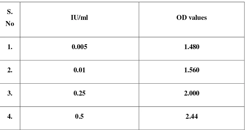
PRINCIPLE OF THE TEST
The Tetanus Toxoid IgG antibody test is based on the principle of the enzyme
immuno assay (EIA). Tetanus antigen is bound on the surface of the microtiter strip.
Diluted patient serum or ready- to- use standards are pipetted into the wells of the
microtiter plate. A binding between the IgG antibodies of the serum and the
immobilized Tetanus Toxoid antigen takes place. After a one hour incubation at room
Introduction
22
unbound material. Then ready-to-use anti-human-IgG peroxidase conjugate is added
and incubated for 30 minutes. After a further washing step, the substrate (TMB)
solution is pipetted and incubated for 20 minutes, inducing the development of a blue
in the wells. The color development is terminated by the addition of a stop solution,
which changes the color from blue to yellow. The resulting dye is measured
spectrophotometrically solution, which changes the color from blue to yellow. The
resulting dye is measured spectrophotometrically at the wavelength of 450 nm. The
concentration of the IgG antibodies is directly proportional to the intensity of the
Introduction
23
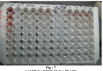
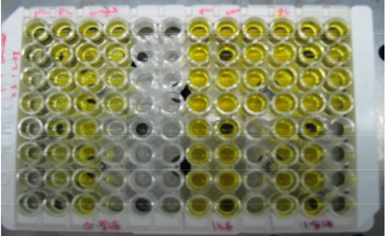
A 96-well microtiter plate being used for ELISA.
Review of Literature
24
REVIEW OF LITERATURE
GREENBERG (1953)13 showed that the problem of assaying tetanus toxoids differs with the two test-animals used , the mouse and the
guinea-pig. In the mouse, the slopes of dosage-response curves of plain
and adsorbed toxoids are clearly not similar, so that separate standards
are required for their assay. In the guinea-pig, the slopes of
immunization curves for these two types are similar and one should
suffice. While a second international standard for adsorbed
preparations is not required, laboratories performing these tests are
advised to establish separate standards- one for plain toxoids and one
for adsorbed.
H. COHEN et al., (1959)21 This study showed that the mouse is a very suitable laboratory animal for the comparison of adsorbed tetanus
toxoids, and that an AIPO4- adsorbed vaccine, which is stable at 4oc, is
a satisfactory reference preparation. The log-dose-response lines of
toxoids adsorbed on different quantities of AIPO4 and on various
quantities of another adsorbent ran parallel to those of the reference
vaccine. The 95% confidence limits for the potencies of tetanus
vaccines, diphetheria-tetanus vaccines, and eiptheria-pertussis-tetanus
vaccines, determined by assay against the reference vaccine in mice,
Review of Literature
25
R MACLENNAN et al., (1965)22 Immunization of pregnant women in New Guinea with three injections of plain toxoid had previously
been shown to prevent neonatal tetanus. In the present study antioxin
levels induced by two oil-adjuvant toxoids (one injection), one AIPO4
toxoid (two injections ) and one plain toxoid (three injections) were
compared with those induced by the same plain toxoid as used in the
earlier study. At term there was no significant difference in the levels
for the five toxoids, but those for the plain toxoids later declined
rapidly. AIPO4-toxoid titres were significantly higher than the titres for
the plain toxoids at the end of the year, but lower than the oil-adjuvant
titres, which were the highest and most persistent. However,
unacceptable side-effects (induced by subsequent lots of oil-adjuvant
toxoids) preclude their routine use at present. The results indicate that a
maternal antitoxin level at delivery of 0.01 unit/ml is protective.
Aluminium-compound toxoid rapidly achieved titres that were better
than this for at least a year, with minimal side-effects. Hence such
toxoids are recommended for maternal immunization to prevent
neonatal tetanus.
W. J. HERBRET (1966)16 Comparative tests were done to find the best conditions under which formalinized sheep red cells can be tanned
and then coated with chromatographically purified
ovalbumin.Variation in the time taken for tanning or coating, the
amount of antigen used, the temperature during coating, the use of
Review of Literature
26
different methods had little effect on sensitivity to agglutination by
antibody A slight increase in sensitivity was obtained by coating at a
low pH, by increasing the quantity of tannic acid employed and by heat
denaturation of the antigen.
E. R. GOLD AND H. H. FUNDENBERG (1967)20 The passive hemagglutation procedure using a chromium chloride solution to bind
antigens and antibodies to red cells has been investigated. The Crcl3
method of coating red cells is rapid and simple, and serologic
sensitivity and immunologic specificity are retained. The present
studies showed that group O human red cells coated with antigen by
the CrCl3 procedure can be used in a number of serologic reactions. In
particular, use of CrCl3 treated cells as immunoadsobent provides a
rapid method for the preparation of monospecific antisera to human
immunoglobulins. The immunoglobulin content of pathologic sera can
be estimated by the degree to which the pathologic seruminhibits an
appropriate agglutination system consisting of cells coated γG-, γA- or
γM-globulin by the CrCl3 method and of homologous antibody.
“Purified” immunoglobulin preparation can be assessed for
contamination by similar inhibition of agglutination of red cells coated
with immunoglobulins by the CrCl3 method.
ARTHUR A. HIRATA et al., (1968)17 Chicken, sheep and rabbit erythrocytes were stabilized with formaldehyde, pyruvic aldehyde or
Review of Literature
27
albumin, insoluble BSA, ovalbumin, rabbit γ-globulin, E. coli
endotoxin, acetylated endotoxin, de-esterified endotoxin, de-esterified
and bromacetylated endotoxin, succinylated endotoxin and synthetic
polyglucose were found to absorb firmly on the stabilized cells. For
optimal coating of stabilized cells with antigens, critical factors
included pH, antigen concentration and the duration of contact with
antigen. These conditions differed somewhat from one antigen to
another.Antiglobulin tests wre applicable in all of the cases tested and
were found to increase the hemagglutination titres 10- to 30- fold.
M.CAROLYN HARDEGREE et al., (1970)26 This paper describes: 1)a continuation of the study reported in 1965 of tetanus antitoxin
titres among women after primary immunization with plain, AlPO4 –
adsorbed or oil adjuvant toxoids; (2) the effect of age and of abscess
formation due to oil adjuvant on antitoxin response;. And (3) a
comparative study of titres in some women of the study groups who
received either a plain or an AlPO 4 Toxiod booster injection in
pregnancy .
MICHAEL F . BARILE ET AL., (1970)27 The mouse toxin-neutralization test procedure described has been used for antitoxin
titration of sera of new Guinea women following primary
immunization with plain and adjuvant tetanus toxoids and following
booster immunization , and for titration of guinea –pig sera . The
responses of guinea –pigs immunized with the same toxoids and
Review of Literature
28
observed for 6 months . The relative antitoxin responses of guinea-pigs
to the different toxoids were similar to those of the women .
M.CAROLYN HARDEGREE et al., (1970)28 Haemagglutination (HA) has been used frequently for the titration of tetanus antitoxin but
published results have varied in relation to the “unitage” determined
by the mouse toxin –neutralization test (TH). This report gives the
results of the titration of a group of sera by haemogglutination and
compares them with the results obtained by toxin neutralization .
Although there was marked variation between HA and TN titres of
individual sera , results indicated that haemagglutination is a useful
procedure for the over – all evaluation of the antitoxin responses to
tetanus toxoids in field studies.
J.D.VAN RAMSHORST (1971)34 Available methods for titrating diphtheria and tetanus antitoxin at low concentrations in human or
animal blood are surveyed, with special attention to the amount of
serum required for the test . In vivo methods, especially the rabbit
or guinea-pigs international test for diphtheria and the mouse test for
tetanus , are precise and reliable . If , however , serum levels as low as
about 0.001IU/ml have to be determined, rather large amounts of
serum are required ; moreover, the test are rather time –consuming
and expensive Tissue culture methods are available only for diphtheria
antitoxin titration . the titres found coincide very well with those form
animal tests. The titrations are less time –consuming and more
Review of Literature
29
replacement of animal tests. Of the real in vitro methods, the
haemagglutinaton procedure has been investigated most thoroughly
and used most frequently. Low titres can be measured using small
amounts of serum , but the titres thus obtained may differ considerably
from those obtained in animal tests ,at least for individual sera. For
mass screening , the method is very suitable .
B.BYCENKO et al., (1971)35 Determination of the level of tetanus immunity in different age groups of a population is important in
relation to the results of mass immunization of the population with
vaccines containing tetanus toxoid. In this study , the level of tetanus
antitoxin in sera from 1053 inhabitants of 6 villages Bosnia and
Herzegovina was determined by means of the passive
haemagglutination test.the results show that children from 1to 14 years
of age regularly immunized with DT and DPT vaccines possessed a
high level of immunity against tetanus (geometric mean antitoxin titre
=0.8 IU/ml) and were protected against tetanus in 80-95% of cases.
The adult inhabitants(men and women over 29 years age)were not
sufficiently protected against tetanus , the concentrations of antitoxin
in their sera being equal to or greater then 0.01 IU/ml in less then 50%
of cases.
K.W.NEWWELL ET AL., (1971)1 To Determine the effectiveness of a method for controlling tetanus neonatorum , a double
–blind controlled trial involving 1618 women was conducted between
Review of Literature
30
of aluminium –phosphate-adsorbed tetanus toxoid obtained from 299
women . sera were titrated for anti-tetanus antibodies by two methods .
A comparison of the clinical and laboratory results showed a close
relationship. It is suggested that the level of protection may be lower
then is at present accepted. Antitoxin levels were inversely related to
age and directly to the interval between injections . two widly spaced
injections may be about as effective as 3 injections . one injection of
specially prepared toxoid with a high immunizing potency might gives
significant protection.
D. D. OURTH 5 (1977)18 This investigation found that the human antibody class of importance in neutralizing tetanus toxin in mice was
IgG, and that toxin neutralization was retained by the F(ab’)2 and Fab’
subunits of the human IgG class. Although human IgM and IgA classes
appeared to neutralize tetanus toxin at very low levels, evidence was
obtained that this neutralization was probably due to IgG
contamination. Human Fabµ, isolated from the IgM class did not
neutralized tetanus toxoid in the indirect hemagglutination (HA) test
with IgG giving the highest HA titre. Rabbit antibodies of the IgG
class also neutralized tetanus toxin, with neutralization being retained
by the F(ab’)2 and the Fab’ subunits of the rabbit IgG class. Absorption
of several rabbit antisera to tetanus toxoid with goat-antirabbit Fc,
which is specific for absorption of IgG from antiserum, rendered
Review of Literature
31
MARGARET M. PEEL (1980)24 Variables affecting the indirect microhaemagglutination technique for measuring tetanus antitoion
were investigated with a view to attaining maximum sensitivity in the
test system by the application optimal conditions. The addition of
2-mercaptoethanol to the diluent used for the preparaion of serial
dilutions of antsera was also investigated as a simple means of
inactivating the IgM component of tetanus antitoxin and so of
improving the correlation between antitoxin titres determined by the
indirect haemogglutination and by the toxin neutralization techniques.
The use of mercaptoethanol in the assay system improved the
correlation between titres determined by haemagglutinaton and T N
test with same sera , but did not always reduce or eliminate the
discrepancies.
MALATI KESKAR et al., (1981)19 Pre and postimmunisation samples of 189 medical students were simultaneously subjected to
hemagglutination (HA) and toxin neutralization (TN) tests. HA method
was found to be simple and economical but lacking in sensitivity and
consistency. Refinement of the technique would be necessary before its
application in the field on a mass scale.
MOIRA E.MELVILLE –SMITH et al.,(1983)5 An enzyme-linked immuniosorbent assay(ELISA) has been developed for the
measurement of tetanus antitoxin in human sera as an alternative to the
toxin neutralization test in mice, the currently accepted method of
Review of Literature
32
required only small amounts of materials. In addition, the assay was
found to give reproducible estimates of antiroxin levels and to measure
antitoxin at levels as low as 0.01 IU per ml, a sensitivity similar to tha
of the neutralization test. Furthermore, a comparison of the results of
the ELISA and the neutralization test involving 80 human sera,
including sera with both high and low antitoxin levels, showed close
agreement in antitoxin levels obtained by the two methods. It was
concluded that ELISA was an acceptable alternative to the toxin
neutralization test in mice for the measurement of tetanus antitoxin
levels inhuman sera.
L.PITZURRA et al.,(1983)6 Antibody titers to tetanus toxin in human sera were assayed by passive haemagglutination with turkey
erythrocytes, enzyme-linked immunosorbent assay and
counterimmunoelectrophoresis. The first two of these tests were shown
to be the most sensitive for antibody detection, having the same range
of sensitivity and reproducibility. The first two of these tests were
shown to be the most sensitive for antibody detection, having the same
range of sensitivity and reproducibility. The antibody levels
determined by these assays were up to 400 fold higher than those
determined by counterimmunoelectrophoresis. The turkey erythrocyte
haemagglutination assay requires only 40 min, whereas the
immunosorbent assay method requires 24 h. These results suggest that
the haemagglutination assay is the more appropriate method for rapid
Review of Literature
33
R.K.GUPTA et al., (1983)14 Serum, samples from 77 guinea pigs immunized against tetanus have been titrated for tetanus antitoxin by a
standardized indirect haemagglutination (IHA) test and the
conventional toxin neutralization (TM) test. These sera were titrated
before and after treatment of the sera with 2-mercaptoethanol(2-ME)
by the IHA test using unfixed sheep erythrocytes and erythrocytes
fixed with glutaraldehyde, formaldehyde and pyruvic aldehyde. The
titres of these se5ra obtained by IHA using unfixed and
glutaraldehyde-fixed sheep erythrocytes before treatment of the sera
with 2-ME were two to six times higher than the TN titres, whereas the
IHA titres using formaldehyde – and pyruvic aldehyde-fixed sheep
erythorocytes were 10 times higher than the TN titres in some of the
sera. There was no statistically significant difference between TN and
IHA titres using unfixed and glutaraldehyde-fixed sheep erythrocytes
after the treatment of the sera with 2-ME.
M.PITZURRA et al., (1983)11 Passive haemogglutination (HA) assays were performed using turkey red blood cells (TRBC-HA) on
sera form normal healthy people , from normal people previously
immunized against tetanus , and from tetanus patients receiving human
using immunoglobulins. The TRBC-HA test was compared with HA
assys using sheep red blood cells (SRBC-HA) and with the TN test
,and was found to be more sensitive then the SRBC-HA test and
showed good correlation with the TN test . While the SRCB-HA assay
Review of Literature
34
be tested , the use of turkey red blood cell does not requireany such
adsorption. In addition, the TRBC-HA assy can be performed in 40
minutes compared with 6 hours for the SRCB-HA assy. All these
advantages make the TRBC-HA assy a more useful test for screening
large numbers of sera in the evaluation of sera in the evaluation of
tetanus immunity of normal people and of patients with wounds when
seen in the emergency room of hospitals.
S.T.CHEN et al., (1983)23 A Study was undertaken to determine the relationship between the timing of maternal immunization with a
commercially available adsorbed tetanus toxoid and the presence of
protective antitoxin in cord blood. Women at various stage of gestation
were given one or two doses of 20IU of toxoid, and the maternal and
cord sera collected at delivery were assayed for tetanus antitoxin by the
I HA and T N techniques. Results indicated that the first injection of a
two –dose schedule should be given at least 60 days, and preferably 90
days or more before delivery, with the second injection 20 days or
more before delivery. The single-dose schedule conferred no
significant
R.K.GUPTA et al., (1984)12 The tetanus antitoxin titres of 174 serum samples from healthy adults were determined by a standardized
indirect haemagglutination test(IHA) and the conventional toxin
neutralization (TN) test. The serum samples were titrated by the IHA
test using glutaraldehyde-fixed and toxioid sensitized sheep
2-Review of Literature
35
mercaptoethanol (2-ME). The IHA method has been found to be very
sensitive and specific for the estimation of tetanus antitoxin in human
sera. The IHA titres before the treatment of the sera with 2-ME were
generally about four times higher than the TN tires and the correlation
coefficient between these titres was 0.94. The IHA titres after the
treatment of the sera with 2-ME were in good agreement with the TN
titres and there was no statistically significant differences between the
titres by the two methods. The tetanus antitoxin titres of 50% of the
sera were below the minimum protective titres of tetanus antitoxin
(0.01 IU /ml). In 19.5% of the sera the antitoxin level (IU / ml) ranged
from 0.01 to 0.1, in 20.1% from 0.1 to 1.0 and in 10.4% from 1.0 to
10.0.
G.GENTILI et al., [1984)32 A micro enzyme linked Immunosorbent assay has been adapted to the quantitation of Specific tetanus
antibodies in commercial preparation pf human immunoglobulins.The
results of the assay are compared with those obtained from the same
samples tested by seroneutralisation tests invivo. Statistical analysis of
the data shows good correlation between the titres obtained with two
tests. Results obtained by indirect haemogglutination are also reported.
It is proposed that all interested laboratories perform the described
immuno enzymatic method invitro for a given period in parallel with
the invitro test to gain sufficient experience of this technique with the
Review of Literature
36
GIULIANO GENTILI et al.,(1985)29 The ability of an enzyme-linked immunosorbent assay (ELISA) to detect tetanus antitoxin in
human sera has been evaluated in comparison with the in vivo
seroneutralizationtest. The results of this study, carried out on 171
serum samples, show that ELISA is a sensitive and specific in vitro test
for immunity to tetanus in man; it reveals the minimum protective
level of 0.01 IU /ml and is well correlated with seroneutralization. A
comparison has also been made with indirect haemagglutination.
Differences in specificity in low tittered sera, although not statistically
significant, have been observed. Reported data suggest tha the ELISA
may be used for the estimation of tetanus antitoxin in sero-
epidemiological surveys and for clinical purposes with reliability
equal-and perhaps superior- to that of IHA.
R.K. GUPTA et al.,(1985)9 The quality of retanus toxin affected the sensitivity of the toxin neutralization(TN) test greatly. By using
purified toxin a minimum level of 0.001 IU/ml only could be detected.
The TN test described whereas with crude toxin a level of 0.025 IU/ml
only could be detected. The TN test described in this report permitted
titration of tetanus antitoxin in twofold dilution steps from levels as
low as 0.001 IU/ml using 0.6 ml of serum only at the L+75000 level of
purified tetanus toxin. Treatment of the sera
with2-mercaptoethanol(2-ME) did not affect the TN titres showing that the TN test detects the
neutralizing antibodies (IgG) which are not affected by 2-ME. The TN
Review of Literature
37
O.SIMONSEN et al., (1986)4 Serum samples form 727 persons with different vaccination histories were assessed for tetanus antitoxin
content in an enzyme linked immunosorbent assay (ELISA) and tested
for tetanus antitoxin neutralization activity in mice in order to compare
the results obtained by the two methods. Neutralizing antibody
activities in sera from individuals previously completely vaccinated
correlated well with results obtained by ELISA and the accuracy
increased with increasing antitoxin concentration in serum. This
correlation was observed in sera from persons vaccinated recently as
well as in sera from persons vaccinated many years ago. In sera from
persons with an incomplete vaccination history ELISA was found to be
an unreliable tool for the prediction of in vivo results. Many of these
sera had antitoxin levels by ELISA far above the in vivo values,
probably due to the presence of non specific or low avidity antitoxin
which is detected in ELISA.
O.SIMONSEN et al., (1987)3 The use of indirect ELISA for the quantitation of tetanus toxin neutralizing antibodies in human sera is
limited by marked overestimations in low tittered sera. The reasons for
the discrepancy between the results obtained by ELISA and by in vivo
assay and modifications of the ELISA to overcome the problem were
investigated. Catching ELISA and indirect ELISA using trays coated
with the contaminant proteins in toxoid preparations indicated that
antibodies to contaminants were only partly responsible for the
Review of Literature
38
the problem. In ELISA competiton experiments withtoxin neutralizing
monoclonal antibodies, the human immunoglobulins irrelevant in toxin
neutralization, byut detectable in indirect ELISA, were found to be
difficult to inhibit in their binding to the solid antigen phase. These
might represent antitoxins bound bivalently to the solid phase but with
affinities in monovalent binding isufficient for toxin neutralization or
other coupled antibodies due to conformational changes of the antigen.
M.GERMAN-FATTAL et al., (1987)8 Immunity to tetanus was investigated in 157 individuals aged between 1 and 77 years using, for
the evaluation of both tetanus antitoxin and antibody to fragment BIIb
(anti-BIIb), an easily performed ELISA that gave reproducible results.
Among these people 72% were protected against tetanus (tetanus
antitoxin titres > 0.06IUml-1 (not protected against tetanus) were
found to have anti-BIIb titres >0.15 Uml-1. These data raise questions
as to the significance and protective effect of anti-BIIb against tetanus.
C.F.M HENDRIKSEN et al., (1988)15 A method for the screening of human sera for tetanus antibodies has been developed and evaluated.
The toxin binding inhibition test(ToBI-test) is based on inhibition of
the binding of tetanus toxin to an antitoxin- coated immunoassay
microtitre plate by tetanus antibodies. Serum samples form 191 healthy
adults with different vaccination histories have been titrated for teranus
antibodies by the toxin neutralization (TN) test in mice, by
toxoid-ELISA and by the ToBi-test. In every respect, the ToBI-test proved to
Review of Literature
39
showed a higher degree of correlation between the ToBI-test and the
TN-test than between the toxoid-ELISA and the TN-test. Furthermore,
no overestimation of antibody content was seen in titrating low titre
sera by the ToBI-test. In contrast, several false positive results were
seen when using the toxoid-ELISA. It is concluded that the ToBI-test
is a reliable and precise alternative to the TN-test and can be performed
under simple laboratory conditions in a short time.
INMACULADA ESPARZA AND THOMAS KISSEL (1992)25 The immunogenic potential of tetanus toxoid (TT) was compared when
either adsorbed to aluminium hydroxide (TT-alum) or entrapped in
microparticles consisting of poly)D,L-lactic-CO-glycolic acid) (PLA:
PGA, 55:45) derived polymers. Furthermore, the effect of
administering the microparticles in an aqueous buffer or water-in-oil
emulsion on the TT immunogencity was also investigated. When mice
were immunized with the different formulations, similar levels of
anti-TT antibodies were observed during the primary IgG response. The
choice of the carrier seemed to play an important role for both the level
and maintenance of the secondary IgG response, attained as a
consequence of a booster immunization with TT-containing
microparticles, resuspended in wate-in-oil emulsions. As expected,
incomplete Freund’s adjuvant (IFA) proved to be a more potent
adjuvant than peanut oil, whereas resuspension of the microparticles in
aqueous solution induced a relatively less efficient antibody response.
Review of Literature
40
since the secondary antibody response was higher and persisted longer
compared with TT-alum priming. These results indicate that in
addition to TT maintaining its antigenicity after microencapsulation,
the microparticles also potentiate its immunogenic properties. This
approach should prove very useful for designing more effective
vaccines.
NILAY COPLU et al., (2004)7 In order to determine a pratically useful quantitative assay method for tetanus antibody in large-scale
seroepidemiological study, a method combining an in-house ELISA
with a particle agglutination test (KPA) was evaluated in comparison
with the in vivo mouse neutralization test. Serum samples with mouse
neutralization antibody titers 0.01 IU/ml (the minimum protective
level) or below showed considerable over-estimation of antitoxin titers
up to 1.0 IU/ml when studied by in –house ELISA alone. On the other
hand, the DPA values were highly correlated with the mouse test, even
in cases of titers equal to 0.01 IU/ml or below. The combination of
these two procedures, in which in-house ELISA values of 1.01 IU/ml
were replaced by KPA values, provided a high correlation in antibody
titers with the mouse test (r=0.968). We applied this combined method
to a tetanus seroepidemiological survey in a province in Turkey. The
survey included347 subjects from the healthy population, and the
qquantitative analyses showed high antibody levels in children and
young adults and significantly low levels among adults aged 40 or
over. A characteristic distribution of antibody titers in each age group
Aim and objective
42
AIM AND OBJECTIVE
The quality control testing of biologicals especially potency test is a subject on
its own. The biological standardizations of each procedure requires meticulous
planning and execution. The potency test for tetanus toxoid as specified in Indian
pharmacopoeia is biological assay of tetanus antitoxin in immunized guinea pigs. An
in vitro serological assay system – ELISA to assess the potency of tetanus toxoid was
standardized and results compared with the conventional biological assay.
ELISA test has the advantages of easy automation and end point readings can
be measured objectively.
A major problem with ELISA is that different scientist assign antibody
concentrations by different terminologies like AU/IU titers etc.
To determine the potency of tetanus toxoid by using ELISA was easy to
perform, sensitive, highly reproducible and require only small quantities of sera and
time saving.
In the present study tetanus antibodies in sera of guinea pigs after
immunization of 2 doses of tetanus toxoid were measured at a various methods by
Plan of work
43
PLAN OF WORK
1. Lf determination of tetanus toxoid by using flocculation method.
2. Selection of test animals guinea –pigs
3. Immunization of guinea pigs
4. Collection of blood and separation of sera
5. Determination of potency
5.1 Toxin neutralization test
5.1.1. Tetanus anti toxin preparation
5.1.2. Reference standard preparation
5.1.3. Test sera preparation
5.1.3.1.. Neat sera preparation
5.1.3.2 Diluted sera preparation
5.2. ELISA
5.2.1. Standardization of in-house ELISA for determination of ELISA IgG
antibodies against TT raised in guinea pigs.
a)Coating of antigen (TT)
b)Preparation of reagents for conduct of ELISA
c)Preparation of test protocols for standardization of
• Antigen
• Conjugate
• Block buffer
• Incubation times
5.2.2.Assessment Of IgG In Guinea-Pig Sera By ELISA
Material and instruments
44
MATERIALS AND INSTRUMENTS
1. Peroxidase conjugate anti guinea
pig IgG
- SIGMA ALDRICH - USA
2. Anhydrous sodium carbonate - SD FINE –CHEM LTD.MUMBI
3. Sodium hydrogen carbonate - SD FINE –CHEM LTD.MUMBI
4. Sodium chloride - FLUKA
5. Potassium dihydrogen phosphate - SD FINE –CHEM LTD.MUMBI
6. Disodium hydrogen phosphate
dehydrate
- MERCK –MUMBAI
7. Potassium chloride - NICE-CHEMICALS COCHIN
8. Citric acid - NICE-CHEMICALS COCHIN
9. Sodium hydrogen - NICE-CHEMICALS COCHIN
10. Dried skimmed milk - BHARATH BIO CHEM LTD
11. Polysorbate 20 - HI MEDIA
12. Diammonium 2,2’-azinobis - HI MEDIA
13. Elisa plates 96 wells - NUNC
14. Sodium citrate - NICE CHEMICALS COCHIN
15. Anti toxin - CDL CRI KASAULI
16. T.T. adsorbed - BETT
17. Glycerin - QUALIGEN, MUMBAI
18. Peptone - HIMEDIA – MUMBAI
19. Gloves - DIAMOND GRIB PLUS
20. Wells - NUNC BRAND PRODUCTS
DENMARK
Material and instruments
45
INSTRUMENTS USED
S.No INSTRUMENTS NAME - COMPANY
1. Auto clave - NEW LAB EQUIPMENT
2. Centrifuge - REMI
3. Deep Freezer - SIEMENS
4. Digital PH meter - NEW LAB EQUIPMENT
5. Hot air oven - GENUINE
6. Incubator - HERA CELL 150
7. Laminar Air flow - GENUINE
8. Refrigerator - LG
9. Shaker - NEW LAB EQUIPMENT
10. Water bath - GENUINE
11. Flocculation apparatus - KING INSTITUTE
12. 5µl Pipette - STERILIN
13. 10µl Pipette - FALCON USA
14. 100µl Pipette - STERILIN
15. 200-1000 µl Pipette - FALCON USA
16. Multi channel pipette - CAPP-FRANCE
17. Cryo tube vials - NUNC
Methodolgy
46
METHODOLOGY
1.Lf DETERMINATION OF TETANUS TOXOID BY USING
FLOCCULATION METHOD.
37PROCEDURE
Preparation of 10% tri Sodium Citrate Solution:
10.0g of tri sodium citrate was weighed and transferred in to the
100 ml bottle and the volume was maked to 100 ml using WFI /Miii Q water. Stored
at room temperature for not more than 6 months.
Test Procedure:
10 ml of the sample was taken in a 15 ml centrifuge tube.
Centrifuged at 2000 rpm for 10 minutes.
Supernatant was collected in another tube and label it as supernatant. The
supernatant contains Lf from the vaccine sample that was left unabsorbed.
0Store at 2-8 C for testing at a later stage.
To the sediment of the test tube, labeled as Elute add 10% tri sodium citrate
solution to make up the make up the volume to 10 ml. Incubated at 35-37 C
for about 16 hours. Tetanus Toxoid shall elute in this mixture and is tested for
Methodolgy
47
10 ml of the sample was taken in another test tube and label it as Total, add
10.0 g of tri sodium citrate. Mixed well and incubated at 35-37°C for about 16
hours. It contains total Lf contributed by both adsorbed as well as unadsorbed
tetanus toxoid.
The Lf in supernatant was estimated by performing the Lf test taking
antitoxin concentration in the range of 2 Lf to 10 Lf with the difference of 1Lf.
Add 0.85% sterile phgysiological saline was added in the tubes to make up
the volume to 1.0 ml. Add 1.0 ml of the supernatant sample to all flocculation
tubes.
The Lf in Elute was estimated by performing the Lf with the difference of 1
Lf. Add 0.85% sterile physiological saline in the tubes to make up the volume
to 1.0 ml. 1.0 ml of the Total sample was added to all flocculation tubes.
Keep the reaction mixture at 45-50°C in water bath in such a way that at least
half of the reaction mixture is in the water.
The tubes were observed for the flocculation reaction to occur.
Mixture flocculating first is that which contains the most nearly equivalent
quantities of Tetanus Toxoid.
2.SELECTION OF TEST ANIMALS ON GUINEA –PIGS2
Healthy guinea – pigs was used from the same stock and weighing between 250
and 350 g. Distribute them into six groups of sixteen each. The guinea – pigs should
Methodolgy
48
the groups. if the challenge toxin to be used has not bee shown to be stable or has not
been adequately to serve as unvaccinated controls.
3. IMMUNIZATION OF GUINEA PIGS 2
Inject subcutaneously on each of two occasions separated by an interval of not
more than weeks, one tenth of the stated human does diluted to 1 ml with saline
solution into each of 9 normal, healthy guinea – pigs weighing between 250 and
350 g. Not more than 2 weeks after the second injection. collect the serum form each
animal and carry out the biological test for tetanus antitoxin, described under Tetanus
antitoxin or any other method approved by National Regulatory Authority.
Animal used Guinea pig Hartley
Weight range 250- 350 g
No. of guinea pigs 12
Dose 0.5ml
Route of inoculation subcutaneous
Date of 1st dose 18.09.08
Date of 2nd dose 17.10.08
Bleeding on 29.10.08
Methodolgy
49
4. COLLECTION OF BLOOD AND SEPARATION OF SERUM
Preparation of serum samples: For preparation of serum samples, the
following technique has been found suitable. Invert the tubes containing blood
samples 6 times and allow to stand 370c for 2 h, then 40cfor 2 hours, centrifuge at room temperature at 800 g for 20 min. transfer the serum to steri