The late gene promoter P
7535of the epidermodysplasia verruciformis-associated human papillomavirus type
8 (HPV8) is regulated by the viral E2 protein. Transfection experiments performed with the human skin
keratinocyte cell line RTS3b and P
7535reporter plasmids revealed transactivation at low amounts and a
repression of basal promoter activity at high amounts of E2 expression vector. This repression was promoter
specific and correlated with the amount of transiently expressed E2 protein. Mutational analyses revealed that
the negative regulation of P
7535activity is mediated by the low-affinity E2 binding site P2, which is separated
by one nucleotide from the P
7535TATA box. Biochemical and genetic analyses suggested that repression is due
to a displacement of the TATA-box binding protein by E2 and an interference of E2 with promoter-activating
cellular factors that specifically recognize the P2 sequence. The high conservation of the P2 sequence among
several papillomaviruses (epidermodysplasia verruciformis-associated HPVs, HPV1, cottontail rabbit
papil-lomavirus, and bovine papillomavirus type 1) in the vicinity of the late gene promoter cap site suggests that an
interplay of E2 and cellular factors at this sequence element is important for the expression of structural
proteins.
Gene expression in papillomaviruses is tightly linked to the
differentiation state of the keratinocyte. In particular, late
genes encoding structural proteins are only transcribed in the
uppermost layers of the epithelium. Expression of
papilloma-virus genes is controlled by a complex interplay of host and
viral transcription factors, which mainly act through sequences
located in the noncoding regulatory region (NCR) between the
L1 and E6 genes (12). The basal promoter activities in the
absence of viral proteins are the net result of both positively
and negatively acting host transcription factors. These
activi-ties can be further modulated by viral proteins, especially by
products encoded by the E2 open reading frame (14, 28).
E2 proteins regulate viral gene expression and DNA
repli-cation by specifically binding as dimers to the DNA sequence
ACCN
6GGT, which occurs several times in the NCRs of all
papillomaviruses (1, 14, 17, 28). Bovine papillomavirus type 1
(BPV1) expresses three different E2 proteins, of which only the
48-kDa protein (E2TA) is able to transactivate viral promoters
(16, 21, 27, 29). The two shorter forms lacking the
transacti-vation domain antagonize the function of E2TA (2, 30). E2TA
activates all but one of the BPV1 early promoters to different
extents (40). Only the P1 (or P
7185) promoter is repressed by
E2TA via E2 binding site 1 (BS1), localized immediately
down-stream of the P1 cap site. Repression seems to occur by
inter-ference with a cellular transcription factor whose target
se-quence overlaps BS1 (37). In the cases of cervical
carcinoma-associated human papillomavirus (HPV) types 16 and 18, both
positive and negative effects of E2 on the activity of the viral
oncogene promoter have been observed under slightly
differ-ent experimdiffer-ental conditions concerning E2 expression vectors
and host cells. HPV16 E2 was shown to transactivate the early
promoter in rodent cells (22). According to a recent work,
full-length HPV16 E2 protein moderately stimulates viral gene
expression in normal and immortalized keratinocytes, but
N-terminally truncated E2 protein forms lead to repression (4).
Other laboratories described repression of HPV16 and -18
promoter activity by homologous full-length E2 in human
ke-ratinocytes (3, 34). Of the four highly conserved E2 binding
sites (E2BSs) among mucosotropic HPVs, only the
promoter-proximal one turned out to be necessary for E2 repression of
the HPV18 E6/E7 promoter (6), whereas both
promoter-prox-imal E2BSs are required for the repression of the HPV16 P
97promoter (34, 41, 42). These E2BSs are spaced 1 and 3 bp,
respectively, from the recognition sequences for the cellular
transcription factors SP1 and the TATA box-binding protein
(TBP), the DNA-binding component of the TFIID complex.
Binding site competition between E2 and both factors causes
repression of HPV16 P
97activity, whereas occlusion of TBP
alone is sufficient for E2-mediated repression in the case of
HPV18 P
105(6, 9, 41, 42). This negative modulation of
onco-gene expression is not restricted to cancer-associated HPVs
but also occurs with the ‘‘low-risk’’ HPV11 (7).
Transcriptional regulation of the late promoter has only
been studied in the case of HPV8. This type belongs to the
group of HPVs associated with the syndrome
epidermodyspla-sia verruciformis (EV) and is regarded as a highly oncogenic
virus (32). EV-associated HPVs differ from other
papilloma-viruses mainly in the structure of the NCR. They all display the
sequence motif M33/AP1 and five E2 recognition sequences
(P0 to P4) with a highly conserved distribution (10, 20). The
late gene promoter P
7535of HPV8 is localized in the
L1-proximal part of the NCR. The initiated mRNAs are
alterna-tively spliced and may code for the major capsid protein L1 or
E2TA (11, 38). Promoter activity is negatively modulated by
the negative regulatory element overlapping E2BS P1 and
pos-* Corresponding author. Mailing address: Institut fu¨r Virologie derUniversita¨t, Fu¨rst-Pu¨ckler-Str. 56, D-50935 Cologne, Germany. Phone: 49-221-4784481. Fax: 49-221-407490.
119
on November 9, 2019 by guest
itively modulated by the M33/AP1 region, E2BS P2, the TATA
box, and viral E2 (19, 25, 38, 39).
The five E2BSs display highly different affinities for the E2
protein. Mutational analyses of E2BSs revealed that four sites
(mainly high-affinity sites P0 and P1 and to a smaller extent
medium-affinity site P3 and low-affinity site P4) serve to
trans-activate the promoter, whereas the single low-affinity site close
to the promoter, P2, is able to downmodulate E2
transactiva-tion (39). In this report, we present data that late gene
pro-moter activity can be repressed by E2 in the human skin
ke-ratinocyte cell line RTS3b (33). This repression seems to be
mediated by the simultaneous interference of E2 with TBP and
a cellular factor(s) specifically binding to P2.
MATERIALS AND METHODS
Cell culture and transient-transfection assays.HT3 cells (ATCC HTB32) were maintained in Dulbecco’s modified Eagle’s medium (DMEM) with supple-ments as described elsewhere (38). The spontaneously immortalized human skin keratinocyte cell line RTS3b contains no papillomavirus sequences (33). It was maintained in F12-DMEM (1:4) supplemented with hydrocortisone (0.4mg/ml), 10210
M cholera toxine, transferrin (5mg/ml), 2310211
M triiodothyronin, 1.8
31024
M adenin, insulin (5mg/ml), epidermal growth factor (10 ng/ml), gen-tamicin (10 mg/ml), and 10% fetal calf serum (FCS). Both cell lines were transfected in 60-mm dishes by the calcium coprecipitation method as described before (39). Briefly, cells at 40 to 50% confluency were incubated overnight with 3 mg of reporter plasmid, the indicated amounts of E2 expression plasmid (pCE2), and the empty expression vector pCB6 to standardize the total amount of expression vector to 1mg. Cells were extracted 24 h after the glycerol shock. Protein extract (5 to 50mg) was assayed in a 1-h reaction for chloramphenicol acetyltransferase (CAT) activity. Luciferase constructs were introduced into
RTS3b cells by liposome-mediated transfection. Cells were seeded 18 to 20 h before transfection at a density of 63105to 73105per 60-mm plate. Four micrograms of DNA and 15ml of Lipofectamin (Gibco BRL, Eggenstein, Ger-many) were diluted in 0.6 ml of OptiMEM medium (Gibco BRL). After incu-bation for 30 min at room temperature, 2.4 ml of OptiMEM was added, and the mixture was applied to the cells which had been washed once with OptiMEM. After 5 h, 3 ml of growth medium containing 20% FCS was added and changed after overnight incubation to medium supplemented with 10% FCS. Cells were harvested 48 h after the addition of DNA in 100 mM potassium phosphate buffer (pH 7.8) containing 1 mM dithiothreitol (DTT) and lysed by freeze-thawing. Extract (30ml) was assayed for luciferase activity in a luminometer (Berthold LB 9501). Transfection experiments were repeated at least three times, and the data in the figures represent averages. For monitoring the expression of E2 in HT3 and RTS3b, both cell lines were transfected by calcium phosphate coprecipita-tion, but nuclear instead of whole-cell extracts were prepared.
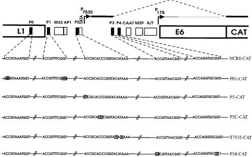
Plasmids and oligonucleotides.The basic construct pNCR8-CAT and its mu-tated derivatives P01-CAT, P34-CAT, P2-CAT, P2C-CAT, and T7535-CAT con-tain the 1,133-bp EcoRI-EcoRV fragment of HPV8 in front of the cat gene (Fig. 1) (38, 39). Enhancer test plasmid pNCR8-tk-CAT is a derivative of pBLCAT2 (23) and contains the same fragment as pNCR8-CAT cloned in the antisense orientation into the BglII site upstream of the herpes simplex virus thymidine kinase (tk) gene promoter linked to the cat gene.
Luciferase reporter constructs were derived from the promoterless pALuc plasmid (8). BamHI-Eco47III fragments containing HPV8 sequences from nucleotides (nt) 7077 to 7628 were excised from plasmids pNCR8-CAT, P01-CAT, P2-P01-CAT, and T7535-CAT and inserted between the BamHI and Ecl136II restriction sites of pALuc upstream of the luciferase gene, giving rise to plasmids p7535-Luc, p7535-P01-Luc, p7535-P2-Luc, and p7535-TATA-Luc, respectively. Plasmid p7535-P2110-Luc is a derivative of p7535-Luc and was constructed by PCR as described before (39). It contains a 10-bp insertion (59 -CATACGCGTA-39) between HPV8 nt 7503 and 7504 (see Fig. 9).
[image:2.612.58.553.75.383.2]HPV8 E2 expression vector pCE2 consists of the whole HPV8 E2 gene under the control of the immediate-early promoter-enhancer of human cytomegalovi-rus (39).
FIG. 1. Schematic representation of the reporter plasmid pNCR8-CAT. The HPV8 fragment driving cat gene expression consists of the NCR flanked by parts of the L1 and E6 genes. The relative amounts of transcripts initiated at the P7535and the P175promoters are indicated by thick and thin lines, respectively. Intron sequences of the P7535transcript are shown as dashed lines. Sequence motifs specific for EV-associated HPVs (M33/AP1, CAAT, M29, and A/T [10, 20]) and the P7535TATA box are shown by open boxes; E2BSs P0 to P4 are shown by solid boxes. The wild-type and mutated DNA sequences (white letters black boxes) of the E2BSs and the P7535TATA box in the context of the corresponding reporter plasmids are shown beneath.
on November 9, 2019 by guest
http://jvi.asm.org/
transfected RTS3b or HT3 cells were prepared by the method of Sealey and Chalkley (36) with the modifications described below. Briefly, cells were scraped into 2 ml of cold phosphate-buffered saline and pelleted at full speed in a microcentrifuge at 48C for 30 s. The pellet was incubated for 5 min on ice in 500
ml of lysis buffer (10 mM HEPES [N-2-hydroxyethylpiperazine-N9 -2-ethanesul-fonic acid, pH 7.9], 300 mM saccharose, 1 mM EDTA, 0.25 mM EGTA [ethylene glycol tetraacetic acid], 1.5 mM MgCl2, 50 mM NaCl, 0.5% [vol/vol] Nonidet P-40 [NP-40], 0.5 mM DTT, 0.5 mM phenylmethylsulfonyl fluoride, 5mg of leupeptin per ml). Nuclei were pelleted at 3,000 rpm for 5 min at 48C, resus-pended in 150ml of lysis buffer, and repelleted. The nuclear pellet was extracted on ice for 30 min with 50ml of elution buffer (25% [vol/vol] glycerol, 10 mM HEPES [pH 7.9], 0.1 mM EDTA, 0.1 mM EGTA, 1.5 mM MgCl2, 525 mM NaCl, 0.5 mM DTT, 0.5 mM phenylmethylsulfonyl fluoride, 5mg of leupeptin per ml). The supernatant, representing the crude nuclear extract, was recovered by cen-trifugation (16,0003g, 5 min, 48C) in a microcentrifuge. Aliquots were snap-frozen and stored at2808C.
Binding reactions with TBP and E2 consisted of an aliquot corresponding to 23104Cerenkov cpm of32P-end-labeled oligonucleotide in 20 mM HEPES (pH 7.9)–135 mM KCl–4 mM spermidine–0.1 mM EDTA–1 mM DTT–0.05% (vol/ vol) NP-40–1 mg of bovine serum albumin per ml–12.5mg of poly(dG-dC) per ml. Binding reactions with TBP alone were incubated in 60 mM KCl. For E2-TBP binding site competition experiments, the labeled DNA was added after the proteins. For binding site competition experiments that included unlabeled DNA, the protein extract was added last. Reactions were incubated at 308C for 30 min and then loaded onto a 5% native polyacrylamide gel (19:1) containing 0.53TBE (Tris-borate-EDTA). Gels were run at 200 V for 2 h, dried, and then autoradiographed overnight at2808C with an intensifying screen. Crude nuclear extracts (5mg) were incubated at room temperature in the same buffer, but 1 mg of poly(dA-dT) per ml was used as the nonspecific competitor.
DNase I footprint analysis.A subcloned HPV8 restriction fragment encom-passing nt 7454 to 7602 was labeled with Klenow enzyme and [a-32P]dATP at the coding and noncoding strand, respectively. An aliquot corresponding to 2.53104 Cerenkov cpm was incubated with TBP (240 ng/ml) under the conditions de-scribed for gel retardation analysis. Reactions were digested with DNase I and processed as described before (39). An A1G sequencing reaction of the corre-sponding fragment was used as a molecular size marker.
RESULTS
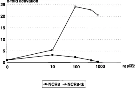
E2-specific repression of HPV8 promoter activity in RTS3b
cells.
To test for differences in E2 modulation between
muco-sal keratinocytes (HT3), which were used in the previous study
(39), and keratinocytes of cutaneous origin, we performed
transfection assays with the human skin keratinocyte cell line
RTS3b. Low input levels (10 ng) of E2 expression vector pCE2
slightly activated CAT expression (3.5-fold) from the reporter
plasmid pNCR8-CAT (Fig. 2). Higher amounts of pCE2
re-duced transactivation and finally repressed basal promoter
ac-tivity. In HT3 cells, CAT expression was not further stimulated
but was never diminished by increasing amounts of E2 (39).
The repression observed with increasing amounts of E2
ex-pression vector may theoretically be due to squelching or
dom-inantly expressed N-terminally truncated E2 repressor forms.
To control for the ability of pCE2 to produce
transactivation-competent E2 even when high amounts of expression vector
were transfected in RTS3b cells, cotransfection experiments
were carried out with an HPV8 enhancer construct (Fig. 2).
Plasmid pNCR8-tk-CAT contains the same HPV8 fragment as
pNCR8-CAT but in the antisense orientation relative to the tk
promoter-cat gene fusion. Titration experiments with this
plas-mid and pCE2 showed a 24-fold stimulation of CAT activity at
100 ng of transfected pCE2 and no significant drop at higher
amounts of HPV8 E2 expression vector. This confirms that the
repression of pNCR8-CAT by E2 is specific for the promoter
configuration.
Transfected HT3 and RTS3b cells contain different amounts
of E2 protein.
The apparent difference between HT3 and
RTS3b cells in the response of pNCR8-CAT to E2 might be
due to different amounts of E2 protein after transfection. We
therefore isolated nuclear extracts from HT3 and RTS3b cells
which had been transfected under conditions identical to those
for the analysis of transient expression of CAT.
Gel retardation assays were performed with equal amounts
of nuclear extracts and
32P-labeled oligonucleotide P1,
carry-ing the highest-affinity E2BS of the HPV8 NCR (39). The two
cell lines displayed the same pattern of three shifted bands
when extracts isolated from the control transfection with the
parental expression vector pCB6 were used (Fig. 3). Increasing
amounts of transfected pCE2 resulted in a new complex
spe-cific for E2. The E2-DNA complex obtained with nuclear
ex-tracts from transfected RTS3b cells was much more prominent
than that obtained with HT3 extracts. This experiment strongly
suggests that the difference in the response to E2 between HT3
and RTS3b cells is mainly due to different amounts of E2
protein after transfection.
E2 repression is mediated by binding site P2.
Experiments
in HT3 cells indicated that P2 is responsible for the
down-modulation of E2 transactivation (39). To test for the
possibil-ity that P2 also mediates the E2 repression of promoter activpossibil-ity
in RTS3b cells, different amounts of pCE2 and pNCR8-CAT
or mutated derivatives thereof (Fig. 1) were transfected, and
CAT activity was determined (Fig. 4). Mutations in P2 or in the
putative TATA box of P
7535decreased basal activity to 20 to
[image:3.612.317.555.77.231.2]30%, which is similar to the reduction of promoter activity in
HT3 cells (39). The mutation of P0 and P1 led to a clear drop
in E2 transactivation, confirming that the weak transactivation
is binding-site dependent. The promoter activity of all plasmids
that contained an intact binding site P2 was repressed below
FIG. 2. Repression of HPV8 promoter activity by E2 is promoter specific. RTS3b cells were transfected with CAT reporter plasmids (NCR8 or NCR8-tk) and different amounts of HPV8 E2 expression vector pCE2, adjusted with the parental expression plasmid pCB6 to a total of 1mg. Cell extracts were prepared after 36 h, and CAT activity was determined with 50mg (NCR8) or 5mg (NCR8-tk) of total cellular protein for 1 h at 378C. The basal activities of pNCR8-CAT and pNCR8-tk-CAT were set to 1.on November 9, 2019 by guest
the basal level at higher amounts of cotransfected E2
expres-sion vector. Knockout of binding site P2 (P2-CAT) resulted in
higher induction by E2 than with pNCR8-CAT and in a release
from E2 repression. The notion that P2 mediates E2 repression
was further substantiated by analysis of plasmid P2C-CAT,
which contains a mutated P2 site with a higher affinity for E2
than the wild type (see Fig. 7) (39). It displayed a reduced E2
induction and was repressed more strongly than all other
con-structs. This confirms that E2 not only downmodulates E2
transactivation via P2 but is able to repress basal promoter
activity if present in sufficient amounts.
The plasmid pNCR8-CAT harbors the late promoter P
7535and the E6 promoter P
175, which was shown to be only
mar-ginally active in HT3 cells (38). In view of the residual activity
of the TATA box mutant (T7535-CAT), one might be
con-cerned that P
175contributes to CAT expression in RTS3b cells.
We therefore performed primer extension experiments to map
RNA initiation sites and constructed reporter plasmids in
which the P
175sequences were deleted. RNA was isolated from
cells transfected with plasmid pNCR8-CAT or T7535-CAT in
the presence or absence of E2 expression vector. An extension
product of 56 nt could be observed in all cases, confirming a
correct initiation at position 7535 (Fig. 5). This indicates that
for P
7535, TBP binding is important for promoter strength but
not essential for precise initiation. The intensities of the bands
correlated well with the results of the CAT assays, suggesting
FIG. 3. RTS3b cells express higher amounts of E2 than HT3 cells. Both celllines were transfected in 100-mm tissue culture dishes with 9mg of reporter plasmid and 30, 300, or 3,000 ng of pCE2 (CE2) or 3,000 ng of pCB6 (CB6). Nuclear extracts were prepared 36 h after addition of the DNA. Gel retardation assays were carried out with32
[image:4.612.112.252.71.240.2]P-labeled oligonucleotide P1 and 15mg of nuclear extract. The E2-DNA complex is indicated by an arrow.
FIG. 4. E2 repression is mediated by P2. RTS3b cells were transfected with pNCR8-CAT (NCR8) or mutated reporter plasmids (P01-, P2-, P2C-, P34-, and T7535-CAT; see Fig. 1) and different amounts of pCE2, and CAT activity was determined in 50mg of extract. The activation or repression by E2 is given relative to the basal activity of the individual constructs. The relative basal activities of the constructs compared with NCR8 (set at 1) are 1.8 for P01, 1.2 for P34, 0.2 for P2, 0.3 for P2C, and 0.3 for T7535.
FIG. 5. Primer extension analysis of RNA isolated from RTS3b cells trans-fected with 9mg of HPV8 reporter plasmid pNCR8-CAT (lanes 1 and 2) or T7535-CAT (lanes 3 and 4) in the absence (lanes 1 and 3) or presence (lanes 2 and 4) of 30 ng of HPV8 E2 expression vector pCE2. Equal amounts of total cellular RNA (45mg) were reverse transcribed after addition of32
P-end-labeled primer 3. Extension products were separated in a 6% polyacrylamide–urea gel, and the gel was analyzed after drying by phosphorimaging. Specific extension products are marked by an arrow. A mixture of32
P-end-labeled fragments of HaeIII-digestedfX174 and MspI-digested pBR322 DNA served as molecular size markers (lane M). Sizes are shown in nucleotides.
TABLE 1. Modulation of P7535-driven luciferase expression by
HPV8 E2a
Reporter
Luciferase expressionbwith pCE2 at:
0 ng 10 ng 100 ng
Relative activity
Relative activity
Fold activation
Relative activity
Fold activation
p7535-Luc 1 2.9 2.9 1.7 1.7
p7535-P01-Luc 1.8 2.3 1.3 1.3 0.7
p7535-P2-Luc 0.3 1.8 6 1.8 6
p7535-TATA-Luc 0.5 1.8 3.7 1.4 2.9
a
RTS3b cells were transfected with 3mg of reporter plasmid and the indicated amount of HPV8 E2 expression vector pCE2 plus pCB6 to keep the amount of expression vector added constant. Luciferase assays were done with 30ml of cellular extract.
b
Relative activity reflects expression relative to that with p7535-Luc. These baseline values are then used to calculate fold activation for each reporter.
on November 9, 2019 by guest
http://jvi.asm.org/
[image:4.612.63.296.507.667.2]exclusive.
To test for the possibility that binding site
competi-tion between HPV8 E2 and TBP is involved in E2 repression
of the late promoter, we investigated the ability of TBP to bind
to the P
7535TATA box. A DNase I footprint experiment was
carried out with purified recombinant TBP and an HPV8
frag-ment encompassing the
2
81 to
1
68 region of the P
7535pro-moter (Fig. 6A). The specifically protected region is centered
over the TATA box sequence at
2
31 to
2
26. To further
elucidate the specificity of TBP binding to the TATA box, band
shift competition experiments were performed (Fig. 6B).
32P-labeled P2 oligonucleotide was incubated with TBP alone or
with an excess of unlabeled oligonucleotides carrying the P2
wild-type, P2mt, P2mtC, or T7535 sequence. Only the T7535
oligonucleotide, with mutations in the CATAAA sequence,
was no longer able to compete for TBP binding. These results,
in conjunction with data from the transfection experiments,
strongly suggest that binding of TBP to the HPV8 late
pro-moter is important for its function.
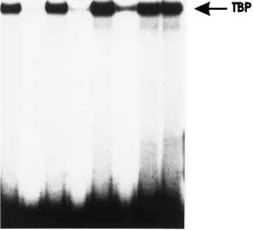
The sequences protected by TBP partially overlap the E2
recognition sequence, indicating that the simultaneous binding
of both proteins might not be possible for steric reasons. To
test for this, gel retardation experiments were carried out (Fig.
7). Aside from the labeled P2 wild-type oligonucleotide,
la-beled P2mtC was used, as this oligonucleotide displays a higher
affinity to E2 than the wild type, while its affinity for TBP is
unchanged (Fig. 6B) (39). Furthermore, in the background of
the reporter plasmid P2C-CAT, this mutant displays the
stron-gest repression by E2. Incubation of both oligonucleotides with
TBP or E2 protein alone resulted in clearly distinguishable
shifted complexes. Binding reactions with both proteins gave
rise to two shifted bands corresponding to TBP-DNA and
E2-DNA complexes. Increasing the amount of E2 in the
pres-ence of a fixed amount of TBP decreased the intensity of the
TBP-DNA complex with both the P2 wild-type and P2mtC
oligonucleotides. This strongly suggests that E2 represses P
7535activity by displacement of TBP.
Specific binding of cellular factors to P2.
Plasmids
[image:5.612.108.290.470.635.2]T7535-CAT and p7535-TATA-Luc are also repressed by E2 despite
the fact that the mutated TATA box is no longer able to bind
to TBP in vitro (Fig. 6B). This suggests that E2 might also
interfere with the function of other factors that are important
for promoter activity. Mutations in E2BS P2 (P2mt and
P2mtC) reduce promoter activity (Fig. 4 and Table 1) (39),
which indicates that it is an important element required for the
FIG. 6. TBP specifically recognizes the P7535TATA box. (A) DNase Ifoot-printing analysis of the P7535promoter region. An HPV8 DNA fragment (nt 7454 to 7602) was labeled at the coding (left half) or the noncoding (right half of the autoradiogram) strand and then incubated with recombinant TBP (TBP) or with buffer only (lanes —). Binding reactions were digested with 40 ng (left TBP lane), 10 ng (right TBP lane), or 2 ng (lanes —) of DNase I. Reaction products were separated in a 6% polyacrylamide–urea gel. An A1G Maxam-Gilbert sequenc-ing reaction of the labeled fragments served as molecular size markers. The positions of the P7535TATA box on both strands are indicated by open boxes. (B) Gel retardation competition analysis of TBP binding to the P2 TATA region (nt 7486 to 7516). The32P-end-labeled P2 oligonucleotide was incubated with
TBP and without (lanes —) or with 100 ng of unlabeled wild-type or mutant oligonucleotides as competitors. Reaction products were separated in a 5% polyacrylamide gel containing 0.53TBE, dried, and then autoradiographed.
on November 9, 2019 by guest
basal activity of P
7535. The gel retardation assays suggested that
the reduced activity of these mutants is not due to a decrease
in TBP binding affinity (Fig. 6B). Specific binding of cellular
factors to P2 has been demonstrated with HeLa nuclear
ex-tracts (26). We carried out gel retardation experiments with
labeled P2 wild-type oligonucleotide and crude nuclear
ex-tracts from RTS3b cells and HT3 cells, which yielded identical
patterns (Fig. 8 and data not shown). Five different complexes
(A to E) could be detected with extracts from both cell lines.
Complexes B to E seem to be specific, as they cannot be
eliminated by an unrelated oligonucleotide (P36). Complex C
was only weakly inhibited by specific oligonucleotides,
indicat-ing that the responsible factor might be present in high
amounts or might be of low specificity. The three specific
complexes B, D, and E are not TBP or TFIID related, as they
were eliminated by the T7535 oligonucleotide. They were not
or only weakly inhibited by oligonucleotides P2mtC and P2mt,
respectively. On principle, the significantly lower binding
af-finity correlates with reduced promoter activity of P2mt and
P2mtC, suggesting that the protein(s) involved is important for
promoter function. However, the P2mt oligonucleotide
seemed to bind slightly better than the P2mtC oligonucleotide
to the specific factors, although the basal activity of P2-CAT is
lower than that of P2C-CAT. This may be due to the fact that
the gel retardation assay does not correctly reflect the in vivo
binding affinities of these factors or may point to the
involve-ment of additional factors that could not be detected in gel
retardation assays. It was not yet possible to demonstrate
com-petition between P2-binding cellular factors (P2Fs) and
in-creasing amounts of purified E2 in vitro with crude nuclear
extracts (data not shown). These experiments have to await the
purification of the P2Fs.
To obtain genetic evidence that E2 repression of P
7535ac-tivity also involves an interference of E2 with the P2Fs, we
created plasmid p7535-P2
1
10-Luc, in which E2BS P2 is
sep-arated from the TATA box by 11 instead of 1 bp (Fig. 9). Steric
hindrance between E2 and TBP should be relieved with this
plasmid, because it has been shown for the HPV18 P
105 [image:6.612.73.285.71.288.2]pro-moter that increasing the distance between the E2BS and the
TATA box from 3 to 8 bp results in vitro in simultaneous
binding of both proteins and in vivo in transactivation instead
of repression by E2 (9, 15). Plasmid p7535-P2
1
10-Luc
dis-played fourfold-higher basal activity than p7535-Luc (Fig. 9).
Cotransfection with different amounts of pCE2 revealed that
this plasmid responds to E2 as p7535-Luc does. It was
trans-activated to a slightly higher degree at 10 ng of pCE2, but this
transactivation dropped at higher amounts of E2 expression
vector (Fig. 9). The E2-mediated negative modulation of
pro-moter activities in the context of plasmids p7535-P2
1
10-Luc
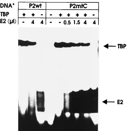
FIG. 7. E2 occludes TBP from binding to the P7535TATA box. Gelretarda-tion assays were carried out with the32P-labeled P2wt or P2mtC oligonucleotide, which were added to the reaction after the protein(s). Binding reactions were incubated with TBP (25 ng) and the indicated amounts of recombinant HPV8 E2 protein affinity purified from insect cells. The TBP-DNA and E2-DNA com-plexes are indicated by arrows.
FIG. 8. Nuclear proteins from RTS3b cells specifically interact with the P2 sequence. Gel retardation assays were carried out with32
P-labeled P2 oligonu-cleotide and crude nuclear extracts isolated from RTS3b cells. Reactions re-ceived no protein (2) or 5mg of extract (1). Prior to the addition of extract, 0, 10, or 100 ng of unlabeled wild-type P2, mutated P2 (P2mt or P2mtC), mutated TATA box (T7535), or unrelated P36 oligonucleotide was added. Reactions were incubated at room temperature for 20 min and then separated in a 5% poly-acrylamide gel containing 0.53TBE. Complexes A to E are indicated by arrows.
FIG. 9. E2 represses a modified P7535promoter (P2110) in which the E2BS P2 is separated by 11 nt from the TATA box. RTS3b cells were transfected with p7535-Luc (7535) or p7535-P2110-Luc (P2110) and different amounts of HPV8 E2 expression vector pCE2, and luciferase activity was determined. The activa-tion or repression by E2 is given relative to the basal activity of the individual constructs. The relative basal activity of P7535-P2110-Luc is 4.0, with that of p7535-Luc set at 1. The DNA sequences of the wild-type (P2) and the mutated (P2110) P2 are shown below; the insertion is underlined.
on November 9, 2019 by guest
http://jvi.asm.org/
[image:6.612.59.298.476.659.2]more pronounced in RTS3b cells, finally leading to repression
of the basal promoter activity. The obviously high levels of E2
in RTS3b cells might interfere with E2 transactivation by
squelching. This was discussed for HPV16 E2 and the HPV16
P
97promoter (4), whereas no squelching was detectable with
HPV16 E2 and an E2-dependent minimal promoter construct
(43). Only a slight reduction in E2 transactivation, from 24- to
20-fold, at high levels of HPV8 E2 expression vector could be
noted with the HPV8 enhancer construct, which could indeed
be due to squelching. However, this cannot account for the
repression of P
7535starting at 100 ng of pCE2. Transactivation
of the binding site mutant P2mt (P2-CAT and p7535-P2-Luc)
was slightly reduced at high E2 levels (Fig. 4 and data not
shown), similar to the enhancer construct, but repression could
not be achieved. This binding-site dependence of the negative
control argues strongly against squelching mechanisms.
The difference in the response of P
7535to E2 between HT3
and RTS3b cells seems to be due to different amounts of E2
protein after transfection. This can be explained by a higher
transfection efficiency of RTS3b cells than of HT3 cells, as
judged by in situ staining after transfection of a
b
-galactosi-dase-expressing plasmid (data not shown). Other mechanisms,
such as differences in the expression level of the
cytomegalo-virus-E2 construct or different stabilities of the HPV8 E2
pro-tein might also be involved.
The E2BS P2 is localized at positions
2
44 to
2
33 relative to
the P
7535cap site. It overlaps recognition sequences for a
cellular factor(s) (P2F [Fig. 8]) which is important for
pro-moter function and is separated by only one nucleotide from
the P
7535TATA box. Mutational and biochemical analyses
strongly suggest that E2 represses promoter activity by
inter-ference with the binding and/or function of cellular
transcrip-tion factors. We have shown on the one hand that E2 is able to
occlude TBP from binding to the promoter in vitro. This has
also been reported to be relevant for the repression of HPV18
P
105activity by BPV1 E2TA (9). E2 repression of plasmids
with a mutated TATA box and p7535-P2
1
10-Luc, with an
increased spacing between P2 and the TATA box, indicated
that there is an interference with promoter activity that is
independent of the occlusion of TBP binding. The TATA box
mutant was negatively regulated by E2 despite the fact that
TBP is not able to bind in vitro to the mutated promoter. The
sequence arrangement of p7535-P2
1
10-Luc, in which E2BS
P2 is separated by 11 bp from the TATA box, should allow the
simultaneous binding of E2 and TBP, as shown for the E6/E7
promoter of HPV18 after introduction of only 5 bp (9, 15), but
it is still repressed by high amounts of pCE2 and not
transac-tivated, as in the case of HPV18 (15). This suggests that
inter-ference of E2 with the P2Fs is also involved in repression of
P
7535. Repression of viral promoters by competition of E2 with
the strategy for transcriptional repression by E2 seems to be
very similar among early and late promoters.
The HPV8 late gene promoter is activated at low E2 protein
concentrations and repressed at higher amounts, which
corre-lates with the observation that P2 is a low-affinity E2BS (39).
The biological significance of this complex regulation is still
unclear, but cDNA analyses suggest that P
7535may drive the
expression of both the L1 and E2 genes (11, 38). It is tempting
to speculate that P
7535might be used at early times of infection
as an autoregulated E2-specific promoter. The observed
regu-latory loop would then serve to maintain a low but constant
level of E2. E2 autoregulation has been described for BPV1
(18) and might also occur with HPV11 and -16, as mRNAs
transcribed from the respective E2-regulated E6 promoters
may encode the E2 gene (31, 35). In the uppermost layers of
the epithelium, transcription of the L1 gene could be achieved
by reduced E2 expression and/or by upregulation of the P2Fs,
preventing E2 from binding. On the other hand, the P2-E2
interaction may be weakened by modification of the E2 protein
or of the P2 sequence, e.g., by DNA methylation.
ACKNOWLEDGMENTS
We thank G. Steger, Institut Pasteur, Paris, for providing the puri-fied recombinant TBP.
This work was supported by grant Hi 291/5-1 from the Deutsche Forschungsgemeinschaft to H.P.
REFERENCES
1. Androphy, E. J., D. R. Lowy, and J. T. Schiller. 1987. Bovine papillomavirus E2 trans-activating gene product binds to specific sites in papillomavirus DNA. Nature (London) 325:70–73.
2. Barsoum, J., S. S. Prakash, P. Han, and E. J. Androphy. 1992. Mechanism of action of the papillomavirus E2 repressor: repression in the absence of DNA binding. J. Virol. 66:3941–3945.
3. Bernard, B. A., C. Bailly, M.-C. Lenoir, M. Darmon, F. Thierry, and M. Yaniv.1989. The human papillomavirus type 18 (HPV18) E2 gene product is a repressor of the HPV18 regulatory region in human keratinocytes. J. Virol. 63:4317–4324.
4. Bouvard, V., A. Storey, D. Pim, and L. Banks. 1994. Characterization of the human papillomavirus E2 protein: evidence for activation and trans-repression in cervical keratinocytes. EMBO J. 13:5451–5459.
5. Chomczynski, P., and N. Sacchi. 1987. Single-step method of RNA isolation by acid guanidinium thiocyanate-phenol-chloroform extraction. Anal. Bio-chem. 162:156–159.
6. Demeret, C., M. Yaniv, and F. Thierry. 1994. The E2 transcriptional repres-sor can compensate for SP1 activation of the human papillomavirus type 18 early promoter. J. Virol. 68:7075–7082.
7. Dong, G., T. R. Broker, and L. T. Chow. 1994. Human papillomavirus type 11 E2 proteins repress the homologous E6 promoter by interfering with the binding of host transcription factors to adjacent elements. J. Virol. 68:1115– 1127.
8. Dong, X.-P., F. Stubenrauch, E. Beyer-Finkler, and H. Pfister. 1994. Preva-lence of deletions of YY1-binding sites in episomal HPV 16 DNA from cervical cancers. Int. J. Cancer 58:803–808.
9. Dostatni, N., P. F. Lambert, R. Sousa, J. Ham, P. M. Howley, and M. Yaniv.
on November 9, 2019 by guest
1991. The functional BPV-1 E2 trans-activating protein can act as a repressor by preventing formation of the initiation complex. Genes Dev. 5:1657–1671. 10. Ensser, A., and H. Pfister. 1990. Epidermodysplasia verruciformis associated human papillomaviruses present a subgenus-specific organization of the reg-ulatory genome region. Nucleic Acids Res. 11:3919–3922.
11. Fuchs, P. G., S. Horn, T. Iftner, M. May, F. Stubenrauch, and H. Pfister. 1993. Molecular biology of epidermodysplasia verruciformis-associated hu-man papillomaviruses, p. 517–529. In W. Doerfler and P. Bo¨hm (ed.), Virus strategies. Verlag Chemie, Weinheim, Germany.
12. Fuchs, P. G., and H. Pfister. 1994. Transcription of papillomavirus genomes. Intervirology 37:159–167.
13. Fuchs, P. G., J. Weninger, T. Iftner, and H. Pfister. 1986. Epidermodysplasia verruciformis-associated human papillomavirus 8: genomic sequence and comparative analysis. J. Virol. 58:626–634.
14. Ham, J., N. Dostatni, J. M. Gauthier, and M. Yaniv. 1991. The papilloma-virus E2 protein: a factor with many talents. Trends Biol. Sci. 16:440–444. 15. Ham, J., G. Steger, and M. Yaniv. 1994. Cooperativity in vivo between the E2
transactivator and the TATA box binding protein depends on core promoter structure. EMBO J. 13:147–157.
16. Haugen, T. H., L. P. Turek, F. M. Mercurio, T. P. Cripe, B. J. Olson, R. D. Anderson, D. Seidl, M. Karin, and J. Schiller.1988. Sequence-specific and general transcriptional activation by the bovine papillomavirus-1 E2 trans-activator require an N-terminal amphipathic helix-containing E2 domain. EMBO J. 7:4245–4253.
17. Hawley-Nelson, P., E. J. Androphy, D. R. Lowy, and J. T. Schiller. 1988. The specific DNA recognition sequence of the bovine papillomavirus E2 protein is an E2-dependent enhancer. EMBO J. 7:525–531.
18. Hermonat, P. L., B. A. Spalholz, and P. M. Howley. 1988. The bovine papillomavirus P2443 promoter is E2 trans-responsive: evidence for E2 au-toregulation. EMBO J. 7:2815–2822.
19. Horn, S., H. Pfister, and P. G. Fuchs. 1993. Constitutive transcriptional activator of epidermodysplasia verruciformis-associated human papillomavi-rus 8. Virology 196:674–681.
20. Krubke, J., J. Kraus, H. Delius, L. T. Chow, T. R. Broker, T. Iftner, and H. Pfister.1987. Genetic relationship among human papillomaviruses associ-ated with benign and malignant tumours of patients with epidermodysplasia verruciformis. J. Gen. Virol. 68:3091–3103.
21. Lambert, P. F., B. A. Spalholz, and P. M. Howley. 1987. A transcriptional repressor encoded by BPV-1 shares a common carboxy-terminal domain with the E2 transactivator. Cell 50:69–78.
22. Lees, E., K. Osborn, L. Banks, and L. Crawford. 1990. Transformation of primary BRK cells by human papillomavirus type 16 and EJ-ras is increased by overexpression of the viral E2 protein. J. Gen. Virol. 71:183–193. 23. Luckow, B., and G. Schu¨tz.1987. CAT constructions with multiple unique
restriction sites for the functional analysis of eukaryotic promoters and regulatory elements. Nucleic Acids Res. 15:5490.
24. May, M., X.-P. Dong, E. Beyer-Finkler, F. Stubenrauch, P. G. Fuchs, and H. Pfister.1994. The E6/E7 promoter of extrachromosomal HPV16 DNA in cervical cancers escapes from repression by mutation of target sequences for YY1. EMBO J. 13:1460–1466.
25. May, M., K. Grassmann, H. Pfister, and P. G. Fuchs. 1994. Transcriptional silencer of the HPV8 late promoter interacts alternatively with viral E2 or a cellular protein. J. Virol. 68:3612–3619.
26. May, M., V. Helbl, H. Pfister, and P. G. Fuchs. 1991. Unique topography of DNA-protein interactions in the non-coding region of epidermodysplasia verruciformis-associated human papillomaviruses. J. Gen. Virol. 72:2989– 2997.
27. McBride, A. A., J. C. Byrne, and P. M. Howley. 1989. E2 polypeptides encoded by bovine papillomavirus type 1 form dimers through the common
carboxyl-terminal domain: trans-activation is mediated by the conserved amino-terminal domain. Proc. Natl. Acad. Sci. USA 86:510–514. 28. McBride, A. A., H. Romanczuk, and P. M. Howley. 1991. The papillomavirus
E2 regulatory proteins. J. Biol. Chem. 266:18411–18414.
29. McBride, A. A., R. Schlegel, and P. M. Howley. 1988. The carboxy-terminal domain shared by the bovine papillomavirus E2 transactivator and repressor proteins contains a specific DNA binding activity. EMBO J. 7:533–539. 30. Monini, P., I. L. Blitz, and E. Cassai. 1993. Cooperative DNA binding of the
bovine papillomavirus E2 transcriptional activator is antagonized by trun-cated E2 polypeptides. J. Virol. 67:5668–5676.
31. Nasseri, M., J. R. Gage, A. Lorincz, and F. O. Wettstein. 1991. Human papillomavirus type 16 immortalized cervical keratinocytes contain tran-scripts encoding E6, E7 and E2 initiated at the P97 promoter and express high levels of E7. Virology 184:131–140.
32. Pfister, H. 1992. Human papillomaviruses and skin cancer. Semin. Cancer Biol. 3:263–271.
33. Purdie, K. J., C. J. Sexton, C. M. Proby, M. T. Glover, A. T. Williams, K. J. Stables, and I. M. Leigh.1993. Malignant transformation of cutaneous lesions in renal allograft patients: a role for human papillomavirus. Cancer Res. 53:5328–5333.
34. Romanczuk, H., F. Thierry, and P. M. Howley. 1990. Mutational analysis of cis elements involved in E2 modulation of human papillomavirus type 16 P97 and type 18 P105promoters. J. Virol. 64:2849–2859.
35. Rotenberg, M. O., C.-M. Chiang, M. L. Ho, T. R. Broker, and L. T. Chow. 1989. Characterization of cDNAs of spliced HPV-11 E2 mRNA and other HPV mRNAs recovered via retrovirus-mediated gene transfer. Virology 172:468–477.
36. Sealey, L., and R. Chalkley. 1987. At least two nuclear proteins bind specif-ically to the Rous sarcoma virus long terminal repeat. Mol. Cell. Biol. 7:787–798.
37. Stenlund, A., and M. R. Botchan. 1990. The E2 trans-activator can act as a repressor by interfering with a cellular transcription factor. Genes Dev. 4:123–136.
38. Stubenrauch, F., J. Malejczyk, P. G. Fuchs, and H. Pfister. 1992. Late promoter of human papillomavirus type 8 and its regulation. J. Virol. 66: 3485–3493.
39. Stubenrauch, F., and H. Pfister. 1994. Low-affinity E2-binding site mediates downmodulation of E2 transactivation of the human papillomavirus type 8 late promoter. J. Virol. 68:6959–6966.
40. Szymanski, P., and A. Stenlund. 1991. Regulation of early gene expression from the bovine papillomavirus genome in transiently transfected C127 cells. J. Virol. 65:5710–5720.
41. Tan, S.-H., B. Gloss, and H.-U. Bernard. 1992. During negative regulation of the human papillomavirus-16 E6 promoter, the viral E2 protein can displace Sp1 from a proximal promoter element. Nucleic Acids Res. 20:251–256. 42. Tan, S.-H., L. E.-C. Leong, P. A. Walker, and H.-U. Bernard. 1994. The
human papillomavirus type 16 E2 transcription factor binds with low coop-erativity to two flanking sites and represses the E6 promoter through dis-placement of Sp1 and TFIID. J. Virol. 68:6411–6420.
43. Ushikai, M., M. J. Lace, Y. Yamakawa, M. Kono, J. Anson, T. Ishiji, S. Parkinnen, N. Wicker, M.-E. Valentine, I. Davidson, L. P. Turek, and T. H. Haugen.1994. trans activation by the full-length E2 proteins of human papillomavirus type 16 and bovine papillomavirus type 1 in vitro and in vivo: cooperation with activation domains of cellular transcription factors. J. Vi-rol. 68:6655–6666.
44. Vande Pol, S. B., and P. M. Howley. 1990. A bovine papillomavirus consti-tutive enhancer is negatively regulated by the E2 repressor through compet-itive binding for a cellular factor. J. Virol. 64:5420–5429.