MANAGEMENT OF INFECTED NON UNION
OF LONG BONES BY ANTIBIOTIC LOADED
PMMA CEMENT COATED NAIL AND BEADS
DISSERTATION SUBMITTED FOR
M.S. DEGREE
(BRANCH II - ORTHOPAEDIC SURGERY)
MARCH 2009
THE TAMILNADU
DEPARTMENT OF ORTHOPAEDICS
MADURAI MEDICAL COLLEGE AND GOVERNMENT RAJAJI HOSPITAL MADURAI.
CERTIFICATE
This is to certify that the dissertation entitled
“MANAGEMENT OF INFECTED NON UNION OF LONG BONES
BY ANTIBIOTIC LOADED PMMA CEMENT COATED NAIL
AND BEADS” is a bonafide record of work done by Dr. K.
VIJAYANTH in the Department of Orthopaedics and
Traumatology, Government Rajaji Hospital, Madurai Medical
College, Madurai, under the direct guidance of me.
Prof. Dr.V. RAVIRAMAN, M.S.ORTHO., D.ORTHO., Professor and Head of the Department
DECLARATION
I Dr. K. VIJAYANTH, solemnly declare that the dissertation
entitled “MANAGEMENT OF INFECTED NON UNION OF LONG
BONES BY ANTIBIOTIC LOADED PMMA CEMENT COATED
NAIL AND BEADS’ has been prepared by me under the able guidance
and supervision of my guide Prof. Dr.V. Raviraman, M.S.ORTHO.,
D.ORTHO., Prof & HOD, Department of Orthopaedics and
Traumatology, Madurai Medical College, Madurai, in partial fulfilment
of the regulation for the award of M.S. (ORTHOPAEDIC SURGERY)
degree examination of The Tamilnadu Dr. M.G.R. Medical University,
Chennai to be held in March 2009.
This work has not formed the basis for the award of any other
degree or diploma to me previously from any other university.
Place : Madurai
Date : DR. K. VIJAYANTH
ACKNOWLEDGEMENT
I express my sincere and heartfelt gratitude towards
Prof. Dr.V. Raviraman, M.S.Ortho, D.Ortho., Professor and Head of the Department of Orthopaedics, Madurai Medical College, Madurai under while guidance and supervision the present work has been carried out.
I am immensely thankful to Prof. Dr.M. Malairaju, for necessary
guidance, advice and his timely help.
My sincere thanks to Prof. P.V. Pughalenthi, for his help during the
study.
I am extremely thankful to Prof. Dr. A. Rajamani, Dr. T.
Chandraprakasam for their advice and guidance during this study.
My profound and heartfelt thanks to Dr.K. Ravichandran, Registrar for valuable help and guidance in preparing this dissertation throughout.
My sincere thanks to my Assistant Professors Dr. S.Ramanathan, Dr. B. Sivakumar, Dr. M.N. Karthi, Dr.N. Thanappan, Dr. P.V.Thirumalai Murugan, Dr. K.P. Saravana Kumar, Dr. T.C.Premkumar, Dr.J. Manikandan, Dr. Duraimurugan for their valuable guidance during this study
I am extremely thankful to Prof. Dr. S.M. Sivakumar, M.S., Dean, Madurai Medical College, Madurai for allowing me to utilize the hospital materials of the hospital.
I am very much indebted to all my patients who lent themselves for carrying out this study.
CONTENTS
S. NO TOPIC PAGE NO
1. INTRODUCTION 1
2. AIM 5
3. CAUSES AND CLASSIFICATION 6 4. PATHOPHYSIOLOGY OF INFECTED NONUNION 12 5. INVESTIGATIONS 15 6. HISTORY OF INFECTED NON UNION TREATMENT 17 7. PRINCIPLES OF MANAGEMENT OF INFECTED
NON UNION WITH ANTIBIOTIC CEMENT
LOADED INTRAMEDULLARY RODS AND BEADS 23
8. REVIEW OF LITERATURE 42 9. MATERIALS AND METHODS 49 10. RESULTS AND ANALYSIS 57
11. DISCUSSION 59
12. CONCLUSION 62
INTRODUCTION
Ununited fractures of long bones are not only a complex
surgical problem but also a chronic and at times debilitating
condition. Infected non-union of long bones are not only a source of
functional disability but also lead to economical hardship and loss of
self esteem. Infected non-union has been defined as a state of failure
of union for 6-8 months with persistent infection at the fracture site.
Infected non-union can develop after an open fracture, after a
previous open reduction and internal fixation (ORIF) or as sequelae
to chronic haematogenous osteomyelitis. The incidence also seems
to be increasing especially in view of increasing high velocity
trauma, which is more frequently treated with internal fixation.
It is difficult to treat infected non-union because of following
reasons.
1. Previous surgeries would have resulted in cicatrisation of
the soft tissue with an avascular environment around the
fracture site.
2. The presence of dead bone or sequestrum at the fracture site
3. Necrosis of bone near the non union site, to a considerable
distance, due to thrombosis of blood vessels of Haversian
canals.
4. Prolonged immobilization, multiple surgeries with fibrosis
of the muscles leading on to a stiff joint / fracture disease.
5. The micro organism may develop resistance to the systemic
antibiotic therapy and poses a problem in controlling the
disease.
Soft tissue loss with multiple sinuses, osteomyelitis, osteoporosis,
systemic antibiotic resistance all complicates treatment and
recovery. These factors make an unfavourable milieu for fracture
union. Even after prolonged treatment and repeated surgeries to
correct this problem, the outcome is unsure and amputation may be
the only alternative left.
Hence the treatment of non union of long bones associated
with infection is a formidable challenge to the treating orthopaedic
surgeons. Bony union is not usually obtained until the infection has
Three entirely different methods of treatment have been
recommended for this difficult problem in the past.
These include the CONVENTIONAL OR CLASSIC,
ACTIVE OR MODERN and treatment by PULSED
ELECTROMAGNETIC FIELDS. The major disadvantages of these
procedures being multiple surgeries, need of an external fixator for
stabilisation, and associated poor patient compliance.
The current management of this kind of infection consists of
two main objectives. Infection control which is usually achieved by
nail removed with debridement, lavage of medullary canal, local
delivery of antibiotics by antibiotic impregnated bead chains and
fracture union, which usually is accomplished by alternative fixation
mostly external fixation.
The principle of antibiotic impregnated beads is to
i) Fill the dead space and
ii) Deliver high concentration of antibiotic to infected site.
The concept of antibiotic impregnated PMMA spacer originated
in prosthetic joint infection. With very good results in prosthetic
indication were extended to infected non union of diaphyseal
fractures.
The advantages of antibiotic impregnated PMMA coated nails
and beads in the treatment of infected non union of long bones being
increased local concentration of antibiotics and stable internal
fixation, which controls infection and promotes bony healing at a
AIM
¾ To discuss the biological advantage of antibiotic cemented
nailing and beads in the management of infection non union of
long bones.
¾ To evaluate the clinical outcome of the study and discuss the
results of cases with infected non-union of femur and tibia
managed by antibiotic impregnated PMMA cemented Rods
CAUSES AND CLASSIFICATION OF NON UNION
Causes:
In 1986 FDA panel defined Nonunion as ‘established when a
minimum of nine months have elapsed since injury and the fracture
show no visible progressive signs of healing for 3 months’. But that
criterion cannot be applied to every fracture. A fracture of shaft of
long bones should not be considered as non-union until at least 6
months after the injury, because often union requires more time,
especially after some local complications such as an infection. Non
union can result from the following causes.7
1. Excess motion
Due to inadequate immobilization
2. Gap between fragments
a) Soft tissue interposition
b) Malposition or over riding or displacement of fragments
c) Loss of bone substance
d) Distraction by hardware or traction.
3. Loss of blood supply
b) Excessive stripping or injury to periosteum and muscles
c) Free fragment, severe communition
4. Infections
a) Bone death (sequestrum)
b) Osteolysis (Gap)
c) Loosening of implants
5. General (pre disposing factors)
Age, nutrition, steroids, radiation, anticoagulants, DM,
immunodeficient states etc.
CLASSIFICATIONS
There are various classifications available for non-union and
infected non-union in the literature.
I - Judet, Muller, Weber and Cech classified non-union broadly into
two types. They are
a) Hyper vascular (hypertrophic) the ends of the fragments
are capable of biological reactions
b) Avascular (atrophic), the ends of the fragments are inert
Hypervascular / Viable / Hypertrophic non-union further
subdivided into
1) Elephant foot type
2) Horse hoof type
3) Oligotrophic type
Avascular nonviable / atrophic non-union further subdivided into
1) Torsion wedge
2) Communited
3) Defect
4) Atrophic
II - Paley et al divided non union clinically and radiologically into 2
major types
Type A (Bone loss < 1 cm)
A1 - Non union with a mobile deformity
A2 - Nonunion with a fixed deformity
A2 -1 Stiff non-union without deformity
A2 -2 Stiff non-unions with a fixed deformity
Type B (Bone loss > 1 cm)
B1 - Nonunion with a bony defect
B2 - Nonunion with loss of bone length
This classification system is further modified by the presence
or absence of infection.
III - Maurizio Catagni’s classification
A1 - Non infected mobile non-union
A1 - Non infected stiff hypertrophic non union without deformity
A3 - Non infected hypertrophic non-union with deformity
B1 - Non infective non-union with bone defect of up to 5 cms
B2 - Non infective non-union with bone defect exceeding 5 cms
B3 - Non infective non-union exceeding 10cms with local scarring
C1 - Infected non-union with atrophy
C2 - Infected non-union with hypertrophy without deformity
C3 - Infected non-union with hypertrophy and deformity
C4 - Infected non-union with bone gap of less than 5 cms
C5 - Infected non-union with bone gap between 5 and 10 cms
C6 - Infected non-union with bone gap exceeding 10 cms
IV - The University of TEXAS Classification
Based on the location of infection and modified by immune
Type 1 : Intramedullary
Type 2 : Superficial
Type 3 : Local
Type 4 : Diffuse with segmental bone loss
Type A : Healthy immune system
Type B : Local or systemic compromise of immune
System
Type C : Severe compromise of immune system
V - WIELAND’S Classification
Type 1 : Bone exposed and soft tissue infection present
Type 2 : Circumferential – cortical and endosteal infection
Present
Type 3 : Cortical and endosteal infection combined with
segmental bone loss
VI - AO - Classification :
1) Infected non draining non-union (Active / Quiescent)
2) Infected Draining non-union
VII - KULKARNI’S Classification
Classification of infected non-union is based on the severity of
Type 1 : Mild infection
Beads of pus on pressing
Fragments in alignment
With or without an implant
No gap
No deformity
No shortening
Type 2 : Moderate infection :
Fragments in apposition
No gap or gap < 2 cm
No deformity
No shortening
Moderate infection with a large or small wound
Type 3 : Severe Infection
Pouring pus
Gap > 2 cms
With deformity
PATHOPHYSIOLOGY OF INFECTED NONUNION
Infection per se, does not cause non-union, as union has been
shown to occur in the presence of active infection. Uncontrolled
infection, however causes non-union, predominantly because
purulent material dissects under pressure within the intramedullary
canal and along the subperiosteal surfaces of bone, resulting in bone
necrosis. The inflammatory response to the infections process may
also lead to an excessive remodelling response causing osteolysis,
which further slows the rate of union. 1,2,3
Pathophysiology of infections after internal fixation of fractures:
Infection complicating internal fixation of fractures is a serious
complication that is difficult to treat whenever metallic implants are
implanted in vivo, successful bio integration requires that host cells
colonize the highly reactive implant surface. Bacteria such as
staphylococci can also became adherent to metallic or polymeric
implants and will compete with host cells for colonization of the
implant surface. Once adherent these bacteria form a bio film and
undergo phenotypic changes that make them resistant to the normal
metallic implants themselves cause specific deficits in the function
of the local immune system that may render the host response to
infection inadequate. Any associated soft tissue injury causes
greater impairment of local immune function. Despite the
potentially detrimental impact of internal fixation, fracture stability
is of paramount importance in achieving fracture union and in
preventing infection. It has been demonstrated in animal models that
contaminated fractures without internal fixation develop clinical
infection more commonly than similar fractures treated with internal
fixation at the time of colonization. Because of the potential for
infection whenever internal fixation is utilized appropriate
prophylactic antibiotic coverage for staphylococci and gram negative
organisms should be provided. Open wounds and severely damaged
soft tissues require aggressive management so that a viable soft
tissue envelope is maintained around the implant. Host factors such
as smoking and malnourishment should be corrected. Early
diagnosis and aggressive treatment of implant related infection with
antibiotics, debridement and maintenance of stable internal fixation
Bacterial adherence to biomaterials and tissue. The significance
of its role in clinical sepsis.
A study by AG Gristina and JW costerton
The direct examination of tissue and biomaterials from
prosthesis related infections of 25 patients showed that the causation
bacteria grew in glycocalyx enclosed bio films that were adherent to
surface of biomaterials and tissue in 76%. This high rate of recovery
of adherent bio film mediated growth suggests that the process
occurs commonly in the presence of a foreign body or biomaterial
related infection. Because of the adherent mode of growth of the
infecting organisms, accurate microbiological sampling was
difficult. The analysis of swabs of excised tissue and of prosthetic
surfaces often yielded only one species from what was a
INVESTIGATIONS
1. Diagnosis of Infection in ununited fractures
Combining imaging with Indium – 111 labelled leukocytes
and technetium – 99m methylene diphosphonate were used were
compared with the results of cultures of open bone at 102 sites of
delayed union and non-union, to determine the effectiveness of this
combination as a preoperative indicator of osteomyelitis.
A sensitivity of 86%, specificity of 84% an accuracy of 82%, a
positive predictive value of 69% and a negative predictive valve of
94%.
2. ESR
Measurement of increased rate of setting of erythrocytes is an
important lab test in evaluation of disease activity in patients with
infections.
Done by Westergren method
Normal valves are 0-9 mm per hour for men, 0-20 mm / hr for
women. In routine orthopaedic procedures, maximum of 25 to 40
mm / hr is reached in 4 days which gradually decreased to normal
3. C – Reactive protein
An acute phase protein that can be used to follow the course of
acute infections
It rises and falls faster than ESR
10 mg per litre is used as the threshold for THR infection,
using this threshold the sensitivity and specificity are both about
90%
4. WBC count
HISTORY OF INFECTED NONUNION TREATMENT
Considerable judgement is required to treat a non-union of an
infected fracture. There entirely different methods of treatment have
been most of recommended for this difficult problem.
1. Conventional or classic method
2. Active or modern method
3. Pulsed electromagnetic fields
Ilizorov method is a more recent method of treating infected
non-union that has similarities to both the conventional and active
methods. 6,7
1. Conventional Treatment :
The conventional treatment is used for many decades.
The object of conventional treatment are to convert an infected and
draining non-union into one that has not drained for several months
and then to promote healing of the non-union by bone grafting. This
method of treatment often requires 1 or more years to complete and
usually results in stiffness of adjacent joints.
The skin over the bone is made as nearly normal as possible.
Stage I :
Wound is thoroughly saucerised and all foreign and infected or
devitalized materials are removed to provide a vascular bed. Any
gross overlapping and displacements of the fragments are corrected.
Fixation is done either by internally or externally. Antibiotics are
used both parenterally and locally after surgery.
Stage II :
After 4-7 days when a thin layer of granulation tissue has
covered the wound, a split thickness skin graft is applied. The split
graft is replaced by a full thickness pedicled skin graft 4-6 weeks
after the wound has healed from the operation.
Stage III :
When the clinical sign of infection have subsided, the skin
over the bone is good and non-union persists, bone grafting must be
considered. Controlling infection before attempting bone grafting
always has been a sound clinical principle in the conventional
treatment of non-union.
2. Active treatment :
The object of active treatment is to obtain bony union early
Cech described this method and much of the following is taken from
their reports. The first step is restoration of bony continuity. This
takes absolute priority over treatment of the infection. The
non-union is exposed through the old scar and tissues. The ends of the
fragments are then decorticated subperiosteally forming many small
osteoperiosteal grafts, any graft that become detached are discarded.
Next all devitalised and infected bone and soft tissues are removed.
Then the fragments are aligned and stabilised usually by an external
fixator. Compression is applied across the non union if possible.
Weber and Cech then inserted autogenous cancellous bone grafts.
Internal fixation with a plate is used only when drainage has already
ceased, and then the approach is away from the area of old drainage
or when no other method of fixation is possible and the infection is
mild. Finally a tube for suction drainage is inserted and as much of
the wound as possible is closed, any remaining open area is covered
by a biological dressing, systemic antibiotics are given.
If necessary for union, a second decortications with or without
Disadvantage :
Both conventional and active methods had their own
disadvantages and faded over time.
1. Required multiple procedures
2. Poor patient compliance.
3. Joint stiffness.
Electric and Electromagnetic stimulation :
External electrical stimulation is especially advantageous in
infected non-union management or when surgical intervention is
contraindicated. At least 3 electrical and electromagnetic methods
are available for the treatment of non-union. These methods are
either invasive, requiring the implantation of electrodes or semi
invasive requiring percutaneous application of multiple electrodes.
Devices that use inductive coupling differ in their configuration
some try to recreate the Helmholtz configuration and others use a U
shaped coil. 80% united if the device was used for more than 3
hours a day. Nonunion healed as readily as those from 9-12 months.
Also infected nonunions were as likely to heal as noninfected
nonunions, and fracture gaps up to 1 cm did not adversely affect the
non-union healed with electrical stimulation compared with
uninfected nonunions. However they recommended that infected
fractures be debrided before electrical stimulation. 7
Ilizorov Method :
According to Ilizorov, to eliminate infection and obtain union,
vascularity must be increased. In his approach this is achieved by
corticotomy and the application of his circular external fixator.
Catagni reported that, although union was obtained, infection was
not always eliminated. For atrophic nonunions with diffuse
infection or sequestrated bone open resection of the infected segment
is performed and bifocal compression is used. 7
Disadvantages of Ilizorov method :
1. Poor patient compliance
2. Cumbersome procedure
3. Pin tract infections
4. Muscle contracture and joint stiffness
Polymethylene methacrylate (PMMA) Antibiotic Beads :
Antibiotic impregnated PMMA beads also can be used to treat
infected nonunions. Head stable antibiotics such as tobramycin and
gentamycin can be mixed with PMMA and used locally to achieve
200 times the antibiotic concentration achieved with iv antibiotics.
Calhoun et al reported the use of antibiotic impregnated PMMA
beads in conjunction with debridement in the management of
infected nonunions. In their series a group of patients treated with
debridement and implantation of antibiotics beads for 4-6 wks were
compared with patients treated with parenteral antibiotics for 4
weeks after debridement. Bone healing and stabilisation were
treated according to local conditions. Infection was successfully
arrested in 89.3% of patients who were treated with gentamicin
PMMA beads and in 83.3% in patients who received in
PRINCIPLES IN THE MANAGEMENT OF INFECTED
NONUNION BY ANTIBIOTIC LOADED PMMA CEMENTED
ROD AND BEADS
Preliminary Steps :
The principles of treatment are infection control, stabilisation
of fracture, soft tissue coverage, and bone grafting of ununited
fractures and large bone defects.
Infection control includes irrigation and debridement, culture
and sensitivity and antibiotic therapy. In chronic osteomyelitis
obtain aerobic, anaerobic and fungal cultures. Recent studies have
advocated taking of multiple deep cultures from purulent material,
soft tissue, and Marrie and Costerton postulated that different
organisms may be growing in isolated microenvironments.
Sampling differences and bacterial viability may influence the
culture results.
Stabilization of the ununited fracture or non-union is essential.
Soft tissue coverage may require the use of local muscle flaps and
free vascularised muscle flaps for soft tissues defects. These may
important in host defence mechanisms, antibiotic delivery and
osseous and soft tissue healing.
Surgical Consideration :
Tourniquets :
Apply a tourniquet whenever possible except in patients with
sickle cell disease or significant peripheral vascular disease. The
tourniquet improves haemostasis and they facilitate identification of
the infection process. In acute cases with swelling, cellulitis, or
abscess formation, elevate the extremity for several minutes before
inflating the tourniquet. In chronic osteomyelitis without significant
cellulitis or abscess, use an elastic bandage to extravasate the
extremity before inflating the tourniquet. 6,7
Debridement :
Thorough debridement of all sequestra and necrotic and
desiccated bone is essential. Do not remove viable infected bone, so
as not to create large bony defects.
Clinically dried out, exposed, desiccated bone is darker than
normal and should be debrided. Necrotic bone that has not been
exposed may appear at surgery more yellowish than viable bone,
The main finding is that viable bone bleeds, whereas necrotic
bone does not. Use of an osteotome to superficially shave the outer
cortex of the questionable bone results in small areas of punctuates
bleeding. Evacuate all pus and abscess and remove all necrotic and
infected soft tissue. 6,7
Irrigation :
Use copious amounts of irrigating fluid, which cleanses the
area of purulent exudates, loose soft tissue and bony fragments and
decreases the bacterial count.
With regard to delivery of irrigation, high pressure pulsatile
lavage appears to be most effective for removal of bacteria and other
contaminants. Sterile saline solution either alone or with an additive
is commonly used for irrigation. The available additives are divided
into 3 general categories.
I Antiseptics - Povidone Iodine
- Chlorhexidine gluconate
- Hexa chlorophene
II Antibiotics - Bacitracin
III Soaps
Soaps remove microbes instead of killing them, and have least
varies. From the available evidence, it is not possible to recommend
any particular additive for the irrigation of medullary canal.
We used 10 litres of normal saline for irrigating the infected
wounds, 2 litres of diluted 5% povidone iodine 1: 20,000 solution as
the final irrigating solution. Povidone Iodine is a broad spectrum
microbicidal agent, acts by iodinating and oxidizing microbial
protoplasm.
After irrigation, the medullary canal is reamed up to its
maximum capacity to allow fresh bleeding and for maximum space
for the prepared nail to be inserted. 6,7
Wound Management :
The decision to leave a wound opens or to close it, requires
careful judgement. In the majority of acute infections and in all
cases in which there is associated abscess formation with cellulitis
and swelling the wound should be left open. In some cases of early
post operative infection, the wound may be closed over drainage
tubes as long as the wound is thoroughly clean and the infection is
not anaerobic.
In some cases in which bone or metal will be exposed if the
desirable as long as an adequate pathway has been provided for
drainage. When there is any doubt, it is safest to leave the wound
open. If the wound is closed, the wound site must be examined daily
for any signs of infection, if such signs appear the wound must be
opened.
Many wounds heal nicely by secondary intention. In case of
large wounds or when delayed closure is preferable, do not attempt
closure until two criteria are met.
First, the wound should appear clinically healthy with clean
granulating tissue and without any purulent exudates or necrotic
tissue. If infected necrotic tissue are present redebride the wound
until it appears healthy. Second, once clinical appearance of the
wound is clear, take quantitative tissue culture and do Gram stains.
Wounds with either a positive Gram stain or quantitative tissue
cultures with a bacterial count greater than 105 organisms should
never be closed. (A positive gram stain implies a bacterial count of
greater than 105 organisms)
With experienced surgical teams, tissue culture is not routinely
debridement followed by an en bloc excision of the wound at closure
or muscle transfer. 6,7
Drains :
When the wounds are closed, silastic (Jackson-Pratt) or
polyethylene (Hemovac) drain may be used. Penrose drains, made
of rubber, are the most reactive, and if left in for long periods can
cause foreign body granulomas. Do not use Penrose drains in
orthopaedic infection management.
Remove the suction drain in 48-72 hrs. The drain allows the
removal of all hematoma and tissue fluid and the collapse of the
potential dead space. The drain should be removed under sterile
conditions and the tip cut off and sent for culture and sensitivity
tests.
In general a positive culture of the drains tip is a bad
prognostic sign. It means that bacteria remain behind. 6,7
Wound Packing :
The purpose of leaving a wound open in to allow drainage.
Make certain that, when packing wounds with gauze or other
materials packing does not obstruct drainage. If it does, purulent
breakdown and necrosis with secondary cellulitis or even abscess
formation. It is best to put wicks perpendicular to the open wound to
allow free drainage. Wicks can be either povidone iodine soaked
gauze, plain gauze or fine mesh gauze. The size varies with the size
of the wound. The ends of the wicks should always protrude
through the skin edges to allow easy access and removal and to
prevent retention. 6,7
Antibiotic loaded PMMA Beads & Intramedullary nails.
Materials used
1. Bone Cement:
Bone cement are orthopaedic acrylic radioopaque sterile
cements that allow an immediate and stable fixation if surgical
implants into the bone. 20,21
Available as
1. Standard viscosity (digital uses)
2. Low viscosity (syringe use)
Bone cements are composed of two components
1. The polymer in powder form
Powder
1. Polymethyl methacrylate – polymer
2. Benzoyl peroxide – initiates polymerisation
3. Barium sulphate – Radio opaque contrast medium for
X ray examination
Liquid
1. Methyl methacrylate – monomer
2. Butyl methacrylate – Co monomer
3. N, N dimethyl p – toluidine – promote cold curing of the
finished therapeutic compound
4. Hydro quinine - prevent premature polymerisation
Preparation of the Bone cement:
To mix, empty the contents of the packet containing the
powder into a sterile inert mixing device. The liquid from the
ampoule is added to the powder. Stirring is done until a dough like
mass is formed. The dough like mass is ready for manipulation.
The mixing and manipulation process should be at least 4 mins. The
completion of polymerisation occurs with an exothermic reaction
with considerable liberation of heat. Temperature occurring during
Hardening time:
From the start of mixing, the final hardening occurs in
standard viscosity bone cement Æ 7.5 – 8.5 min
Low viscosity bone cement Æ 9.5 – 10.5 min
Adverse reaction:
Most serious reaction
1. Cardiac arrest
2. Myocardial infarction
3. Pulmonary embolism
4. Cerebrovascular accident
Most frequent reactions
1. Transitory fall in BP
2. Thrombo phlebitis
3. Haemorrhage
4. Loosening and displacement of prosthesis
5. Surgical wound infection
6. Trochanteric bursitis
Storage:
Store in dark below 25o C
Flammable – keep away from the source of ignition
Pack presentation:
o Sterile packet contain 20 gm or 40 gms of sterile powder
polymer
o Sterile ampoule containing 10 ml or 20 ml of sterile liquid
monomer. 23,24,25
2. Antibiotics in Bone Cement:
The infection rate in early series was up to 5% or more. The
initial contamination, soft tissue injury immunity status of the
patient, and theatre sterility all affect the outcome of surgery. With
increasing experience the infection rate is now down to 1-2 % in the
average orthopaedic unit.
Short courses of perioperative systemic antibiotics have helped
to reduce postoperative sepsis. In 1969, Buchholz and Engelbrecht
first proposed a totally different approach incorporating antibiotic
data from Marks, Nelson and Lautenschlager and invitro
pharmacokinetic studies of Wahlig and Dingeldein (1980).
Mechanical testing of the antibiotic impregnated bone cement
confirmed that the antibiotic when incorporated in the amounts
usually used for clinical purposes had no significant influence as the
strength of the bone cement both in comparison and tension. 26,27
Antibiotic impregnated bone cement has been used for long
time in orthopaedic joint replacement surgeries. Now we have tried
this in infected non union of long bones.
Antibiotic Agents:
Antibiotic agents that are heat stable, available in powder from
and active against suspected pathogen are appropriate choices for
local therapy. Amino glycosides and vancomycin accept these
criteria.
Fluroquinolones, tetracycline and polymyxin B, are broken
down during the exothermic process of cement hardening and
IDEAL ANTIBIOTIC
1. Should be available in powder form
2. Broad spectrum – effective against gram positive cocci and
MRSA
3. Thermo stable
4. Less toxic
5. Less used – less resistance
6. Cost effective
7. Non allergic
Antibiotics commonly used
1. Vancomycin 7. Teicoplanin
2. Gentamicin 8. Erythromycin
3. Tobramicin 9. Colistin
4. Cefazolin 10. Cefotaxime
5. Clindamycin 11. Amphotericin B
6. Ticarcillin
Microorganism in infected non-union:
Currently most infections are cause by gram positive cocci and
aureus (MRSA) has recently emerged as a potential cause of
infection.
Vancomycin:
It is a glycopeptide antibiotic assumed special significance due
to its efficacy against MRSA, Strep viridians, Enterococcus and
Clostridium difficile. It is bactericidal to gram positive cocci,
Neisseria, Clostridia and Diphtheroids. 27,28
It acts by inhibiting bacterial cell wall synthesis.
It is not absorbed orally. After iv administration it is widely
distributed, penetrates serous cavities, inflamed meninges and is
excreted by glomerular filtration, with a t ½ of 6 hours.
Toxicity:
1. It causes plasma concentration dependent nerve deafness
2. Kidney damage is also dose related
3. Skin allergy, thrombophlebitis, fall in BP, are other
problems.
4. Rapid iv injection has caused chills, fever, urticaria,
intense flushing called Redman syndrome. Available as
o Vancomycin has much slower and more consistent elution
characteristics.
o As much as 4 gms of vancomycin can be used for batch of
cement.
o In the study by Evans et al, the authors used 4 gms of
vancomycin per 40 gms batch of cement in 54
periprosthetic joint infections.
o At 2 years follow up there were no renal, vestibular or
hearing changes.
o Note that the cost for a 1 gm vial is about 40 US dollars.
o In the report by Springer et al total antibiotic load of 10.5
gms of vancomycin was clinically safe, with no evidence of
acute renal insufficiency or other systemic side
effects.28,29,30
Antibiotic loaded beads were first introduced by Klaus,
Klemm for use on osteomyelitis. Henry, Seligson and Ostermann
introduced a physician made antibiotic bead pouch for use in open
fractures. Microbe specific antibiotics can be added to either palacos
or simplex PMMA. The antibiotic elution has been reported by
most antibiotics may be added to PMMA depending on the
microbial sensitivity results, vancomycin, tobramycin and other
amino glycosides are the most commonly used antibiotics.
We used 2 gms of vancomycin to 40 gms of Palacos PMMA,
an amount sufficient to make enough bead chains to fill large
defects. We made 6 or 7 mm beads string on 24 or 26 mm stainless
steel wire. Bead size should be small because increased surface area
allows for better antibiotic elution. The advantages of the antibiotic
beads used in this fashion are high local antibiotic levels with low
systemic toxicity and less chance for secondary contamination
because the wound is covered. Also important is patient comfort, as
dressing changes are not required, and there are decreased
requirements for wound care.
Preparation of Antibiotic Loaded Cemented Rods:
2 gms of vancomycin powder is mixed with 40 gms of PMMA
polymer powder, the liquid monomer is added in a stainless steel
bowl, mixed thoroughly until it forms a doughy consistency. In the
mean time the debridement and irrigation of medullary canal is done.
K nail or interlocking nail of size 2 mm smaller than the diameter of
bone cement is coated on the nail surface, rolled over to create a
smooth surface, the excessive bone cement is cleared off from the
surface by using K nail gauze, so that an ultimate antibiotic
cemented nail of diameter equal to that of the medullary canal
reamed is prepared. The nail is allowed to set till it gets hardened.
8,9,10,11,12,18
Preparation of Antibiotic Loaded Cement beads:
When the Vancomycin mixed PMMA bone cement attains its
doughy consistency it is rolled over manually into balls of 6 mm to
7mm diameter. A string of 30 beads are made over 20 or 22 gauze
stainless steel wire. They are allowed to get set, and thus beads are
made ready for implantation. 16,17,18,19
Advantages of Antibiotic loaded Cement beads and Rods:
1. High local concentrations of antibiotic - 200 times greater
than systemic drug delivery. (Ref. Campbell’s Text book of
Orthopaedics, 10th edition, volume 3, page 3136.)
2. Least systemic toxicity
3. Primary wound closure, reduced post operative morbidity
4. Painful inflammatory response subsides rapidly and there is
increased patient comfort.
5. No daily dressing, as wound is closed.
6. Long stay in hospital avoided thereby decreased hospital
acquired infections.
7. Cost effective 12,13,14
Insertion of Antibiotic cemented Rods and beads implantation
The prepared antibiotic cement loaded nail is inserted into the
medullary canal of femur or Tibia through the same portal of
previous surgery. Care must be taken, not to insert the nail before it
hardens or else the cement may get debonded from the nail.
The prepared beads are folded and packed within the soft
tissue around the infective foci. Primary wound closure is done.
The antibiotic is leached from the PMMA beads and rod into the
postoperative wound hematoma and secretion, which acts as a
transport medium. The use of suction drains is debatable, because
their presence may diminish the concentration of antibiotic in the
wound hematoma.
Beads Removal:
Long term - Left up to 80 days
We recommend removal of PMMA beads at 4-6 weeks
Rationale of early removal:
Local bactericidal antibiotic level lasts only between 2-4
weeks after implantation.
Once all the antibiotics has leached out of the bead, a foreign
body remains that may be colonised by glycocalyx forming bacteria.
PMMA also has been shown to inhibit local immune response by
impairing various phagocytic immune responses.
Bone grafting:
Early cancellous bone grafting is recommended in few cases
where there is no evidence of any purulent discharge. Cancellous
bone grafting can be performed as early as 4-6 weeks after initial
surgery along with beads removal. The autografts are considered to
be osteoinductive as well as osteoconductive. Posterolateral bone
grafting (Harmon’s procedure) is a special procedure applied in
infected non-union of Tibia with indolent ulceration and draining
sinuses anteriorly. Bone morphogenic protein (BMP) contains bone
growth factors necessary to stimulate new bone formation. It
triggers a cascade of cellular events that resemble endochondral
Treatment of Complications:
For cases, with persistent infection, the antibiotic cemented
nails were removed after bony union within an average period of
16-24 months. The patients were followed and there were no
evidence of further infection. In a few cases we did sequestrectomy
at the time of nail removal. In patients with knee stiffness knee
mobilisation exercises was encouraged and in patients with limb
shortening heal and sole rise was given. In cases with difficulty in
beads removal the patients were followed up for 12 months and
beads were found to be inert and there were no evidence of
REVIEW OF LITERATURE
1. Calhoun et al reported the use of antibiotic – impregnated
PMMA beads in conjunction with debridement in the management
of infected non-union.
In their series a group of patients treated with debridement and
implantation of antibiotic beads for 4-6 weeks was compared with
patients treated with parenteral antibiotics for 4 weeks after
debridement. Bone healing and stabilization were treated according
to local conditions. Infection was successfully arrested in 89.3% of
patients also were treated with gentamicin PMMA beads and in
83.3% in patients who received IV antibiotics.
2. Chirurgie de la main vol 26, Issues 4-5 Aug-Oct 2007, page
243-246. Institute of orthopaedics and traumatology Prof. Dr.Carlos
E. Otto lenghi “Hospital Italiano Buenos Aires, Argentina.
Study : Infected non-union of humerus treated with an antibiotic
cement rod. Case report : the authors present a case of an infected
non-union of humerus treated initially with reaming of the medullary
canal followed by the introduction of an antibiotic impregnated IM
rod. Reconstruction of humerus with bone graft was done at second
functional result with no evidence of recurrence of infection at a 25
months follow up.
3. NaG KH, Park SJ, Han SK, Sung HS, Choi NY, Department of
orthopaedic surgery, St Paul’s hospital, The Catholic University of
Korea, college of Medicine, Seoul, Korea.
J. Korean Fracture SOC 2003, Oct 16 (4) : 511-518. Study :
Treatment of infected non-union of long bones : Comparison
between fixation by antibiotic cement loaded IM nailing and fixation
by antibiotic cement loaded external mono fixator.
Among the 15 case of infected non-union of long bone shaft, 6
cases treated with fixation by antibiotic cement coated IM nailing
and 3 cases treated with fixation by IM nailing along with antibiotic
cement beads insertion were divided as group I (n=9) and other 6
cases treated with fixation by external monofixator along with
antibiotic cement beads insertion were divided as group II (n=6)
Conclusion : In the treatment of infected non-union of long bones
with mild bone loss and shortening of less than 1 cm, the fixation by
IM nailing with use of antibiotic cement prefers to the fixation by
velocity of union, control of infection and in clinical aspects such as
alignment, early ambulation and joint stiffness.
4. Gianluca Giavaresi Velonica Borsari, Milena Fini, Roberto
Giardeno, Vitlorio sambri, Paolo Gaibani, Revizo soffiatti J orthop
Res. : 2008 Jan 9.
Study : Preliminary investigations as a new gentamicin and
vancomycin coated PMMA nail for the treatment of bone IM
infections an experimental study in rabbits.
To evaluate a new gentamicin – vancomycin impregnated (2:1)
PMMA coated nail drug delivery device to treat bone and IM
infected MRSA was used to induce femoral osteomyelitis in 20
Newzealand male rabbits. 4 weeks after inoculum, the animals were
submitted to debridement of infected femoral canal.
Group I : Insertion of a steel IM nail
Group II : Insertion of GM vancomycin impregnated PMMA Nail
Group III : No therapy
Group IV : No fixation, 1 week systemic antibiotic therapy
At 7 weeks after inoculum the radiological score showed that
the lowest and best cure was obtained in Group II.
5. Susan M. Rapp orthopaedics Today 2008, 28: 24
Study : Antibiotic coated rods treated infection, stabilize defects
In 2002, co way devised the technique and in 2004, she
decided to study the initial results in 32 cases. She fills silicone
tubing with an inner diameter of 12-5 mm or less with cement,
places the rod inside, lets the cement set and then cuts away the
tubing.
Cultures showed most patient had MRSA. Investigations
mixed 3.6 gm of Tobramycin and 1 gm of vancomycin into a 40 gm
package of bone cement. Conway found nails coated in antibiotic
cement were an effective treatment with a 73.1% success rate.
6. Zhang Qiang, Pan Zhijun, Xu Jian Jie, Littang, Li Jian birg, and Li
Fang cae. Archieves of orthopaedic and Trauma surgery volume
127, Dec 2007.
Study :
We received 19 infected patients also underwent removal of
the nails, excision of sinus tract, debridement of the canal and
cases achieved bony healing, 6 cases showed partial union. We
conclude that antibiotic cement rods could be a relatively effective,
simple and inexpensive method of treating intramedullary infections
after nailing.
7. Han SK, NT,, Park SJ, Lee SK, Jang G, Lee IJ, J. Korean orthop
Assoc 2000 Oct 35 (5) : 699-703, Korean.
Study :
Our study was again to evaluate the results of treatment of
antibiotic cement coated unreamed nailing for infected non-union of
long bones 10 cases, 6 femurs and 4 tibia were included in our study.
All of the 10 cases had bony union. Union time was average 31.5
weeks in femur and 26.4 in tibia. Early weight bearing ambulation
and motion of adjacent joint were beneficial.
8. Thonse R. Conway J. Musgrove Park Hospital, Belfast ; United
Kingdom. Journal of orthopaedic Trauma 2007 Apr 21(4) : 258 – 68.
www.ncbi. n/m rib. Govt / pub med / 1741 4554.
Study :
Chronic infection of bone with non union is traditionally
treated by a 2 stage procedure involving initial debridement and
with antibiotic cement coated. Interlocking nails are prepared in
operating room with the use of nails and materials that generally are
available is here in described. This technique was used in a series of
20 patients. In 17 patients, the goal of bony union was achieved
(85%). In the remaining 3 patients (15%) the goal of control of
infection was achieved with stable non union (1 patient) and stable
non-union with cement spacer (2 patients) 4 patients (20%)
experienced cement nail debinding during removal.
9. Qiang Z, Jun PZ, Jie XJ, Hang L, Bing LJ, Cai LF. Arch Orthop.
Trauma Surg. 2007, Dec ; 127 (10) ; 945-51. E Pub 2007, Mar 27.
Study :
Use of antibiotic cement rod to treat intramedullary infection
after nailing : preliminary study in 19 patients. We use self made
antibiotic cement rod to treat intramedullary infections. Compared
with the beads it provides some limited mechanical support and can
be preserved in the canal for a long time. We reviewed 19 infected
patients who underwent removal of the nail excision of sinus tracks,
debridement of the canal and insertion of the rods. No recurrent
infection occurred in 18 cases, 11 cases achieved bony union, 6
underwent amputation because of severe primary trauma and long
term infection. The rod was removed between 35 and 123 days after
implantation we conclude that the antibiotic cement rod could be a
relatively effective, simple and inexpensive method of treating IM
infectious after nailing.
10. Rodriguez Hugo, Ziran Bruce H, University San Jote del
Rosario, Bogota, Colombia. St. Elizabeth Health centre,
Youngstown, OH, ETATs – UNIS. Journal of clinical orthopaedics
and related research ISSN 0009 – 921x CODEN CORTBR 2007, no
454, pp 270-274.
Study :
Temporary antibiotic cement covered gamma nail spacer for
an infected non-union of the proximal femur.
We reported the case of an infected non union of proximal
femur in an elderly patient. An impromptus use of a
cephalomedullary nail coated with antibiotic – laden bone cement is
described, followed by implantation with a revision type proximal
MATERIALS AND METHODS
A prospective study of 20 patients of infected non-union of
long bones of lower limb managed by antibiotic cement loaded nails
and beads were done.
The period of study and follow up extends from November
2006 to November 2008 at Government Rajaji Hospital, Madurai.
We took infected non union of femur and Tibia in our study.
Inclusion Criteria:
1. Mild and moderate infection (according to Kulkarni’s
classification)
2. Shortening < 2 cm
3. With no deformity
4. Age group between 25 and 50 years
Exclusion criteria:
1. Severe infection
2. Shortening > 2 cm
3. With deformity
4. With poor skin and soft tissue condition.
5. Age < 25 years and > 50 years
The basic surgical technique in my study as discussed earlier
included the removal of infected nail , debridement and irrigation of
medullary canal, preparation of Vancomycin loaded PMMA
cemented nail and beads, insertion of antibiotic cemented nail into
the medullary canal and implantation of beads at infected site.
Post operatively the beads were removed in most of the cases
at 4 to 6 weeks interval; and 4 patients did not turn out for early
beads removal and beads removal was done at 8 to 10 weeks
interval. Bone grafting was done in 12 cases. We followed the cases
for 12 to 24 months period. All cases united with an average time of
4.5 months. In 4 cases infection persisted in spite of union which
STATISTICAL ANALYSIS
Table : 1 Age Distribution
In our study the age group varied from 25 to 50 years with a
mean of 35 years. Incidence was observed maximum between 25-40
years.
Age group No. of cases Percentage
25-35 10 50%
35-45 7 35%
45-50 3 15%
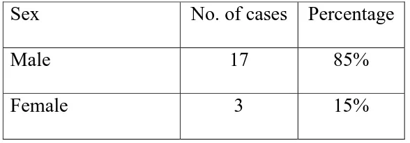
Table: 2 - Sex Distribution:
Among the 20 cases, males were predominant with a male to
female ratio of 9: 1
Sex No. of cases Percentage
Male 17 85%
[image:57.612.170.464.576.679.2]Table: 3- Site of Application
In our study series we took only infected non union of femur
and tibia for analysis as they are more prone for infected non union.
We operated up to 16 cases of infected non-union of femur and
4 cases of Tibia.
Site No. of cases Percentage
Femur 16 80%
Tibia 4 20%
Table: 4. Severity of Infection:
We graded the severity of infection based upon Kulkarni’s
classification of infected non union.
Severity Femur Tibia Percentage
Mild 11 1 60%
[image:58.612.144.492.546.645.2]Table: 5 Side of infected non union
Of the 20 cases, the incidence of non union was more with
femur than with tibia.
Of them the right side was more commonly involved.
Side Side
Right Left Total
Femur 10 6 16
Tibia 3 1 4
Table-6 Number of Infected non union with previous procedures
With implants No implants
Femur 15 1
Tibia 4 0
Out of the 20 cases of infected non union, 19 cases occurred after
previous procedures (18 cases after intramedullary nailing and 1 case
after plating) and in 1 case of infected non-union of femur it
[image:59.612.159.479.451.549.2]Table - 7. Implant used for antibiotic cemented nailing:
Implant
Femur
IL Nail
Femur
K Nail
Tibial
IL nail
No. of cases 2 14 4
IL Nail – Interlocking nail K Nail - Kuntcher Nail
Both proximal and distal lockings was done in all cases in
[image:60.612.176.455.396.528.2]which interlocking nails were used.
Table - 8 Time of Union :
Time of Union No. of cases %
< 4 months 12 60%
4- 6 months 5 25%
> 6 months 3 15%
Table - 9. Patients required Bone grafting
No. of cases Percentage
Femur 10 62.5%
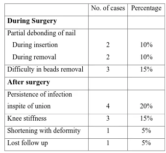
[image:60.612.179.452.595.696.2]Table 10 : Complications
No. of cases Percentage
During Surgery
Partial debonding of nail
During insertion During removal 2 2 10% 10%
Difficulty in beads removal 3 15%
After surgery
Persistence of infection
inspite of union 4 20%
Knee stiffness 3 15%
Shortening with deformity 1 5%
Lost follow up 1 5%
Table – 11 :
Second surgery for persistence of infection In spite of union:
No. of cases
Antibiotic cemented nail
removal
3
Sequestrectomy with nail
removal
[image:61.612.195.441.518.682.2]FOLLOW UP
Post operatively, the patient is kept on appropriate intravenous
antibiotics based on the culture report for one week followed by oral
antibiotics for 6 weeks. Follow up includes complete blood count,
erythrocyte sedimentation rate, C – reactive protein for activity of
infection, and x rays for signs of bony union.
The follow up period was 12-24 months.
Early hip and knee mobilisation exercise started to prevent
post operative knee stiffness on the 1st post operative day.
If interlocking nails is used touchdown weight bearing is
allowed in the 1st post operative day.
Quadriceps setting and straight leg raising exercise are begun
before hospital discharge.
Weight bearing up to 40 lbs is allowed as callus formation
RESULTS AND ANALYSIS
o 85% of patients were male
o 50% of patients were between 25-35 year age group
o Right side was more common
o Femur infected non-union (80%) more reported than Tibia
non-union cases
o Average time interval between primary procedure and
antibiotic cemented nailing was 8 months
o Average time for bone union was 4 ½ months
o Overall percentage of complications was low compared to
other methods
o 60% of patients required bone grafting.
o 95 % of patients went on for union.
Complications :
During Surgery :
1. Partial debonding of nail during insertion – 2 cases
2. Partial debonding of nail during extraction – 2 cases
3. Difficulty in beads removal – 3 cases
After surgery
1. Knee stiffness – 3 cases
2. Persistence of infection in spite of bony union – 4 cases
3. Shortening with equinus deformity - 1 case
DISCUSSION
Antibiotic impregnated cement was first used to treat infection
associated with Hip arthroplasty in the early 1970s. Antibiotic bead
chains were subsequently introduced by Klemn in 1974 and have
been widely used in established bony and soft tissue infections.
Their proposed use as a prophylaxis against infection in introduction
of the antibiotic bead pouch technique has reduced the incidence of
infection in the management of open fractures.
The pathomechanics of infection after plating are different
from those of infection after intramedullary nailing. Infection after
plate osteosynthesis usually involves the fracture site and causes
local sequestration. The infection is primarily extramedullary and
the medullary cavity involvement is limited to the segment where
the plate was. The intramedullary canal proximal and distal to the
infection usually is not involved. Placement of antibiotic beads
delivers, the antibiotics locally in a higher concentration and helps
control the infection. Infection after intramedullary nail fixation
usually involves the entire medullary canal. Infection spreads along
Infection is primarily intramedullary. This is the rationale of
placement of intramedullary antibiotic bead chains.
Placement of an intramedullary antibiotic bead chain from the
nail insertion site, after nail removal, poses some practical
problems. Antibiotic bead chains are difficult to place into the
medullary canal. They are incompatible with external fixators, as
the chain cannot be introduced after pin placement. If placed before
the pins, the chains cannot be removed easily. To overcome this,
Klemn introduced the PMMA stick, which was fabricated by
extruding antibiotic laden PMMA on a continuous monofilament
wire. This was easy to introduce and remove. It could be passed
around the fixator pins and if necessary could be exchanged as a
minor procedure. The main disadvantage of the PMMA stick are the
fact that it does not provide any stability to the fracture, requires
external fixation and has not been evaluated in a prospective trail.
The antibiotic impregnated cement nail is an extension of the
antibiotic PMMA stick used by Klemn. The antibiotic nail is not
only used for the delivery of antibiotics but to provide some stability
to the fracture. The antibiotic cement nail has several advantages
contact with the medullary canal and hence more elution of
antibiotics to the endosteal surface. It can be inserted through the
same portal of entry as the original nail. The nail transverses the
entire medullary canal and enables a more effective delivery of the
antibiotics. It is easy to remove and subsequent exchange nailing is
technically easier than bead chain. The major advantage of this
modality of treatment is that it is a single staged procedure and
avoids systemic toxicity of antibiotics.
Based on our study and the report by Ohtsuka et al, we believe
the antibiotic impregnated acrylic cement nail is an effective means
of treating post intramedullary infection. Although our experience is
limited to a small group of patients, future controlled prospective
CONCLUSION
.
Control of infection and stability to promote union in infected
non-union of femur and tibia has traditionally been provided by
multiple surgical procedures which have proved to be not
efficacious, with poor cooperation and compliance from patients.
However the single staged procedure of antibiotic cemented nailing
has achieved the goals of infection control and fracture union with
good patient compliance. It avoids systemic toxicity of antibiotics,
provides high concentration of local antibiotics and avoids multiple
procedures with good patient compliance.
Hence we conclude that antibiotic cemented rods and
beads could be a relatively effective, simple and inexpensive method
BIBLIOGRAPHY
1. Patzakis MJ, Wilken J, Kumar J et al, Comparison of the
results of bacterial cultures from multiple sites in chronic
osteomyelitis of long bones JBJS Am 1994 ; 76, 664.
2. Perry CR, Pearson RL, Miller GA. Accuracy of cultures of
material from swabbing of the superficial aspect of the
wound and needle biopsy in the preoperative assessment of
osteomyelitis JBJS Am 1991 ; 73 : 745.
3. Hodler NM, Granovelter DA, Phlogistic properties of
bacterial debris. Semin Arthritis Rheum 1978 ; 8 : 1.
4. Klemn K. Die Behandlung chronischer Knochenin.
Fektionen mit Gentamycin – PMMA Kugeln.
Cufallichirurgie, 1977 ; 1 : 20.
5. Klemm K. Indication and Techhik zur, Enlarge von
Gentamicin – PMMA- Kur geln bei Knocher – and
Wechteilin fekten AK tuelle probl Chir Ortho P 1979 : 12.
6. Chapman’s orthopaedic surgery Third edition – volume 3, P
7. Champbell’s operative orthopaedics Tenth edition – volume
3 ; chapter 56 ; P 3125 – 3128, 3134-3137.
8. Henry SL, Ostermann FAW, Seligson D. The antibiotic
bead pouch technique. The management of severe
compound fractures. Clin orthop 1993 ; 295 : 54.
9. Ostermann PAW, Seligson D, Henry S. Local Antibiotic
therapy for severe open fractures. A review of 1085
consecutive cases ; JBJS Br 1995 ; 77 : 93.
10. Buchholz HW, Gartmann HD. Infection prevention and
surgical management of deep insidious infection in total
endoprosthesis (German) chirurg 1972 ; 43 : 446-453.
11. Carlesson AS, Josefsson G, Land berg L. Revision with
gentamicin impregnated cement for deep infectious in total
Hip Arthroplasties JBJS Am. 1978 ; 60 – 1059 – 1064.
12. Klemm K. Gentamicin – PMMA beads in treating bone and
soft tissue infections (German) Zentral bl chir ; 1979 ; 104 :
934 – 942.
13. Klemm K. Treatment of chronic bone infection with
Gentamicin – PMMA chains and beads In. Contzen H, ed.
14. Henry SL, Ostermann PH, Seligson D. The antibiotic bead
pouch technique. The management of severe compound
fractures clin. OrthoP 1993, 295 : 54 – 62.
15. Osterman PH, Henry SL, Seligson D. The role of local
antibiotic therapy in the management of compound
fractures. Clin orthop 1993, 295 : 102 – 111.
16. Henry Sl, Ostermann PA, Seligson D. The prophylactic use
of antibiotic impregnated beads in open fractures. J.
Trauma 1990 ; 30 : 1231-1238.
17. Seligson D, Klemm K. Treatment of infection following
intramedullary nailing. In Browner BD ed. The science and
practice of intramedullary nailing 2nd ed. 1996.
18. Ohtsuka H, Yokoyama K, Higasti K et al use of antibiotic
impregnanted bone cement nail to treat septic non union
after open tibial fractures. J. Trauma 2002 : 52 : 354-366.
19. Seligson D, Popham GJ, voo SK, Henry SL, foglure M.
Antibiotic leaching from PMMA beads. JBJS Am 1993 ; 75
20. Gresser JD, Trantolo DJ, “Bone cement Part -1 Bio
polymer for Avulsive maxillo facial repair” Human
biomaterials applications, Human press Inc., Totowa, NJ.
21. Gresser JD, Trantolo DJ, “Bone cement part-2 –
Biomaterials to restore function in people with physical
disabilities, “Human biomaterials applications, Humana
Press Inc, Totawa, NJ.
22. Polymethacrylates, “Ullmann’s Encyclopedia of industrial
chemistry” vol A21, VCH publishers Inc. 1992.
23. http // www. Bone cement. com.
24. http // www. Immedica. Com
25. Debs, Vazquez B, “Effect of cross linking agents on acrylic
bone cements containing radio opacifiers”, Biomaterials 22
(2001) pp – 2177-2181.
26. Mizumuma H, Hidaka N, Higashitani M et al, physical and
chemical characteristics of Vancomycin hydrochloride.
Antibiotic chemother. 1992 : 8 : 1142 – 50.
27. Bertazzoni Minelli E, Caveiari C, Benini A, Release of
antibiotics from polymethyl methacrylate cement. J.
28. Penner MJ, Masri BA, Duncan CP. Elution characteristics
of Vancomycin and Tobramycin combined in acrylic bone
cement. J. Arthroplasty, 1996, 11 : 939 – 44.
29. Gristina AG, Shibata Y, Giridhar G, Kreger A, Myrik QN.
The glycocalyx, biofilm, microbes and resistant infection,
Semin Arthroplasty, 1994 : 5 : 160-70.
30. Verhayen CC, Dhert WJ, de Blick – Hogervorst J, Van der
Reijden TJ, Petit PL, de Groot K. Adherence to a metal
polymer and composite of staphylococcus aureus and
CONSTITUENTS OF ANTIBIOTIC CEMENTED NAIL AND BEADS
PREPARATION OF ANTIBIOTIC CEMENTED NAIL
DEBRIDEMENT OF MEDULLARY CANAL AND INFECTED NON UNION SITE
INSERTION OF ANTIBIOTIC CEMENTED NAIL IN MEDULLARY CANAL
CASE - 1
32 yr old female pt. with
5 months old infected 4 months post operative non-union right femur
CASE – 2
26 yr old pt with 6 months old 10 weeks of post operative infected non union right femur
CASE – 3
29 yr old male pt. with 8 months 3 months post operative old infected non union right tibia
CASE - 4
31 yr old male patient with
11 months old infected non-union 10 weeks post operative right proximal femur
4 months post operative Full weight bearing of right lower Limb at 10weeks Post operative
CASE – 5
25 yr old pt with 18 months old 9 weeks post operative infected non-union right femur