PREVALENCE OF ENTEROPATHOGENIC
INFECTIONS IN HIV PATIENTS
DISSERTATION SUBMITTED FOR
BRANCH – XII-A - M.D. DEGREE
(DERMATOLOGY, VENEREOLOGY & LEPROSY)
MARCH 2009
THE TAMILNADU
DR.M.G.R. MEDICAL UNIVERSITY
BONAFIDE CERTIFICATE
This is to certify that the dissertation entitled
“PREVALENCE OF ENTEROPATHOGENIC INFECTIONS IN HIV PATIENTS” submitted by Dr. S. UMA to the Tamil Nadu Dr. M.G.R. Medical University, Chennai in partial fulfillment of the
requirement for the award of M.D degree Branch – XII A
(Dermatology, Venereology & Leprosy) is a bonafide research work
carried out by her under direct supervision & guidance.
Dr.S. Krishnan. M.D., D.D. Dr.N. Nagarajan. M.D.,D.D.
Professor & HOD Professor & HOD
Dept. of Dermatology, Dept. of STD,
Madurai Medical College, Madurai Medical College,
DECLARATION
I, Dr. S. UMA declare that, I carried out this work on, “PREVALENCE OF ENTEROPATHOGENIC INFECTIONS IN HIV PATIENTS” at the Department of Sexually Transmitted Diseases, Govt. Rajaji Hospital during the period of June 2007 to August 2008. I also
declare that this bonafide work or a part of this work was not submitted by
me or any others for any award, degree or diploma to any other University,
Board, either in India or abroad.
This is submitted to The Tamilnadu Dr. M. G. R. Medical University,
Chennai in partial fulfillment of the rules and regulations for the M.D.
Degree examination in Dermatology, Venereology & Leprosy.
Place : Madurai Dr. S. UMA
ACKNOWLEDGEMENT
I wish to express my gratitude to the DEAN, Govt. Rajaji
Hospital, Madurai for permitting me to utilize the resources and
materials for the conduct of this study.
At the outset I wish to express my respect and my sincere
gratitude to my beloved teacher Dr. Amalraja. M.D., D.V., Asst.
Professor of Sexually Transmitted Diseases, for the valuable guidance
for this study.
I express my heartful thanks and respect to Dr. N. Nagarajan,
M.D., D.D., Professor and Head of the Department of Sexually
transmitted diseases, Dr. S. Krishnan. M.D., D.D., Professor and Head
of the Department of Dermatology, for their constant encouragement
throughout the study.
I would like to express my sincere gratitude to Dr. T. Uma.
M.D., Professor, Department of Microbiology for her guidance and
valuable suggestions for this study.
I am thankful to Mrs. Katheeja Bi, Lab Technician for her
assistance in laboratory work in this study. I am grateful to my unit
PG’s and CRRI’s for their help in this study. Finally I thank my
CONTENTS
S.No.
TOPIC
PAGE NO.
1.
Introduction
1
2.
Aims & objectives
3
3.
Review of Literature
4
4.
Materials and Methods
17
5.
Results
31
6.
Discussion
42
7.
Summary
51
8.
Conclusion
54
Annexure i)
Bibliography
ii)
Proforma
iii) Master chart
ABBREVIATIONS
WHO
-
World Health Organisation
HIV
-
Human Immuno Deficiency Virus
AIDS
- Acquired Immuno Deficiency Syndrome
CD
-
Cluster Differentiation
ELISA
-
Enzyme Linked Immuno Sorbent Assay
ART
-
Anti Retroviral Therapy
AFS
-
Acid Fast Staining
INTRODUCTION
HIV / AIDS is becoming a major threat to the human population
across the Globe. According to UNAIDS report, globally there were an
estimated 33 million people living with HIV in 2007. There were about
2.7 million new HIV infections and about 2 million AIDS related
deaths in 2007. The rate of new HIV infections has fallen in several
countries, but has increased in developing countries.
Patient infected with HIV suffer from various opportunistic
infections, the most commonly encountered in our setting being
Tuberculosis, Candidiasis, Pneumocystis jiroveci Pneumonia and
diarrhoea due to various pathogens. Intestinal opportunistic infections
present commonly as diarrhoea.
Studies indicate that diarrhoea occurs in 30-60% of AIDS
patients in developed countries and more than 90% in developing
countries.
Chronic diarrhoea is responsible for considerable morbidity and
mortality in such patients. Several species of protozoa and other
bacterial infections have been associated with acute and chronic
Only a few studies regarding the prevalence of intestinal
opportunistic infections in HIV infected patients are available from
South India at present.
I took up this study in our institution, to evaluate the prevalence
of such infections in HIV patients in our setup and to ascertain the
importance of stool examination for the detection of enteropathogens
that aids in treatment, thereby decreasing the morbidity and mortality
AIM OF THE STUDY
1. To study the prevalence of enteropathogenic infections in HIV
positive patients.
2. To study the correlation between CD4 and the prevalence of
intestinal parasites.
3. To study the association between the gastrointestinal
REVIEW OF LITERATURE
India has the dubious distinction of having the largest number of
people living with Human immuno deficiency virus in the world.(1)
with an adult prevalence rate of 0.91% (2).
People with advanced HIV induced immuno suppression are
vulnerable to infections called “Opportunistic infections” because they
take advantage of the opportunity offered by weakened immune
system. Since the beginning of the HIV epidemic, opportunistic
infections have been recognized as common complications of HIV
infections. (6) (7) (8) Opportunistic infections cause substantial morbidity
and hospitalization, necessitate toxic and expensive therapies, and
shorten the survival of people with HIV infection (9,10).
The relative frequencies of specific opportunistic diseases may
vary in different countries and even in different areas within the same
country. (12). The identification of such pathogens is very important for
HIV & AIDS case management.
Diarrhoea is one of the most common presenting complaints in
symptomatic HIV infected individuals. The infectious aetiological
chronic or frequent gastro intestinal disease and non-opportunistic
agents, that usually cause acute, treatable diarrhoeal illness. (3). It is
associated with a 3.3 fold increased risk of disease progression. (3)
Chronic diarrhoea, defined as persistence of diarrhoea beyond
four weeks (4) is a common symptom in HIV infected patients in the
tropics. The World Health Organisation (WHO) defines diarrhoea
wasting syndrome along with a positive HIV serology test to be an
AIDS defining illness. (5).
Opportunistic parasitic gut infections cause severe diarrhoea and
profoundly compromise the absorptive function of the small intestine,
leading to significant mortality.(13)
Protozoan parasites, namely Cryptosporidium parvum, Isospora
belli, Cyclospora cayetanensis, Microsporidia, Entamoeba histolytica /
Entamoeba dispar, and Giardia lamblia account for a significant
number of cases of diarrhoea in this population(14). They are described
in detail below. Giardiasis is not more frequent or severe than in HIV
Cryptosporidium (spp) :
This small, coccidian parasite was considered a veterinary
pathogen but not an important human pathogen until the early 1980’s,
when it was recognized in patients with AIDS. Subsequently, it has
become recognized as a major cause of diarrhoeal illness both in
immunocompromised and immunocompetent individuals. (18)
Only two species are commonly seen in humans,
Cryptosporidium hominis, which has anthroponotic transmission and
Cryptosporidium parvum, which is seen in humans mainly in outbreak
settings, and has a zoonotic transmission cycle. Other species are
rarely seen in immunocompetent individuals, but multiple species have
been described in HIV infected individuals in developing countries.
Cryptosporidium hominis causes acute watery diarrhoea in
immuno competent individuals and protracted life threatening
diarrhoea in immuno deficient individuals. This is considered an AIDS
defining illness. Recently, following multiple out breaks of diarrhoea,
including one affecting over 4,00,000 people following contamination
of water supplies, it has also been described as a water borne pathogen,
This coccidian has a worldwide geographical distribution with
increased incidence in developing countries. Prevalence rates of over
12% are seen in asymptomatic individuals. Transmission can be by
faeco-oral route, water borne, due to contact with farm animals and by
drinking unpasteurized milk. (31)
The incubation period ranges from 1 to 14 days. Typically acute
infection is characterized by watery diarrhoea, crampy epigastric
abdominal pain, weight loss, anorexia, malaise and flatulence. (21) (22)
Spectrum varies from asymptomatic shedding to chronic
diarrhoea depending on the CD4 count. Median CD4 count of
individuals who develop chronic severe diarrhoea is well under 50
cells / mm3. (21, 22)
All patients with CD4 count > 200 cells / mm3 are able to clear
cryptosporidial enteritis. (23, 36)
A subset of patients with AIDS, cryptosporidiosis develop biliary
tract involvement associated with right upper quadrant pain, nausea &
vomiting (22, 24). This is a difficult to treat complication in individuals
with exceedingly low CD4 count and long standing chronic
The most common site of cryptosporidia infection is the small
intestine, although it is frequently present in the colon and the biliary
tract of persons with immunodeficiency. It infects only the epithelial
surface of the mucosa and it does not invade the submucous layer or
cause ulcerations. (23) (26) (27).
Lab Diagnosis :
The stage seen in faeces is the oocyst which measures 4-6 µm
and contains 4 fully developed and infectious sporozoites. 80% are
thick walled and 20% thin walled.
Oocysts stain with modified acid fast stain and auramine phenol
stains, appearing bright orange red or fluorescing golden yellow under
ultraviolet illumination. Other stains used include methanamine silver,
Giemsa and periodic acid Schiff stains. Polyclonal and monoclonal
antibody conjugates with flourescein isothiocyanate for direct
identification of oocysts in stool are also available commercially.
Identification of antibodies to cryptosporidium measures exposure of
the population to this parasite, but is not useful in diagnosis. (28).
A large number of studies on treatment of this infection have not
included antimotility agents, Paromomycin, Spiramycin and
Azithromycin (29).
Recently, Nitazoxanide has been recommended as specific
therapy for cryptosporidiosis but appears effective only in immuno
competent patients. Anti retroviral treatment resulting in restoration of
immune function results in prevention and resolution of disease.(30)
Somatostatin analogues especially octreotide are found to be useful in
the treatment of Cryptosporidial diarrhoea.
Isospora belli :
First described in 1860 by Virchow, this diarrhoeal disease
pathogen is a host specific coccidian protozoan parasite. There is no
evidence that the human Isospora species is capable of infecting non
human hosts or that animal species affect humans (31).
Infection with this opportunistic parasite is considered an AIDS
defining illness if present for over 4 weeks.(5). It has a world wide
geographical distribution.
Transmission may be by faeco-oral, water borne or sexual routes.
The incubation period ranges from 1-4 days and can be upto 1 week.
The clinical features of infections with Isospora belli are diarrhoea,
Diagnosis of infection is by examination of stool samples.
Oocysts can be demonstrated in wet preparation and stained with
modified acid fast stains and direct fluorescent stains. Duodenal
aspirates can also be examined. Small bowel biopsies may show
developmental stages of the parasite.(31)
Oocysts are the diagnostic forms of the parasite found in humans.
They are elliptical in shape and measure 20-33 µm by 10-19 µm.
Each oocyst contains two sporoblasts, each of which contains
four sporozoites.
Treatment with trimethoprim and sulphamethoxazole for 10 days
have been shown to eradicate infection, although there is a 50% risk of
relapse if treatment is not fully completed. Pyrimethamine can also be
used. (17).
Cyclospora cayetanensis :
This organism was first described in humans in Papua New
Guinea in 1979. It is a coccidian parasite of the genus Cyclospora. (31).
This parasite has a world wide distribution. Transmission by
contaminated water supplies has been shown to be responsible for out
breaks in many settings, and it is likely that this is also transmitted by
Ingestion of the mature oocyst results in watery diarrhoea,
weight loss, abdominal pain, fatigue, anorexia, bloating, flatulence and
fever. The illness may be prolonged, lasting 2-6 weeks. It is chronic
and severe if untreated especially in patients with AIDS (31).
The life cycle is incompletely described. The unsporulated
oocyst is shed in the faeces and upto 40% of these sporulate in 1-2
weeks. The oocyst is round, 8-9 µm in diameter similar to
cryptosporidia but larger, when examined under ultraviolet
illumination, exhibiting a blue fluorescence. (33).
Diagnosis of infection is by identification of the oocysts in wet
preparations and by staining with modified acid fast stains as for
cryptosporidium. It does not stain with monoclonal antibodies specific
for cryptosporidium. They have also been observed in duodenal
aspirates and small bowel biopsies by electron microscopy (31).
Microsporidia :
The first human case was reported in 1959.
Five main species that affect humans are enterocytozoon,
encephalitozoon, microsporium, pleistophora and Nosema. They are
mainly seen in immunocompromised hosts, although some
Ingestion with microsporidia may be
i) Latent asymptomatic or chronic mild symptomatic in adults
with normal immunity.
ii) Acute, potentially fatal in neonates.
iii) Proliferative disease in the absence of competent host
defences.
The route of transmission is not known, although infection by
aerosols or ingestion from animals and social partners have been
proposed as potential modes.
Laboratory diagnosis of these infections is by demonstration of
microsporidia in infected tissue or body fluids. Stool specimens can be
examined by the modified trichrome stain, chemifluorescent stain or
the Giemsa stain on concentrated specimens.
Microsporidia can also be identified in tissue specimens by
electron microscopy. Immunoflurescence using polyclonal antibodies
has also been done. ELISA, Indirect immuno fluorescence have been
shown to be useful for antibody detection.(34).
Albendazole is the most promising agent. The drug is
Prophylaxis is indicated in any HIV infected patient with CD4 counts
less than 200 cells / mm3 (31)
HIV and other Helminths
The most common pathogenic helminth associated with AIDS is
Strongyloides stercoralis. Other helminths reported were Ascaris
lumbricoides,Trichuris trichura, Ankylostoma duodenale, Schistosoma
mansoni , Hymenolepsis nana.
Strongyloides stercoralis
A number of early case reports documented disseminated
strongyloidiasis in HIV infected patients, but later studies seemed to
indicate that the risk of disseminated disease was not associated with
HIV related immuno deficiency. This has been proposed to be due to
the association of declining CD4 counts with lower larval maturation
in the gut. This would diminish the risk of autoinfection which in turn
is required for hyperinfection (34).
However, possible immune reconstitution disease with
Strongyloides stercoralis hyperinfection is now being reported in
patients started on anti retroviral therapy. Symptoms may have been
because immune recovery facilitated dissemination of
stongyloidiasis(35).
Entamoeba histolytica
Entamoeba histolytica in the immuno compromised patients is
believed to be usually a commensal belonging to nonpathogenic
zymodemes (16).
Intestinal infection is quite common among homosexuals, the
incidence is 20-30%. Presence of trophozoites in fresh stools is
pathognomonic.
Clinical presentation of the patients is similar to that reported in
non HIV patients.
Blastocystis hominis
This pathogen is also frequently identified in human stools, but
the role of this anaerobic protozoan as a human pathogen is
controversial and as yet unresolved (17).
Ascaris lumbricoides :
Co-infection of HIV with roundworms is particularly important.
Adult round worms reside in the small intestine, but larvae migrate
Ascaris polarizes the immune response in young adults to Th2,
which increases the risk of sexual transmission of HIV.
Ascaris also suppresses IL-2, a Th1 cytokine that can be used for
AIDS / HIV because it improves count of CD4 T cells and restores
immune function substantially.
Bacterial entero pathogenic Infections in HIV / AIDS
Bacteria may be responsible for secondary infection of the gastro
intestinal tract. Infections with enteric pathogens such as Salmonella,
Shigella and Campylobacter are more common in homosexual men
and are often more severe and more apt to relapse in patients with HIV
infection. Patients with untreated HIV have approximately a 20-fold
increased risk of infection with Salmonella typhimurium.
They may present with a variety of non specific symptoms
including fever, anorexia, fatigue and malaise of several weeks
duration. Diarrhoea is common but may be absent. Diagnosis is made
by culture of blood and stool (37).
HIV infected patients also have an increased incidence of
Salmonella typhi infection in areas of the world where typhoid is a
intestinal disease in HIV infected individuals and upto 50% of patients
will develop bacteremia (34).
Campylobacter infections occur with an increased frequency in
patients with HIV infections. While Campylobacter jejuni is the strain
most frequently isolated. Infections with many other strains have been
reported. Patients usually present with crampy abdominal pain, fever
and bloody diarrhoea.
Infection may present as proctitis. Stool examination reveals the
presence of faecal leucocytes. Systemic infection can occur, with up to
10% of infected patients exhibiting “bacteremia”. Most strains are
MATERIALS AND METHODS
Setting :
The study was conducted in both in-patients and out-patients of
Govt. Rajaji hospital.
Collaborating Departments :
i) Institute of microbiology, Madurai Medical College.
ii) Anti retroviral therapy centre / Out patients
Design of Study :
Prospective analytical study
Study period :
June 2007 to August 2008, a period of 15 months
Sample size :
100 consecutive HIV positive patients were included irrespective
of the symptomatology, CD4 count and ART status.
Ethical Clearance :
Obtained
Consent :
Selection Criteria :
Inclusion criteria :
i) All men and women who were positive for HIV by two rapid
tests and ELISA who presented with or without diarrhoea
were included in the study.
ii) All patients were above 15 years of age.
Exclusion Criteria :
1. Patients under 15 years of age
2. Endocrine disorders
3. Malignancy
4. Collagen vascular disorders
5. Other organ disorders
6. Patients on steroid therapy
7. Pregnancy
8. Protein losing enteropathy
9. Crohn’s disease
10. Irritable bowel syndrome
11. Unconscious / Bedridden
12. Non co-operative and non-willing
13. Patients who were on antidiarrhoeal or on antimotility drugs
14. Patients using liquid paraffin / laxatives
15. Renal disorders
Materials :
Data Collection :
Socio demographic and clinical data were collected.
- CD4 count was done in all the patients
- General investigations were done, and physician’s
opinion to rule out the other potential causes of
gastrointestinal disturbances was obtained for each
patient.
Patients were explained about the proposed study and asked to
collect stool samples. Stool samples were collected in two containers.
One containing glycerol phosphate buffer and other containing formol
saline. For suspected cases of cholera, stool samples were sent in
Venkatraman Ramakrishnan Medium. Stool samples were subjected to
the following methods for evaluation of parasites
1. Wet saline method
2. Wet Iodine method
3. Floatation technique
4. Sedimentation technique
5. Modified acid fast technique
Collection of stool :
For stool sample collection essential criteria followed were
1. Fresh stool samples
2. Receptacle was kept clean and without antiseptics
3. Patients were instructed not to mix urine with stool
Samples were taken to the microbiology department, within 30
minutes.
Preparation of Materials :
Two smear preparations, one unstained preparation and another
stained with Lugol’s Iodine were made.
a) Wet Saline method :
Minute portion of faeces is diluted with normal saline 0.9% and a
drop of it is taken on a clean microscopic glass slide. A
coverslip No. 1 or No. 0 is then gently put over it to spread out
the emulsion. Unstained preparation is specially useful for
demonstration of actively motile forms of parasites like
b) Wet Iodine Mount :
A drop of Lugol’s Iodine is added to minute portion of faeces on
a clean microscopic glass slide. A cover slip is placed and
examined under microscope. This method helps in the
visualization of the cysts of protozoa.
c) Floatation technique :
Faecal material was dissolved in a medium of higher density than
the eggs and cysts of parasites. The eggs and cysts that float to
the top are collected by placing a glass slide on the surface of the
meniscus at the top of the tube.
d) Sedimentation techniques :
Faecal material was dissolved in a solution of density below that
of eggs. So eggs will be concentrated at the bottom.
e) Modified Acid Fast staining Method :
It was adopted to detect cryptosporidiosis and isosporidiosis.
Preparation of smear :
1) With Diamond pencil lab numbers were written in full. One
glass slide was used for each specimen.
2) Using 5mm internal diameter 24 SWG Nichrome wireloop
smears were prepared with a portion of thickest part on about
2/3rd of slide
3) Slides were air-dried
4) The slides were transferred to hot plate 80C and were fixed
for 10 mts.
Staining : ‘Kinyoun’ staining
This staining technique was adopted for Acid Fast Staining method
1. The slides were placed on the staining rack with the smear
part upper most, the slides not touching each other.
2. The slides were flooded with carbol fuchsin stain. The slides
were heated till carbol fuchsin starts steaming. More carbol
fuchsin is added to prevent the slides from drying. The slides
were allowed to stain for 9 minutes.
3. The slides were washed well with running tap water.
4. Slides were decolorized by covering completely with 1%
5. Slides were washed again gently with running tap water.
6. Slides were counter stained with 1% methylene blue for one
minute.
7. Slides were washed as before with water and the slides were
dried with filter paper and the smears were read.
Cryptosporidium appeared as round or oval shaped pink coloured
oocysts measuring 4-6 µm.
Isospordium appeared as mature or immature oocysts measuring
20-40 µm
Staining for
microsporidia
: Strong Trichrome stain
Faeces fixed in 10% formalin were used for a minimum of
one hour for staining microsporidia.
1. A suspension of faeces in 10% formalin (approx. 1 : 1) was
prepared. A thin smear was made along a microscope slide
using an orange stick. Some thick areas were made at one end
of the slide. The slides were air dried and fixed in methanol for
5 minutes.
2. Slides were stained for 10 minutes in strong Trichrome stain
which has been pre-heated to 500 C, or 1 hour in cold strong
3. Rinsed briefly in acid alcohol, for 1-2 seconds. Rinsed briefly
in 95% ethanol for 1-2 seconds.
4. Slides were dehydrated in 95% ethanol for 5 minutes and in
absolute alcohol for 5 minutes.
5. Microsporidia stain strong pink and measure 1.5 to 2.0 micro
meter with a clear vacuole and membrane fold.
Stool Culture for Entero pathogenic bacteria :
For enteric pathogens culture was done on Nutrient agar and Mac
conkey agar plates. Further identification was done by biochemical
reactions.
Analysis :
Data were entered in Microsoft Excel Spreadsheet and were
analysed using statistical package.
Conflict of interest : Nil
Financial support : Nil
Limitations of the Study :
1) Infective status of the parasitic infestations were not supported
by serological studies
3) Therapeutic outcome of the patients with intestinal parasitosis
were not studied.
4) Post ART follow up of the patients receiving ART was not
analysed in relation to non ART patients.
The results of similar studies conducted across the Indian subcontinent were as follows :
Kava Mohandas et al. Dept of Parasitology and Dept. of Int. Medicine, PGI Chandigarh. Prevalence of Intestinal parasitic pathogen in HIV positive patients in Northern India. Jpn, J. Infect. 2002. Dis : 55 : 83-84.
Of 120 HIV positive patients 30% were found to harbour an
intestinal parasite. Cryptosporidium parvum was the most common
(10.8%), followed by Giardia lamblia (8.31%). Cyclospora
cayetanensis and Blastocystis hominis each were detected in 3.3% of
the patients, while Isospora belli and Enterocytozoon bieneusi were
each detected in 2.5% of the patients. The other parasite observed
were Entamoeba histolytica in two cases and hook worm ova in one
Gupta et al. Indian Journal of Medical Microbiology(2008) 26(2) : 172-5 (All India Institute of Medical Sciences)
Out of 113 HIV positive patients, enteric parasites were detected
in 55.8% with diarrhoea compared to 16.4% in patients without
diarrhoea.
Isospora belli was found in 41.1% of chronic diarrhoea, 6.3% in
non diarrhoeal cases. Cryptosporidium was detected in 20.6% of
chronic diarrhoea and 2.5% in non diarrhoeal cases. Cyclospora
cayetanensis associated diarrhoea was detected in only one case of
chronic diarrhoea (2.9%)
The mean CD4 T cell count was lower (180cells/µl) in diarrhoeal
patients as compared to non diarrhoeal patients(261.3cells/µl).
Coccidian parasites were seen at a mean CD4 T cell count of 186.3
cells / sec. This study concluded that Isospora belli was the
predominant parasite followed by cryptosporidium and both were
strongly associated with diarrhoea among HIV patients.
Satheesh Kumar, S. Ananthan et al. Dept. of Microbiology, Dr. ALM PG Institute of Basic Medical Sciences, Chennai, India.
During may 2000 to Jan 2001, 152 stool samples from (43 with
HIV seropositive individuals were examined for enteric coccidian and
other intestinal parasites by microscopy and special staining methods.
A total of 52 enteric parasites, 15 from patients with acute
diarrhoea and 24 from patients with chronic diarrhoea, 7 from patients
infected with HIV without diarrhoea and 6 from normal individuals
without diarrhoea.
Isospora belli was detected in 13.7% with acute and chronic
diarrhoea. The association with diarrhoea among HIV positive
individuals was significant. Cryptosporidium was detected in 7 patients
each with acute and chronic diarrhoea. It was also detected in 4
patients with HIV infection without diarrhoea. Hence its association
with diarrhoea among HIV patients was found to be not significant in
the present study. Cyclospora and microsporidia each were detected
in only one HIV positive patient with chronic diarrhoea.
The study revealed that the coccidian parasites are one of the
etiological agents of diarrhoea especially of chronic diarrhoea among
HIV positive patients.
Isospora belli was found to be a frequent enteric parasite
1. Nilanjan Chakraborty, Anirban Mukherjee, et al, ICMR,
Virus unit, Kolkata, ID & BG HDS petal, Kolkata, India. Jpn. J. Infect. Dis, 61, 49-53; 2008. In this study of 125 patients, Enteropathogenic vibrio (47%), cryptosporidial diarrhoea (43%).
Escherichia coli infection (42%).
Results of similar studies in Other countries :
Mora CA, Altieri R, Davario M, Lasala M.B. Division of infectious disease, J.M. San martin Hospital school of medicine, Buenos Aires Argentina. 11th International AIDS conference, Int.
Conf. AIDS, 1996. July 7-12 : 11: 296 Abstract No 4624.
According to the study out of 376 HIV positive patients who
were followed from 1-1-1987 to 12-3-1993, parasites were found in
171 patients of which Entamoeba histolytica were found in 48 patients
(12.77%). Escherichia coli were found in 46 patients (12.23%),
Giardia were found in 39 patients (10.37%). Dientamoeba Fragilis in
23 patients (6%), cryptosporidiosis were found in 11 patients (3%) and
Isospora belli in 4 patients (1%). There is significant correlation
between cryptosporidiosis and diarrhoea when CD4 count is below 200
Alijandro Carabello, Indhira Ozozco, Bol.Chil Parasitol Intestinal parasitic infection in HIV positive individuals in south eastern Venezuela.
According to this study cryptosporidiosis were found in 22.8%,
Ascaris lumbricoides in 14.2%, Hookworm in 8.6% and Trichuris
trichuria in 8.6%. Isospora belli in 2.9% According to them 56.5%
cryptosporidiosis is found and CD4 count < 200 cells/mm3 and 32%
between 200-500 cells/mm3 and 12% when CD4 count above 500
cells / mm3 Entamoeba histolytica were found in 28.9% when CD4 <
200 cells/mm3 and 17.4% when CD4 count between 200-500 cells/mm3
and 53.7% when CD4 count above 500 cells/mm3.
AR Meamar et al, Iranian J. Parasitol : vol. 2, No.1 2007, pp 1-6. Dept of Parasitology and Mycology, Faculty of Medicine, Iran University of Medical Sciences and Health services, Tehran, Iran.
A total of 781 HIV + / AIDS patients were submitted to copro
parasitological examination from 2003 to 2005. 191 individuals were
at AIDS stage. The prevalence of intestinal parasites was 11.4%.
The prevalence of infection for each helminith and pathogenic
Blastocystis hominis - 6.1%
Giardia lamblia - 4.2%
Cryptosporidium Spp - 0.9 %
Isospora belli - 0.26 %
Strongyloides stercoralis - 0.26%
Hymenolepsis nana - 0.13 %
Rhabditis axei - 0.13%
All cases of Cryptosporidium spp and Isospora belli with clinical
picture of severe diarrhoea were exclusively found in those HIV
positive patients who were at AIDS stage.
In all patients with Cryptosporidium Spp, the mean number of
CD4 lymphocyte was 50.1 + 8.8/mm3, whereas in 2 patients infected
R E S U L T S
Symptomatology & Sex Distribution : Table – 1 With Gastro
intestinal symptoms
Without Gastro intestinal symptoms
Total
Males 36 18 54
Females 25 21 46
Total 61 39 100
Out of 100 patients, 54 were males and 46 were females
Out of 54 males 66 %were symptomatic and out of 46 females
54% were symptomatic.
The symptoms included diarrhoea, vomiting, abdominal pain,
flatulence, dyspepsia etc.
Marital Status :
Among the 100 patients, 95 patients were married. Only 5
patients were unmarried. Spouses of 62 patients were also HIV
positive by ELISA and were under follow up. 34.7% of the couples
Sexual Behaviour
96% of men had history of promiscuous sexual behaviour and
4% of men and all women denied history of promiscuity.
Among men, 7.8% had history of homosexual behaviour.
Anogenital sex was the mode adopted by these homosexuals.
Distribution of cases in relation to age
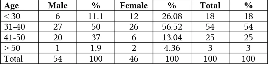
Out of 100 HIV positive patients, 72% were below the age group
of 40. The age range varied from 25-55 years. The mean age was 40.
There was no significant difference among gender with reference to the
[image:38.595.80.526.459.566.2]age. The results are tabulated below.
Table - 2
Age Male % Female % Total %
< 30 6 11.1 12 26.08 18 18
31-40 27 50 26 56.52 54 54
41-50 20 37 6 13.04 25 25
> 50 1 1.9 2 4.36 3 3
Total 54 100 46 100 100 100
Occupation :
Various occupational categories were noted. Most of the males
were drivers and agricultural workers. Others were load men, skilled
Socio-economic Status :
95% of our patients belonged to the low socio economic status.
The remaining were of moderate socio-economic status.
Educational Status
The educational status was classified into those who completed
their primary school education, high school, higher secondary,
graduates, technical education and illiterates.
[image:39.595.81.523.387.535.2]The following were the observation.
Table - 3
Occupation Males Females
Illiterates 10 10
Primary school 12 8
High school 14 14
Higher secondary 10 13
Graduates 5 1
Technical 3
-Total 54 46
Domicile Pattern :
Table - 4
Male Female Total
Rural 32 24 56
Urban 22 22 44
[image:39.595.83.520.662.736.2]Out of 100 patients, 44 patients were from urban areas in around
Madurai and 56 patients were from rural side mostly from villages
around Madurai.
57.2% of males and 42.8% of females were from rural areas and
42.8% of males and 57.2%of females were from urban areas.
Water sources :
All patients were dependent on protected water supplied by the
local bodies. The knowledge about using boiled water was very poor
in illiterate patients when compared with the literate.
Hand washing :
Hand washing habits and other hygiene practices were far from
satisfactory among our patients, especially among the illiterates.
Table - 5
CD4 > 500 CD4 201-499 CD4 < 200
Oral candidiasis 1 4 56
Tuberculosis (Pulmonary
and extra pulmonary)
- 3 18
Pneumocystis jiroveci
pneumonia
- 1 8
Oral hairy leukoplakia - 1 28
Oral candidiasis was the predominant opportunistic infection
seen in our patients.
Staging of the Disease :
The staging was done based on the WHO staging for the year
[image:41.595.85.536.79.238.2]2007 and are tabulated as follows.
Table - 6
Stage Male % Female % Total
I 10 18.5 11 23.9 21
II - -
-III 27 50 20 43.4 47
IV 17 31.5 15 32.6 32
Total 54 46 100
Prevalence of Other STDs among the study group
Table – 7
Male Female Total
Genital herpes 6 6 12
Anogenital warts 2 3 5
Candidiasis 2 5 7
Latent syphilis - 3 3
Bacterial vaginosis - 2 2
Pelvic inflammatory disease - 3 3
Genital herpes was the most common STD in both males and
females.
PREVALENCE OF ENTERO PATHOGENS FROM OUR STUDY
Prevalence of Enteric pathogens from our study Sole isolates :
Cryptosporidium - 33
Entamoeba histolytica - 3
Escherichia coli - 1
Isospora belli - 3
Ankylostoma duodenale - 1
Microsporidia - 1
Cryptosporidia + Entamoeba histolytica - 8
Cryptosporidia + Escherichia coli - 9
Cryptosporidia + Ankylostoma duodenale - 1
Cryptosporidia + Klebsiella - 1
Cryptosporidia+ Enteamoeba histolytica+Klebsiella –1
Cryptosporidia + Escherichia coli + Ankylostoma
duodenale - 1
Giardia lamblia + Entamoeba histolytica - 1
Escherichia coli + Escherichia histolytica - 1
Percentage prevalence of enteric pathogens
% of patients harbouring enteric pathogens - 65%
Sole isolates :
Cryptosporidium - 50.76%
Isospora belli - 4.62%
Microsporidia - 1.54%
Entamoemba histolytica- 4.62%
Ankylostoma duodenale- 1.54%
Escherichia coli - 1.54 %
Table – 8
Enteric pathogens in relation to symptoms With
gastrointestinal symptoms
Without gastrointestinal
symptoms
Total %
Cryptosporidium 27 ( 41.53%) 6 (9.23%) 50.76 %
Isospora 2 (3.08%) 1 (1.54%) 4.62%
Microsporidia 1 (1.54%) - 1.54%
Entamoeba
histolytica
1 (1.54%) 2(3.08%) 4.62%
Ankylostoma
duodenale
1 (1.54%) - 1.54%
Escherichia Coli 1 (1.54%) - 1.54%
Mixed Infections 19 (29.23%) 4 (6.15%) 35.38%
Table – 9
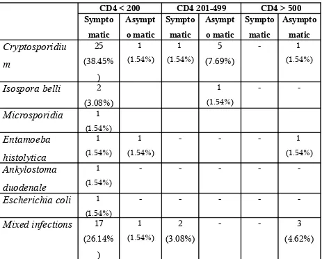
Pathogens in relation to CD4 Count
CD4 < 200 CD4 201-499 CD4 > 500
Sympto matic Asympt o matic Sympto matic Asympt o matic Sympto matic Asympto matic Cryptosporidiu m 25 (38.45% ) 1 (1.54%) 1 (1.54%) 5 (7.69%) - 1 (1.54%)
Isospora belli 2 (3.08%) 1 (1.54%) - -Microsporidia 1 (1.54%) Entamoeba histolytica 1 (1.54%) 1 (1.54%)
- - - 1
(1.54%) Ankylostoma duodenale 1 (1.54%) - - - -
-Escherichia coli 1 (1.54%)
- - - -
-Mixed infections 17 (26.14% ) 1 (1.54%) 2 (3.08%)
- - 3
DISCUSSION
Enteric pathogens are among the most common opportunistic
infections and are a major cause of morbidity and mortality in HIV
positive individuals world wide.
The prevalence of enteric pathogens shows wide geographic
variations and their isolation and treatment carries importance while
treating HIV infected patients especially in advanced stage of immuno
suppression.
A clear knowledge of the prevalence of various enteric pathogens
in a particular area would be of immense help in choosing empirical
antimicrobial regimens in resource poor settings.
In the present study enteric pathogens were recovered from 65%
of patients. Study conducted by Kava Mohandass et al (14) at PGI
Chandigarh whose sample size closely matched our study, showed
only 30% prevalence of enteric pathogens, when compared with an
alarmingly high prevalence in our study (65%). The CD4 count and
prevalence of pathogens in relation to CD4 count or stage of the
disease has not been mentioned in their study. The probable
were in stage III & IV of the disease, which could account for the
increased prevalence of enteric pathogens in our patients.
Variations in socio economic status, personal hygiene and quality
of water supply may also have a role in the disparity.
The study conducted by Gupta et al(39) from All India Institute of
Medical Sciences showed a prevalence of intestinal parasites in about
28.3% of 113 patients with HIV. CD4 count was done only for 48
patients due to financial constraints. The prevalence in this study again
did not match our study probably due to same reasons mentioned
earlier.
The study conducted by Kumar S et al(43) at Chennai in 152 HIV
positive patients showed a prevalence of 34.21% of enteric parasites,
which is also very low compared to our studies, despite geographic
proximity to our centre. This study also does not mention the CD4
count of the patients included.
Studies conducted in Iran by AR Meamar et al(40) showed a
prevalence of only 11.4% and studies conducted by Morra et al(41) from
Argentina showed a prevalence of 45.4% when compared with our
availability of treatment facilities would account for the gross variation
between different countries.
80% of our patients, who harboured an enteric parasite had
gastro intestinal symptoms like diarrhoea, vomiting, nausea, belching,
flatulence, colicky abdominal pain. Diarrhoea was the predominant
symptoms seen in all patients. A similar finding was also recorded in
the other studies.
Kava Mohandass et al(14) also showed a similar picture where
75% of patients with an enteric pathogen had diarrhoea.
Gupta et al(39) showed that 55.8% of patients with enteric
pathogens were symptomatic, the percentage was comparatively lower
than our study.
Kumar et al(43) showed 72% of patients to be symptomatic which
also closely correlated with our study.
Studies from abroad did not mention about the symptomatology,
or their prevalence with relation to CD4 count.
The most prevalent pathogen isolated in our study was
cryptosporidium species. Out of 100 patients screened cryptosporidium
was the sole pathogen isolated in 33 patients and in 21 patients it was
50.76%. This is by far the highest prevalence reported so far in studies
conducted across the world.
The study conducted by Nilanjan et al(42) showed a prevalence of
43% of cryptosporidial diarrhoea in a sample size of 125. It was the
most common entero pathogen isolated as in our study, and the
percentage prevalence also closely matched our study.
Kava Mohandas et al(14) reported cryptosporidium as the
commonest pathogen isolated with a prevalence of 10.8% which was
very less when compared with our study, even though both the studies
showed cryptosporidium as the predominant pathogen.
Gupta et al(39) reported Isospora belli to be the predominant
pathogen with a prevalence of 41.1% with chronic diarrhoea and 6.3%
in non diarrhoeal cases. Cryptosporidium was detected in 20.6% of
chronic diarrhoea and 25% of non diarrhoeal cases.
Kumar et al(43) reported isospora to be the most prevalent
pathogen (13.7%). Cryptosporidium was present in 7% patients.
Though closely related in sample size and geographic area there is a
gross difference between the species isolated and the percentage of
prevalence. This is intriguing, and simultaneous, larger, multicentric
AR Meamer et al(40) Iran showed a very low prevalence of
cryptosporidia of about 0.9%. Mora et al(41) Buenos Airs, Argentina
showed a prevalence of cryptosporidia in 3% of patients. In both these
studies cryptosporidium was not the predominant pathogen and the
prevalence was very low.
Alijandro carabellow et al(40) showed a prevalence of
cryptosporidium to be 22.8% (South Eastern Venezuela)
cryptosporidium was the predominant pathogen isolated, though in a
lower percentage prevalence.
The type of coccidian parasite isolated differed in different
geographic regions, majority of the studies showed cyrptosporidia to
be the most common pathogen isolated, as in our study. However, the
percentage varied. Our study showed the highest prevalence.
Though the isolation of cryptosporidia was seen in 33 of our
patients (50.76%), Isospora was found only in 3 of our patients
(4.62%), which was comparable with studies by Kava Mohandass et
al(14) (2.5%) but studies by Anandan et al(43) revealed higher percentage
of (13.7%) of isospora and itwas the predominant pathogen isolated in
Gupta et al(39) showed a 16.8% of isospora and again it was the
predominant pathogen isolated in their study which was not correlating
with our study.
AR Meamar et al(40) reported 0.26% of isospora. The probable
explanation that could be offered for this disparity may be as follows.
An over whelming 79% of our study population were on cotrimoxazole
prophylaxis, started fairly early in the course of the disease, as per the
WHO guidelines, irrespective of their CD4 counts, which could have
offered protection against this coccidian parasite. The percentage of
cotrimoxazole prophylaxis in the other studies was not available for
comparison.
Microsporidia was isolated in only one patient (1.54%). Study
by Kumar et al(43) showed a prevalence of 0.65% in their study. The
prevalence closely correlated with our study, whereas other studies did
not isolate microsporidia at all.
Other protozoans isolated in our study was Entamoeba
histolytica (4.62%) Ankylostoma duodenale (4.61%). Giardia lamblia
Kava Mohandas et al(14) showed a higher prevalence of Giardia
lamblia (8.3%) and a lower prevalence of Entamoeba histolytica
(1.6%) when compared with our study.
Kumar et al(43) did not isolate any of these protozoans in their
study.
Gupta et al(39) showed a prevalence of Giardia lamblia 2.6% and
Entamoeba histolytica 0.88%. They isolated strongyloides
stercocaralis in one patient which was not found in our study.
AR Meamear et al(40) showed a prevalence of Giardia lamblia in
4.2% which was higher when compared with our study.
Strogyloides stercoralis was isolated in 0.26% and
Hymenolepsis nana in 0.13% which were not isolated in our study.
Mora et al(40) showed Entamoeba histolytica in 12.77% which
was lower and Giardia in 10.37% which was higher than our
prevalence. Their study also isolated Dietamoeba fragilis in 6% of
patients which was not isolated in our patients.
E. coli was the most common bacterium isolated with prevalence
of 1.54% as the sole isolate. Klebsiella was isolated in 2 patients as a
Nilanjan chakraborty et al(42) reported enteropathogenic vibrio
(47%) as the most common bacterium isolated. The probable reason
for isolating vibrios as the predominant pathogen was that the study
was conducted in Kolkata which is known to be endemic for cholera.
E.coli was reported in 42% which was very high when compared to our
study. The other Indian studies have not made a mention about the
bacterial isolates in their study. Studies abroad by Mora et al(41) showed
a prevalence of E.coli in 12.33% which closely correlated with our
study.
Mixed infections were isolated in 23 patients (35.38%).
Cryptosporidium was the predominant pathogen isolated in the mixed
infections.
Out of 23 patients 18 (78.2%) had CD4 count < 200. 82.6% of
patients with mixed infections were symptomatic. These features
highlight the importance of immune system in clearing the enteric
pathogens and advanced immuno suppression provides a favourable
environment for mixed enteric pathogens.
None of the studies mentioned across the country and abroad did
Majority of enteric pathogens were isolated in patients with CD4
count < 200 / mm3 . The statistics is shown in the table and most of the
patients with low CD4 count were symptomatic.
Gupta et al(39) showed a similar finding as in our study. CD4 T
cell count was lower (180 cells/mm3) in diarrhoeal HIV patients as
compared to non diarrhoeal patients(261.3 cells / mm3). Coccidian
SUMMARY
Entero pathogenic infections cause significant morbidity &
Mortality in HIV patients especially in advanced states of immuno
suppression.
Socio economic conditions, educational status, unhygienic
practices contribute for intestinal parasitosis.
There are wide geographic variations in the prevalence of entero
pathogenic infections due to variations in the standards of living,
hygiene practices, availability of safe drinking water and literacy rate.
Since large number of our patients belonged to low poverty line,
an attempt was made to study the prevalence of enteric pathogens in
our population.
A total of 100 consecutive HIV positive patients (54 males ; 46
females) were selected. After thorough clinical examination and
investigations to rule out other systemic illnesses, their stools were
examined for the gastrointestinal pathogens.
Enteric pathogens were isolated in 65% of our patients.
Cryptosporidium was the most common pathogen isolated which
The other pathogens isolated were Isospora belli (4.62%),
Microsporidia (1.54%) Entamoeba histolytica (4.62%), Ankylostoma
duodenale (1.54%), Escherichia coli (1.54%), Mixed infections in
35.38%.
The prevalence of infections was high in patients with CD4
count < 200 / mm3 . Most of the patients with a lower CD4 count were
symptomatic.
The isolation of Isospora belli was very less. It may be explained
by the cotrimoxazole prophylaxis that is offered to our patients. There
are wide geographic variations among the prevalence of enteric
pathogens even in areas which are in close proximity to us, in city like
Chennai.
Therefore large multicentric studies need to be conducted in wide
geographic areas to assess the prevalence of such parasites, so that
appropriate therapeutic strategies can be planned for the treatment of
such patients.
Since personal hygiene and good standards of living are essential
to prevent acquiring these parasites, proper health education regarding
hand washing habits, drinking boiled water needs to be emphasized to
HAART is mandatory as boosting up the immunity leads to
spontaneous clearance of most of the enteric pathogens especially the
CONCLUSION
The prevalence of enteropathogenic infections in 100 conseutive
HIV positive individuals were studied and the following conclusions
were arrived at.
1. Enteric pathogens were isolated in 65% of our study population,
and the coccidian parasites were the most common gastro
intestinal pathogens isolated.
2. Cryptosporidium is the most common enteropathogen isolated.
3. Isospora was isolated only in three of our patients.
4. Escherichia coli was the most common bacterial isolate.
5. Cotrimoxazole prophylaxis appears to offer a significant
protection against isospora infection.
6. The prevalence of enteropathogens varies significantly between
different centres, despite geographic proximity.
7. 80% of patients who harboured enteropathogens were
8. Mixed infections were commonly seen in profoundly immuno
suppressed patients. This signifies that advanced immuno
suppression paves way for mixed infections.
9. Patients with advanced illness were more symptomatic than those
in the early stages.
10.Enteric pathogens were isolated predominantly when the CD4
BIBLIOGRAPHY
1. UNAIDS – AIDS Epidemic update : 2007– Available from :
http : // www. Data. Unaids.org
2. HIV / AIDS epidemiological surveillance & estimation report
for the year 2005, NACO, April 2006. Available from :
http : // www. naco online. Org.
3. Smith PD, Lane HL, Gill VG, Manilchewitz JF, Quinnan GV,
fauci AS, et al. Intestinal infections in patients with AIDS:
Etiology and respose to therapy. Ann. Intern Med. 1988 ; 108
; 328 – 33.
4. Thomas PD, Forbes A, Green J, Howdle P, Long R, Playford
R, et al. Guidelines for the investigation of chronic diarrhoea,
2nd edition. Gut 2003 ; 52 : 11-15.
5. WHO case definitions of HIV for surveillance and revised
clinical stain and immunological classification of HIV related
disease in adults aged 15 years or older. SEARO publications
on HIV / AIDS : 2006. Available from : http : // www.
6. Kanabus, A. Fredrikson- Bass, J and Noble R (2006) : HIV
related opportunistic infection : prevention treatment, AIDS –
care – watch 3, 1-11.
7. Centres for Disease, control (1982) : Update on acquired
immune deficiency syndrome (AIDS) – United states morb.
Mortal. Wkly Rep., 31, 507-514.
8. Selix, R.M., Haverkis, H.W. and Curren, J.W. (1984).
Acquired immune deficiency syndrome (AIDS) Trends in the
United States, 1978-1982, Am. J. Med., 76, 493-500.
9. Moore, R.D. and Chassion, R.E. (1996) : Natural history of
opportunistic disease in an HIV – infected urban clinical
cohort. Ann. Intern Med., 124, 633-642.
10. Finkelstein, D.M., Willilams, P.I. Molenbergs, G., et al,
Pattern of opportunistic infections in patients with HIV
infection. J. Acquire Immuno Deficiency syndrome. Hum
Retrovirol., 12, 38-45.
11. Stein, D.S., Korvick, J.A. and Vermun, S.H., (1992) : CD4 +
Lymphocytes cell numeration for prediction of clinical course
of human immuno deficiency virus disease : a review J.
12. Kaplan, J.E., Hk, D.J., Holmes, KK et al (1996) : Preventing
opportunisitic infections in human immunodeficiency virus
infected persons : implications for the developing world. Am.
J. Trop. Med. 149, 55, 1-11.
13. Escobedo AA, Nunez FA, Prevalence of intestinal parasites
en Cuban AIDS patients Acta Trop. 1999 ; 72 : 125-30.
14. Mohandas K, Sehgal R, Sud A, Malla N. Prevalence of
intestinal parasitic pathogens in HIV seropostive individuals
in Northern India. Jpn J. infect Dis. 2002 ; 55 : 83-4.
15. Janoff En, Smith PD (1988) Perspectives on gastrointestinal
infections in AIDS. Gastroenteral clin North Am 17 : 451-63.
16. Allason – Jones E, Mindel A, Sargeaunt P, Katz D (1988)
Outcome of untreated infection with Entamoeba histolytica in
homosexual men with and without HIV antibody Brit. Med. J
297 : 654-7.
17. Kain KC, Keystones JS (1995). Intestinal infection with other
protozoa. In : Infections of the gastro intestinal tract Eds
18. Gatei W et al (2002). Zoonotic species of cryptosporidium
are as prevalent as the anthroponotic in HIV infected patients
in Thailand. Ann Trop Med Paraseitol 96 : 797-802.
19. Muthusamy et al (2006). Multi locus genotypes of
cryptosporidium spp. Isolates from HIV infected individuals
in South India. J. clin Microbial 44 : 632 -4.
20. Mckenzie WR et al (1994). A massive outbreak in Milwaukee
of cryptosporidium infection transmitted through the public
water supply. J. Engl I Med. 331 : 161-167.
21. Good game RW. Understanding intestinal spore forming
protozoa : Cryptosporidia, Microsporidia, Isospora, Ann.
Intern Med. 124 : 429, 1996.
22. Hashmey & Smith NH et al. Cryptosporidiosis in Texas a
report of 95 cases. Medicine 76 : 118, 199A.
23. Flanigan et al. Cryptosporidium infection and CD4,
T lymphocyte count. Ann Intern Med. 116 : 840, 1992.
24. Ducreux et al. Diagnosis and prognosis of AIDS related
25. Vakil et al, Biliary cryptosporidium in HIV infected people
after the water borne out break of cryotosporidiosis in
milvaukee. N Engl J. Med. 334 : 19, 1996.
26. Kotter. D. Francisco et al. Small intestinal injury and
parasitic diseases in AIDS. Ann Inter Med 113 : 444, 1990.
27. Kotter D. Francisco et al. Effects of enteric parasitosis and
HIV infection upon small intestinal structure and function in
patients with AIDS, J. Clin Gastro enterol 16 : 10, 1993.
28. Casemore DP (1991) ACP Broad sheet 128 : Laboratory
methods for diagnosis cryptosporidiosis J Clin pathol 44 :
445 – 451.
29. Adal et al (1995) Cryptosporidium related species In :
Infections of the gastrointestinal tract. Raven Press, New
York, PP 1107-1128.
30. Abubakar I, et al. Treatment of cryptosporidium in
immunocompromised individuals. Systematic review and
meta analysis. Br. J. Clin Pharmacol 63 : 387-98.
31. Gegandeep Kang ; CMC Vellore. J. Clin Microbiology 2006,
32. Casemore DP (1994) Cyclospora : another ‘new’ pathogen J
Med. Microbiol 41 : 217-219.
33. Ortega et al. Cyclospora species (1993) new protozoan
pathogen of humans N. Engl. J. Med 328 : 1308-1312.
34. Viney et al (2004) why does HIV infection not lead to
disseminated strongyloidiasis. J Infect Dis. 190 : 2175-2180.
35. Lawn S., Wilkinson RJ 2006. Immune reconstitution disease
associated with parasitic infections following antiretro viral
treatment. Parasit Immunol 28 : 625-633.
36. Vaj payee M, et al I, Spectrum of opportunistic infections and
profile of CD4+ counts among AIDS patients in North India
Infection 2003 ; 32 : 336-40.
37. Kaplan Je et al : Guidelines for preventing oppourtunistic
infections among HIV infected persons – 2002.
Recommendations of the US Public Health Service and the
Infectious Diseases Society of America. MMWR Recomm
Rep. 51 (RR-8) : 1, 2003.
38. Chaisson RE, Gallant JE, Keruly JC, Moore RD. Impact of
opportunistic disease on Survival in patients with HIV
39. Gupta et al – Chronic diarrhoea in HIV patients : Prevalence
of coccidian parasites. Indian Journal of Medical
Microbiology, (2008) 26 (2) : 172-5.
40. AR Meamar et al – A comparative analysis of intestinal
parasitic infections between HIV + / AIDS patients and non
HIV infected individuals. Iranian J. Parasitol : Vol. 2, No.1,
2007, pp1-6.
41. Mora et al, 11th International AIDS conference 1996, July
7-12 : 11 : 296, Abstract no. 4624.
42. Nilanjan Chakraborty et al, ICMR, Kolkatta ; India, Current
trends of opportunistic infections among HIV Sero positive
JM. J. Infect Dis, 61, 49-53 ; 2008. Patients from Eastern
India.
43. Kumar et al, Role of coccidian parasites in causation of
diarrhoea in HIV infected patients at Chennai. Indian J. Med.
Res. 116, September 2002, pp 85-89.
44. Alijandra Carabello et al, Intestinal parasitic infection in HIV
positive individuals in South Eastern Venezuela.
PROFORMA
ENTEROPATHOGENS IN HIV
1. S. No. :
2. MVD / FVD No. :
3. Name :
4. Age / Sex :
5. Occupation :
6. Education :
7. Socio-economic status :
8. Marital status : Married / Unmarried
Divorced / Separated Widow / Widower
9. High risk behaviour :
a) Age at 1st intercourse :
b) Promiscuous / Non promiscuous: Premarital / extra marital
c) No.of partners :
d) Protected / Unprotected :
e) Hetero sexual : Anogenital / Genito genital /Orogenital
f) Homosexual : Anogenital / Orogenital
Active / Passive / both
10. Others : Alcoholism
IVDA
11. Date of diagnosis as HIV positive :
Age at Diagnosis :
Purpose for which HIV testing was done :
12. Blood VDRL :
13. Other STD’s :
14. CD4 count :
15. WHO staging :
16. Presenting complaints :
17. Positive clinical findings:
18. Motion Smear : Wet Mount :
Modified AFB staining :
Motion culture :
19. USG :
20. Other Investigations :
21. Other opportunisitic Infections :
22. Treatment given :
PCP prophylaxis : Yes / No
ART :
Others :
MASTER CHART ABBREVIATIONS
P Positive Neg Negative
UM Un married NR Non Reactive
C Cryptosporidium E Escherichia Coli
EH Entamoeba histolytica Anky Ankylostoma duodenale
Kleb Klebsiella Gia Giardia Lamblia
I Isospora belli WNL With in normal limits
A Absent P Present
OC Oropharyngeal candidiasis PT Pulmonary tuberculosis PCP Pneumocystis Carinii pneumonia GW Genital Warts
VVC Vulvo vaginal candidiasis BP Balano posthitis
GH Genital herpes Ade Adenitis
BV Bacterial Vaginosis PID Pelvic inflammatory disease CRF Chronic renal failure PA/LN Para aortic lymphadenopathy MPC Muco purulent cervicitis SpM Splenomegaly