COMPARATIVE STUDY OF CLONIDINE AND
DEXMEDETOMIDINE AS ADJUNCTS TO ROPIVACAINE IN CAUDAL ANALGESIA IN CHILDREN
DISSERTATION SUBMITTED FOR DOCTOR OF MEDICINE
BRANCH X (ANAESTHESIOLOGY) APRIL -2013
THE TAMILNADU DR MGR MEDICAL UNIVERSITY
CHENNAI
BONAFIDE CERTIFICATE
This is to certify that this dissertation entitled
“COMPARATIVE STUDY OF CLONIDINE AND DEXMEDETOMIDINE
AS ADJUNCTS TO ROPIVACAINE IN CAUDAL ANALGESIA IN
CHILDREN” is a bonafide record work done by Dr.KASIRAJAN.G under my direct supervision and guidance, submitted to The Tamil Nadu Dr. M.G.R. Medical University in partial fulfillment of University regulation for MD, Branch X –Anaesthesiology.
PROF. Dr. S. C. GANESH PRABU, M.D, D.A,
Director,
Institute
Of
Anaesthesiology,
Madurai Medical College &
Govt . Rajaji Hospital,
DECLARATION
I Dr.KASIRAJAN.G declare that the dissertation titled
“COMPARATIVE STUDY OF CLONIDINE AND DEXMEDETOMIDINE
AS ADJUNCTS TO ROPIVACAINE IN CAUDAL ANALGESIA IN
CHILDREN” has been done by me. This is submitted to The Tamil Nadu Dr. M.G.R. Medical University, Chennai, in partial fulfilment of the requirement for the award of M.D. Degree, Branch X–Anaesthesiology degree Examination to be held in April 2013. I also declare that this dissertation, in part or full was not submitted by me or any other to any other University or Board, either in India or abroad for any award, degree or diploma.
Place: Madurai
ACKNOWLEDGEMENT
I am deeply indebted to Prof. Dr. S. C. GANESHPRABU, MD., DA.,
Director & Institue of Anaesthesiology, Madurai Medical College, Madurai for his able guidance, inspiration and encouragement he rendered at every stage of this study.
I express my heartful gratitude to Prof. Dr. T.Thirunavukkarasu,
MD., DA., Prof. Dr. R. Shanmugam, MD., Prof.Dr.A. Paramasivan,
MD.,DA., and Prof. DR. Evlyn Asirvatham, M.D, DGO., Professors of
anaesthesiology, for their able guidance in doing this study.
My profound thanks to Dr. N. Mohan, M.S., Dean, Madurai Medical
College,Govt Rajaji Hospital and Dr.Swaminathan, M.S., Medical
Superintendent, Government Rajaji Hospital, Madurai for permitting me to
utilize the clinical materials of this hospital in the completion of my dissertation.
I express my profound thanks to assistant professor
Dr. S. Vijayaragavan, M.D., D.A., for his valuable assistance and technical
guidance in doing this study.
I am also thankful to my other assistant professors and my post graduate colleagues of Institute of anaesthesiology and department of Pediatric surgery for their kind co-operation in doing this study.
CONTENTS
S. No TITLE Page No.
1. INTRODUCTION 1
2. HISTORY 4
3. AIM OF THE STUDY 5
4. ANATOMY RELATED TO CAUDAL BLOCK 6 5. PHYSIOLOGICAL CONSIDERATION 16 6. APPLIED PHARMACOLOGY
A. PHARMACOLOGY OF RPOIVACAINE B. PHARMACOLOGY OF Α2 ADRENORECEPTORS C. PHARMACOLOGY OF CLONIDINE
D. PHARMACOLOGY OF DEXMEDETOMIDINE
22 29
30
38
7. REVIEW OF LITERATURE 51
8. MATERIALS AND METHODS 56
9. STASTISTICAL ANALYSIS 61
10. OBSERVATION AND RESULTS 62
11. DISCUSSION 76
12. SUMMARY 81
13. CONCLUSION 82
1
1.
INTRODUCTION
Pain as an unpleasant sensory and emotional experience associated with actual or potential tissue damage or described in terms of such damage.
Pain is an unpleasant subjective sensation which can only be experienced and not expressed, especially in children who would seem to conceal their feelings when suffering from pain. The primary reason to treat or prevent pain is humanitarian and this becomes even more important in children who rely completely on their parents or care givers for their well being.
Acute pain is associated with a brief episode of tissue damage or inflammation such as that caused by trauma or surgery. In most of the cases the intensity of the pain diminishes steadily over a period of time. The various methods of pain relief have their own disadvantages which prohibit their use in children for eg: narcotics in children because of their respiratory depression and other analgesics which cannot be given for sometime after general anaesthesia due to the fear of vomiting and aspiration, the objection to the needles in the case of parenterally administered analgesics.
2
In this study caudal block was done after the induction of general anaesthesia and was used as an adjunct to intraoperative anesthesia as well as postoperative analgesia in children undergoing surgical procedures below the level of the umbilicus. Caudal analgesia reduces the amount of inhaled and intravenous anaesthetic drug requirement attenuates the stress response to surgery, facilitates a rapid, smooth recovery and provides good postoperative analgesia. In order to decrease intra operative and postoperative analgesic requirements and to prolong duration of analgesia after single shot caudal epidural blockade, various additives such as morphine, fentanyl, clonidine and ketamine with local anaesthetics have been studied.
Ropivacaine is a new long acting amino amide local anaesthetic agent. It is structurally related to bupivacaine and has been used for pediatric caudal analgesia. It provides pain relief with less motor blockade. Literature suggests that ropivacaine is less cardiotoxic than bupivacaine, hence ropivacaine may be a more suitable agent for caudal epidural analgesia especially in day care surgery.
Dexmedetomidine is a α2 adrenergic receptor agonist. It has increased
affinity to α2 adrenergic receptors than clonidine and less α1 adrenergic receptor
actions. The main advantage of the dexmedetomidine is it higher selectivity were compared in Clonidine for α2A receptor is responsible for analgesic,
3
The objective of this study was to compare of the analgesic action of clonidine and dexmedetomidine combined with ropivacaine in caudal analgesia for children undergoing infra umblical surgical procedures.
4
2. HISTORY
1901 – SICARD and CATHELIN described epidural injection through sacral hiatus
1933 – CAMBEL M.F first described sacral epidural block in children and infants
1957 – Synthesis of Ropivacaine.
1960 - Clonidine hydrochloride was introduced.
1965 - Melzock and Walts propounded the Gate Control Theory of pain.
1974 - KAY B used caudal block for post operative pain relief in children.
1992 - Ropivacaine was introduced in clinical practice. 1999 - Dexmedetomidinewas introduced in clinical practice.
5
3. AIM OF THE STUDY
1. To assess and compare the efficacy of dexmedetomidine and clonidine used as adjuncts to ropivacaine in caudal analgesia for children.
6
4. ANATOMY RELATED TO CAUDAL BLOCK
Anatomy of sacrum
Sacrum is a large wedge shaped bone. It is formed by fusion of the five sacral vertebrae. It articulates above with 5th lumbar vertebra and below with the coccyx .It has an anteriorly concave and posteriorly convex surface. The anterior surface bears four transverse lines which terminate on each side in four anterior sacral foramina. The anterior primary rami of the first four sacral nerves emerge from the anterior sacral foramina. The posterior surface is convex and in midline runs a bony ridge, the median sacral crest with 3 or4 rudimentary spinous processes. The lamina of the 5th and sometimes the 4th sacral vertebra fuse fail to fuse in the midline and thus the deficiency, formed is known as
“sacral hiatus”. The lateral margins of this space each bear a prominence called
“sacral cornu” which represents the inferior articular process of the 5th sacral
vertebra. (Figure1)
Sacral canal
Figure 1: Anatomy of sacrum and hiatus
[image:12.595.169.453.108.417.2]7
continuous with the lumbar epidural space. Its lower limit is the sacral hiatus closed by posterior sacrococcygeal membrane. Fibrous bands may be present in the canal and divide the epidural space into loculi which prevent the spread of solution and these may account for occasional incomplete anaesthesia.
Contents of the sacral canal
Sacral and coccygeal nerve roots with their dorsal root ganglia. The filum terminale which is the continuation of piameter.
Epidural plexus of veins formed by the lower end of vertebral veins. These vessels are numerous in the anterior aspect than the posteriorly.
Loose areolar and fatty tissue, which is more dense in males than in females.
Sacral hiatus
8
When local anaesthetic is injected into the sacral canal, it ascends upwards in the sacral epidural space for a distance proportional to the volume of the solution, force of injection, amount of leakage through the eight sacral foramina and the connective tissue in the space.
CAUDAL ANAESTHESIA
Selection of equipment:
Reliability of the technique and the incidence of complications largely depend on the characteristics of the needle used. The four important characteristics of the needle are – bevel, internal &external diameter, length, presence of the stylet.
Sharp bevelled needle:
Advantage – traverse easily through the tissues
Disadvantage
¾ Characteristic “give way” while puncturing sacrococcygeal membrane may not be clearly felt with sharp needles.
¾ Sharp needles have long bevel which may have to be advanced further into the epidural space so that it lies entirely within it.
¾ Cartilaginous sacrum can be easily traversed by a sharp and long
bevelled needle that may lead to rectal puncture or iliac vessel puncture.
9
Diameter:
Small needles may bend & break during procedure 21G, 22G, 23G are ideal because it is rigid and large enough to allow reflux of blood or CSF.
Length:
Proximity of dural sac makes it dangerous to use very long needles. The distance between skin and epidural space is almost always less than 20mm even in adults. So it is not advisable to use a needle longer than 30mm.Needle with stylet if used prevents formation of an epidermoid tumour due to skin tag. Epidural needle with 20G, 21G and 22G are employed when one intends to use an epidural catheter via caudal route to achieve anaesthesia at higher level after radiographic confirmation.
Determination of the volume of local anaesthetics:
Height of block – depends on the volume injected Formula based on weight/ age:
Armitage formula
High sacral – 0.5 ml/kg High lumbar – 1ml/kg Thoracic level – 1.25ml/kg
10
Weight is the best predictor. To calculate the total volume to be injected. Volume required in ml = 0.65 X number of segments to be blocked x body weight (kg)
Spiegal formula:
Total volume of injection (ml)= 4+(D-15)/2,Where D is the distance between from the spinous process of 7th cervical vertebra to the sacral hiatus in cm.
Modified Spiegal formula:
Volume of injection (ml) = 4+(D-13)/2
Despite larger volumes of local anaesthetics used in children as compared to adults, peak plasma levels of the local anaesthetics in children remain far below the toxic levels than in adults. As the child grows space become less compliant and hence large volume can cause high spread of solution and an increase in the CSF concentration.
Maximum volume recommended for injection is 20ml.
Patient position:
Three positions can be used for caudal anesthesia; 1. Prone position - Most often chosen in adults.
11
prone position and the landmarks are more easily palpable than in adults.
3. Knee-chest position – This is infrequently used.
Anatomical landmarks:
Classically hiatus is described as the apex of the triangle formed by joining the two posterior superior iliac spine and tip of the coccyx. (fig 2&3).
The point of puncture is at the midpoint of this triangular space. Intergluteal fold is not an ideal landmark because it will not always correspond to the midline. Left forefinger is placed in coccyx tip,the hiatus corresponds to the second crease of the finger palpation of this membrane gives a characteristic feel of a membrane under tension similar to that of a fontanelle.
TECHNIQUE
Figure 2 caudal technique.
[image:18.595.129.453.110.399.2]
12
CONFIRMATION OF SPACE
Whoosh test:
It is done by injecting air via the needle and another person should auscultate just proximal to the injection site. If the needle is correctly positioned in the caudal space, then the characteristic whoosh sound is heard when air is pushed.
Swoosh test
If the needle is correctly positioned in the caudal space, while injecting local anaesthetics, Swoosh sound is heard at a site just proximal to the hiatus.
It is suggested to avoid injection of air in children as it may cause patchy block and in a rare complication of pneumocephalus ,venous air embolism may also occur.
Other techniques commonly used to identify the space are:
• Easy injection of drug • No resistance to injection • No subcutaneous bulge
INJECTION OF DRUG:
13
Transient increase in intracranial pressure with transient loss of consciousness and headache can occur .On the other hand, too slow an injection increases the chance of lateralisation of the block or a lower level of anaesthesia as the drug tends to leak through the foramina and increases the risk of needle displacement.
INDICATIONS:
Ideal for lower abdominal and lower limb surgeries
Emergency: testicular torsion, strangulated hernia repair, paraphimosis, wound debridement of pelvis/ lower limbs.
Elective:
Repair of inguinal hernia, umbilical hernia, hydrocele, Orchidopexy, anorectal, genitourinary surgeries, pelvic, hip, phimosis and lowerlimb surgeries.
CONTRAINDICATIONS:
a) Hydrocephalus. b) Seizure disorders.
c) Vertebral osteosynthesis. d) Local skin infection. e) Pilonidal sinus near hiatus.
14
COMPLICATIONS
Possibilites due to error in the technique:
1. Subcutaneous injection.
2. Vascular puncture: 10-15%. Since epidural veins are valveless, injection may be immediately followed by convulsions, arrhythmias, hypotension and respiratory depression.
3. Dural puncture: If dura is punctured, withdraw the needle immediately. Second attempt can be made provided the drug is injected slowly under low pressure.
4. Subarachnoid injection may lead to total spinal even along cranial nerve distribution.
5. Bone marrow, rectal and intraosseous injection
6. Complete or partial failure of the block More common in > 7 years old. 7. Lateralization: occurs in 1 in 1000 cases When caudal is performed in
lateral decubitus, 50% have a level of anaesthesia 2 dermatomes higher on the dependent side Slow injection may cause a difference of more than 4 dermatomes ; may be due to presence of a complete plica mediana dorsalis.
8. Un anaesthetised dermatomes.
15
10. Neurological complications Urinary retention – most common if narcotics are used in caudal. Time to micturate may be delayed but not troublesome
11. Nerve injury is rare.
12. Other complications Vomiting, epidural infection , meningitis, shivering.
16
5.
PHYSIOLOGICAL CONSIDERATION
Classification of sensory fibers
Sensory fibres Speed of transmission Sensory function Myelination
C fibres 0.5 -2m/sec Pain,cold, heat, touch. Unmyelinated
A-Alpha
fibres 70 -120m/sec
Noxious chemical thermal, mechanical
stimuli Myelinated
A-Beta fibres 30 -70m/sec
Light touch,
pressures,vibration
proprioception Myelinated A-Gamma
fibres 30-70m/sec
Proprioception, Motor to muscle spindle
Myelinated
A-Delta fibres 12 -30 m/sec Pain, cold, touch Myelinated
B fibres 3 -15 m/sec
Pre ganglionic autonomic (sympathetic)
Myelinated
PAIN PATHWAYS
17
A delta conducted pain is felt quickly and is well localised. ‘C’ fibres are very fine non-myelinated fibres which conduct at a very slow rate 2-3m/sec or less. Their threshold for stimulation is higher and is responsible for delayed and truly noxious burning or throbbing pain.
The activation of two different type of fibres (A delta &C) by noxious stimuli explains the double sensation for pain evoked in the human by a single short noxious stimulus, rapid pricking pain(0.1sec, latency, fast pain) carried by A delta fibres is followed approximately one second later by a burning pain (slow pain) mediated by C fibres.
Peripheral sensory nerves have their cell bodies in the dorsal root ganglion and the central projection of A delta and C fibre neurons enter the dorsal horn in the lateral division of the dorsal root.
[image:24.595.173.459.460.669.2]
18
In the grey matter of the spinal cord cell bodies are arranged in a
series of laminae, some of which have classical names but which are more
simply given roman numerals by Rexed, starting with I at the tip of the
dorsal horn. A delta and C primary afferent fibres terminate principally in
the marginal layer (lamina I) and the substantia gelatinosa (lamina II) of
the spinal cord (fig.4). Some of the neurons of the lamina I which synapse
with A delta fibres, give off axons which ascend in the contralateral
anterior columns without synapsing with neurons from deeper layers. The
majority of the pain fibres, however synapse in the substantia gelatinosa
into intermediate neurons which sent projections to deeper layers or with
the dendrites of neurons whose cell bodies reside in deeper layers,
principally in lamina V.
The central projection from cell bodies in lamina IV, V & VI with contribution from lamina I, cross midline in the anterior commissure to form the spinothalamic tract, which ends in thalamus, principally in the ventroposterior nucleus, sending a few fibres enroute to the periaqueductal grey matter. The ventroposterior nucleus of the thalamus projects to the post central gyrus, the sensory cortex.
19
Spinothalamic Pathway
While it appears that the thalamus is involved in the experience of
pain, the post-central gyrus is necessary for its accurate localization and prefrontal cortex for unpleasant affective reactions to it.
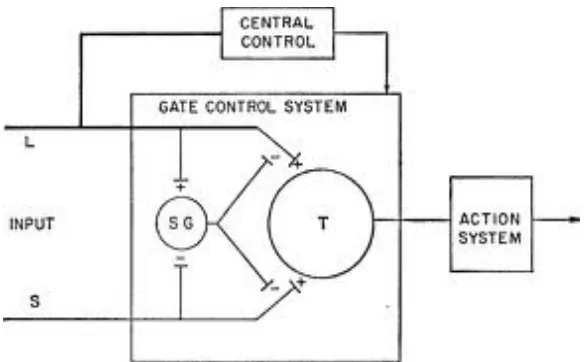
INHIBITORY PATHWAYS:
1. Large primary afferent fibres, which mainly ascend in the dorsal columns and whose cell bodies lie in the dorsal root ganglion send collaterals to synapse with and activate inhibitory inter neurons in the dorsal horn. These inturn inhibit release of transmitters along pain pathways. Thus, stimulation of large A beta cutaneous afferents may inhibit pain transmission (gate theory of Melzack and Wall).
20
PHYSIOLOGICAL CHANGES
21
[image:28.595.150.442.74.255.2]
Figure 5: Gate control theory.
L.glutamate is the only excitatory aminoacid which is concentrated in the dorsal horn. There are a number of receptors for glutamate namely NMDA (N methly D aspartate), AMPA (α amino3 hydroxy 5 methyl- isoxazole 4 propionate), L-2 amino-4-phospono butyrate(LAP4), kainite and metabotropic receptors. The majority of the opioid receptors in the dorsal horn are mu receptors although delta and kappa receptors are present.
22
6. APPLIED PHARMACOLOGY
A. PHARMACOLOGY OF ROPIVACAINE
Ropivacaine is the new amino amide local anaesthetic. It is a derivative of pipecoloxylidide. Pipecoloxylidide was first synthesized in 1957. Pipecoloxylidide are chiral drugs due to asymmetric carbon atoms and form two groups of S and R enantiomers. Ropivacaine is a pure S enantiomer with chiral purity of 99.5%. Ropivacaine is prepared by alkylation of S enantiomer of dibenzoy-l-tartaric acid
PHYSIOCHEMICAL PROPERTIES:
Chemical name as S-1-propyl-2,6- pipecoloxylidide hydrochloride monohydrate. (Figure 6)
23
Molecular weight - 274
Pka - 8.07
pH - 7.4
Protein binding - 94% Partition coefficient (lipid solubility) - 8.7 Mean uptake ratio - 94
T1/2 - 111 minutes, Clearance - 10.3 L/minutes. Solubility in H2O at 250C - 53.8g/L
Specific gravity - 1.002 to 1.005 at 25oC.
MECHANISM OF ACTION:
Ropivacaine acts through inhibition of sodium channel. It inhibits the conduction of sodium ions through the channel and also potassium channel. Thus it blocks the generation and conduction of impulses across the nerve fibres. This type of block is reversible.
PHARMACOKINETIC PROPERTIES:
Absorption
24
the slow absorption from the epidural phase. Thus it has longer duration of action through the epidural route.
Distribution
The steady state plasma concentration after intravascular injection is about 59+7 liters in total volume of distribution. The protein bound fraction is about 94%. It mainly binds to α–1- acid glycoprotein. There is an increase in bound form of drug in post operative state due to increase in α-1-acid glycoprotein from stress response in surgery. This is especially so after continuous epidural infusion. Ropivacaine can easily cross the placenta and equilibrium is reached.
Metabolism
25
Elimination
Ropivacaine is mainly eliminated through the kidney as various metabolites.About 86% of the total drug is excreted through the kidneys. The total clearance is about 387 ml/min. The mean half life is about 1.8 hours after intravascular injection and about 4.2 hours after epidural injection.
PHARMACODYNAMIC PROPERTIES:
Action on Nervous system
The type of blockade produced by ropivacaine depends upon the concentration of drug used. In low concentration it blocks both Aδ and C fibres which is more potent than that of equal concentration of bupivacaine. In high concentration, the blockade of Aδ fibres’ is less than that of bupivacaine while the blockade of C fibres is similar. The penetration of ropivacaine into myelin sheath is less due to low lipid solubility compared to bupivacaine. Thus it preferentially blocks C fibres than Aδ fibres. This causes less potent motor blockade.
The addition of epinephrine does not influence the type of blockade produced. In toxic doses, it causes initial excitation of nervous system manifesting as restlessness, tremor, and convulsions. Later it leads to depression of medullary centre and coma.
Effect on Cardiovascular system
26
Effect on respiratory system
Ropivacaine does not have any marked effect on the respiratory system in normal doses. Higher doses leading to toxicity of drug produces respiratory depression secondary to medullary depressant effect.
INDICATIONS
Surgical anaesthesia:
a) Spinal anaesthesia. Epidural anaesthesia. Caudal anaesthesia.
Peripheral nerve block and infiltration anaesthesia.
Pain management:
a) Labour analgesia – intermittent bolus or continous infusion a. more in walking epidurals
Post operative pain management – epidural infusion as i. Intermittent bolus.
ii. Continous infusion.
iii. Patient controlled analagesia.
Pain management in paediatrics:
a) Caudal epidural block.
27
CONTRAINDICATIONS
a) Known cases of allergic reactions to amide type of local anaesthetics. b) Intravenous regional anaesthesia (Bier’s block).
c) Obstetric Para cervical anaesthesia. d) Hemodynamic instability.
e) Septicemia.
f) Local site infection.
ADVERSE EFFECTS
The adverse reactions to ropivacaine are related to excessive plasma levels due to excess dosage, unintentional intravascular injection or slow metabolic degradation. The mean doses of plasma level when toxicity begin to occur are about 4.3 and 0.6 μg/ ml of total and free plasma concentrations respectively. The toxic levels are reached in cases of continuous epidural infusion as the drug is administered for long times.
THE VARIOUS POSSIBLE SIDE EFFECTS :
g) Cardiovascular System – bradycardia, hypotension, vasovagal reaction, syncope, arrhythmias. Due to low lipid solubility the cardiotoxic potential is less than that seen with bupivacaine.
h) Central and peripheral nervous System – dyskinesia, hypokinesia, neuropathy, vertigo, tremors, paresis, neropathy and coma.
28 k) Hepato - Biliary System – jaundice l) Musculoskeletal System – myalgia.
m) Psychiatric Disorders - agitation, confusion, nervousness, amnesia, hallucination, emotional liability, insomnia, nightmares.
n) Skin Disorders - rash, urticaria.
o) Urinary System Disorders- urinary incontinence, micturition disorder. p) Vascular - deep vein thrombosis, phlebitis, pulmonary embolism.
AVAILABILITY
Ropivacaine is available in ampoules of isobaric solution in concentration of 0.2%, 0.5%, 0.75% and 1%. The solutions are prepared in preservative free form.
DOSAGES
a) Caudal - 1ml/kg of 0.2% solution.
b) Epidural - 15 to 20 ml of 0.2% or 0.5% solution.
c) Spinal - 3 to 4ml of 0.5% or 0.75% in adults. 0.3 to 0.5mg/kg of 0.5% solution in children.
29
B.
PHYSIOLOGY OF
α
2-ADRENOCEPTORS
α2 - receptors are found in many sites throughout the body. α2
adrenoceptors are found in peripheral and central nervous systems. It is also present in effector organs such as the liver, kidney, pancreas, eye vascular smooth muscles and platelets. Physiologic responses mediated by α2
adreno-ceptors vary with location and can account for the diversity of their effects.
The classification of α2 - receptors based on anatomical location is
complicated since these receptors are found in presynaptic, postsynaptic and extrasynaptic locations. α2 - adrenoceptors are divided into three subtypes; each
subtype is responsible uniquely for some of the actions of α2 - receptors. (Fig 7)
α2A is the predominant subtype in CNS and responsible for the
sedative, analgesic and sympatholytic effect.
α2B is found mainly in the peripheral vasculature and responsible
for the short-term hypertensive response.
α2C is found in the CNS and responsible for the anxiolytic effect. All
the subtypes produce cellular action by signaling through a G-protein which couples to effector mechanisms. This coupling appears to differ depending on the receptor subtype and location. The α2A-adreno-ceptor subtype seems to
couple in an inhibitory fashion to the calcium channel in the locus ceruleus of the brainstem, whereas in the vasculature the α2B-adrenoceptor sub type couple
30
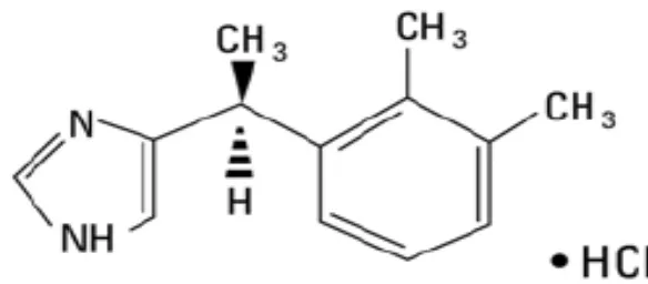
C. PHARMACOLOGY OF CLONIDINE HYDROCHLORIDE
[image:38.595.167.397.124.265.2]
Figure 8. Chemical structure of clonidine hydrochloride.
Introduction:
31
manner. Clonidine potentiates the anaesthetic action of the local anaesthetics with fewer side effects in peripheral nerve blocks and central neuraxial blockade.
Availbility:
Available as one ml ampoule containing 150 micrograms. It should be stored below 25 degree Celsius. It also available as tablet.
Mechanism of action:
Clonidine is a centrally acting partial α2 adrenergic agonist with a selectivity ratio of 220: 1 in favour of α2 receptors.When clonidine was injected into epidural space it is penerates the blood brain barrier and reaches to the hypothalamus and medulla. It is stimulates the inhibitory α2 adrenergic receptors to reduce the central neural transmission in the spinal neurons.
32
Clonidine reduces the anaesthetic requiremet and its analgesic effects due to modification the potassium channels in the central nervous system and hyperpolarisation in the cell membranes.
Clonidine is neuraxial inhibits of spinal substance-P release is believed to be involved in the analgesic effect. Another contribution to analgesic effect may be through the release of acetylcholine in the neuraxial region. The α2 adrenergic agonist also enhances analgesia from intraspinal opioids. Sedation is produced by its action on locus ceruleus.
The α2 adrenoreceptors terminals are presented centrally, peripheraly, superficial laminae of the spinal cord and brain stem neuclei is believed to involved analgesic effects after neuraxial administration of clonidine.it also reduce the cold response threshold and increase the sweating threshold may inhibition of the shivering response.
Clonidine affects the blood pressure in a complex fashion after neuraxial or systemic administration because of the opposing action at multiple sites. In the nucleus tractus solitories and locus ceruleus of the brain stem, activation of post-synaptic α2 adrenoreceptors decrease the sympathetic actions. producing hypotension and anti- arrythmogenic action.
33
Pharmacokinetics:
Clonidine well absorped from orally and bioavailability of clonidine is 75 to 95%. It is peak plasma half life is 60 to 90 minutes, Plasma half life is 12 to 33 hours.50% of the drug is mainly metabolized in the liver whereas it is excreted in an unchanged form by the kidney 40 to 60%, and its half life can increase in the presence of renal dysfunctions. A transdermal delivery system is available in which the drug is released at a consant rate for a week. Three or four days are required to achieve steady state concentration.
After 300µgm/ kg intravenously infused over 10 min produces: Distribution t½: 11± 9 minutes
Elimination t½: 9± 2 hours, 41 hours in severe renal dysfunction. Volume of distribution: 2.1± 0.4 l / kg
Plasma protein binding: 20 - 40% in vitro.
DOSAGE REGIMEN:
Oral: 3-5μg/kg.
Intramuscular: 2 μg/ kg. Intravenous: 1-3μg/kg. Spinal: 50 -100μg. Caudal: 1-2μg/kg. Epidural: 1-2μg/kg.
34
PRECAUTIONS:
1. In renal insufficiency patients, lower dose is needed.
2. Sudden withdrawal of prolonged continuous epidural infusion roduces hypertensive crisis. So gradually discontinue over 2 to 4 days to prevent this complication.
3. Use with caution in patients with cerebrovascular or coronary insufficiency.
4. Patient on beta blocker therapy,beta blocking drugs should be withdrawn several days before the epidural clonidine.
CONTRAINDICATIONS:
1. Known hypersensitivity to clonidine or components of the product. 2. Brady arrhythmia or AV block.
3. Severe cardiovascular disease.
4. Cardiovascular / hemodynamic instability.
INTERACTIONS:
1. Clonidine may potentiate the CNS- depressive effect of alcohol, barbiturates or other sedative drugs.
2. Clonidine potentiate the hypotensive action with narcotic drugs.
35
4. Concomitant administration of drugs with a negative chronotropic or dromotropic effect (beta blocker, digoxin) can cause or potentiate bradycardia and rhythm disturbances.
5. Beta blockers may potentiate the hypertensive response seen with clonidine withdrawal.
USES:
1. Caudal anaesthesia: 1to2μg/kg of clonidine combined with local anaesthetic agents prolong the duration of analgesia by 2 or 3 times without hemodynamic side effects.
2. Epidural block: Clonidine as sole agent or in combine with opioids or local anaesthetics to provide excellent analgesia in labour analgesia.
3. Spinal anaesthesia: Clonidine combined with local anaesthetics improves the quality and duration of the block, minimize the tourniquet pain during lower limb surgery, and prevents shivering.
4. Pre anasthetic medication : Oral clonidine is 5 micg/kg.
a. Blunts reflex tachycardia associated with direct laryngoscopy for intubation of trachea.
b. Reduce the intraoperative instability of the blood pressure and heart rate. c. Plasma catecholamine concentrations levels decreased.
36
5. In Peripheral nerve blocks: Clonidine prolongs the duration of anaesthesia and analgesia with local anaesthetics by two times in a dose of 75 to 150 micro grams.
6. In Bier’s Block: 150 microgram of clonidine enhances the tolerance of tourniquet.
7. It is also used in intra articular analgesia.
8. Protection against perioperative myocardial ischemia: Clonidine decreases myocardial ischemia, infarction and mortality following cardiovascular surgery.
9. In the management of hypertensive crisis clonidine is useful.
10. Treatment of shivering; Administration of clonidine75μg IV abolishes shivering by inhibiting thermoregulatory control.
11. Clonidine is useful in the treatment of opioid and alcohol withdrawl syndrome.
Side effects;
1. The most common side effects are sedation and xerostomia.
2. Cardiovascular complications are bradycardia, hypotension, and sinusnode arrest, junctional bradycardia, high degree AV block and other arrhythmia are reported rarely. Occasionally bradycardia may requires treatment with I.V anticholinergics.Orthostatic hypotension occurs rarely.
37
last dose. Symptoms of the rebound hypertension are headache, abdominal pain, tachycardia, nervousness, and diaphoresis, which often precede the actual increase in systemic blood pressure. Labetalol is useful in the treatment of rebound hypertension.
4. Skin rashes occur frequently. 5. Impotence occurs occasionally.
Treatment for over dosage:
38
D. PHARMACOLOGY OF DEXMEDETOMIDINE
Dexmedetomidine is an α2 adrenergic agonist. The α2adrenergicreptors
agonists produce sedation, anxiolysis, hypnosis, analgesia, and sympatholysis.It is a nonselective α2 agonist with a 1600 greater selectivity for α2 receptor as
compared to α1 receptor. Introduced in clinical practice 1999 and approved by
FDA only for short-term, less than24 hours sedation and analgesia for ICU adult patients. Now a days used off-label outside of the ICU for sedation, adjunct analgesia, sedation for short term diagnostic procedures. It decreases the sympathetic tone and also attenuates the stress response during intubation and surgery. They are also used as adjuvants during intravenous regional anaesthesia.
Physicochemical Characters:-
It is a potent, highly selective α2 adrenergic agonist. Its freely water soluble. Molecular formula is C13H16N2.HCl.
Molecular weight: 236.74.
The chemical name as 4-(S)-[1-(2,3dimethylphenyl)ethyl]-1H-imidazole monohydrochloride.
vessels release cord. I and an F MECH recepto helices adenyl channe Postsynap s produce e of norep
The α2 ad
It inhibits nalgesia. (F
[image:47.595.130.422.279.407.2]
Figure:9.
HANISM
α2 adreno
ors, which s.α2 adre lcyclase, w els and thu
ptic α2 adr
e vasocon inephrine drenorecep the neuro Figure.9) Chemical
M OF ACT
oreceptors h contains energic re which in us augmen renorecept nstriction. and leads ptors also onal firing l structur TION s belongs s a charac eceptors
turn inh nts the hyp
39 tors which
Presynap s to fall in located in , causing
re of dexm
to the la cteristic st
activation hibits the perpolariz
h are locat ptic α2 ad
blood pre n central n
hypotensi
medetomid
arge fami tructure o n are th opening zing potass
ted in the drenorecep essure and
nervous sy ion, brady
dine hydr
ily of G of seven tr e decrea of voltag sium ion c
periphera ptors inhi d heart rate ystem and ycardia, se
rochloride
protein c ransmemb sed activ e gated c channel ac l blood ibit the e. d spinal edation, e. coupled brane α
40
METABOLISM AND PHARMACOKINETICS
Dexmedetomidine is 94% protein bound.It undergoes an extensive hepatic metabolism. It is rapidly distributed and extensively metabolized in the liver by glucuronidation, conjugation, N-methylation, or hydroxylation and cytochrome P450 mediated metabolism. Metabolites are excreted in the urine (about 95%) and in the feces(4%). Dose needs to be adjusted in patients with liver impairment, but dose need not be adjusted in renal dysfunction patients. Large doses can produce the vasoconstriction leading to reduced drug volume of distribution.
Dexmedetomidine follows the nonlinear pharmacokinetics.
Elimination half life:- 2 to 3 hours. Distribution half life: - 6 minutes.
Volume of distribution (vss):-118 litres.
Context-sensitive half-time: - Ranges from 4 minutes after a 10-minutes infusion to 250 minutes after an 8-hour infusion.
It has shorter duration of action compared to clonidine. The most frequently observed side effects in patients receiving dexmedetomidine include hypotension, hypertension, nausea, bradycardia and atrial fibrillation.
41
Dexmedetomidine does not affect the synthesis, storage or metabolism of neurotransmitters and does not block the receptors, thus providing the possibility of reversing the hemodynamic effects with vasoactive drugs or the specific α2antagonist, Atipamezole which acts by increasing the central turnover of norepinephrine. Its duration of action is 2 hours.
DOSAGE
It is available as100 mcg in1 ml ampoule or 2 ml vial. It is containing 100mcg/ml. It is generally diluted in 50ml of 0.9% normal saline to obtain concentration of 4mcg/ml.
DRUG INTERACTIONS
42
ADVANTAGES
• It is a Sedative and analgesic drug that produces sympatholysis without any respiratory depression.
• It is also an antisialogogue.
EFFECTS ON THE CENTRAL NERVOUS SYSTEM
Sedation
It produces its effect by acting on the α2 receptors present in the locus
caeruleus and analgesic effect due to its action on α2 receptors within the locus caeruleus and spinal cord. It produces natural sleep with minimal respiratory depression. The quality of sedation produced by dexmedetomidine seems different compared with that produced by other sedatives acting through the GABA systems like benzodiazepines.
Patients receiving dexmedetomidine infusions of sedation regimen in ICU setting have been described as being very easy to wake up and having the ability to follow commands but are able to recall the events in the ICU. It is easy to perform the daily wake up tests. In this critical test, mechanically ventilated ICU patients are taken off all sedatives to assess their mental status and titrate sedation.
The α2 agonists act through the endogenous sleep-promoting pathways to
exert their sedative effect.
43
increase in GABAergic and galanin release in the tuberomammillary nucleus, producing a decrease in histamine release in cortical and sub cortical projections.
The α2 agonists inhibit ion conductance through L-type or P-type calcium
channels and facilitate conductance through voltage-gated calcium-activated potassium channels.
Dexmedetomidine can produce profound sedation, and it has been used as a total IV anaesthetic when given at 10 times the normal sedation concentration range.
It produces cerebral protection by decreasing cerebral blood flow without affecting CMRO2, decreases cerebral blood flow velocity.
It has minimal effect on the cortical evoked potentials. It also ablates memory in a dose dependent manner.
The α2 agonists have the advantage that their effects are readily reversible
by α2-adrenergic antagonists (e.g., atipamezole). It is administered in the dose of
50µg/kg I.V.
Atipamezole is not currently approved for human use. Similar to other adrenergic receptors, the α2 agonists also show tolerance after prolonged
administration.
44
Analgesia:-
When dexmedetomidine is injected into the epidural space, it rapidly diffuses into the CSF. The effects on blood pressure are slower in onset with an epidural injection than with an intrathecal administration. Onset of action via epidural route is 5 to 20 minutes. The primary site of analgesic action is thought to be at the spinal cord. It produces prolonged analgesia and increased duration of sensory and motor blockade when administered intrathecally along with bupivacaine.
RESPIRATORY EFFECTS:-
Dexmedetomidine throughout a broad range of plasma concentration has minimal effects on the respiratory system. Co administration of dexmedetomidine with other sedatives, hypnotics or opioids is likely to cause additive effects. It decreases the minute ventilation in a dose dependent manner. At lower concentrations, no effect on arterial oxygenation and pH. With increasing concentration, increased respiratory rate is observed.
CARDIOVASCULAR EFFECTS:-
45
PERIOPERATIVE USES OF DEXMEDETOMIDINE
A. Premedication
Dexmedetomidine is anxiolytic, sedative, analgesic, antisialogogue and sympatholytic properties, which makes it suitable as a premedication agent. Dexmedetomidine decreases the thiopentone(30%) dose requirement, decreases the haemodynamic response to intubation, decreases volatile anaesthetic requirement .It should be administered at a dose of 0.3-0.6µg/kg intramuscular injection given 15minutes before surgery The administration of intramuscular dexmedetomidine at a dose of 1 μg/kg for premedication in outpatient cataract surgery resulted in sedation, and decrease in intraocular pressure without significant hypotension or bradycardia.Also the administration of dexmedetomidine for premedication decreases oxygen consumption intraoperatively by 8% and postoperatively by 17%.
Indications for the use of dexmedetomidine as premedication include patients susceptible to preoperative and peri operative stress, drug addicts and alcoholics, chronic opioid users and hypertensive patients.
B. Intra operative uses of dexmedetomidine
46
1. Used an adjunct to general anesthesia
The use intraoperative dexmedetomidine may increase hemodynamic stability because of attenuation of the stress-induced sympathoadrenal responses to intubation,decreased level of plasma catecholamines during surgery and during emergence from anesthesia.
Administration of intravenous dexmedetomidine decreases the narcotic consumption intraoperatively and post operatively when compared to propofol.
It reduces the vasoconstriction and the shivering threshold and is associated with a lower incidence of shivering.
It decreases the muscle rigidity caused by high doses of opioids. It also decreases the cardio stimulatory effects and post anaesthetic delirium produced by ketamine.
2. Used for regional anesthesia
47
3. Used in monitored anesthesia care
Dexmedetomidine confers arousable sedation with ease of orientation, anxiolysis, mild analgesia, lack of respiratory depression and hemodynamic stability at moderate doses. These properties allow dexmedetomidine to be an almost ideal agent for MAC despite its lack of amnesia and poor controllability because of its slow onset and offset.
Dexmedetomidine in MAC was used successfully in many situations: when patient arousability needed to be preserved, as for awake craniotomy , awake carotid endarterectomy and for vitreoretinal surgey. In addition, dexmedetomidine was used for sedation in difficult airway patients; during fiberoptic intubation, and for sedation of a patient with difficult airway undergoing lumbar laminectomy surgery in the prone chest position under spinal anesthesia.
4 .Use of dexmedetomidine as a sole anesthetic agent
Dexmedetomidine used as a sole anesthetic agent. Does not produce any respiratory depression, hypotension and severe bradycardia. These effects were maintained at higher doses without hemodynamic instability.It can be safely used in patients who are susceotible to narcotic induced respiratory depression.
C. Use of dexmedetomidine in postoperative period
48
spontaneously breathing patient. The possibility of ongoing sedation and sympathetic block could be beneficial in reducing high rates of early postoperative ischemic events in high-risk patients undergoing non-cardiac surgery.
Perioperative administration of dexmedetomidine could be beneficial in chronic opioid users and alcoholics, in high-risk patients as well as in cardiac patients with good to moderately decreased left ventricular function.
D. Use of Dexmedetomidine in the pediatric-age group
In pediatric-age group uses of intraoperative dexmedetomidine at different doses with the goal of reducing the post sevoflurane agitation in children aged 1-10 years. The optimal dose of dexmedetomidine was 0.3 μg/ kg and its use did not result in adverse effects. When compared with propofol for sedation during MRI, dexmedetomidine provides adequate sedation during the scan but has a slower recovery profile.
One of the major advantages of dexmedetomidine over other sedatives is its respiratory effects, which are minimal in adults and children. It does not lead to extreme hypoxia or hypercapnia. Indeed, respiratory rate, CO2 tension, and
49
CONTRAINDICATIONS
A. Severe bradycardia and cardiac conduction abnormalities. B. Ventricular dysfunctions - ejection fraction<30%.
C. Patients with hypovolemia or hypotension.
ADVERSE EFFECTS
1. Central and Peripheral nervous system: Dizziness, headache, neuralgia, neuritis, speech disorder, Convulsion
2. Gastrointestinal System :
Abdominal pain, diarrhea, vomiting, nausea, dry mouth 3. Cardiovascular systems:
Arrhythmia, ventricular arrhythmia, bradycardia, hypoxia, atrioventricular block, cardiac arrest, extrasystoles, atrial fibrillation, heart block, Twave inversion, tachycardia, supraventricular tachycardia, ventricular tachycardia
4. Hepato Biliary System:
Increased gamma glutamyl transpepsidase, hepatic function abnormal, hyperbilirubinemia, alanine transaminase, aspartate aminotransferase.
5. Metabolic and Nutritional Disorder:
50 6. Psychiatric Disorders:
Agitation, confusion, hallucination, illusion. 7. Blood: Anemia.
8. Renal system: Blood urea nitrogen elevated, oliguria.
51
7. REVIEW OF LITERATURE
1. Journal of clinical anaesthesiolgy and pharmacology.2010, 26(2).149-53. Dhurjoti prosad et al. Studied 90 children for caudal analgesia in age group between one to six years and divided into three groups. Group R received 0.25%of ropivacaine 1ml/kg , Group C received 0.25%of ropivacaine 1ml/kg with clonidine 1 mcg/kg and group D is received 0.25% of ropivacaine 1ml/kg with dexmedetomidine1mcg/kg. This study results concluded the duration of analgesia was 6 ± 0.46 hours in Group R , Group C is 13.17 ± 0.68 hours and Group D 15.26±0.86 hours.Addition of dexmedetomidine and clonidine with ropivacaine in caudal anaaethesia better than ropivacaine alone and without any hemodynamic instability.
2. Journal of acta anaeathesia scandinavica.2009.53 (2), 251-256. Boles, Saadway et al , studied the effect of dexmedetomidine combined with bupivacaine in caudal anaesthesia in children . In this study results were excellent intraoperative and postoperative analgesia and without any adverse effects.
52
caudal anaesthesia. This study showed that dexmedetomidine group had prolonged duration of post operative analgesia than the clonidine group, without any significant side effects.
4. Anaesthesia and analgesia 2007, 14.1356-1363.Thomas et al. a comparative study of single shot caudal epidural clonidine ,morphine and hydromorphine with ropivacaine in caudal anaesthesia in children age between 6 months to six years .It was a double blind randomized controlled study divided into three groups, clonidine 2mcg/kg, morphine 50mcg/kg, hydromorphine 10mcg/kg with 0.25%of ropivacaine in adrenaline. Results of in this study was clonidine combined with ropivacaine to produce the increases duration of analgesia without significant side effects. Caudal opioid produced the postoperative nausea and vomiting.
53
6. European journal of anaesthesia. 2010. 27, (11), 560. Erbek et al studied comparison between bupivacaine 0.5% and dexmedetomidine sedation for septoplasty procedure. The patients divided randomized into two groups. Group B patients received 0.5% bupivacaine alone and grooup BD patients received 0.5% bupivacaine with dexmedetomidine 2mcg/kg. Results of this study were bupivacaine combined with dexmedetomidine is better intraoperative, postoperative pain relief and reduce the intraoperative bleeding than bupivacaine alone.
7. Indian journal of anaesthesiology. 2010, 53(3) page 226-230. Jabir kaur et al. studied to determine the quantitative and qualitative of caudal epidural anaesthesia, hemodynamic effects and postoperative duration of analgesia. This is randomised controlled double blind study for children age between one to nine years divided into two group, group I received 0.25% of ropivacaine and group II received 0.25% of ropivacaine with clonidine 2mcg/kg .the results of this study is duration of analgesia in group II is prolonged the duration of postoperative analgesia and efficient intraoperative analgesia without significant hemodynamic changes.
54
0.25% of bupivacaine 0.75ml with clonidine 2mcg/kg. Postoperatively monitoring for OPS score, sedation score, pulse rate and blood pressure were recorded. This study concluded that the duration of analgesia in group C was 10.25 hours compared with group B was 4.55 hours and without hemodynamic instability.
9. British journal of anaesthesia.2011. 106. N Kumar et al. This is randomized controlled prospective study was selected 50 patients for upper abdominal surgeries and divided into two groups. Group BM patients are received 0.25% of bupivacaine 1.25ml/kg with morphine 30mcg/kg and Group BC 0.25% of bupivacaine 1.25ml/kg with clonidine2 mcg/kg. In this study was concluded group BC was prolonged duration of post operative analgesia than group BM. The morphine group was produced more nausea and vomiting.
55
56
8. MATERIALS AND METHODS
This Prospective, randomised, double blind, comparitive study was done to compare the efficacy and safety of dexmeditomidine and clonidine as ajuvants to caudal ropivacaine in postoperative analgesia for children. The study was carried out in 60 children at Government Rajaji hospital, Madurai for surgeries of lower abdomen and perineum in the year 2012. The children in the age group of 1-6 years and weighing 5-20Kgs were selected for the study.
INCLUSION CRITERIA:
Elective infra umbilical surgeries Both sexes
Age: Between one to six years. ASA: I
Weight: 5-20kg
Exclusion CRITERIA:
1. Known allergic to LA. 2. Local sepsis.
57
Only ASA I physical status patients were chosen to avoid the influence of the associated diseases. The sixty children were divided into two groups of thirty each.
Group RC: Received 1ml/kg of 0.25% ropivacaine and 1μg/ kg clonidine. Group RD: Received 1ml/kg of 0.25% ropivacaine and 1 μg/ kg dexmedetomidine.
Pre anaesthetic evaluation
1. History.
2. Clinical examination.
3. Relevant investigations – haemoglobin, urine analysis. 4. Informed consent from parents.
5. All children were kept nil per oral for 6 hrs prior to surgery.
58
Anaesthesia was maintained with 60%, nitrous oxide in40% oxygen and 0.6%.Sevoflurane using controlled ventilation. The patients were positioned in left lateral position. After aseptic draping, a 23G needle was introduced into caudal space and either or ropivacaine with clonidine (Group RC) or ropivacaine with dexmedetomidine (group RD) was administered. At the end of the operation, residual neuromuscular block was reversed by appropriate doses of neostigmine 40µg/kg & atropine 10µg/kg and tracheal extubation was performed. Postoperatively they were monitored in postanasthetic care unit. Pulse rate, MAP and SPO2 were recorded throughout the operation at an interval of five minutes
Intraoperative monitoring:
Pulse rate, blood pressure, saturation. Decrease of mean arterial blood pressure and pulse rate more than 30% from the baseline values were defined as severe hypotension and bradycardia, respectively which were treated injection atropine sulphate 20mcg/kg. At the beginning of skin closure anaesthesia was discontinued.
Postoperative monitoring was done in post anaesthetic care unit where vital parameters and pain, sedation scoring done every 15 minutes for 3 hours and then every one hour.
1. Time from caudal block to end of the surgery. 2. Sedation was assessed by 4 point scale.
59 4. Duration of post-op analgesia. 5. Pulse rate, map and spo2. 6. Complications.
The following parameters were assessed
4 point sedation scale:
1. Barely arousable. (Sleeps Needs shaking or shouting to arouse). 2. Asleep. (Eyes closed arousable with soft voice or light touch). 3. Sleepy. (Eyes open but less active and responsive).
4. Awake.
CRIES pain scale:
0 1 2 Crying No High pitched Inconsolable Requires O2 for
SPO2 >95% No < 30% of O2 > 30% of O2 Increased vital
signs
No increase in HR and MAP
↑ HR or MAP < 20%
↑ HR or MAP > 20%
Expression None Grimace Grimace/grunt
60 Score 0- signifies excellent analgesia. Score 10- indicate ineffective analgesia.
61
9. STASTISTICAL ANALAYSIS
Statistical Tools (To be included at the end of Materials and Methods) The information collected regarding all the selected cases were recorded in a Master Chart. Data analysis was done with the help of computer using Epidemiological Information Package (EPI 2010) developed by Centre for Disease Control, Atlanta.
62
10. OBSERVATION AND RESULTS
[image:70.595.65.529.256.436.2]The two groups were compared in characteristics like demographic data and basic vital parameters (pulse rate, MAP, saturation) and duration of surgery, duration of postoperative analgesia, complications.
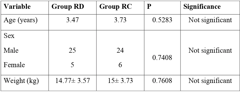
Table-1 DEMOGRAPHIC VARIABLES
Variable Group RD Group RC P Significance
Age (years) 3.47 3.73 0.5283 Not significant Sex
Male Female
25 5
24
6 0.7408
Not significant
Weight (kg) 14.77± 3.57 15± 3.73 0.7608 Not significant
The mean age of children in group RD was 3.47 years and in group RC was 3.73 years which is found not to be statically significant with P value of 0.5283.
The sex distribution in group RD 25 males and 5females and in group RC were compared and found 24 males and 6 females. Both group were compared and found not to be statistically significant.
GRAPH 1: AGE DISTRIBUTION
0
1
2
3
4
GROUP
RC
GROUP
RD
3.73
3.47
GRAPH 2: SEX DISTRIBUTION
0% 20% 40% 60% 80% 100%
GROUP RC
GROUP RD
24 25
6 5
63
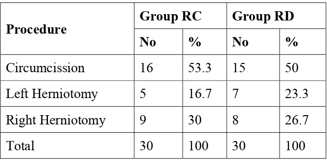
TABLE 2: PROCEDURE DONE
Procedure Group RC Group RD
No % No %
Circumcission 16 53.3 15 50 Left Herniotomy 5 16.7 7 23.3
Right Herniotomy 9 30 8 26.7
Total 30 100 30 100
GRAPH 4: PROCEDURE DONE 0 4 8 12 16 20 GROUP RC GROUP RD 16 15
5 9 7 8
N u m b er o f cases
64
[image:76.595.55.543.206.780.2]B: HAEMODYNAMIC CHANGES
Table 3: Preoperative, Intra operative and postoperative
changes in pulse rate
Pulse rate
Pulse rate of
‘p’
Group RC Group RD
Mean SD Mean SD
Pre op 0 hour 106.0 4.8 106.9 5.2 0.4399 Not significant
Intra op 5min 104.5 4.8 104.2 4.2 0.9289
Not significant
10 min 95.8 1.6 95.9
0.6476
Not significant 15 min 93.8 2.1 94.0 0.6644
Not significant 20 min 92.5 1.4 92.8 0.988
Not significant 25 min 92.5 1.9 92.1 0.2787
Not significant 30 min 92.3 1.6 91.8 0.2975
Not significant 45 min 91.3 1.4 91.1 1.3 0.5577
Not significant Post op
60 minutes 91.2 1.7 90.9 1.4
0.6357
Not significant 75min 91.2 1.8 90.8 1.5 0.2985
Not significant 90 min 90.8 1.5 90.7 1.3 0.7373
Not significant 105 min 90.7 1.7 90.5 1.4 0.659
Not significant 120min 91.4 1.6 90.6 1.3 0.0476
65
135min 90.5 1.4 90.4 1.3 0.7342
Not significant 150 min 90.4 1.4 90.4 1.4 1.0
Not significant 165 min 90.7 1.5 90.4 1.0 0.7111
Not significant 180 min 91.1 1.9 90.3 1.4 0.1023
Not significant 4 hours 90.7 1.7 90.5 1.4 0.659
Not significant 5 hours 91.4 1.6 90.6 1.3 0.0476
Not significant 6 hours 90.5 1.4 90.4 1.3 0.7342
Not significant 7 hours 90.4 1.4 90.4 1.4 1.0
Not significant 8 hours 90.7 1.5 90.4 1.0 0.7111
Not significant 9 hours 91.1 1.9 90.3 1.4 0.1023
Not significant 10 hours 93.4 1.8 90.6 1.5 0.0001Significant 11 hours 100.1 2.9 90.6 1.3 0.0001Significant
12 hours 103.0 3.3 90.3 1.2 0.0001Significant
GRAPH 5: CHANGES IN PULSE RATE PRE,
INTRA, POST OPERATIVE PERIOD
90 93 96 99 102 105 108 0m in 5 min . 10 m in 15 m in 20 m in 26 m in 30 m in 45 m in 60 m in 75 m in 9o m in 105 m in 120 m in 135 m in 150 m in 165 m in 180 m
in 4h 5h 6h 7h 8h 9h
10 h 11 h 12 h 13 h 14 h 15 h 16 h 17 h
M
ean
P
u
lse R
a
te
66
Table 4: Pre operative and intra operative, post operative
changes in mean arterial pressure
MAP
MAP of
‘p’
Group RC Group RD
Mean SD Mean SD
Pre op.
0min 85.7 3.7 84.7 3.1
0.3529
Not significant Intra op
5min
81.3 2.3 80.5 1.7 0.1764 Not significant
10 min
75.4 1.8 74.7 2.1 0.2209 Not significant
15 min
74.4 1.9 74.8 1.8 0.3302 Not significant
20 min
73.8 1.4 74.0 1.5 0.8255 Not significant
25 min
74.2 1.1 74.4 1.2 0.7653 Not significant
30 min
73.0 1.2 73.3 1.1 0.3183 Not significant
45 min
73.0 0.9 73.0 0.9 0.8695 Not significant Post op
60 minutes
73.1 1.0 72.9 0.9 0.5754 Not significant 75 minutes 73.0 1.0 72.9 0.9 0.6754
Not significant 90mins 72.9 0.8 72.8 0.8 0.5855 Not significant
105 min 72.9 1.0 72.7 0.9 0.3395 Not significant 120min 73.1 1.1 73.2 0.8 0.9319
67
150min 72.7 0.8 72.8 0.7 0.6318 Not significant 165 min 73.0 0.9 73 0.7 0.9556 Not significant
180 min 72.8 0.9 72.9 0.9 0.9317 Not significant
4 hours 73.1 1.1 73.2 0.8 0.9319 Not significant 5 hours 72.8 1.0 72.7 0.9 0.6701
Not significant 6 hours 72.7 0.8 72.8 0.7 0.6318
Not significant 7 hours 73.0 0.9 73 0.7 0.9556
Not significant 8 hours 72.8 0.9 72.9 0.9 0.9317 Not significant
9 hours 72.7 0.9 72.6 0.7 0.6499 Not significant
10 hours 72.8 1.0 72.7 0.9 0.6701 Not significant 11 hours 72.7 0.8 72.8 0.7 0.6318 Not significant
12 hours 73.0 0.9 73 0.7 0.9556 Not significant
GRAPH 6: MEAN ARTERIAL PRESSURE 70 72 74 76 78 80 82 84 86 0 m in 5 m in . 10 m in 15 m in 20 m in 26 m in 30 m in 45 m in 60 m in 75m in 90m in 105m in 120m in 135m in 150m in 165m in 1 80m
in 4h 5h 6h 7h 8h 9h
10h 11h 12h 13h 14h 15h 16h 17h
Me
a
n
M A
P
68
Table 3 & 4
69
Table 5: Pre operative and Intra operative changes in SPO2
SPO2
SPO2
‘p’
Group RC Group RD
Mean SD Mean SD
Pre op. 0 hour 97.8 1.1 97.6 1.1 1.0Notsignificant
Intra Op 5 min
98.6 0.5 98.4 0.4 1.0 Not significant 10 minutes 98.6 0.5 98.3 0.4 1.0
Not significant 15 minutes 98.7 0.5 98.7 0.5 1.0
Not significant 20 minutes 98.6 0.5 98.5 0.4 1.0
Not significant 25 minutes 98.8 0.4 98.7 0.4 1.0
Not significant 30 minutes 98.8 0.5 98.5 0.4 1.0
Not significant 45 minutes 98.6 0.4 98.7 0.5 1.0
Not significant 60 minutes 98.7 0.4 98.4 0.4 1.0
Not significant
75min 98.7 0.4 98.4 0.4 1.0
Not significant
90min 98.8 0.4 98.6 0.4 1.0
Not significant 105 min 98.8 0.4 98.5 0.4 1.0
Not significant
120MIN 98.9 0.3 98.6 0.3 1.0
Not significant
135min 98.9 0.3 98.6 0.3 1.0
Not
significant 150 min 98.9 0.3 98.8 0.3 1.0
Not significant
165min 98.9 0.3 98.5 0.3 1.0
Not significant
180min 98.9 0.3 98.4 0.3 1.0
70
Table 5 (a) Post operative changes in SPO2
Post op. SPO2 at
Post operative SPO2 of
‘p’
Group RC Group RD
Mean SD Mean SD
4 hours 98.9 0.3 98.8 0.2 1.0 Not significant 5 hours 98.9 0.3 98.7 0.2 1.0 Not significant 6 hours 98.9 0.3 98.8 0.2 1.0 Not significant 7 hours 98.9 0.3 98.7 0.3 1.0 Not significant 8 hours 98.9 0.3 98.6 0.3 1.0 Not significant 9 hours 98.9 0.3 98.8 0.3 1.0 Not significant
10 hours 99.6 0.2 99.4 0.2 1.0 Not significant
11 hours 99.4 0.2 99.3 0.2 1.0
Not significant 13 hours 98.9 0.3 98.7 0.3 1.0 Not significant 14 hours 99.0 0.2 98.9 0.3 1.0 Not significant
15 hours 99.2 0.2 99.2 0.2 1.0 Not significant 16 hours 99.4 0.3 98.9 0.3 1.0 Not significant
17 hours 99.0 0/2 98.9 0.3 1.0 Not significant
[image:84.595.56.542.99.605.2]GRAPH 7: SPO2
71
[image:86.595.123.467.147.291.2]C: COMPARATIVE EFFICACY
Table 6: Duration of surgery
Parameter
Duration of surgery ( in minutes)
Group RC Group RD
Range 20-45 20-45
Mean 31.0 30.0
SD 8.14 7.88
‘p’ 0.6244 Not significant
Table 6
GRAPH 8: DURATION OF SURGERY 0 5 10 15 20 25 30 35 40
GROUP RC GROUP RD
72
Table 7: Duration of postoperative analgesia
Parameter Duration of Post Operative
analysis ( in hours)
Group RC Group RD
Range 8-12 13-17
Mean 9.8 14.67
SD 1.4 1.15
‘p’ 0.0001 Significant
Table 7
GRAPH 9: DURATION OF POSTOPERATIVE ANALGESIA
73
TABLE 8 : 4 POINT SEDATION SCORE
TIME IN HOURS
GROUP RD GROUP RC
1 2 3 4 1 2 3 4
1hr 30 0 0 0 30 0 0 0
3hr 30 0 0 0 25 5 0 0
6hr 24 6 0 0 18 10 2 0
12hr 0 20 10 0 0 15 12 3
17hr 0 0 10 20 0 0 8 22
P VALUE 1.0 NOT SIGNIFICANT
74
Table 9: CRIES Pain scale
Pain scale Group RC Group RD
No % No %
0 14 46.7 20 66.7
1 15 50 10 33.3
2 1 3.3 - -
Total 30 100 30 100
Range 0-2 0-1
Mean 0.57 0.33
SD 0.57 0.48 ‘p’ 0.1039 Not significant
75
Table 10 : Complications
Complications Group RC Group RD
No % No %
Nausea 1 3.3 2 6.7
Nausea & Vomiting 1 3.3 - -
Pruritis 1 3.3 1 3.3
Nil 27 90 27 90
Total 30 100 30 100
‘p’ 1.0 Not significant