CHARACTERIZATION OF BACTERIAL ISOLATES WITH
DETECTION OF METHICILLIN RESISTANT
STAPHYLOCOCCUS AUREUS
AND EXTENDED
SPECTRUM BETALACTAMASES PRODUCERS IN
ADULT PNEUMONIA
Dissertation Submitted To
THE TAMILNADU DR. M.G.R. MEDICAL UNIVERSITY
CHENNAI
In partial fulfillment of the regulations For the award of the degree of
M.D. (MICROBIOLOGY)
BRANCH IV
GOVT. KILPAUK MEDICAL COLLEGE
CHENNAI
CERTIFICATE
This is to certify that this dissertation entitled “CHARACTERIZATION OF
BACTERIAL ISOLATES WITH DETECTION OF METHICILLIN RESISTANT
STAPHYLOCOCCUS AUREUS & EXTENDED SPECTRUM BETALACTAMASES
PRODUCERS IN ADULT PNEUMONIA” is the bonafide original work done by
Dr. P. HEMA SUGANYA, Post graduate in Microbiology, under my overall
supervision and guidance in the Department of Microbiology, Govt. Kilpauk
Medical College, Chennai, in partial fulfillment of the regulations of The Tamil
Nadu Dr. M.G.R. Medical University for the award of M.D Degree in
Microbiology (Branch IV).
Dr. RADHIKA KATRAGADDA, M.D.,
Professor & H.O.D Department of Microbiology Govt. Kilpauk Medical College
Chennai-600010
Dr.R.NARAYANA BABU, M.D., D.C.H.,
The Dean
CERTIFICATE
This is to certify that the dissertation entitled “CHARACTERIZATION OF
BACTERIAL ISOLATES WITH DETECTION OF METHICILLIN RESISTANT
STAPHYLOCOCCUS AUREUS & EXTENDED SPECTRUM BETALACTAMASES
PRODUCERS IN ADULT PNEUMONIA” is a bonafide research work done by,
Dr. P. HEMA SUGANYA, Post graduate in Microbiology, under my guidance in
the Department of Microbiology, Govt. Kilpauk Medical College, Chennai, in
partial fulfillment of the regulations of The Tamil Nadu Dr. M.G.R. Medical
University for the award of M.D Degree in Microbiology (Branch IV).
Dr. THYAGARAJAN RAVINDER M.D.
DECLARATION
I solemnly declare that this dissertation “CHARACTERIZATION OF
BACTERIAL ISOLATES WITH DETECTION OF METHICILLIN RESISTANT
STAPHYLOCOCCUS AUREUS &EXTENDED SPECTRUM BETALACTAMASES
PRODUCERS IN ADULT PNEUMONIA” is the bonafide work done by me at the
Department of Microbiology, Govt. Kilpauk Medical College and Hospital,
Chennai, under the guidance and supervision of Dr.RADHIKA KATRAGADDA,
M.D., Professor & H.O.D of Microbiology, Dr. K.V. LEELA, M.D., DGO.,
Professor, Department of Microbiology and Dr. THYAGARAJAN RAVINDER,
M.D., Professor, Department of Microbiology, Govt. Kilpauk Medical College,
Chennai-600010. This dissertation is submitted to The Tamil Nadu Dr. M.G.R.
Medical University, Chennai in partial fulfillment of the University regulations for
the award of Degree of M.D. Branch IV Microbiology examinations to be held in
April 2017.
Place: Chennai. Dr. P. HEMA SUGANYA
ACKNOWLEDGEMENT
My heartfelt thanks and deepest sense of gratitude to
Dr.R.NARAYANA BABU, M.D., D.C.H., Dean, Government Kilpauk Medical
College and Hospital for giving me permission to carry out my dissertation work
and also to avail all the facilities available in the department.
I am deeply indebted to Dr. RADHIKA KATRAGADDA, M.D., Professor
and H.O.D., Department of Microbiology for her relentless efforts, valuable
advice, excellent guidance and encouragement given to me throughout this study.
I am immensely grateful to Dr. K.V.LEELA, M.D., Professor, Department
of Microbiology for her unflinching interest, effort and motivation extended to me
during my study.
I owe my sincere gratitude to Dr. THYAGARAJAN RAVINDER, M.D.,
Professor, Department of Microbiology for his valuable guidance, his timely advice,
motivation and encouragement throughout the period of this study.
My sincere and special thanks to Dr. S. USHA LAKSHMI, M.D., Professor
and HOD, Department of Medicine, Govt. Kilpauk Medical College and Hospital
I extend my sincere thanks to Dr.M.SUGANTHI,M.D.,
Dr.S.HEMALATHA,M.D., Dr.K.LAVANYA,M.D., Dr.B.RAVICHANDRAN,M.D.,
Dr.C.AMUTHA,M.D., Assistant Professors, Department of Microbiology for their
help, support, interest and valuable hints.
I also thank all my department Postgraduates for their timely help,
cooperation and support. I express many thanks to all the technical staffs and other
staff members of the Department of Microbiology for their kind co-operation to
carry out this work successfully.
I also extend my thanks to all the patients who participated in my study.
SL.NO. TITLE PAGE NO.
1. INTRODUCTION 1
2. AIMS AND OBJECTIVES 5
3. REVIEW OF LITERATURE 6
4. MATERIALS AND METHODS 35
5. RESULTS 51
6. DISCUSSION 70
7. SUMMARY 84
8. CONCLUSION 88
9. ANNEXURES
I) PROFORMA II) APPENDIX
INTRODUCTION
Sir William Osler in year 1898 had described pneumonia in the elderly as “The
friend of the aged, allowing them a merciful relief from those cold gradations of
decay, that make the last state of all so distressing”. Pneumonia is one of the most
common infectious illness encountered in the clinical practice1.
Pneumonia is an infection in lung parenchyma due to proliferation of
pathogenic microorganisms at alveolar level and due to varied response of host
towards these pathogens results in varied clinical symptoms 2.
In general population, infection that is frequent and accounting for larger
number of working days lost is the respiratory tract infection3. Pneumonia being the
major cause of morbidity worldwide mainly in developing countries with varied
etiology pathogenesis, clinical presentation and epidemiology, early diagnosis of
etiological agents and administration of specific antimicrobial treatment reduces
complication and improves the prognosis. As per WHO estimation of adult
pneumonia prevalence in South East Asia region, India accounts for about 4% 4.
Even though pneumonia accounts for more numbers of morbidity and mortality, it
still has been underestimated, not properly diagnosed and treated2.
Clinically pneumonia can be classified as Community acquired pneumonia
(CAP) and Nosocomial pneumonia. Nosocomial pneumonia is further classified in
to Hospital acquired pneumonia (HAP), Ventilator associated pneumonia(VAP)
According to Infectious disease society of America (IDSA), Community
acquired pneumonia (CAP) is defined as an acute onset of infection in the lung
parenchyma associated with few symptoms, radiological evidence, auscultatory
signs pertaining to pneumonia in patients who is not admitted in hospital or in any
health care facility for greater than 14 days prior to the symptom onset3.
Microorganisms causing Community acquired pneumonia includes Streptococcus
pneumoniae, Staphylococcus aureus, Haemophilus influenzae and Gram negative
bacilli such as Klebsiella pneumoniae, Pseudomonas aeruginosa2. Delay in
diagnosis and associated risk factor of community acquired pneumonia may lead to
complications like pleural effusion, lung abscess, bacteremia and empyema 4.
Hospital acquired pneumonia (HAP)- As per IDSA, pneumonia occurring
after more than fourty eight hours of duration of stay in hospital without any
previous symptoms of pneumonia 5. Multidrug resistant pathogen (MDR) and
polymicrobial infections associated with anaerobes is a complicating microbial
etiology in HAP2 .It increases the duration of stay in hospital of about seven to nine
days per patient with also a economical burden to hospital7.
Ventilator associated pneumonia (VAP) - Infection occurring in patients
admitted in Intensive Care Unit (ICU) who is on mechanical ventilation for more
than 48 hours of intubation 5,6,8,9. Due to occurance of Multidrug resistant pathogen
(MDR) such as MRSA, Pseudomonas aeruginosa, Acinetobacter spp and with
inadequate antibiotic therapy results in poor patient prognosis which can be
corrected by early diagnosis and administration of specific antibiotics at the earliest,
Health care associated pneumonia (HCAP)- Due to multiple risk factor in
patients , they should be considered individually because of the emergence of
multidrug resistant pathogen commonly MRSA. Recurrent pneumonia is a
complication due to necrotizing infection of lungs2.
In all categories of pneumonia, multidrug resistant pathogen includes
Methicillin resistant Staphylococcus aureus (MRSA) in Gram positive isolates and
Extended spectrum betalactamases (ESBL) producers in Gram negative bacilli has a
major role in morbidity and mortality. Gram negative bacteria is the leading cause
of pneumonia in the hospitalized patients due to longterm intake of antibiotics,
surgery, trauma, malnourishment, tumor 10. Ineffectiveness of antibiotics and
increase in the severity of illness is due to quick spread of Extended spectrum
betalactamases resistant pattern in to various pathogenic strain causing
pneumonia11.
Methicillin resistant Staphylococcus aureus (MRSA) expresses modification
in pencillin binding protein (PBP2a) which leads to resistance of betalactam
antibiotics. Change in resistance pattern of MRSA has lead to changes in antibiotic
policy, health awareness with education and infection control measures which will
reduce the disease burden of MRSA 12 .
Pneumonia remains a significant medical problem despite the advent of
antibiotics, improved diagnostic and microbial techniques and sophisticated
respiratory support system. Inorder to circumvent this problem due to resistance
important to treat the infection with appropriate antibiotics and this can be achieved
by conducting a local surveillance program13.
This study aimed at studying the organisms distribution pattern in each
categories of pneumonia by isolation, identification and characterization by
standard microbiological technique with their antibiotic sensitivity pattern . The
resistant organisms was screened for MRSA and ESBL followed by confirmation as
per CLSI guidelines. Thus early diagnosis and effective treatment will reduce the
local burden of the disease pattern, complications of pneumonia and knowing the
antibiotic sensitivity pattern will help to formulate an antibiotic policy institutional
patient care.
AIMS AND OBJECTIVE
1. To isolate, identify and characterize bacteria from adult patients with
pneumonia.
2. To find out the antibiotic susceptibility pattern of the bacterial isolates.
3. To detect the Extended Spectrum Beta Lactamases (ESBLs),
Metallobetalactamases, AmpC Beta Lactamases producers from Gram
negative bacterial isolates.
4. To find the prevalence of Methicillin Resistant Staphylococcus aureus
REVIEW OF LITERATURE
About 2500 years ago Hippocrates described pneumonia and Sir William
Osler said that pneumonia as “Captain of the men of death”. Pneumonia is an
infection in lung parenchyma due to proliferation of pathogenic microorganism at
alveolar level and due to varied immune response of host towards these pathogens,
it presents as clinical symptoms 2 .
EPIDEMIOLOGY:
Approximately more than six lakh individual admitted in hospital and 45
thousand death due to Pneumonia observed worldwide annually. Pneumonia is the
5th leading cause of death in the world14. Estimated prevalence of Pneumonia
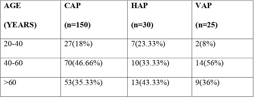
affecting adults in India is about 4%4. Due to the association with risk factor such as
COPD, smoking, males are commonly affected and people between age group of
51-60 yrs are also affected due to chronic ailments.
In India, mortality rate of Community acquired pneumonia CAP lies between
3.3-11%4. Hospital acquired pneumonia (HAP) accounts for about 13-18% of all
nosocomial infection and affects only 0.5- 2%15 of hospitalized patients with
mortality rate from 20-50%. (Hoffkenet al)16,17. In the study done by Vasuki et al18
in Tamilnadu, the incidence of HAP is 10.3%. Patient on mechanical ventilation
have rates of pneumonia 7 to 21 folds higher than patient not on ventilator support.
Rate of infection is twice high in teaching institution when compared to small
institution. Ventilator associated pneumonia (VAP), overall incidence in ICU ranges
CAUSATIVE ORGANISMS OF PNEUMONIA23:
Bacteria - Streptococcus pneumoniae, Haemophilus influenzae,
Mycoplasma pneumoniae, Chlamydophila pneumoniae, pseudomonas aeruginosa,
Enterobacteriaceae, Peptococcus, Prevotella, Actinomyces, Nocardia spp,
Coxiella burnetii, Mycobacteria spp.
Virus - Respiratory syncytial virus, Influenza, Parainfluenza virus type1,2,3.
Rhinovirus, Human Metapneumovirus, Adenovirus (type 4and7).
Parasites – Paragonimus westermani, Ascaris lumbricoides,
Strongyloides stercoralis, Toxoplasma gondii.
Fungi – Histoplasma capsulatum, Coccidioides immitis, Mucor spp, Pneumocystis jirovecii, Rhizopus spp, Absidia spp.
PATHOPHYSIOLOGY 2:
The lung has been frequently exposed to the particulate things, gaseous
mixture and numerous microorganism present in inspired air. In addition, seeping
down of oral secretions from the upper respiratory tract occurs which leads to
microaspiration. The lower respiratory tract are maintained sterile due to defense
mechanisms of the respiratory tract (nasal hair, mucociliary clearance, gag reflex,
cough mechanism).
The acute pulmonary infection is developed due to either a defect in host
defenses, exposure to a particularly virulent microorganism .Infectious agents gain
entry to the lower respiratory tract through aspiration of upper airway resident flora,
from blood. Alveolar macrophages ingest the pathogens and initiate an
inflammatory response which triggers the clinical symptoms of pneumonia.
PATHOLOGY2,24 :
Classic pneumonia evolves through a series of pathologic changes.
Congestion /Edema- with the presence of a proteinaceous exudates, and
bacteria in the alveoli.
Red hepatization - presence of erythrocytes, neutrophils in the cellular
intraalveolar exudate . During this phase, occasionally bacteria may be seen
in the culture.
Gray hepatization- no new extravastion of erythrocyte and those already
existing are lysed and degraded. Neutrophil is the predominant cell,
abundant fibrin deposition, and absence of bacteria which is sign of
improving from infection.
Resolution -The dominant cell type in the alveolar space is the macrophage,
and bacteria, fibrin, debris of neutrophils has been cleared.
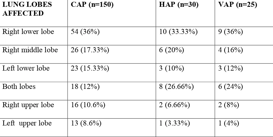
Because of the microaspiration mechanism, bronchopneumonia pattern is
mostly seen in nosocomial pneumonias, whereas a lobar pattern in bacterial
CAP.
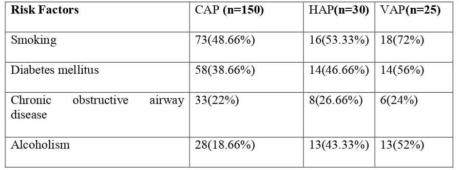
RISK FACTORS 2, 25, 26:
With advanced age group, smoking, alcohol consumption and the presence
of coexisting illness like COPD, diabetes, chronic lung disease, renal failure,
disease in CAP25. Addition to these prolonged intakes of antibiotics and increased
hospital stay influences the outcome of disease in nosocomial pneumonia.
CLASSIFICATION OF PNEUMONIA:
Pneumonia may be classified according to its anatomical location in the lung24:
Lobar pneumonia occurs in one part, or lobe, of the lung.
Bronchopneumonia tends to be scattered throughout the lung.
Clinically pneumonia can be classified as per place of acquisition,
Community acquired pneumonia (CAP) and Nosocomial pneumonia. Nosocomial
pneumonia is further classified in to Hospital acquired pneumonia (HAP),
Ventilator associated pneumonia (VAP) and Health care associated pneumonia
(HCAP)2.
1. COMMUNITY ACQUIRED PNEUMONIA:
CAP can be defined both on clinical and radiographic findings27. According
to Infectious disease society of America(IDSA), Community acquired
pneumonia(CAP) is defined as an acute onset of infection in the lung parenchyma
associated with few symptoms, radiological evidence, auscultatory signs pertaining
to pneumonia in patients who is not admitted in hospital or in any health care
facility for greater than 14 days prior to the symptom onset3.
CASE DEFINITION OF CAP25, 28, 29:
New or progressive infiltration in Chest Xray with atleast any two of the
following symptoms: cough (>4 weeks), purulent sputum production, fever
ETIOLOGY AGENTS IN CAP2:
Outpatient setting- Streptococcus pneumoniae, Haemophilus influenzae, Mycoplasma pneumoniae, Chlamydophila pneumoniae and other respiratory
viruses [Respiratory syncytial virus, Influenza, Parainfluenza virus type1,2,3.
Rhinovirus, human metapneumovirus, Adenovirus (type 4and7)].
Inpatient
setting-NON –ICU-- Streptococcus pneumoniae, Haemophilus influenzae,
Chlamydophila pneumoniae ,Mycoplasma pneumoniae, Legionella pneumophila.
ICU- Streptococcus pneumoniae, Legionella pneumophila, Haemophilus
influenza, Staphylococcus aureus, Gram negative bacilli.
The causative bacterial agents of CAP in India varies with geographical
distribution.for example, the leading causative agent in Shimla and Delhi is
Streptococcus pneumoniae whereas in Ludhiana Pseudomonas aeruginosa
predominates 25.
2. HOSPITAL ACQUIRED PNEUMONIA:
HAP is an inflammation of lung tissue by a pathogen neither present nor at
incubation period during the time of hospital admission27.As per IDSA, pneumonia
occurring after more than fourty eight hours of duration of stay in hospital without
any previous symptoms of pneumonia6. Due to HAP, duration of hospital stay is
CASE DEFINITION OF HAP27, 30:
New or progressive infiltration in Chest X-ray with atleast any two of the following symptoms: cough (>4 weeks), purulent sputum production, fever
(temp.>37.8ºC) or total WBC count >10,000/mm3 occuring in patients admitted in
hospital for after more than fourty eight hours without any prior symptoms of
pneumonia.
ETIOLOGY AGENTS IN HAP31, 32, 33:
Streptococcus pneumoniae, Staphylococcus aureus, Escherichia coli,
Klebsiella pneumoniae, Proteus spp, Serratia marcescens, Haemophilus influenza.
3. VENTILATOR ASSOCIATED PNEUMONIA34:
Ventilator associated pneumonia(VAP)-Infection occurring in patients
admitted in Intensive Care Unit(ICU) who is on mechanical ventilation for more
than 48 hours of intubation8, 7. VAP can be acquired in many routes including
aspiration of oropharyngeal organism, hematogenous spread, MRSA and MDR
pathogens from hands of health care workers, contaminated medical equipment7 .
Classification of VAP7,8 :
Based on duration of mechanical ventilation 19,
A. Early onset VAP- Occurs in first four days on ventilator with possible
causative agents being Enterobacteriacea and Staphyococcus aureus ,carries
B. Late onset VAP- occurs in patient with five days or more on ventilator
mainly due to nonfermenting GNB associated with MDR pathogens.
mortality and morbidity rate is high.
Estimated risk of VAP with hospital stay is high in early as 3% /day for first
five days of ventilation, 2% / day during 5-10 days of ventilation and 1%/day after
10 days of ventilation7.
Case definition of VAP 13,35:
Patient is on mechanical ventilation for more than 48 hours with suspected VAP ,
New and persistant chest infiltrate in chest radiograph with any of these 2 criteria:
Fever (temp.>38ºC) or hypothermia (<36 ºC).
WBC Count ≥10000 mm3 or ≤ 4000mm3.
Purulent tracheal secretion.
ETIOLOGY AGENTS IN VAP 2:
MDR Pathogens- MRSA, Pseudomonas aeruginosa, Acinetobacter
baumanii, ESBL-positive strains Klebsiella pneumoniae, Antibiotic-resistant
Enterobacteriaceae, Legionella pneumophila, Enterobacter spp, Burkholderia
cepacia.
Non-MDR Pathogens - Streptococcus pneumonia, MSSA, Klebsiella spp,
Haemophilus influenza, Escherichia coli, Proteus spp, Serratia marcescens,
Clinical criteria of VAP7, 8, 36, 37, 38:
The Pugin’s modified Clinical Pulmonary Infection Score (CPIS), which
combines clinical, radiographic, physiological and microbiological data into a single
numerical result, is used as a diagnostic tool for VAP. Modified CPIS > 6 is
considered as diagnosis of pneumonia.
Microbiological Criteria of VAP37:
>25 PMN cells with few epithelial cells in Gram stained smear of ETA
sample and Significant quantitative culture (colony count ≥105 cfu/ml).
4.HEALTH CARE ASSOCIATED PNEUMONIA(HCAP)39:
Patient admitted in hospital for ≥2 days within 90 days of diagnosis of
pneumonia, receiving I.V. antibiotics, residing in health care facility <30 days or
received hemodialysis. Multidrug resistant pathogens are the major causative
organism.
CLINICAL FEATURE OF PNEUMONIA2:
Symptoms - fever, cough (productive/non productive), shortness of breath, pleuritic
type of chest pain.
Signs - tachycardia, tachypnoea, dull note on percussion, rales/ronchi/diminished breath sounds on auscultation.
Criteria for hospital admission:
To provide an efficient patient care, criteria has been laid down for patient in
1.Pneumonia Severity Index (PSI) scoring:
The pneumonia severity index (PSI) was developed to categorize patients for
hospital care 39.
Patient characteristic Points
Demographic
Age(years):
Male: age -
Female: age -
Nursing home resident +10
Co- morbidities
Neoplastic disease +30
Liver disease +20
Congestive heart failure +10 Cerebrovascular disease +10
Renal disease +10
Examination findings
Altered mental status +20 Respiratory rate 30/minute +20 Systolic blood pressure <90 mmHg +20 Temperature <35ºC or 40ºC +15
Pulse rate125/minute +10
Laboratory findings
pH <7.35 (do ABG only if hypoxic or COPD) +30 BUN >10.7 mmol/ L +20
Sodium <130 mEq/L +20
Glucose 13.9 mmol/L +10
Hematocrit <0.30 +10 PaO2 <60mmHg or oxygen saturation <90% +10
Pleural effusion +30
Patients with a higher risk are defined as being in PSI risk class V (PSI-V)
PSI score > 130.
2.CURB 65 Criteria: Confusion, Urea(>7mmol/L), Respiratory rate ≥30/mt,
COMPLICATION OF PNEUMONIA24:
Abscess formation particularly seen in type 3 pneumococci, Klebsiella
pneumoniae , Staphylococcus aureus.
Bacterial dissemination to brain, kidney ,spleen and joints.
Empyema - infection spreading to pleural cavity and most common
with infection of pneumococcus40.
LABORATORY DIAGNOSIS4:
Total Leucocyte Count, Differential Count, Erythrocyte sedimentation rate ,
Sputum Microscopy for Gram Stain, Acid Fast Bacilli, Sputum culture and
sensitivity, Chest X-ray PA view, Computerised Tomography Thorax (if necessary),
Arterial blood gas analysis(VAP), Biomarkers-C-Reactive protein, Procalcitonin,
sTREM-1(soluble triggering receptor expressed on myeloid cells)5.
MICROBIOLOGICAL INVESTIGATION COLLECTION OF SPECIMEN41 :
1. SPUTUM42, 43, 44:
Sputum is a mixed collection of bronchial secretion and inflammatory
exudates from affected lobe of lung parenchymal tissue that is coughed up in to the
mouth and expectorated. Sputum should be differentiated from saliva, as sputum is
purulent, opaque, viscus and yellow to green coloured whereas saliva is clear and
Instruction for sputum collection 41,45,46:
1. Sputum is collected prior to the administration of antibiotics.
2. Sputum is collected in the early morning as soon as the patient awakes and
asked to brush the teeth and then rinse the mouth with water before
collection.
3. Sputum is collected in a disposable, sterile, wide mouthed, screw capped
plastic container of about 100ml capacity.
4. Sputum can be collected by asking the patient to deeply cough out the
sputum spontaneously or induced by administering saline nebulisation,
postural drainage or by appropriate physiotherapy.
5. Sputum sample collected in the container is transported to laboratory for
processing within 2 hours of collection.
Processing of sputum: Homogenisation:
Purulent part of the sputum has the appropriate pathogen that is overlaid by
clear to mucoid secretion. Homogenisation is done to make uniform mixture of the
relavant pathogen to be present in the sample , so as each drop of the sample will
contain some amount of pathogen that can be suitable for smear preparation and
culture.
a. Equal volume of sputum sample and sodium dithiothreitol is mixed in a
vortex mixture for about fifteen seconds and allow it to stand for about
b. Equal volume of sputum sample and buffered pancreatin solution is mixed
continuosly and gently in a machine that tilt to and fro and incubated at
37ºC for thirty minutes.
2) ENDOTRACHEAL ASPIRATE (ETA)6,45,47
Endo Tracheal aspirate (ETA) was collected from the patient who has been
in mechanical ventilation for more than fourty eight hours with suspected VAP. One
milliliter of ETA was collected in a sterile screw capped plastic container by using
twenty two inch Ramson’s 12 F suction catheter with a mucus extractor was
introduced slowly in to the endotracheal tube for about a distance of twenty five to
twenty six centimeter.
3) BRONCHOALVEOLAR LAVAGE (BAL)48:
30-50ml of sterile saline is injected in to the fibreoptic bronchoscope which
is threaded to peripheral bronchiolar ramification and it is aspirated, collected in a
sterile container.
4) BLOOD45,48 :
Blood culture was performed in all cases of suspected pneumonia with fever
(temp.>37.8ºC) prior to starting antibiotics. Sterile gloves were worn prior to the
procedure and a patch of skin prepared approx. 5-cm in diameter over the proposed
veni-puncture site. This area was cleansed thoroughly with 70% isopropyl alcohol,
followed by povidone iodine, and followed again by 70% isopropyl alcohol in a
concentric circles moving outward from the centre.
The skin was allowed to dry for at least 1 minute before the sample is
and transferred after removing the needle into the blood culture bottle containing 50
ml of Brain heart infusion broth, maintaining sterile aseptic precautions.
Direct Microscopy:
Smear is made from the homoginsed or purulent material of the sputum.
Gram staining was done and examined under oil immersion field for the relative
number of squamous epithelial cells and neutrophil.
BARLETT’S GRADING:
Average number of neutrophils and epithelial cells for 20-30 LPFs was
calculated and the total score arrived. A score of 0 or lessthan 1 is indicative of
contamination and a score of 1 and above was considered an acceptable quality of
sample.
MURRAY AND WASHINGTON GRADING48:
Epithelial cells/lpf Leukocytes /lpf
Group 1 25 10
Group 2 25 10 - 25
Group 3 25 25
Group 4 10 – 25 25
Group 5 <10 25
Only Group 5 specimen is accepted for culture.
Number of epithelial cells
Grade
10 - 25 -1 >25 -2
Number of neutrophil (lpf)
Grade
<10 0
10 – 25 +1
An acceptable ETA samples in Gram stained smear shows less than ten
squamous epithelial cells per low power or organisms under oil immersion field.
Best ETA samples in Gram stained smear showed >25 polymorphonuclear
leucocytes per low power field with minimal squamous epithelial cells8,45
3. CULTURE41,45,48:
Sputum samples were then plated into the following agar media: Nutrient
agar, 5% Sheep blood agar, Chocolate agar and Mac Conkey agar. All cultures were
incubated at 37°C under aerobic condition and addition to this blood agar and
Chocolate agar also require 5-10% carbon dioxide atmosphere. Plates were
evaluated for growth at 24 and 48 hours.
Endotracheal aspirate, bronchoalveolar lavage specimens were subjected to
quantitative culture. All material is resuspended in the fluid and three serial
dilutions are made (1/10, 1/1000, 1/100,000). These dilutions 0.01 ml is plated out
in 5% Sheep Blood agar and incubated at 37°C under 5-10% carbon dioxide
atmosphere for about 18-24 hours6,49 .
The number of bacteria in the quantitative culture of ETA samples were
expressed in colony-forming unit (cfu) per milliliter.(cfu/ml = number of colonies
×dilution factor ×inoculation factor) Quantitative threshold of organism is colony
count of ≥105 cfu/ml in ETAand ≥104 cfu/ml BAL is consistent with pathogen and
not a colonizer8,50. Bacterial isolates grown in culture were identified by means of
Gram’s staining and biochemical reactions by standard microbiological techniques51
Blood culture:
The inoculated blood culture bottles were incubated at 37°C and examined
after 18 to 24 hours for any turbidity, discoloration or clotting. The first subculture
was done onto Nutrient agar, blood agar and Macconkey agar plates and incubated
aerobically at 37°C with 5-10% CO2 for about 18-24 hours. These bottles were
reincubated and checked for turbidity twice daily.
ANTIBIOTIC SUSCEPTIBILITY TESTING52
Antibiotic sensitivity testing was done on Mueller Hinton agar using Kirby
Bauer disk diffusion method. Interpretation of the results was done by measuring
the sizes of the zone of inhibition according to CLSI guidelines 2015(M-100-S25).
SEROLOGY-Antigen Tests2,53,54,55:
Legionella antigens in urine detects only serogroup1 (accounts for
community acquired infection) with 90% sensitivity and 99% specificity.
Chlamydial antigen (LPSAg) can be demonstrated by ELISA or micro
immunofluorescence method.
Pneumococcal urine antigen test is also quite sensitive and specific (80% and
>90%, respectively).
MANAGEMENT OF PNEUMONIA2,5:
1.COMMUNITY ACQUIRED PNEUMONIA56:
Outpatients-
without comorbidities: Azithromycin [500 mgPO once, then 250 mg qd] or
with comorbidities: respiratory fluoroquinolone (levofloxacin [750 mg PO
qd] moxifloxacin [400 mg PO qd], gemifloxacin [320 mg PO qd]) or
β-lactam amoxicillin [1g tid] or amoxicillin/clavulanate [2g bid] or
ceftriaxone [1–2 g IV qd] , cefuroxime [500 mg PO bid]) cefpodoxime [200
mg PO bid], with a macrolide.
Inpatients:
Non-ICU- respiratory fluoroquinolone (levofloxacin [750 mg PO qd]
moxifloxacin [400 mg PO qd] , gemifloxacin [320 mg PO qd]) β-lactam
antibiotics (ampicillin [1–2 g IV q4–6h], cefotaxime [1–2 g IV q8h], .,
ceftriaxone [1–2 g IV qd], ertapenem [1 g IV qd]) with a macrolide (oral
clarithromycin or azithromycin or IV azithromycin[1 g once, then 500 mg
qd])
ICU- β-lactam antibiotics (ampicillin-sulbactam [2 g IV q8h], ceftriaxone
[2 g IV qd], or cefotaxime [1–2 g IV q8h]) with either fluoroquinolone or
azithromycin .
Special Consideration:
CA-MRSA: Add vancomycin (15 mg/kg q12h) or linezolid (600 mg IV
q12h). Pseudomonas aeruginosa- antipseudomonal β-lactam antibiotics
(Meropenem [1 g IVq8h]) , Imipenem [500 mg IV q6h], Piperacillin/tazobactam [4.
5 g IV q6h], Cefepime [1–2 g IV q12h], with either of fluoroquinolone
{Levofloxacin (750 mg IV qd) or Ciprofloxacin (400 mg IV q12h)}or an
2.NOSOCOMIAL PNEUMONIA57:
a.Patient without risk of MDR pathogen-
Ampicillin /sulbactam I.V. - 3g 6th hourly or Cefotaxime (q 6hr)
/Ceftriaxone(q 24hr) -2g I.V or Moxifloxacin 400mg q24 hr or Ertapenem
I.V.-1g q24 hr.
b.Patient with risk of MDR pathogen-
Linezolid (600 mg IV q12h) or Vancomycin (15 mg/kg q12h) for Gram
positive bacteria along with β-lactam Ceftazidime (2 g IV q8h) or cefepime (2 g IV
q8–12h) or Piperacillin/tazobactam (4. 5 g IV q6h) orImipenem (500 mg IV q6h or
1 g IV q8h), or meropenem (1 g IV q8h) plus Ciprofloxacin (400 mg IV q8h) or
levofloxacin (750 mg IV q24h), Gentamicin or tobramycin (7 mg/kg IV q24h) or
amikacin (20 mg/kg IV q24h) for Gram-negative bacteria.
BACTERIAL RESISTANCE58:
Mechanism by which microrganism showing resistance to antibiotics are due
to production of certain enzymes (β-lactamases) destroy active form of drug are
produced by some microorganism, alteration in the drug permeability, alteration in
the target site or metabolic enzymes responsible for drug action. Drug resistance
may be non genetic (showing phenotypic resistance)or genetic(mutation in
BETALACTAMASES
This is a heterogeneous group of penicillin recognizing proteins. They
belong to members of super family of active site serine protease. These enzymes
inactivate β-lactam antibiotics (Penicillin, Cephalosporins).
CLASSIFICATION OF BETA LACTAMASES
Schemes of functional classification that were accepted by β-lactamase
researchers include:
(i) In 1968, Cephalosporinases and penicillinases were grouped on the basis of
reaction to specific antibody (Sawai et al).
(ii) In 1973, the Richmond and Sykes scheme classified the enzymes into five
main divisions based on the substrate profile and the gene coding for
β-lactamase.
(iii) In 1989, Bush scheme classified β-lactamase on the basis of molecular
structure and the substrate inhibition.
(iv) In 1980, Ambler was the first to propose the Molecular structure
classifications.
(v) More recently, Bush, Jacoby, and Medeiros devised a classification scheme
based on the sequence of nucleotide on the genes for placing β-lactamases
into functional groups and on the enzyme’s biochemical properties and
(Classification schemes for bacterial b-lactamases) Bush- Jacoby-Medeiros group 1989 Bush group Richmon d-Sykes class Mitsuhas hi-Inoue type Molec ular class Preferred substrates Inhibited by: Representative enzyme CAb EDTA
1 1 Ia,Ib,Id Csasea C
Cephalosporins
- -
AmpC from Gram negative
bacteria, MIR-1
2a 2a
not
included Pcase V A
Penicillin
+ -
Penicillinases from Gram Positive bacteria
2b 2b III Pcase I A Penicillins, Cephalosporins + - TEM-1, TEM-2, SHV-1
2be 2b'
not included except K1
in class
IV
Cxase A
Penicillins, Narrow
spectrum and
extended spectrum Cephalosporins,
Monobactams. + -
TEM-3 to TEM-26, SHV-2
to SHV-6, Klebsiellaoxytoca
K1
2br not included not included not included A Penicillins ± - TEM-30 to TEM-36, TRC-1
2c 2c II,V Pcase IV A Penicillins,Carbenicillins + - PSE-1, PSE-3, PSE-4
2d 2d V Pcase II, Pcase III D Penicillins, Cloxacillin ± - OXA-1 to OXA-11, PSE-2 (OXA-10)
2e 2e 1c Cxase A
Cephalosporins
+ -
Inducible cephalosporinases
from Proteus vulgaris
2f not included not included not
included A
Penicillins, Cephalosporins,
Carbapenams + -
NMC-A from Enterobacter
cloacae, Sme-1 from Serratia marcescens
3 3
not included
not
included B
Mostβ lactams, including
carbapenams - +
L1 from Xanthomonasmaltophilia
CcrA from Bacteroides
Fragilis
4 4
not included
not
included NDc
Penicillins
-
Penicillinase from Pseudomonas
Cepacia
a Csase, cephalosporinase; PCase, penicillinase; CXase, cefuroxime-hydrolyzing b-lactamase.
b CA, clavulanic acid.
c ND, not determined.
EXTENDED SPECTRUM BETA LACTAMASES (ESBL)
ESBL are plasmid mediated betalactamases that produce resistance to broad
spectrum betalactam antibiotics like 3rd and 4th generation cephalosporins, extended
METHODS FOR DETECTION OF EXTENDED SPECTRUM BETA LACTAMASES61,62
SCREENING OF ESBL-Disc Diffusion method52,63
The disc diffusion methods are the screening test for ESBL production by
Escherichia coli, Klebsiella and Proteus mirabilis as proposed by CLSI 2015
guindelines use cefotaxime, ceftazidime, cefpodoxime, ceftriaxone and astreonam
for the screening of ESBL production. Screening test is positive for ESBL is that the
isolates showing resistant to 2 or more 3rd generation cephalosporin and it should be
confirmed by phenotypic confirmatory test. The resistant zone size for ESBL as per
CLSI 2015 guidelines: Cefotaxime ≤ 27mm, Ceftazidime ≤ 22mm, Ceftriaxone ≤
27mm, Cefpodoxime ≤ 17mm and Aztreonam ≤ 27mm respectively.
CONFIRMATORY TEST FOR ESBL:
1. Phenotypic confirmatory disc diffusion test11,52,64
The CLSI advocates the phenotypic confirmatory test for the detection of
production of ESBL by Klebsiella and Escherichia coli which use the cefotaxime or
ceftazidime discs (30μg) with or without clavulanate (10μg). A Semiconfluent
growth of test organism on Mueller Hinton agar shows difference of 5 mm along
the cephalosporin with clavulanate disc compared to cephalosporin disc alone.
2.Minimum Inhibitory Concentration
Agar dilution method :
Minimum inhibitory concentration was performed by agar dilution method
ceftazidime 2μg to 2048μg/ml and cephalosporin with 4μg of clavulanic acid
ranging from 0.5μg to 2048μg/ml of agar was tested with isolates. MIC is the least
concentration at which there is no visible growth and it was obtained as eight fold
decrease in CAZ-CL compared to ceftazidime.
Broth Micro dilution:
Disc potentiation test can also be done using broth microdilution assays by
using ceftazidime (0.25to128μg/ml), ceftazidime with clavulanate (0.25/4 to128/4
μg/ml), cefotaxime (0.25 to64μg/ml), and cefotaxime with clavulanate (0.25/4 to
64/4 μg/ml) decrease in MIC of two fold serial dilution of cephalosporin with
clavulanate compared to the MIC of cephalosporin alone suggests positive for
ESBL production.
COMMERCIAL METHODS AVAILABLE TO DETECT ESBL
(i) Epsilometer-Test for ESBLs65,66
Plastic drug impregnated strips are produced by AB bio disk in which one
end contains a gradient of ceftazidime (MIC test ranges from 0.5μg - 32μg/ml) and
with a ceftazidime gradient and constant concentration of clavulanate (4μg/ml). As
per CLSI guidelines 2015, MIC value of ceftazidime – clavulanate should be ≥ 8
fold decrease in concentration than MIC value of ceftazidime alone.
(ii) Vitek ESBL67
Vitek ESBL cards contain cefotaxime and ceftazidme alone and
cephalosporin plus constant concentration of clavulanate. Cards are inoculated in
soon as the growth in the control well has attained a set threshold. A prefixed
reduction in the growth of cefotaxime and ceftazidime plus clavulanic acid
containing wells is compared with the growth in the wells having
cefotaxime/ceftazidime alone, indicates positive for ESBL producer. The sensitivity
and specificity of the test is more than 90%.
GENOTYPIC METHOD TO DETECT ESBL67,68,69:
Detection of the common ESBL gene such as TEM, SHV and CTX-M by
molecular method10,68.
Test Advantages Disadvantages
DNA
Probes Specific for gene family(e.g., TEM or SHV) Labour distinguish between ESBLs and intensive, cannot non ESBLs, and between variants of TEM or SHV
PCR Easy to perform, specific for gene family(e.g.,TEM or SHV)
Require technical skill and expensive.
Oligotyping Detects specific TEM variants Requires specific oligonucleotide probes, labour intensive, cannot detect new variants.
PCR- RFLP
Easy to perform, can detect specific nucleotide changes
Nucleotide changes must result in altered restriction site for detection.
Nucleotide sequencing
Gold standard, can detect all variants
Labour intensive, can be technically challenging, can be difficult to interpret manually.
Real Time PCR
Rapid identification, minimum cross contamination
METALLOBETALACTAMAES70:
Metallo-β-lactamases (MBL’s) are carbapenemases produced mainly by
Pseudomonas aeruginosa which require zinc at the site of action. They are
designated in Ambler’s Class B and Bush-Jacoby Medeiros Group 3. They
hydrolyze virtually all β-lactam agents such as penicillin, cephalosporin, including
the carbapenams.
Till now seven major types of MBL were described worldwide – IMP, SPM,
VIM, GIM, SIM, AIM-1 and NDM-1. Among them, blaIMP and blaVIM are the
most common types of MBLs that are prevalent worldwide. From India, only
blaVIM and NDM-1 have been reported in P. aeruginosa in the past.
TESTS TO DETERMINE METALLO BETA LACTAMASES AMONG NON FERMENTERS
SCREENING FOR MBL
An isolate of P. aeruginosa was considered screen-test positive for MBL
when it was Imipenem resistant: 10 μg(IPM) and/or Meropemem: 10 μg (MRP)
and/or Ceftazidime: 30 μg (CAZ) . Antibiotic sensitivity was done by the
Kirby-Bauer disc diffusion method as per CLSI recommendation.
CONFIRMATION OF MBL PRODUCTION
MODIFIED HODGE TEST71:
MHA plate is streaked with the ATCC Escherichia coli 25922 and an
imipenem disc is placed in the centre. Imipenem resistant isolates are inoculated
read. Imipenem hydrolyzing strains produce distortion on the zone whereas non
hydrolyzing zones do not produce any effect.
Amp C BETALACTAMASES72
METHODS TO DETECT Amp C PRODUCTION73
All Enterobacteriaceae isolates were screened for Amp C betalactamases
production by disk diffusion method.
SCREENING OF Amp C PRODUCTION74,75:
All isolates were screened for cefoxitin susceptibility and those which had a
zone diameter of ≤ 18 mm were suspected to be AmpC producers.
Amp C DISK TEST7,70,76 :
In a MHA plate, a lawn culture of E. coli ATCC 25922 was made. Sterile
saline (20μl) was put on the sterile disks (6 mm) which was later inoculated with
several colonies of test organism. Cefoxitin 30µg disk (almost touching) was placed
on a fresh inoculated plate. The inoculated disk was then placed adjacent to
cefoxitin disk and incubated overnight at 35°C. The inference was made as follows:
Flattening or indentation of the cefoxitin inhibition zone in the vicinity of the
test disk was taken as positive. A negative test had an undistorted zone.
MANAGEMENT OF INFECTION WITH ESBL – PRODUCING ORGANISMS60,77
ESBL producers are treated with Piperacillin – Tazobactam ,Cefoperazone –
sulbactam in case of mild infection ,whereas severe infection are treated with
Amp C producers are usually resistant to cephamycins and oxyimino-beta
lactams but they are sensitive to carbapenams but diminished porin expression
makes them resistant to carbapenam as well which can be treated with Polymixins,
Tigecycline, Fosfomycin or Colistin.
METHICILLIN RESISTANT STAPHYLOCOCCUS AUREUS48,78
Staphylococcus aureus is the most important human pathogen present in the
external environment and in the anterior nares of 20- 40% of adults. It is also seen
in the axillae, intertriginous skin folds, the perineum, and the vagina. It is
responsible for mild infections to severe life threatening infections. Penicillin was
the drug of choice for the treatment of serious S.aureus infections. The advent of
penicillin resistance in the S.aureus was due to the acquisition of plasmid borne
genetic elements coding for β lactamase production. Later, Penicillinase-resistant,
semisynthetic penicillins such as oxacillin, methicillin was the drug of choice due to
its incorrect use, MRSA showing difference in penicillin binding protein known as
PBP2a from a chromosomal gene (mecA) has emerged. Initially1970s MRSA was
accounting to 40-60% of all nosocomial infection but after 1990 it has been
associated with population in the community who has no contact with hospital
known as community- associated MRSA (CA-MRSA) strains which has caused
death of 4 children due to necrotizing pneumonia79. MRSA prevalence in India has
MECHANISM OF RESISTANCE
The chromosomally localized mecA gene responsible for methicillin
resistance acts by synthesizing Penicillin binding protein 2a in turn downregulates
the cross linking of peptidoglycan layer, through which it shows resistance to
betalactam antibiotics. Four different SCC mec elements have been recognized.
SCCmec type I, II and III is associated with Health care associated MRSA
(HA-MRSA). Community associated MRSA tend to carry SCC Type IV element and
Panton- Valentine leukocidin as virulence factor.
METHODS TO DETECT MRSA
PHENOTYPIC METHOD
1. Cefoxitin disc diffusion test12,76-
The test was performed by placing 30μg of Cefoxitin disc in the Mueller
Hinton Agar plate inoculated with test organism. The plate was kept in incubator at
a temperature of 37˚C. The zone of inhibition was determined after 24 hrs and the
zone size was interpreted as Susceptible ≥ 22mm and Resistant ≤ 21 mm.
2. Oxacillin screen agar method80
Oxacillin screening is done by using 6 µg/ml of oxacillin in Mueller Hinton
agar to confirm all methicillin resistant strains .The strains which grow in this
3. MIC determination
(i) Agar dilution method
4-5 discrete colonies were emulsified onto 4-5 ml of nutrient broth which is
adjusted to 0.5 McFarlands standard.0.0001ml is used as the final inoculum. The
concentration of oxacillin used is 32μg-0.015μg/ml. After drying,1μl of inoculum is
inoculated in the plates using a calibrated loop . The plates are incubated at 37˚C for
24hrs.MIC is the lowest concentration at which no visible growth occurs.
Susceptible - ≤ 2μg/ml, Resistant - ≥ 4μg/ml
(ii) Broth dilution method
To a Mueller Hinton broth with 4% NaCl, serial dilution of oxacillin is
added. Few colonies of S. aureus are emulsified into fresh peptone water and
adjusted to match 0.5 McFarlands standard which is used as inoculum. It is
incubated at 33-35c or 24 hrs. Oxacillin MIC <2μg/ml sensitive and Resistant>
2μg/ml.
(iii) E-TEST12,52,81:
MIC test should be performed to differentiate Vancomycin susceptible
isolates of S.aureus from Vancomycin intermediate isolates. Plastic drug
impregnated strips are produced by in which a gradient concentration of
Vancomycin (MIC test ranges from 0.016μg - 256μg/ml) was applied on a MHA
plate on which MRSA isolates were swabbed and the plates were incubated at 37ºC
overnight. Interpretation of Vancomycin MIC E-test for MRSA as per CLSI
guidelines 2015 are as follows 52: <2 µg/ml is sensitive,4-8 µg/ml is Intermediate
GENOTYPIC METHODS 82
Multiplex PCR for MRSA detection of mec A and fem B genes, coag genes,
ccr genes, nuc genes, toxin genes.
1. Pulsed Field Gel Electrophoresis
From an overnight grown culture of a single colony, a bacterial pellet is
processed and the restriction fragments are separated on the gel. Gel is stained with
ethidium bromide. The photo is taken under Ultra Violet light. Strain relatedness
among CA-MRSA and HA-MRSA isolates can be investigated.
2. Real time PCR
MRSA isolates are detected directly from blood culture bottles using real
time PCR assays. Based on melting curve analysis, the assay differentiates into
clusters.
3. Multi locus Sequence Typing (MLST)
The clonal evaluation of MRSA is detected by MLST. Sequential analysis
from 7 Staphylococcus aureus shows the housekeeping genes as follows i.e., aroE,
arcC, glpF, gmK, pt, tpi and yqil. Each isolate is defined by all the alleles of the
seven genes. This results in an allele profile / gene sequence type (ST).
4. Microarray Analysis
Multiplex PCR products can be used as hybridization samples. After
hybridization at the test site of the microarray, detection of fluorescence is done
automatically by the instrument images of the array. Automatically captured image
Management of MRSA infections60, 77
The drug of choice for serious infections caused by Methicillin Resistant
Staphylococcus aureus is glycopeptide antibiotics (Vancomycin or Teicoplanin).
PREVENTION:
VACCINATION2, 31, 83, 84, 85 :
Vaccination against influenza, pneumococcus, Haemophilus influenza b is
used to reduce the burden of pneumonia.
1. Influenza vaccines are of two types intranasal live-attenuated cold-adapted
vaccine (not indicated in immunocompromised patients) and intramuscular
inactivated vaccine. During an influenza outbreak, vaccination given immediately
with chemoprophylaxis (zanamivir or oseltamivir for 2 weeks).Efficacy is about it
prevents 53% pneumonia, 50% from hospitalization and 68% from death.
2. Pneumococcus- Pneumococcal infection severe due to its invasive property
and drug resistance. two types of pneumococcal vaccine - PPV23 pneumococcal
polysaccharide vaccine contains capsular material from 23 pneumococcal
serotypes and PCV13, protein conjugate pneumococcal vaccine contain capsular
polysaccharide from 13 protein commonly affecting children. PCV13has an
immunogenic protein which induces the production of T cell–dependent antigens
for long-term immunologic memory. PCV13 vaccine is given for children,
immunocompromised patients and elderly. VAP-oral care, hand hygiene, use of
prophylactic agent of gastric ulcer, gloves usage, protocol directed weaning
MATERIALS AND METHODOLOGY
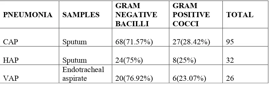
The study of bacterial isolates causing Pneumonia in 205 adult patients with
suspected Pneumonia includes 150 Community Acquired Pneumonia (CAP), 30
Hospital Acquired Pneumonia (HAP) and 25 Ventilator Associated Pneumonia
(VAP).
STUDY DESIGN : Cross sectional study
STUDY PERIOD : January 2015 to June 2016
STUDY PLACE : Government Kilpauk Medical College and Hospital, Chennai.
INCLUSION CRITERIA
1. Clinically suspected and radiologically proven cases of pneumonia.
2. Patient above 18 years of age (adult).
EXCLUSION CRITERIA:
1. Patient with active tuberculous leision was excluded.
2. Patient taking antibiotics currently and past for the period of about two
weeks were excluded.
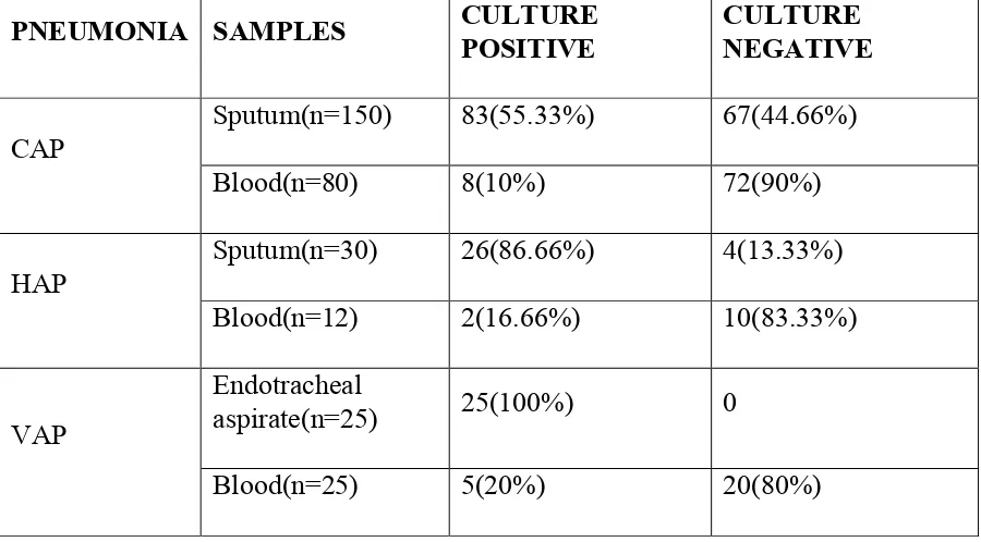
SAMPLE COLLECTION, TRANSPORT AND PROCESSING:
Totally 205 respiratory samples from Pneumonia in adult patients attending
Government Kilpauk Medical College and Hospital includes 150 sputum samples
from Community Acquired Pneumonia(CAP), 30 sputum samples from Hospital
Acquired Pneumonia(HAP) and 25 Endotracheal samples from Ventilator
Blood samples was collected from pneumonia patients having symptoms of
fever (Temp ≥37.8ºC )86 were included 80 samples from CAP, 12 samples from
HAP and 25 samples from VAP.
After obtaining informed consent from these patients respiratory samples
and blood were collected under sterile precaution and transported immediately to
the laboratory in appropriate settings and sample processing done.
SAMPLE COLLECTION 46, 87:
1. Sputum – expectorated or induced41,3,28
Deeply coughed out or when the sputum is scanty it was induced with saline
nebulisation and was collected in a disposable leak proof sterile ,wide mouthed
container with tight fitting lid after giving proper instruction to the patient.
2. Endo Tracheal aspirate (ETA)6,45
Endo Tracheal aspirate (ETA) was collected from the patients who have
been in mechanical ventilation for more than fourty eight hours with suspected
VAP. One milliliter of ETA was collected in a sterile screw capped plastic
container by using twenty two inch Ramson’s 12 F suction catheter with a mucus
extractor which was introduced slowly in to the endotracheal tube for about a
distance of twenty five to twenty six centimeter.
3. Blood86:
Blood culture was performed in all cases of suspected Pneumonia with fever
(temp.≥37.8ºC) prior to starting antibiotics. Under aseptic precaution, 5 ml of
blood was collected by venipuncture using sterile syringe and transferred after
dilution)45of Brain heart infusion broth. Samples collected were sent immediately to
Microbiology laboratory without delay.
SAMPLE PROCESSING
a) Macroscopic examination41:
The sputum was examined for colour (rusty, red currant jelly was noted),
consistency, purulent/non purulent to distinguish it from saliva.
b)Direct Microscopy:
The Sputum, Endotracheal aspirate specimens were subjected to the following
microscopic examination using standard laboratory techniques. Gram staining was
done and examined for the presence of relative number of polymorphonuclear cells
and squamous epithelial cells.
Criteria for assessing the quality of respiratory samples48:
Bartlett’s grading:
Number of neutrophil (lpf)
Grade
<10 0 10 - 25 +1
>25 +2 Presence of mucus
+1
Number of epithelial cells
Grade
10 - 25 -1
>25 -2
Total number of polymorphonuclear cells and epithelial cells and in 20-30
LPFs was calculated and averaged the total score was arrived. A final score of 0 or
less indicated lack of active inflammation or contamination, and a score of 1 and
than ten squamous epithelial cells per low power field in Gram stained smear of
ETA samples was accepted for culture 6.
CULTURE 4:
Sputum culture:
Sputum samples were then plated into the following agar media: Nutrient
agar 5% Sheep blood agar, Chocolate agar and MacConkey agar. All cultures were
incubated at 37°C under aerobic condition and addition to this blood agar and
Chocolate agar plates were kept under 5-10% carbon dioxide atmosphere. Plates
were evaluated for growth at 24 and 48hours.
Endotracheal aspirate culture:
Endotracheal aspirate specimens were subjected to quantitative culture 6,88,89.
Colony count of ≥105 cfu/ml was consistent with pathogen and not a colonizer7,8.
Endotracheal aspirate sample was resuspended in the fluid and three serial
dilutions were made (1/10, 1/100, 1/1000). Of these 0.01 ml from 1/1000 dilutions
was plated on to Blood agar. The number of bacteria in culture of ETA samples
were expressed in colony-forming unit (cfu) per milliliter.(cfu/ml = number of
colonies ×dilution factor ×inoculation factor)Presence of single colony in 0.01ml of
1/1000 dilution indicate ≥105 colonies49 . Bacterial isolates grown in culture were
identified by means of Gram’s staining and biochemical reactions by standard
microbiological techniques.
Blood culture45,48:
The inoculated blood culture bottles were incubated at 37°C and examined
was done onto Nutrient agar, blood agar and Macconkey agar plates and incubated
at 37°C for 18 to 24 hours, meanwhile in addition to these blood agar plate was
kept under 5-10%CO2. These bottles were reincubated and checked for turbidity
twice daily.
ANTIBIOTIC SUSCEPTIBILITY TESTING52
Antibiotic sensitivity testing was done on Mueller Hinton agar using Kirby
Bauer disk diffusion method. Interpretation of the results was done by measuring
the sizes of the zone of inhibition according to CLSI guidelines
2015(M-100-S25).Quality control strains used are as follows7:ATCC 25922 Escherichia coli ,
ATCC 27853 Pseudomonas aeruginosa and ATCC 25923 Staphylococcus
aureus .
DETECTION OF EXTENDED SPECTRUM BETA LACTAMASES
All Enterobacteriaceae isolates were screened for betalactamases production
by disk diffusion method and confirmed by phenotypic confirmatory disc diffusion
test.
Disk diffusion methods-screening for ESBL52:
Disk diffusion test was done for all Enterobacteriaceae isolates against
Cefotaxime (30 μg), Ceftriaxone (30 μg), and Ceftazidime (30 μg) antibiotic disks
for the screening of the isolates for potential ESBL production.
Overnight incubation was done at 37˚C after which the zone size was read as
per CLSI recommendations for ESBL screening criteria in which the isolates
Antibiotics Zone of inhibition –
interpretation
Cefotaxime (30µg) ≤27mm
Ceftriaxone(30µg) ≤25mm
Ceftazidime(30µg) ≤22mm
Quality controls were performed using
ATCC 700603 Klebsiella pneumoniae - Positive control.
Phenotypic confirmatory disc diffusion test11,52
This test was done for all Enterobacteriaceae isolates against Ceftazidime
(30 μg) antibiotic discs with and without clavulanic acid (10 μg). These discs were
placed on a Mueller –Hinton agar plate inoculated with bacterial suspension
equivalent to 0.5 McFarland standards. Overnight incubation was done at 37°C
after which the result was interpreted as follows:
If the zone diameter of Ceftazidime with clavulanic acid was increased ≥ 5
mm when compared with Ceftazidime alone was taken as positive for ESBL
production.
MIC determination – E Test method65
Minimum inhibitory concentration was calculated for all isolates of ESBL by
Epsilometer-Test for ESBL 10,52
Plastic drug impregnated strips are produced by Himedia in which one end
contains a gradient of ceftazidime (MIC test ranges from 0.5μg - 32μg/ml) and
Ceftazidime+clavulanic acid (MIC test ranges from 0.064 μg - 4μg/ml) on the other
end was applied on a MHA plate on which ESBL isolates are swabbed. The plates
were incubated at 37ºC overnight.
As per CLSI guidelines 2015, MIC value of ceftazidime – clavulanate
should be ≥ 8 fold decrease in concentration than MIC value of ceftazidime alone.
(Manufacturer recommends MIC value in the ratio of CAZ:CAC ≥ 8).
DETECTION OF ESBL PRODUCERS BY POLYMERASE CHAIN REACTION (PCR) 10
DNA Extraction methods
DNA extraction was done with the help of DNA Purification kit (PureFast
Bacterial Genomic DNA purification kit) and polymerase chain reaction master
mix.
Constituents of Master Mix 2X
Taq DNA Polymerase - 2Units.
10X Taq reaction buffer
2mM Magnesium Chloride.
10mMdNTPs mix - 1μl.
Agarose for the purpose of Gel Electrophoresis - Agarose, 50XTAE buffer,
6Xgel loading buffer, Ethidium bromide were used.
PRIMERS
CTX-M primer( Product size - 269bp )
5'-TTATGCGCAGACGAGTGCGGTG-3'
5'-TCACCGCGATAAAGCACCTGCG-3'
SHV primer(Product size- 276bp )
5'-CGCCGCCATTACCATGAGCGAT-3'
5'-ACCCGATCGTCCACCATCCACT-3'
TEM primer(Product size - 250bp)
5’-CCAAACGACGAGCGTGACACCA-3’
5’-AGCGCAGAAGTGGTCCTGCAAC-3’
Procedure of DNA Extraction
One ml of overnight culture of Klebsiella pneumoniae was centrifuged at
6000 rpm for five minutes and supernatant was discarded.Pellet was suspended in
200μl of Phosphate Buffer Saline (PBS).
To the suspension twenty microlitre of lysozyme(10mg/ml) , 180μl of
lysozyme digestion buffer was added and incubated at 37ºC for fifteen minutes.
Mixing with 400μl of binding buffer ,5 μl of internal control template and 20 μl of
Protienase K was done by inverting the tube several times and then incubated at
300μl of ethanol was added and mixed well. Whole lysate was transferred
into Pure Fast spin column. It was then centrifuged for one minute at 10000 rpm.
Discard flow through and 500μl of Wash Buffer-1 was added and centrifuged
at 10000 rpm for 1Minute. Discard flow through and 500μl of Wash Buffer-2 was
added and centrifuged at10000 rpm for 1minute. This procedure was done for two
times.
Discard flow through centrifuged column for 1 more minutes so that any
residual ethanol will be removed. The content in spin column was transferred to a
1.5 ml micro-centrifuge tube. 100μl of Elution Buffer was added to elute the DNA,
and centrifuged for 2 minute.
Procedure of Polymerase Chain Reaction
1. Reaction was done with the components in PCR vial-10μl of Master
Mix, 5μl Genomic DNA, 5μl Primer mix which constitutes about 20 ml
of total volume.
2. All these were mixed gently and spinned down briefly.
3. They were then placed in the PCR machine and programmed;
Initial Denaturation: 95ºC for 5 minutes
Denaturation: 94ºC for 30seconds in cycles of 35
Annealing: 58ºC for 30 seconds in cycles of 35
Extension: 72ºC for 30 seconds in cycles of 35