A PROSPECTIVE OBSERVATIONAL STUDY TO DETERMINE
THE AETIOLOGY OF POSTMENOPAUSAL BLEEDING AND
CORRELATION OF ENDOMETRIAL THICKNESS IN
ENDOMETRIAL CARCINOMA IN OUR POPULATION.
CERTIFICATE
This is to certify that this dissertation,
“A PROSPECTIVE OBSERVATIONAL STUDY TO DETERMINETHE AETIOLOGY OF POSTMENOPAUSAL BLEEDING AND CORRELATION OF ENDOMETRIAL THICKNESS IN ENDOMETRIAL CARCINOMA IN OUR POPULATION” is the bonafide work of Dr. Carolin Solomi. V. under my supervision in the Department of Obstetrics and Gynaecology, Christian Medical College Vellore in partial fulfilment of the requirements for the award of M.S, Obstetrics and Gynaecology Examination of the Tamil Nadu Dr. M.G.R Medial University to be held in April 2017 and no part thereof has been submitted for any other degree.
Dr. Jessie Lionel,
Professor and Head of Unit-1,
Department of Obstetrics and Gynaecology,
Christian Medical College,
CERTIFICATE BY THE HEAD OF THE DEPARTMENT/ PRINCIPAL
This to certify that this dissertation,
“A PROSPECTIVE OBSERVATIONAL STUDY TO DETERMINE THE AETIOLOGY OF POSTMENOPAUSAL BLEEDING AND CORRELATION OF ENDOMETRIAL THICKNESS IN ENDOMETRIAL CARCINOMA IN OUR POPULATION” is the bonafide work of Dr. Carolin Solomi .V. under the supervision of Dr. Jessie Lionel, Professor and HOU in the Department of Obstetrics and Gynaecology Unit 1, Christian Medical College Vellore in partial fulfilment of the requirements for the award of M.S, Obstetrics and Gynaecology Examination of the Tamil Nadu Dr. M.G.R Medial University to be held in April 2017 and no part thereof has been submitted for any other degree
Dr. Anna B. Pulimood Principal,
Christian Medical College, Vellore.
Dr. Annie Regi,
Professor and Head of the department, Department of Obstetrics and Gynaecology, Christian Medical College,
DECLARATION
I, Carolin Solomi .V. , do hereby declare that the dissertation titled “A PROSPECTIVE OBSERVATIONAL STUDY TO DETERMINE THE AETIOLOGY OF POSTMENOPAUSAL BLEEDING AND CORRELATION OF ENDOMETRIAL THICKNESS IN ENDOMETRIAL CARCINOMA IN OUR POPULATION” is a genuine record of research done by me under the supervision and guidance of Dr Jessie Lionel, Professor and HOU of Department of Obstetrics and Gynaecology Unit 1, Christian Medical College, Vellore and has not previously formed the basis of award of any degree, diploma, fellowship or other similar title of any university or institution.
Vellore Dr. Carolin Solomi. V.
Acknowledgement
I acknowledge my dependence and gratitude to GOD Almighty in successful completion of my dissertation.
I express my sincere and heartfelt gratitude to Dr Jessie Lionel, Professor and HOU, Department of Obstetrics and Gynaecology Unit-1, Christian Medical College, Vellore for her tireless efforts and guidance during the study. Her valuable views and ideas along with her meticulous correction have brought me so far to complete my dissertation.
I express my sincere thanks to Dr. Elsy Thomas, Professor, Department of Obstetrics and Gynaecology Unit-1, for her timely support and guidance during the study period.
I acknowledge Dr. Anita Thomas, Professor, Department of Gynae-Oncology, Christian Medical College, Vellorefor her help during the study period.
I am extremely grateful to Mrs Narayani and her sonology technicians‟ team without whose help this dissertation would not have been successfully completed. Their willingness to always help me in recruiting patients will always be remembered by me.
I am also thankful to all my colleagues and nursing staff in Gynaecology wards and OPD for their efforts in recruiting patients.
I acknowledge the valuable help from Mrs Mahasampath Gowri from the Department of Bio-statistics.
I specially thank Dr. Thambu David, Professor in the department of Medicine, who took special steps in teaching me the basics of research methodology which has helped me a lot in my dissertation.
I thank all the patients who consented to be part of this study without whom it would have been impossible to have all this done.
CONTENTS
INTRODUCTION
...
01
AIMS AND OBJECTIVES
...
04
REVIEW OF LITERATURE
...
06
MATERIALS AND METHODS
...
37
RESULTS
...
43
DISCUSSION
...
82
LIMITATIONS
...
88
CONCLUSION
...
90
BIBLIOGRAPHY
...
93
1
Introduction:
Postmenopausal bleeding (PMB) is an alarming symptom, which is associated with many gynaecological problems and is seen in 4-11% of menopausal women (1). PMB constitutes for 10% of the cases in gynaecology outpatient clinic. It is one of the most common symptom for which menopausal women seek medical care and evaluation. (1). Postmenopausal bleeding is indicative of underlying malignancy until proven otherwise”, is a golden dictum which is accepted by all gynaecologists. A woman who bleeds after menopause has a 10-15% risk of developing endometrial carcinoma (1-4). 90% of patients with endometrial cancer will have abnormal uterine bleeding, most commonly postmenopausal bleeding. Hence any woman presenting with postmenopausal bleeding should beconsidered for further evaluation, irrespective of the amount of bleeding or its episodic character (5). Unlike other malignancies, endometrial cancer presents at an early stage and curative treatment is available if diagnosed earlier (4).
2 (9).Hence this study is done to determine the commonest cause of postmenopausal bleeding in our population.
The primary goal in the diagnostic evaluation of postmenopausal bleeding is to exclude endometrial carcinoma (10). The initial evaluation of the uterine cavity in a woman with postmenopausal bleeding can be done by Trans vaginal ultrasound (10). Endometrial cancer can be excluded when the endometrium is thin and homogeneous and endometrial biopsy can be deferred for them (10). Studies done by various study groups including, Tabor et al, Osmers et al, Nasri et al, have showed variable endometrial thickness cut-off viz 4mm, 5mm, 6mm as significant for having endometrial carcinoma(11-13)
4
AIMS ANDOBJECTIVES:
1. To determine the most common cause of postmenopausal bleeding in our menopausal women.
6
REVIEW OF LITERATURE:
Definitions:
Menopause
WHO defines Menopause as permanent cessation of menstruation due to loss of ovarian follicular activity (14).
According to The International Menopause Society Menopause is defined as a period after 12 consecutive months of amenorrhea where there is no obvious pathological/ physiological cause. (15)
The International Menopause Society also defines menopause as a period known in certainty only after one year from the Final menstrual period (FMP).(15)
Post menopause:
WHO defines post menopause as dating from the FMP, regardless of whether the menopause was induced or spontaneous. (14)
Postmenopausal bleeding:
Postmenopausal bleeding is defined as bleeding from the genital tract that occurs 12 months after the final menstrual period (FMP). (14)
Introduction:
7 menopause is 47.5 years (16). It isfound in the recent past decades that the lifeexpectancyof Indian women is increasing, and they spend about one third of their life in the menopause period. During this period of life, the menopausal women experience a wide range of menopausal symptoms starting from the menopausal transition period .The distressing menopausal symptoms include hot flushes, urogenital symptoms, mood swings, osteoporosis, postmenopausal bleeding. In addition, menopause perse itself contributes few health risks for these women like cardiovascular problems, cancers etc. Of all these menopausal symptoms explained above, the one which is of major concern is postmenopausal bleeding (PMB) (16).
Global epidemiology:
8 in women (18). The life time risk of developing endometrial cancer among whites is 2.4% and among blacks is 1.3 %( 18).
Cervical cancer is the second most common malignancy worldwide (17). In comparison with developed countries, there is disproportionate increase in the incidence of cervical cancer in developing countries. The mortality related to it is mainly due to lack of infrastructure which is required to screen and detect the pre invasive lesions of Cervix (17).
The third most common malignancy is ovarian malignancy; its prevalence is the same in developing and developed world (17).
Vaginal and Vulval cancers contribute to less than 5% of gynaecologicalmalignancies (17).
Choriocarcinoma accounts for less than 1% in the group of gynaecologicalmalignancies (17).
Indian Epidemiology:
9 Endometrial cancer is not very common in India when compared to its prevalence in Western countries. In India, data suggest a prevalence rates of 4.3/1, 00,000 in hospital based population in Delhi, 4.2/1, 00,000 and 2.8/1, 00,000 in Bangalore and Mumbai respectively (20).
Aetiology of Postmenopausal bleeding:
Postmenopausal bleeding is bleeding from any part of the genital tract, the uterus, cervix, vagina, vulva, fallopian tubes, or due to ovarian pathology. Uterine source is the most common cause of postmenopausal bleeding. The origin of bleeding can also involve non gynaecologic sites, such as urethra, bladder, rectum and bowel. Hence, the causes of postmenopausal bleeding can be classified on the basis of the origin of the bleeding as, non-genital, genital, uterine or extra uterine (6).
As per the literature, studies done by Bani et al, has shown that the commonest aetiology of postmenopausal bleeding is atrophic endometrium which accounts for 60-80%, followed by oestrogen replacement therapy by 15-25%, endometrial polypswhich constitute about 2-12%, endometrial hyperplasia by 5-10% and lastly the endometrial carcinoma by another 10%(8).
10 cause. In their study, endometrial carcinoma accounted for 12.5% and cervical carcinoma for 11.5%. Other benign aetiologies included cervicitis (12.9%), atrophic vaginitis (12.3%), cervical polyp (6.7%), hyperplasia (3.1%), urethral caruncle (2.5%) and oestrogen replacement therapy (1.8%) (9).
Endometrial Atrophy:
11
Endometrial Cancer:
Of all the women with postmenopausal bleeding, approximately 10% of women will have endometrial cancer (8). All women with endometrial cancer presents with postmenopausal bleeding but the vice versa is not true. Hence, every woman with postmenopausal bleeding should be evaluated to rule out endometrial cancer.
Endometrial Polyps:
Endometrial polyps are seen in women at their late reproductive and early menopause age (22-24). 2-12% of women with endometrial polyp presents with postmenopausal bleeding. Endometrial polyps are outgrowths from the endometrial lining in localized areas due to excessive stimulation of oestrogen (24, 25). The exact aetiology of endometrial polyps is unknown but polyps generally arise due to exogenous hormone replacement therapy or tamoxifen therapy (26, 27).
Polyps are usually benign although few may be precancerous or malignant (23). About 0.5% of endometrial polyps will have adenocarcinoma cells.
Endometrial hyperplasia:
12 manifest clinically as abnormal uterine bleeding in perimenopausal and post-menopausal women. Endometrial hyperplasia in postmenopausal women can be attributed to endogenous production of oestrogen from either ovarian or adrenal tumours or exogenous oestrogen therapy or tamoxifen therapy (28). Obese postmenopausal women also have high levels of endogenous oestrogen which contribute to endometrial hyperplasia (29).
Types of endometrial hyperplasia are (28):
1. Simple hyperplasia without atypia
2. Complex hyperplasia without atypia
3. Simple hyperplasia with atypia
4. Complex hyperplasia with atypia.
Histopathologically, endometrial hyperplasia is considered when the following features are present (30):
1. Disordered or irregular proliferation of endometrial glands.
2. Increase in gland to stromal ratio. Even though proliferative endometrium also has increased gland to stromal ratio, it is more pronounced in endometrial hyperplasia.
Postmenopausal hormone therapy:
13 Menopausal hormone therapy is given in two forms as, Sequential hormone therapy or continuous combined hormone therapy.
Sequential hormone therapy:
Normal menstrual cycle is characterized by oestrogen predominance during follicular phase and progesterone dominance during secretory phase. Sequential pills are pills which contain oestrogen alone during the first half of cycle (15 pills) and the rest (6 pills) containing both oestrogen and progesterone.
According to K Freely et al, among the postmenopausal women who are using sequential hormone therapy, the endometrium showed weak secretory activity in 70% of cases and proliferative activity in 15% of cases. About 5% of women showed atrophic endometrium (31).
Sequential hormone therapy in postmenopausal women converts the proliferative endometrium to secretory endometrium and thereby decreases the chance of endometrial hyperplasia and malignancy.
14 progesterone duration in the sequential pill should be at least 12-14 days to prevent endometrial hyperplasia or malignancy (33-35).
Continuous Combined Hormone therapy:
Large prospective study done by Wells et al on usage of continuous combined hormone therapy in postmenopausal women has shown that there is no evidence of hyperplasia or malignancy after five years of itsusage (36). The mechanism by which the continuous combined hormone therapy prevents endometrial hyperplasia and malignancy is by arresting the mitosis in the glands and leading to atrophic endometrium (34). However, in literature there are anecdotal reports of endometrial malignancy in postmenopausal women using continuous combined hormone therapy (37). The reasons attributed are (37-40):
1. Prior use of unopposed oestrogen,
2. Inadequate dose of Progestin in the pill,
3. Use of less effective Progesterone,
4. Poor compliance,
15
Endometrial effects of SERMS:
Another important cause of postmenopausal bleeding is the usage of tamoxifen for breast cancers. Selective oestrogen receptor modulators (SERMS) are group of drugs that act on the oestrogen receptors as competitive partial agonists (41). Different tissues have different degrees of sensitivity to the oestrogens, thus having agonist on some and antagonist on others (41).
CLASSIFICATION OF SERMS (41):
I Generation: Triphenylethylenes (Tamoxifen)
II Generation: Benzothiophenes (Raloxifene)
III Generation: Nafoxidene, Lasofoxitene, Ospemifene.
16 endometrium. Several large trials found an increased incidence in endometrial carcinoma among users of Tamoxifen for breast cancer. The risk of endometrial cancer in women treated with tamoxifen is dose and time dependent (42). According to ACOG, when used in the standard doses of tamoxifen, it causes endometrial hyperplasia, polyp formation and carcinoma (42). According to the study done by National Surgical Adjuvant Breast and Bowel Project, the endometrial cancer among women with tamoxifen users was 1.6/1000(41).
Uterine sarcomas account for only 8% of all uterine cancers as compared to the women treated with tamoxifen, in whom sarcomas accounted for 17% of cases (41).
Mechanism of action of Tamoxifen in endometrium:
Tamoxifen has proliferative action on the endometrium. It causes hyperplasia of the endometrial glands and hypertrophy of the stroma (41). The molecular mechanisms behind the action of Tamoxifen on endometrium are (41, 43-44):
1. Endometrium of women who are on Tamoxifen has been consistently found to have increased oestrogen and progesterone receptors.
2. Following receptors including oestrogen receptors, c-fos and glyceraldehyde phosphate dehydrogenase mRNA will be upgraded by Tamoxifen.
17
Ultrasound findings in Tamoxifen treated endometrium:
1. There is an increase in the thickness in tamoxifen treated endometrium. It is found that the endometrial thickening in these patients is due to the sub endometrial stromal oedema and enlargement of stromal cells (41, 46).
2. Few patients on Tamoxifen may also develop cysts (7%) and endometrial polyps (12-25%). (41, 46)
Herbal and dietary supplements: There are few case series to state that soya and other
phyto oestrogens in large doses may cause estrogenic stimulation of the endometrium (47). One series reports the association of soya with polyp and leiomyoma growth (48). A randomized trial on 376 postmenopausal women who received soya versus placebo showed a significant increase in endometrial hyperplasia over a five-year period (48).
Pathogenesis of endometrial cancers:
The status of normal postmenopausal endometrium:
18 From the pathological point of view, a normal postmenopausal endometrium will have only 50% of inactive/atrophic glands and the rest have weak proliferative glands. These proliferative glands will be either focal or diffuse but, will be exerting a low level of effective estrogenic stimulation on the endometrium (49, 50).
Mechanism of Hormonal feedback system:
In response to the decrease in oestrogen levels, the hypothalamus releases GnRH pulses which trigger the anterior pituitary to secrete FSH (52). FSH executes its action in the ovaries by stimulating the ovarian stroma and aids in androgen production. The androgens produced are converted to oestrone by peripheral aromatization and this result in continuous estrogenic stimulation of the endometrium which makes the endometrial glands to switch from their inactive state to a weak proliferative state (52).
Endometrial cancers in menopausal women are most often well differentiated endometriodadenocarcinomas (86%), which arise from the weakly proliferative type of endometrial glands (53).
19 In the past, endometrial cancers in menopausal women were grouped as: (53)
1. Grade 1 Endometriod adenocarcinoma (55%).
2. Grade 2, 3 Endometriod adenocarcinoma (20%)
3. Mosaic of serous papillary and clear cell carcinoma (15%)
4. Non endometriod carcinoma (10%)
20 The current classification of endometrial tumours is by their histological type and
by their degree of tumour differentiation (Low grade vs. high grade).
The molecular level expressions are also different between the two groups of tumours. In low level malignant potential tumours there is (53):
1. Inactivation of PTEN function,
2. Deletion of tumour suppressor genes like EMX2,
21 4. Microsatellite instability.
In high level malignant potential tumours there is:
1. Mutations in p53 tumour suppressor gene,
2. Reduced expression of epithelial cadherin (E-cadherin).
Risk factors for endometrial cancer:
Risk factor is defined asany characteristic of a person that increases the chance of developing a disease or cancer. The risk factors can be modifiable or non-modifiable. Factors like age, family history, age of menarche, menopause, etc. are non-modifiable. Some factors like obesity, diabetes, drug intake, etc. are modifiable riskfactors.The various risk factors which influence the risk of developing endometrial cancer are as follows.
1. Obesity
2. Increasing age
3. Early menarche and late menopause
4. Anovulation- Polycystic ovarian disease.
5. Diabetes and hypertension
22
Obesity:
Obese postmenopausal women have increased risk for developing endometrial cancer (54, 55). It is associated with 2-5 fold increase in the risk of endometrial cancer (55). They develop cancer mainly due to the excess production of oestrone and to a lesser extent due to the decreased levels of sex hormone binding globulin (SHBG) in them(55,56).
The presence of excess fat cells in obese women increases the peripheral conversion of androgens (androstenedione) from ovaries and adrenals to oestrone. Moreover, the concentration of SHBG is decreased in obese women leading to excess unbound oestrone causing endometrial changes (56).
Age:
23
Exogenous hormone therapy:
Unopposed exogenousoestrogen administration is another important risk factor. It is associated with 8-15 fold increase in endometrial cancer. When combined contraceptive pills are used instead of oestrogen alone, the risk of developing endometrial cancer is decreased (53).
Long term usage of Tamoxifen has 6 fold increase in risk of endometrial cancer. The risk is increased drastically when it is used for more than 5 years (53).
Reproductive factors:
Early menarche, late menopause, nulliparity, history of polycystic ovarian disease is associated with prolonged oestrogen exposure to endometrium, making them high risk for developing endometrial cancer (53).
Lifestyle factors:
Less physical activity and diet containing excess fat indirectly increase the risk of endometrial cancer by increasing the BMI (58).
Medical comorbidities:
24
Miscellaneous:
Cervical stenosis, Pyometra and Ichthyosis uterus are taken as risk factors for developing squamous cell variant of endometrial cancer.
Prior radiation therapy can cause endometrial cancer (Secondary carcinoma).
Genetic causes-Lynch Syndrome.
Factors with reduced risk for developing endometrial cancer (61-63):
1. Cigarette smoking.
2. Coffee intake
3. Intrauterine contraceptive device usage.
Diagnostic Workup and Evaluation:
Endometrial evaluation:
25
Ultrasound in evaluation of Postmenopausal bleeding:
Over the past years, it is found that transvaginal ultrasound is used to accurately diagnose endometrial pathologies. In women with postmenopausal bleeding measurement of endometrial thickness helps in differentiating those who are at risk for endometrial cancer.
TVS which is used as the initial investigation to evaluate the endometrium in women with postmenopausal bleeding is less invasive and has excellent high negative predictive value. TVS is also used in women whom endometrial sampling was performed but tissue was insufficient for diagnosis (64).
The International Endometrial Tumour Analysis (IETA) group has come forward with the following recommendations for an universal standardized technique in measuring the endometrial thickness and determining intracavitrarylesions (65, 66).
Technique of measuring Endometrial Thickness:
26 plane as maximum anteroposterior thickness of the endometrial echo (64, 66). It is the maximum measurement between the two endometrial layers (double endometrial layer) (66). The callipers should be placed in the endometrial-myometrium interface in a magnified image (64, 66).
While measuring the endometrial thickness if the angle of insonation between the endometrium and the ultrasound beam is 90 degrees, the quality of the image will be better (66). The endometrial thickness measurement should be reported in millimetres.
In cases where the endometrium is asymmetrically thickened, the endometrial thickness is reported as the sum of the largest anterior and posterior endometrial thickness (66).
Terms and Definition in interpretingendometrialthickness (66):
The IETA group in the World Congress of Ultrasound in Obstetrics and Gynaecology has recommended standard terms and definitions in interpreting ultrasound reports (66).They are as follows:
1. Echogenicity:
27 cases of atrophic endometrium (66). If the endometrium appears heterogeneous, asymmetrical or cystic it is reported as non-uniform echogenicity.
2. Endometrial – myometrial Junction:
The endometrial –myometrial junction should be reported as regular, irregular, interrupted, (or) not defined (65).
In 10% of cases, where the endometrium is not visualized clearly, the report should be interpreted as „non-measurable‟ (66).
Radiological appearance of postmenopausal endometrium:
The normal postmenopausal endometrium is thin, homogeneous and echogenic. Generally, a uniform, thin homogeneous endometrium of <4 mm without focal thickening excludes malignancy and is suggestive of atrophy. Any endometrial thickness of > 4 mm or non-uniform endometrium with focal thickening should be further investigated by endometrial biopsy or hysteroscopy. The presence of postmenopausal bleeding with endometrial thickness >5mm is 92% sensitive and 57% specific for endometrial cancer (67,). ACOG Committee Opinion also supports the above statement (64).
28 persistent postmenopausal bleeding even if endometrial thickness is less than 4mm (64). The supporting evidence behind this is, a thin, homogeneous endometrium does not reliably exclude type 2 endometrial cancer (69, 70).
A study done by Wang et al reviewed the preoperative ultrasound reports of 52 postmenopausal women who were diagnosed to have type 2 endometrial cancer. Out of the fifty two postmenopausal women, 9 (17%) had endometrial thickness less than 4mm and another 9 (17%) had indistinct endometrium. In these 18 women, in addition to the thin endometrium there were additional ultrasound findings such as adnexal mass, intracavitary growth /fluid etc. Hence, it was concluded that any women with postmenopausal bleeding despite a thin endometrium when associated with other ultrasound abnormalities will require an endometrial sampling (69).
Ferrasi et al and Karlson et al have shown that using 4mm as the cut off for endometrial thickness, the transvaginal ultrasound has 96-98% sensitivity and 36-68% specificity in detecting endometrial cancer. The false positive rates are between 44 and 66%. However, the same studies have picked up 4 cases of endometrial cancer when the endometrial thickness was less than 3.5mm (71, 72).
29 of ultrasonography in detecting endometrial carcinoma was as high as 97%. Hence, it was concluded that women with postmenopausal bleeding with endometrial thickness of less than 3mm are less likely to have endometrial cancer and further investigations can be avoided on them (73).
Invasive methods for endometrial evaluation in women with postmenopausal
bleeding (74):
1. Dilatation and curettage.
2. Office endometrial sampling.
3. Hysteroscopy directed biopsy.
4. Saline sonosalphingography.
Dilatation and curettage:
30 Hence, to avoid all these complications newer endometrial sampling methods have come into practice.
Office Endometrial sampling (74):
First Generation methods:
1. Vabra aspirator
2. Novak curette.
Second Generation methods:
1. The Pipelle device
2. The Pipette
3. The Z-Sampler
4. The Tis- U- Trap.
Technique of endometrial sampling:
31 inner core which on withdrawing outwards helps to create negative pressure to aspirate tissue(78).
It is found that, of all the newer sampling devices, Pipelle is better, safe, accurate and economical in sampling the endometrial tissue in detecting endometrial pathologies. (74).
The Pipelle:
The Pipelle sampling device was first devised by Cornier.E in 1984(79). It is a 23 cm long flexible, plastic polypropylene sheath with the outer sheath diameter 3.1mm and a piston in the inner sheath with diameter 2.6mm. The outer sheath of the Pipelle has graduated markings from 4cm to 10cms from the extreme distal rounded tip of the device. The tip of the device has a small opening of 2.2mm in diameter through which the endometrial tissue will get aspirated when the proximal end of the piston is pulled outwards(79).
It is said that the office endometrial sampling by Pipelle, samples adequate endometrial tissue as compared to the traditional dilatation and curettage. The diagnostic accuracy in detecting endometrial pathologies by Pipelle was found to be 95.5 %.( 76, 77)
32 histopathological results from curettage and Pipelle sampling coincided in 87% of women. Hence, it was estimated that endometrial sampling by Pipelle had 75% sensitivity and 100% specificity in diagnosing endometrial pathologies. When ultrasound is used along with Pipelle, its sensitivity increased to 90 %( 84). Goldschmidt et al in their analysis also supported that in 90% of cases the results from endometrial sampling by Pipelle correlated with D&C results (85). Using transvaginal ultrasound along with Pipelle sampling increases the sensitivity but decreases the specificity in detecting endometrial disease.
Even though endometrial sampling by Pipelle has excellent outcomes, a recent study done by Rezk et al contradicts this. This prospective observational study was done to assess the safety and acceptability of Pipelle endometrial sampling in postmenopausal women when compared to Pipelle sampling in premenopausal women. Primary outcomes taken were safety and adequacy of the Pipelle sampler. The conclusions of this study were endometrial sampling by Pipelle is more painful, lessacceptable and less adequate in postmenopausal women (86).
33 Gudio et al found that, when 65 women with known endometrial cancer underwent Pipelle endometrial sampling cancer was missed in 11 out of 65 women. Endometrial cancer can be a focal disease involving 25% or 50% of the endometrial cavity. Hence, blind sampling can cause errors (88).
3. Hysteroscopic guided Biopsy:
Dilatation and Curettage and endometrial sampling methods, being blind procedures can miss cases of endometrial polyps, myomas, focal hyperplasia and neoplasia. In view of all these constraints hysteroscopy is considered as the preferred method in evaluating the endometrium in women with postmenopausal bleeding (74). It has high sensitivity and specificity in detecting endometrial pathology (74). It visualizes the focal endometrial abnormalities and thereby directed biopsies can be taken in the same sitting (89). In the recent years, due to the advancements in the technology and surgical expertise hysteroscopy is considered as a safe, simple out-patient procedure which is easily performed without any discomfort to the patient (89). Hysteroscopy along with endometrial biopsy helps in accurately diagnosing the endometrial pathology in a woman who is presenting with postmenopausal bleeding. Office based hysteroscopy except in being a diagnostic tool, it is also used in surgically treating the cause of bleeding in the same setting (89, 90).
34 bleeding and concluded them to be 95.1%, 100%, 81%, 92% and 100% respectively(90). In 69% of patients the cause of bleeding was also removed hysteroscopically in the same sitting thus facilitating treating with diagnosis (90).
Even though hysteroscopy has high sensitivity, specificity and excellent diagnostic accuracy in detecting endometrial pathologies, it is not cost effective and easily applicable as transvaginal ultrasound (89). Hysteroscopy is not superior to D&C and other sampling methods in its sensitivity to detect endometrial hyperplasia and malignancy (89). Even though, hysteroscopy is superior to other diagnostic modalities in detection of endometrial polyps, it should be used with caution in women suspected to have endometrial cancer because of the possibility of retrograde spilling of malignant cells into the peritoneum during hysteroscopy (89, 90).
Therefore, in developing countries like India where there is lack of infrastructure, hysteroscopy is not considered as first line gold standard modality in the evaluation of postmenopausal bleeding. When endometrial polyps is suspected and in grossly thickened endometrium the routine endometrial sampling procedure can be bypassed and diagnostic hysteroscopy followed by biopsy can be performed (74).
4. Saline infusion sonohysterography:
35 inflating balloon placed in the cervix (92). The expansion of uterine cavity is directly observed through the transvaginal ultrasound. In an out-patient setting, the feasibility of saline infusion sonography is similar to diagnostic hysteroscopy (93). It helps in discriminating high risk patients who need further evaluation (93). Abeera Choudry et al, did a cross sectional study on 77 women with postmenopausal bleeding to estimate the diagnostic accuracy of saline infusion sonography in them and concluded that saline infusion sonography along with endometrial biopsy can be used as a standard procedure in the evaluation of women with postmenopausal bleeding because of its excellent sensitivity (92%), specificity (79%) and good patient acceptability (93).
36
MATERIALS AND
37
Materials and Methods:
Thestudy was a prospective observational study which was conducted in Christian Medical College Hospital, Vellore between February 2016 to July 2016 in the department of Obstetrics and Gynaecology after the Institutional EthicsCommitteeclearance. We included all postmenopausal women who presented any time after one year of menopause with postmenopausal bleeding. Detailed history, clinical examination, per speculum and per vaginal examination was done systematically to evaluate the clinical diagnosis of postmenopausal bleeding. A structured proforma was made and details of the patient including her age, age of menarche, age of menopause, parity, body mass index, amount of bleeding, number of episodes of postmenopausal bleeding, associated co morbidities and any drug intake like hormone therapy, and anticoagulants are noted, following which, the diagnostic evaluation for postmenopausal bleeding is done by using transvaginal ultra sonogram and the endometrial thickness was determined. Endometrial biopsy is done after ultrasound and the histopathological report was correlated.
38
Participants:
Eligibility Criteria:
Inclusion criteria:
All women who have attained natural menopause after 45 years of age, presenting with postmenopausal bleeding after one year of menopause.
Exclusion criteria for the second aim of the study:
1. Women with other obvious causes of bleeding from cervix and vagina. 2. Women with known case of bleeding disorders
3. Premature menopausal women.
4. Postmenopausal women with postmenopausal bleeding who have been treated with hormones elsewhere.
5. Transvaginal ultrasound showing adnexal pathology.
39
Flow diagram 1:
Setting:
The materials of the study were obtained from the patients who presented with postmenopausal bleeding to the Obstetrics and Gynaecologyoutpatient clinic. All postmenopausal women who presented with postmenopausal bleeding from February 2016 to July 2016 were recruited. Patient recruitment and data collection through a structured proforma was done by the principal investigator. Following data collection, a transvaginal ultrasound was done in the Out Patient department ultrasound room by a trained and experienced senior sonology technician and by
All women with PMB after one year of menopause are recruited
Data collection done using a structured proforma
Patients were subjected to Transvaginal ultrasound and Pipelle biopsy
Correlation of endometrial thickness with the histopathology report of endometrial carcinoma and set a cut-off thickness
Determine the most common cause of
40 the principal investigator. After the ultrasound, endometrial sampling using pipelle was done by the principal investigator in minor operation theatre.
A woman who is recruited for the study was considered postmenopausal when one year has elapsed from the final menstrual period. No hormonal investigation like FSH was done to confirmher menopausal status. All these women except women who had a clinical diagnosis of carcinoma cervix underwent a transvaginal scan followed by endometrial sampling by Pipelle.
The endometrial samples were classified into following histopathological categories: 1.Endometrial carcinoma, 2.Endometrial hyperplasia, 3.Atrophic endometrium, 4. Proliferative endometrium, 5.Secretory endometrium, 6. Endometrial Polyps, 7. Shed endometrium with no hyperplasia./malignancy, 7.Sample inadequate for evaluation.
Statistical methods and sample size calculation:
Data were summarized using mean (S.D), and median (range) for continuous variables and by frequency (percentage) for categorical variables.
Prevalence of carcinoma was presented with 95% CI. (Exact method was used).
41 An ROC analysis was done to obtain the cut-off of endometrial thickness discriminating carcinoma and non-carcinoma. For the cut-off decided, the diagnostic accuracies with 95% CI were presented.
All analysis was done in STATA 13.1/I C.
Sample size calculation:
To detect a prevalence of 10% (1, 6, 7, 94), we collected a sample of 144 postmenopausal women, with 95% CI and 5% precision. The following formula was used.
n = 4pq/d²
Where p denotes prevalence, taken as 10, q is 100-p,
d is precision, which is taken as 5 Hence,
n = 4 x 10 x 90/25
43
Results:
The baseline characteristics of our study population are as follows.
[image:56.612.68.544.219.698.2]The baseline characteristics of our study population are tabulated below:
Table 1:
No. of patients Percentage (%) Age of menarche
Less than 11 years 4 2.78% More than 11 years 144 97.22% Age of menopause
45-50 years 84 58.33% 50-55 years 59 40.97% >55 years 1 0.69% Parity
Nulliparous 7 4.86% Multiparous 137 95.14% BMI
44
Age of presentation:
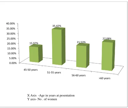
In our study, of the 144 women recruited with postmenopausal bleeding, 15.97% of them were between 45-50 years, 35.42% between 51-55 years, 21.53% between 56-60 years, and 27.08% beyond 60 years of age.
Table 2: Table showing the distribution of patients according to the age of presentation:
Age No. of patients Percentage
45-50 years 23 15.97%
51-55 years 51 35.42%
56-60 years 31 21.53%
45
Figure 1: The bar diagram below depicts the study population according to their age groups.
It is evident from the above bar diagram, postmenopausal bleeding seems to occur most commonly within the next few years of menopause (i.e.) at 51-55 years constituting 35.4% and the next peak after 60 years of age at 27%.
0.00% 5.00% 10.00% 15.00% 20.00% 25.00% 30.00% 35.00% 40.00%
45-50 years
51-55 years
56-60 years
>60 years 15.97%
35.42%
21.53%
27.08%
[image:58.612.74.507.67.431.2]46
Age of menarche:
97% of women had menarche beyond 11 years and only 2.7% had early menarche. The mean age of menarche was found to be 14 years.
Age of menopause:
58% of women in our study group had attained menopause between 45-50 years and 41% of them between 50-55 years and only one woman beyond 55 years. The mean age of menopause in our study group was found to be 48.7 years which is consistent with data from the Indian Menopause Society where the mean age of menopause for Indian women was noted to be 47.5 years (16).
Parity:
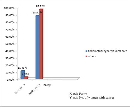
Multiparous women constituted 95% of our study population whereas 7 women (4.86%) were nulliparous. Among the women with endometrial hyperplasia and malignancy, 11.43% were nulliparous.
Table 3: Table showing the distribution of endometrial cancer/hyperplasia according to parity
Parity Cancer Others p value Nulliparous
4(11.43%) 3(2.78%)
[image:59.612.92.480.579.713.2]47
Figure 2: Bar diagram showing the prevalence of endometrial cancers/hyperplasia
according to parity.
BMI:
On considering the BMI, about 36% of our women were overweight, 29.8% obese and 3.4% morbidlyobese. The mean BMI in our study population was 28.15 kg/m2.
Table 2 below shows the number of women with endometrial hyperplasia/cancer on the basis of their BMI:
0.00% 10.00% 20.00% 30.00% 40.00% 50.00% 60.00% 70.00% 80.00% 90.00% 100.00%
11.43%
88.57%
2.78%
97.22%
Parity
Endometrial hyperplasia/cancer others
X axis-Parity
[image:60.612.72.505.69.434.2]48
Table 4:
BMI(Kg/m2) Frequency Percentage p value
< 25 7 20%
0.0554
26-30 13 37.14%
31-35 11 31.43%
36-40 1 2.86%
>40 3 8.57%
Figure 3: Picture showing the distribution of endometrial hyperplasia/cancer cases on the
basis of BMI. 0% 5% 10% 15% 20% 25% 30% 35% 40% 20% 37.14% 31.43% 2.86% 8.57%
Women with endometrial hyperplasia and
cancer
Women with endometrial hyperplasia and cancer
X Axis -BMI
49
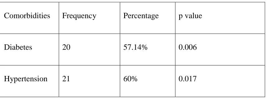
Diabetes and hypertension:
Medical comorbidities like diabetes and hypertension were found in 38% and 43% of our study women respectively.
[image:62.612.71.500.368.526.2]The following table shows the number of diabetics and hypertensives with endometrial cancer/hyperplasia:
Table 5:
Comorbidities Frequency Percentage p value
Diabetes 20 57.14% 0.006
50
Figure 4: Figure showing the prevalence of endometrial cancer/hyperplasia among
diabetics and hypertensives.
Age:
The mean and median age of women with postmenopausal bleeding was found to be 56.6 and 55 years respectively in our study population .The mean age noted in the
0% 10% 20% 30% 40% 50% 60% 70% 80% 90% 100%
57.14% 60% 42.86% 40%
Others
Women with hyperplasia and cancer
[image:63.612.68.506.81.472.2]51 various studies ranged from 47.43 to 57.5 years (95). In the prospective study done by Thomas Gredmark et al the mean age quoted is 61.4 years (96).
In our study, it was found that, the age at which women presents with postmenopausal bleeding peaks at 51-55 years. Even though, there is a slight decrease in women presenting with postmenopausal bleeding after 56 years of age there is definitelyan increase in the incidence of malignancy in this group as compared to women in the early menopausal years.Studies done by Gredmark T et al, Bani et al and Youssef et al had a similar finding (8, 96, 97).Youssef et al, in his study showed that 25 patients (69%) who presented with postmenopausal bleeding were between 50-60 years where as only 5 patients (13%) were above 70 years and at the same time the incidence of malignancy was more after 60 years (97).
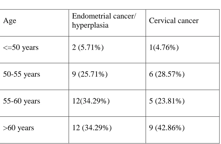
The frequency of postmenopausal bleeding decreased with increasing age but, the number of women with cancer (endometrium and cervix) increased proportionately. There were only 2 cases (5.71%) of endometrial malignancy in the age group of less than 50 years whereas there were 9 patients (25.71%) between 50-55 years and 12 patients (34.29%) between 55-60 years and another 12 patients (34.29%) beyond 60 years.
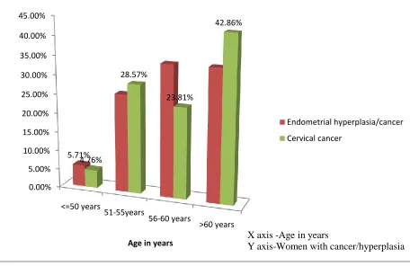
52 On comparing endometrial malignancy and cervical malignancy, endometrial malignancy rates increased beyond 55 years of age and cervical malignancy peaked beyond 60 years.
[image:65.612.67.429.336.571.2]The number of endometrial and cervical malignancies in each age group is summarized in table 6 and Figure 5:
Table 6:
Age Endometrial cancer/
hyperplasia Cervical cancer <=50 years 2 (5.71%) 1(4.76%)
50-55 years 9 (25.71%) 6 (28.57%)
55-60 years 12(34.29%) 5 (23.81%)
53
Figure 5:
It was also found in our study that, the mean age of developing premalignant and malignant diseases of the uterus i.e. endometrial hyperplasia and malignancy was found to be 58.8 years where as mean age for developing other benign conditions like atrophic endometrium, endometrial polyp etc. was found to be 55.71 years. Hence, it is evident in our study that, as age increases the chance of malignancy increases which is statistically significant with a p value of 0.0233. This is in consistent with the study done by Thomas Gredmark et al where he found the incidence of endometrial cancer was found in older women between 65-69 years (96, 98).
0.00% 5.00% 10.00% 15.00% 20.00% 25.00% 30.00% 35.00% 40.00% 45.00% <=50 years 51-55years 56-60 years >60 years 5.71%4.76% 28.57% 23.81% 42.86%
Age in years
Endometrial hyperplasia/cancer Cervical cancer
X axis -Age in years
54
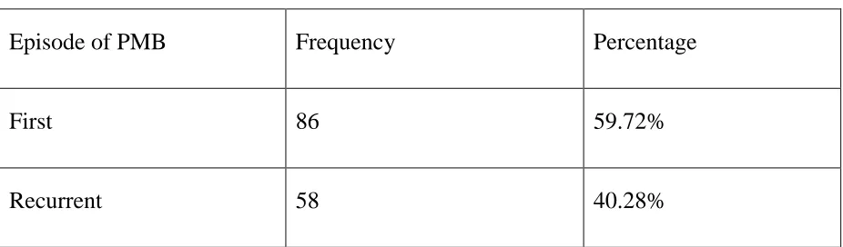
Other characteristics:
[image:67.612.67.530.496.632.2]In our study, 86 women (59.7%) presented to the health care facility in the first episode of postmenopausal bleeding. The rest 40% of women had recurrent episodes of bleeding before they presented to the health care facility. The number of recurrent episodes ranged from 2 to 21 episodes. This is in consistent with the study done by Choo et al where 70% of women presented with the first episode of postmenopausal bleeding(98).The mean age at which our women in the study group experienced the first episode of postmenopausal bleeding is 55.85 years which is about 8-10 years post menopause. This is in consistent with the study done by Bani et al and Choo et al where the mean duration of menopause was 10 years (8, 98).
Table 7:
Episode of PMB Frequency Percentage
First 86 59.72%
55
Figure 6: The above bar diagram shows the number of women with first and recurrent episodes of bleeding.
Amount of bleeding:
The bleeding was described as scanty bleeding in 46% of women, moderate bleeding in 35% of women and heavy bleeding in 18% of women. This is shown in the following table.
0.00% 10.00% 20.00% 30.00% 40.00% 50.00% 60.00%
First
Recurrent 59.72%
40.28%
Episode of postmenopausal bleeding
[image:68.612.67.528.66.329.2]56
Table 8:
Type of bleeding Frequency Percentage
Scanty 67 46%
Moderate 51 35%
Heavy 26 18%
Figure 7: Bar diagram showing the distribution of study population based on amount of
bleeding. 0.00% 5.00% 10.00% 15.00% 20.00% 25.00% 30.00% 35.00% 40.00% 45.00% 50.00%
Scanty
Moderate
Heavy 46%
35%
18%
Amount of postmenopausal bleeding
[image:69.612.68.528.108.648.2]57
Menopausal hormone therapy:
No women in our study group had menopausal hormone therapy.
Drugs:
Of the 144 women, 8 women (5.55%) had exposure to drugs like anticoagulants, antiplatelet and Tamoxifen. Among the 8 women, 2 women had tamoxifen use, 4 ofthem had aspirin use and the rest two of them had anticoagulant use.
Aspirin:
Of the 4 women who were using aspirin, 2 had endometrial polyps, 1 patient had endometrial hyperplasia and the other had cancer cervix as their cause for postmenopausal bleeding.
Anticoagulants:
Of the 2 women who were using anticoagulants, one patient had cancer cervix and the other patient had endometrial hyperplasia.
58
Tamoxifen:
Among the 2 women who were using Tamoxifen, one patient hadtamoxifen usage for 32 months and had endometrial polyp, whereas the other patient had tamoxifen usage for 48 months and had endometrial cancer.
The following table shows the cause of postmenopausal bleeding in the women who were on aspirin, anticoagulants and Tamoxifen:
Table 9:
Drugs Endometrial cancer
Endometrial
hyperplasia Cancer cervix
Endometrial Polyp
Aspirin - 1(25%) 1(25%) 2(50%)
Anticoagulants - 1(50%) 1(50%) -
Tamoxifen 1(50%) - - 1(50%)
59
Figure 8: Bar diagram showing the endometrial pathologies in women with medications
intake.
Family h/o malignancies:
Of the 144 women in our study population, 21 women (14.5%) had family h/o malignancies, like oral, upper GI, colonic, breast and uterine cancers. Of the 21 women, 12 women had malignancies, uterus -6 (28.5%), breast- 4 (19%) , colon-2 (9.5%) which are shown to have greater association with endometrial malignancy. 3 out of 12 (25%) of
0% 5% 10% 15% 20% 25% 30% 35% 40% 45% 50% Endometrial cancer Endometrial hyperplasia
60 those who have given h/o first degree relative having uterus, breast and colonic cancer had endometrial cancer.
Table 10 below shows the number of endometrial cancer in women with family h/o uterine, breast and colonic cancer.
Table 10:
Family h/o malignancy Endometrial cancer Others
Uterine cancer 1 5
Breast cancer 1 3
Colon cancer 1 1
Aetiology of postmenopausal bleeding:
61
Table 11:
Histopathological report No. of patients Percentage (%)
Atrophic endometrium 28 19.44%
Endometrial polyp 24 16.66%
Endometrial malignancy 22 15.27%
Carcinoma Cervix 21 14.58%
Endometrial hyperplasia 14 9.72%
Ovarian cancers 6 4.16%
Secretory endometrium 4 2.77%
Proliferative endometrium 3 2.08%
62
Figure 9: Bar diagram showing the aetiology of postmenopausal bleeding in our study population.
In our study, histopathological reports from the endometrial sampling by Pipelle were only included. Of the 144 samples, 123 patients had the endometrial sampling by Pipelle. Benign conditions including endometrial polyps, secretory endometrium, proliferative endometrium were found in 31 patients which accounted for 21.5%.
Premalignant conditions which included endometrial hyperplasia with/without
atypia and Cervical intraepithelial neoplasia were seen in 14 patients (9.72%). Atrophic endometrium was evident in 28 patients (19.44%).
0% 2% 4% 6% 8% 10% 12% 14% 16% 18% 20% 19.44% 16.66% 15.40% 14.58% 9.72% 4.16% 2.77% 2.08% 15.27% X axis -Causes of PMB
[image:75.612.71.533.76.408.2]63 The malignancies diagnosed in our study were endometrial cancers, cervical cancers and ovarian malignancies. Among our study population, 22 women (15.27%) had endometrial malignancy, 20 (14.58%) had cancer Cervix and 6 patients (4.16%) had ovarian cancers.
[image:76.612.74.507.339.606.2]Of the 144 women with postmenopausal bleeding, 22 women (15.27%) had no histopathological diagnosis for their bleeding and was reported as endometrium with no evidence of hyperplasia/malignancy.
Figure 10: Pie chart showing the major attributable causes of postmenopausal bleeding.
36.80%
19.44% 9.72%
33.30%
Etiology of postmenopausal bleeding
64
Atrophic endometrium:
Of the 144 women with postmenopausal bleeding, 28 women (19.44%) had atrophic endometrium. Even though atrophic endometrium is found to top the list, it constituted only 19.44% which is in contrast to the other studies done by Choo et al, Praghathi et al, Goodmann et al where the incidence of atrophic endometrium was seen in 50%, 32%, 60-80% respectively(95,98,99). However, our results were consistent with the an Indian study done in a teaching hospital in Andhra Pradesh by Kavitha et al where atrophic endometrium was seen only in 16% of patients(5). In an Ethiopian study done by Wondwossen et al in 475 patients with postmenopausal bleeding, only 4.4% cases were reported as atrophic endometrium (100).
Figure 11: Bar diagram showing the incidence of atrophic endometrium in various
studies. 0% 10% 20% 30% 40% 50% 60% 70% 80% Choo et al Praghathi
et al Goodman
et al Kavitha et
al Our study 50%
32%
80%
16% 19.40%
Incidence of atrophic endometrium
[image:77.612.68.508.393.641.2]65
Endometrial Polyps:
The second most common cause of postmenopausal bleeding in our study group is endometrial polyps, which accounted for 16%.This is in consistent with the retrospective study done by Pl So et al on 265 women with postmenopausal bleeding, where benign endometrial polyps were seen in 16% of patients (101). In studies done by Gredmark T et al, polyps accounted for 9%.(96) In the retrospective study by Kavitha et al and Praghathi et al the prevalence of endometrial polyps in their study population was very low which was only 3-4% (5,99).
Endometrial carcinoma:
66 bleeding it was found that malignancies predominated the benign conditions and the endometrial cancer was seen in 12% of patients(94). However, few more Indian studies done by Pragathi et al and Kavitha et al reported a lower incidence of endometrial malignancy in their study population (5, 99).
Figure 12: Figure showing the prevalence of endometrial cancer in various studies.
Endometrial hyperplasia:
Endometrial hyperplasia which is a premalignant condition was found in 9.7% of women in our studygroup.This is in consistent with the study done by Gredmark et al and kavitha et al where it was found in 10% of women with postmenopausal bleeding.(5,96). Another study done by Lidor et al in 226 women with postmenopausal bleeding also found a similar incidence of endometrial polyps. (6).
0% 5% 10% 15% 20% 25%
Bani et al Siyal/Kouser J et al
Alberico et al Our study 7% 16% 24% 15.47%
Prevalence of endometrial cancer2
67 Cervical cancer:
Malignancy is the most important factor which should be ruled out in the
evaluation of a woman with postmenopausal bleeding. In the various Indian studies done
on the aetiology of postmenopausal bleeding, malignancy was noted as the most common
cause .The reported prevalences were 40% by Nirupama et al, 63.6% by Pamela et al and
44% by Asif et al(94,105). In our study, it is found that the prevalence of malignancy in
our study population was 34%, Of which cervical cancer was 14.5% and endometrial
[image:80.612.70.507.333.597.2]cancer was 15.27%.
Figure 13: Bar diagram showing the prevalence of malignancies in women with
postmenopausal bleeding 0% 10% 20% 30% 40% 50% 60% 70% Nirupama et al Pameela et al
Asif et al Our study 40.00% 64% 44% 34% P r e a l e n c e o f m a l i g n a n c y
Name of studies
68 Of the 144 women with postmenopausal bleeding, cervical cancer was seen in 14.5% of women. Studies done in developed countries by Pl So et al found that cervical cancer was seen in only 0.8% of patients in their population (101).
Since cancer cervix is predominantly seen in developing countries, many Indian studies have looked on its prevalence among patients with postmenopausal bleeding. Indian studies done by Kavitha et al ,Pragathi et al found the prevalence of cancer cervix in their women with postmenopausal bleeding was found to be 6.6% and 6.4% respectively(5,99). The earlier studies done by Sengupta et al (1990), Naik et al (2004) has shown a higher prevalence of cervical cancer 32% and 39% respectively (106,107).
Another retrospective study done in Singapore by Lee et al, in 163 women with postmenopausal bleeding, the researchers found that out of the malignant causes of postmenopausal bleeding, Cervical cancer is the most common malignancy and it contributed to 12.9%.(9) .
69
Figure 14: Figure showing the prevalence of cervical cancer in women with
postmenopausal bleeding
The reason for the increased prevalence of malignancy in the developing countries are poor accessibility to the modern health care services, screening programmes and lack of education and awareness about the important aspects of health. Moreover, it is difficult to estimate the prevalence of malignancy in postmenopausal women in developing
countries because many women do not seek medical care for the symptom of
postmenopausal bleeding (100).
In the developed countries, the incidence of cancer cervix among postmenopausal women is very low due to the excellent screening services and infrastructure available in those
0% 10% 20% 30% 40% 50% 60% Sengupta et al
Lee et al Wondwosen et al Our study 32% 12.90% 52.60% 14.58%
Prevalence of cervical malignancy
X axis-Name of the studies
[image:82.612.73.540.74.384.2]70 countries. In a Swedish study done by Gredmark T et al, of the 460 postmenopausal women with postmenopausal bleeding only 6 cases of cancer cervix was found (96).
Ovarian cancers:
71 Figure 15: Pie chart showing the histopathology of ovarian cancer in women with postmenopausal bleeding.
Literature review shows ovarian malignancy to be one among the least causes for postmenopausal bleeding ranging from 1-3%. Gredmark et al, has reported 2% of postmenopausal bleeding to be associated with ovarian malignancy (96).
Others:
Secretory endometrium was found in 2.7% of patients in the present study, which
is in parallel with the other studies by Gredmark T et al, Pragathi et al, Pl So et al where
secretory endometrium was seen in 1%, 4%, 0.5% of patients respectively(96,99,101). It
is being postulated that the reason behind the presence of secretory endometrium in
2
2 1
1
Ovarian malignancy
Granulosa cell tumour
Serous cell carcinoma
Borderline tumour
[image:84.612.70.508.93.354.2]72
postmenopausal women is, as the ovarian function declines in menopause, the
progesterone from the follicular remnants fluctuate leading to secretory endometrium
(96).
Proliferative endometrium is seen in 2% of women in our study. This is in
consistent with the retrospective studies done by Gredmak T et al, Praghathi et al, where
they found it in 4% of patients (96, 99). Pl So et al in theirretrospective study on 265
patients with recurrent postmenopausal bleeding found that proliferative endometrium
was the cause for the postmenopausal bleeding in 4% of cases (101).A analysis done by
Kavitha et al on the aetiology of postmenopausal bleeding found proliferative
endometrium was the most common cause of postmenopausal bleeding which accounted
for 36% in their population (5).
Sample Inadequate for evaluation:
In our study, we found that out of 123 women who required endometrial sampling
using Pipelle, 9 (i.e.) 7.3% of women had histopathology reported as sampling failure
(or) tissue inadequate for evaluation. Among the nine women with sampling failure, 6
patients (4.8%) had endometrial thickness less than or equal to 5mm, whereas the
remaining 3 patients (2.4%) had a thick endometrium. In a prospective cohort study done
by Visser et al in 356 women with postmenopausal bleeding 29.8% of patients had tissue
73
Figure 16: Pie chart showing distribution of pipelle samples in our study population
Diagnostic evaluation:
Evidence from the literature review suggests that the transvaginal ultrasound has excellent sensitivity, specificityand predictive value in the diagnostic evaluation of women with postmenopausal bleeding. Hence, it is noteworthy to start our initial evaluation of postmenopausal bleeding with transvaginal ultrasound.
In our study, all women with postmenopausalbleeding except women with clinical examination finding suggestive of cancer cervix underwent transvaginal ultrasound by an experienced sonology technician. Out of 144 women, 139 women underwent transvaginal ultrasound. During the transvaginalultrasound, endometrial thickness, growth/ polyp in
92.60% 7.30%
00
Pipelle
74 the endometrial cavity and adnexal mass if any are noted. After TVS, all these women underwent office endometrial sampling using Pipelle.
The mean endometrial with thickness in women endometrial cancer was found to be 13.81+/- 9.93mm. Of the 35 women who were diagnosed with endometrial cancer/hyperplasia, no women had cancer when the endometrial thickness was <=3mm.
[image:87.612.69.531.351.709.2]The table below shows the no.of cases of endometrial cancer/hyperplasia diagnosed with each endometrial thickness
Table 12:
Endometrial Thickness Frequency Percentage
4mm 3 8.57%
5mm 2 5.71%
6-10mm 10 28.57%
11-15mm 10 28.57%
16-20mm 6 17.14%
75
Figure 17: Bar diagram shows the distribution ofendometrial hyperplasia/cancer women
corresponding to each endometrial thickness.
When a subgroup analysis was madebetween women with endometrial hyperplasia , endometrial cancer and atrophic endometrium it was evident that all women with hyperplasia with or without atypia had endometrial thickness between 6-15mm. The mean endometrial thickness in women with endometrial hyperplasia was found to be 11.7 +/-3.41mm.
Women with atrophic endometrium had endometrial thickness up to 10mm, and the mean endometrial thickness was found to be 5.07 +/- 2.23mm. Among the 28 women who had atrophic endometrium 10.7% had 2mm endometrial thickness , 14.2% women
0.00% 5.00% 10.00% 15.00% 20.00% 25.00% 30.00%
8.57%
5.71%
28.57% 28.57%
17.14%
8.57%
Endometrial hyperplasia/cancer
Endoetrial hyperplasia/cancer
76 had 3mm, 21.4% of them had 4 and 5mm and 32% of them had thickness between 6-10mm.
[image:89.612.69.531.252.554.2]The following table and bar diagram summarizes the women with atrophic endometrium and their endometrial thicknesses:
Table 13:
Endometrial thickness Frequency Percentage
2mm 3 10.71%
3mm 4 14.29%
4mm 6 21.43%
5mm 6 21.43%
77
Figure 18:
0.00% 5.00% 10.00% 15.00% 20.00% 25.00% 30.00% 35.00%
2mm 3mm 4mm 5mm 6-10mm
10.71%
14.29%
21.43% 21.43%
32.14%
Atrophic endometrium
Atrophic endometrium
X axis-Endometrial thickness
78 Figure 19 below depicts the mean endometrial thickness in women with endometrial cancer, hyperplasia and atrophic endometrium:
Figure 19:
[image:91.612.74.541.144.425.2]79 The ROC curve is depicted below:
Figure 20: ROC curve comparing endometrial thickness Vs. endometrial
cancers/hyperplasia
On comparison of the endometrial thickness 2mm, 3mm, 4mm all the three had good sensitivity of 96.97% whereas the specificity was very low in 2mm and 3mm (nil and 6% respectively). But, when 4mm is considered, the specificity increased to 15.58%. At endometrial thickness of 5mm, even though the specificity further increased to 23%, the sensitivity decreased to 87%.
[image:92.612.73.495.112.399.2]