COMPARATIVE STUDY OF HAEMATOLOGICAL
INDICES IN CORD BLOOD VS PERIPHERAL VENOUS
BLOOD IN PREDICTING EARLY ONSET NEONATAL
SEPSIS
Dissertation submitted to
THE TAMILNADU DR.M.G.R. MEDICAL UNIVERSITY, CHENNAI
with partial fulfillment of the regulations for the award of the degree of
MD BRANCH VII PAEDIATRIC MEDICINE
GOVT.KILPAUK MEDICAL COLLEGE & HOSPITAL, CHENNAI
BONAFIDE CERTIFICATE
Certified that this dissertation entitled “COMPARATIVE STUDY OF
HAEMATOLOGICAL INDICES IN CORD BLOOD VS PERIPHERAL
VENOUS BLOOD IN PREDICTING EARLY ONSET NEONATAL SEPSIS”
is a bonafide work done by DR.MAHALAKSHMI.P postgraduate
student of paediatric medicine , Government Kilpauk Medical college & Hospital,
during academic year 2015-2018.
PROF.Dr.K.DEVI MEENAKSHI M.D.,DCH,
Professor of pediatrics, Department of Pediatrics,
Govt. Kilpauk Medical College
Chennai-600 010
PROF.DR.K.SUGUNA,M.D,DCH.,
Professor and Head,
Department of Pediatrics,
Govt.Kilpauk Medical College/
Govt.Royapettah Hospital,
Chennai- 600 014.
PROF. P.VASANTHAMANI, MD., DGO., MNAMS., DCPSY., MBA DEAN
CERTIFICATE
Certified that this dissertation entitled “COMPARATIVE STUDY OF
HAEMATOLOGICAL INDICES IN CORD BLOOD VS PERIPHERAL
VENOUS BLOOD IN PREDICTING EARLY ONSET NEONATAL SEPSIS”
is a bonafide work done by DR.MAHALAKSHMI.P, postgraduate student of
paediatric medicine , Government Kilpauk Medical college & Hospital, during
academic year 2015-2018, Under the guidance of Prof.Dr.K.DEVI
MEENAKSHI M.D.,D.C.H.
PROF.DR.K.DEVI MEENAKSHI M.D.,D.C.H.
Department of Paediatrics
Kilpauk Medical College and Hospital,
DECLARATION
I declare that this dissertation entitled “COMPARATIVE STUDY OF
HAEMATOLOGICAL INDICES IN CORD BLOOD VS PERIPHERAL
VENOUS BLOOD IN PREDICTING EARLY ONSET NEONATAL SEPSIS”
has been conducted by me at Government Kilpauk Medical College and Hospital.
It is submitted in the fulfillment of the award of the degree of M.D (Paediatrics) for
the May 2018 examination to be held under the Tamilnadu DR.M.G.R Medical
University, Chennai. This has not been submitted by me for the award of any
degree or diploma from any other university.
PLACE:
CERTIFICATE - II
This is to certify that this dissertation work titled “COMPARATIVE
STUDY OF HAEMATOLOGICAL INDICES IN CORD BLOOD VS
PERIPHERAL VENOUS BLOOD IN PREDICTING EARLY ONSET
NEONATAL SEPSIS” of the candidate Dr.P.MAHALAKSHMI, Post graduate
in PAEDIATRICS with registration Number 201527152 for the award of
M.D.PAEDIATRICS in the Branch IV. I personally verified the urkund.com
website for the purpose of plagiarism check. I found that the uploaded thesis file
contains from introduction to conclusion pages and result shows 2% percentage of
plagiarism in the dissertation.
ACKNOWLEDGEMENT
I express my sincere thanks and deep gratitude to my beloved
Dean,Prof.Dr.P.VASANTHAMANI MD, DGO, MNAMS, DCPSY, MBA,
Government Kilpauk Medical college and Hospital, for permitting me to utilize
the infrastructure and resources needed to conduct this study.
I express my sincere gratitude to Prof Dr.C.HEMACHANDRIKA,Vice
principal,for the timely help and support to conduct this study.
I express my special thanks to Prof.Dr.K.SUGUNA,
M.D.,D.C.H.,Professor and HOD , Department of Paediatrics ,Govt Royapettah
hospital/Kilpauk Medical college and Hospital, for permitting me to conduct this
study.
I would like to thank wholeheartedly to my Guide, Prof.Dr.K.DEVI
MEENAKSHI ,M.D,D.C.H, Professor, Department of Paediatrics, Govt. Kilpauk
Medical college and Hospital, for her valuable guidance ,encouragement and
constant motivation throughout the study.
I sincerely thank to my Professors, Prof.Dr.K.
SUBRAMANIAN,M.D,D.C.H, and Prof.Dr.V.E.VIVEKANANDHAN
I would like to thank the Assistant professors of Department of Paediatrics
,Kilapuk medical college & hospital, Dr.N.ADALARASAN M.D,D.C.H,
DrK.C.SUNDAR M.D, Dr.S.SRIDEVI M.D,D.C.H, Dr.A.PADMAVATHY
M.D,D.C.H, Dr R.RAJI M.D,D.C.H, Dr.M.SUGANYA,M.D,D.C.H.,
Dr.D.SELVAKUMAR,M.D for their valuable suggestions and guidance
throughout the study.
I also thank Department of Obstetrics and Gynaecology, Pathology,
Microbiology and Statistican for their valuable support throughout my dissertation
work.
I also thank my colleagues, friends and staff of our hospital for their support.
Finally I would like to thank the parents of the newborns who whole
heartedly gave their consent on behalf of their newborn for this study and the
newborns who bore the pain of venipuncture without whom this study would not
TABLE OF CONTENTS
S.No Chapter Page no.
1 INTRODUCTION 1-3
2 NEONATAL SEPSIS 4-23
3 TREATMENT 24-30
4 COMPLICATIONS AND PREVENTION OF NEONATAL SEPSIS
31-35
5 REVIEW OF LITERATURE 36-44
6 AIMS AND OBJECTIVES 45
7 MATERIALS AND METHODS 46-50
8 RESULTS AND ANALYSIS 51-71
9 DISCUSSION 72-76
10 CONCLUSION 77
11 ANNEXURES
BIBLIOGRAPHY
PROFORMA
INFORMED CONSENT
ABBREVIATION
MASTER CHART
INTRODUCTION
Neonatal sepsis is a common and major risk factor for neonatal morbidity
and mortality in the developing countries (1).
As per National Neonatal Perinatal Database (NNPD) 2002-2003, the
incidence of neonatal sepsis in India was 30 per 1000 live birth (2).
The incidence of EOS was 8-10 per 1000 live births and it constituted
55.4% of overall sepsis (1).
Neonatal Sepsis can be divided into two groups named as early onset (first
72 hours of life) and late onset sepsis (>72 hours) (3).The case fatality rate is
higher in early onset sepsis as compared to late onset sepsis. The neonates have
prematurity of immune system, hence it is agreed that neonatal sepsis is a
syndrome, expressing both metabolic and haemodynamic impairments, brought
usually non-specific; thereby diagnosis of sepsis only by clinical findings is
difficult (4). Thus, diagnosis of sepsis relies on combination of various laboratory
tests.
The usual practice is, after development of clinical sepsis i.e. significant
multisystem disease; blood sample is collected by venipuncture from the neonates
and sent for culture and haematology. Collection of blood sample can induce pain
to the neonates. If this process were used, babies would not need to have blood
drawn and would experience less pain and antibiotics can be started earlier to avoid
neonatal morbidity. . Shortly after delivery, well infants at risk for sepsis are
removed from their family, thus interrupting the bonding process. This is
distressing to the family, knowing that their newborn infant is about to have a
painful procedure out of their presence and comfort. Often an infant will be
admitted into a triage bed for evaluation. If the venipuncture is unsuccessful, it
may need to be repeated multiple times (5). Often it is difficult to obtain an
adequate volume of blood from a newborn which may cause a delay in bacterial
growth or may be difficult to interpret (6). The volume of blood obtainable from
the umbilical cord is usually more than adequate.
The use of umbilical cord blood would allow the entire evaluation to be
earliest possible time, allowing rapid institution of antibiotic therapy. The method
is noninvasive and nontraumatic and may be performed by a less skilled member
of the health care team, and an adequate volume of blood could be easily obtained
(6).
AIM OF THE STUDY
To study the haematological indices in cord blood of infants
at risk of early onset sepsis.
NEONATAL SEPSIS
DEFINITION
Neonatal sepsis is a clinical syndrome of bacteremia characterized by
systemic signs and symptoms of infection and documented by a positive blood
culture in the first four weeks of life. Older textbooks may refer neonatal
sepsis as "sepsis neonatorum”.
CLINICAL SEPSIS is defined as neonates presenting with clinical symptoms
and signs of sepsis without laboratory findings consistent with sepsis.
PROBABLE SEPSIS : clinical and laboratory findings are consistent with sepsis
but blood culture is negative.
PROVEN SEPSIS : neonate with clinical picture suggestive of sepsis and positive
culture ( blood/urine/CSF).
INCIDENCE
Incidence of neonatal septicemia differs among hospitals, depending on the
factors such as obstetric and nursery practices, prenatal care, the health and
As per National Neonatal Perinatal Database (NNPD) 2002-2003, the
incidence of neonatal sepsis in India was 30 per 1000 live birth (2).
The incidence of EOS was 8-10 per 1000 live births and it constituted 55.4%
of overall sepsis (1).
Neonatal sepsis was one of the common causes of neonatal mortality
contributing to 23% of all neonatal deaths.
Klebsiella and staphylococcus aureus were the two most common organisms
isolated in the developing countries like india. Developed countries did have gram
negative organism as predominant pathogen decades ago . In India ,it is quite
possible that with passage of time,gram negative organisms may be replaced by
Group B streptococcus.indication of the same has come from few centres
,including CMC Vellore of South India,reporting an increasing incidence of Group
B streptococcus.(7).
CLASSIFICATION OF NEONATAL SEPSIS
Neonatal sepsis can be divided into two types depending upon whether the
EARLY ONSET SEPSIS
Although the term early onset neonatal sepsis had been used to refer to
infections occurring as late as one week of age, it should be restricted to those
infections with a perinatal pathogenesis, the usual onset of which occur within 72
hours. Early - onset sepsis is caused by organisms prevalent in genital tract or in
the labour room. Ascending infection and transplacental hematogenous spreads are
important mechanisms implicated in the acquisition of infection by the neonate.
The organisms enter the body through the umbilicus, skin or mucosa.
Due to poor immunity of the new born, even local infections tend to become
generalised. Early onset sepsis can manifest as a fulminant disease with immediate
onset of respiratory distress soon after delivery or on day one to three of postnatal
life after an asymptomatic period. Infections are more commonly seen in preterm
and low birth weight babies.
LATE ONSET SEPSIS (LOS)
It usually presents after 72 hours of age. The source of infection in LOS is
either nosocomial (hospital-acquired) or community-acquired and neonates usually
present with septicemia, pneumonia or meningitis (8,9).
Low birth weight,
Preterm labour,
Admission in intensive care unit
Mechanical ventilation,
Invasive procedures
Administration of parenteral fluids.
Factors that may increase risk of community-acquired late onset sepsis
include poor hygienic practice by the caretaker, poor cord care, use of
bottle-feeding and prelacteal feeds. Breast-bottle-feeding, on the other hand, prevents infection
in neonates.
Risk factors for early onset neonatal sepsis: (
10,11)1. Low birth weight (<2500 grams) or preterm baby
2. Febrile illness in the mother within 2 weeks prior to delivery.
3. Foul smelling and/ or meconium stained liquor amnii.
4. Prolonged rupture of membranes >24 hours.
5. More than 3 vaginal examinations during labor
7. Perinatal asphyxia (Apgar score <4 at 1 minute or age) or difficult
resuscitation
8. Pathological evidence of funisitis or presence of polymorphs ( > 5 / HPE )
in the gastric aspirate.
Neonates with presence of foul smelling liquor or three of the above
mentioned risk factors should be considered to have early onset sepsis and treated
with antibiotics.
Presence of 2 risk factors should be investigated with a septic screen and
treatment should be initiated accordingly.
Clinical features
Non-specific features
The earliest signs of sepsis are often subtle and nonspecific; thus, a high
index of suspicion is needed for early diagnosis and preventing complications.
Neonates with sepsis may present with one or more of the following symptoms
and signs.
Hypothermia or fever – hypothermia is more common in low birth weight
and preterm babies.
Poor perfusion, prolonged capillary refill time
Hypotonia, absent neonatal reflexes
Brady/tachycardia
Respiratory distress, apnea and gasping respiration
Hypoglycemia, hyperglycemia, Metabolic acidosis.
Specific features related to various systems
Central nervous system (CNS): Bulging anterior fontanelle, blank look, high-pitched cry, lethargy, excess irritability, comatose, seizures, neck retraction.
Presence of these features should raise a clinical suspicion of meningitis .
Cardiovascular system tachycardia, Hypotension, poor perfusion with capillary filling time > 3 seconds, cyanosis,shock
Gastrointestinal: poor feeding, Feed intolerance, excessive prefeed residue, vomiting, diarrhea, abdominal distension, paralytic ileus, necrotizing enterocolitis
(NEC).
Hepatic:Hepatomegaly, direct hyperbilirubinemia (especially with UTI)
Renal: oliguria,Acute renal failure
Skin changes: Multiple pustules, abscess, sclerema, mottling, umbilical redness and discharge.
Swollen/tender joints/bones.
INVESTIGATIONS
It is important that the supportive and antimicrobial therapy of a neonate
with sepsis should be instituted quickly.Hence minimum and rapid investigations
should be done as early as possible.
Direct methods of screening include the following
Examination of gastric aspirates for polymorphonuclear cells had been used
as a screening procedure for noenatal sepsis. Aspiration to be done within 1 hour of
birth before starting the feeds.
It is not useful if it is contaminated with blood or meconium.
Aspirate should be stained with Leishman stain.
5 polymorphs / HPF is taken as abnormal.
Microscopy of buffy coat smear stained with methylene blue.
Indirect markers to diagnosis sepsis:
Among them commonly and widely used indirect markers of infection are
2. Absolute neutrophil count
3. (Band forms) Immature to total neutrophil ratio > 0.2
4. C-reactive protein (CRP)
5. Micro ESR
When these are studied collectively, called ‘Sepsis Screen “
SEPSIS SCREEN ABNORMAL VALUES
COMPONENTS ABNORMAL VALUES
Total leucocyte count < 5000/ cu mm
ANC As per Manroe and Mouzinho chart
Micro ESR >15 mm in 1sthr
Platelet count < 150000/ cu mm
I/T RATIO >0.2
Total leucocyte count
Normal count ranges from 9000 to 30,000 cells/mm2 at the time of birth and the differences in the site of sampling and activity of the baby can affect the value.
M.Xanthou in 1970 studied leucocyte blood picture in healthy full term and
full term during first 10 days of life and on 14 preterms during first 30 days of life.
The main changes in the leucocyte count during the neonatal period were as
follows – an increase in polymorphonuclear neutrophils after birth reaching a peak
at 12 hours, thereafter dropping to a figure which remains fairly constant from 72
hours onwards12.
The micro-Erythrocyte sedimentation rate
Micro ESR is performed by measuring in millimeters ,the settling of
erythrocytes in a vertically placed capillary tube in 1 hr.
Normal value increases with post natal age and are equal to the day of life
plus 3mm/hr, upto a maximum of 15mm/hr.
The micro Erythrocyte sedimentation rate is a nonspecific indicator of tissue
damage and is known to be elevated in infective states. The rate of increase
depends on the severity of the morbid process.
False positive reactions can occur with hemolysis and even in physiological
jaundice, whereas false negative results may be due to disseminated intravascular
coagulation with consumption of fibrinogen which affects roleaux formation(13).
Anita Sharma et al (14) in their study of 65 clinically suspected cases of
neonatal septicemia reported the elevated C-reactive protein and elevated micro –
ESR compared to controls at the time of diagnosis, but micro – ESR had no
Absolute neutrophil count and Immature to total neutrophil ratio
• Knowing the total leukocyte count and differential count, absolute neutrophil count can be calculated.
• Then the absolute neutrophil count and was noted in monroe’s chart. Absolute neutrophil count outside the normal as seen in munroe’s chart was taken as positive
Neutrophil indices like absolute neutrophil count and the ratio of band form
to total neutrophil count(I/T ratio)has proven more useful than other indices.
The lower level of neutrophil count is 1700cells/mm2 at birth raises to
7200cells/mm2 by 12 hrs of life. It declines to 1720/mm2 by 72 hrs of life. (15).
Immature neutrophil or band form is a neutrophil in which the width of the
narrowest segment of the nucleus is not less than one third of the broadest segment.
Immature neutrophils (Band cells + myelocytes + metamyelocytes) to total
neutrophils ratio (l/T) > 0.20 means that immature neutrophils are over 20 percent
of the total neutrophils because bone marrow pushes even the premature cells into
The absolute band form count also undergoes similar changes postnatally. It
attains peak value of 1400cells/mm3 at 12 hr of life and then declines. I/T ratio is
maximum at birth 0.16 and then decline to a value of.12 at 72 hrs of life.
The reference ranges of each of these indices were established by Monroe et
al (15)in 1979.
Mouzinho A et al (16) observed that very low birth weight babies i.e
<1500gm (<30 week) often had neurtophil indices that did not fall within the range
Monroe et al (15) in 1979 observed that mild or early-onset of infection
caused a significant increase in absolute value of neutrophils. The values were as,
high as 17,500/cmm.
Monroe et al. (15) also observed a 100% negative predictive value if the
PLATELET COUNT
Thrombocytopenia is classified as
Mild - 100000 to 150000 / cu mm
Moderate - 50000 to 99000 / cu mm
Severe - < 50000 / cu mm
Mean platelet count are lower in preterm neonate than term or near term
neonates.(18).
Incidence of thrombocytopenia is inversely correlated to the gestational age,
reaching approximately 70 % among neonates born with a weight < 1000 gram
(19).
Blood culture
It is the gold standard for the diagnosis of sepsis and should be done in all
cases of suspected sepsis prior to starting antibiotics. A positive blood culture and
sensitivity of the isolate is the best guide to antimicrobial therapy. Therefore blood
culture should be strictly taken under aseptic precautions to avoid
contamination.
The person involved should wear sterile gloves for the procedure and
veni-puncture site.This area should be cleaned thoroughly with alcohol followed
by povidone-iodine, followed by alcohol again. Application of povidone-iodine
should be done in concentric circles moving outward from the centre. The skin
should be allowed to dry for a minimum of 1minute before the sample is collected.
A one-ml sample of blood should be withdrawn for a blood culture bottle
containing 5-10 ml of culture media. Blood cultures should be collected from a
fresh veni-puncture site because samples collected from indwelling lines and
catheters are likely to be contaminated.
It is now possible to obtain bacterial growth within 12-24 hours by using
improved techniques such as BACTEC and BACT/ALERT blood culture systems.
These techniques are having the advantage of detecting the bacteria at a
concentration of 1-2 colony-forming unit (cfu) per ml.
Mathur et al. in 1991(20)found that the Klebsiella (38.6%), Staphylococcus
aureus (21.5%) were the most commonly isolated pathogens with higher mortality
ininfections with Gram-negative (63.5%) than with Gram-positive organisms
(19.1%). Blood cultures should be observed for a period of atleast 72 hours before
they are reported as sterile.
Piyush Gupta et al. in 1993(21) in their study concluded that Klebsiella
evidenced by the rising incidence. Klebsiella septicemia affects the most
vulnerable, has more incidences of complications and carries high mortality rate.
Moreno et al. in 1994 (22) in their study of 577 cases with culture proved
sepsis found that Gram-negative bacilli particularly species of Klebsiella and
E-coli were responsible for 61% of infections, whereas Gram-positive isolates
especially Staphylococci and Candida were responsible for 37% and 2%
respectively. Case fatality rate was 32%.
Mortality was greater in infants with early-onset sepsis than in those with
late onset sepsis.
Endo et al. in 1996 found that Klebesiella pnuemonae and E-coli have been
replaced by Staphylococcus aureus and Pseudomonas aeruginosa as the
predominant isolate in newborn with sepsis (18).
Among the intramural live births, Klebsiella pneumoniae was the most
frequently isolated pathogen (31.2%) followed by Staphylococcus aureus (17.5%).
Among extramural babies, Klebsiella pneumoniae was the commonest
organism (36.4%), followed by Staphylococcus aureus (14.3%) and Pseudomonas
Lumbar puncture (LP)
Since clinical features of meningitis are non-specific in neonates, it is likely
that it may be present without specific symptomatology along with sepsis.
Kaftan H et al 1998 (23) in their study, the incidence of meningitis in
neonatal sepsis has from 0.3-3% and 0.5% according to the NNPD 2002-2003
data.
However the morbidity involved with a delayed or a missed diagnosis of
meningitis probably due to the extra precaution involved in performing lumbar
punctures in neonates suspected of septicemia.
In early onset sepsis, a lumbar puncture is indicated in the presence of either
a positive blood culture or presence of clinical picture of septicemia. It is probably
not indicated if antibiotics have been started solely due to the presence of risk
factors only.
In late onset sepsis, a lumbar puncture is indicated in all infants with signs
and symptoms prior to starting antibiotics. The lumbar puncture should be
Table - 2
CSF COMPONENTS NORMAL RANGE
Cells /cu mm 8 ( 0 – 30 )
PMN % 60 %
Proteins mg/dl 90 ( 20 – 170 )
Glucose mg/dl 52 ( 34 - 119 )
Protein/glucose % 5 ( 44 - 248 )
Radiology
Chest x-ray in the presence of respiratory distress/ apnoea
Abdominal x-ray is indicated if abdominal signs are present and/ or
suspicion of necrotizing enterocolitis (NEC).
Urine culture
Francisco J. Garcia et al, in 2002 done a study and found the incidence of
UTI between 5% and 11%.(24).
In early onset sepsis, urine cultures have a low yield and are not Indicated.
Suprapubic bladder puncture sample or bladder catheterization sample has been
recommended in all cases of late onset sepsis, the procedure is painful and the
yield is very poor.
However, neonates at risk for fungal sepsis and very low birth weight
babies with poor weight gain should have a urine examination to exclude urinary
infection.
Urinary tract infection may be diagnosed in presence of one of the following:
(a) >10 WBC/mm3 in a 10 ml centrifuged sample
(b) >104 organisms /ml in urine obtained by catheterization
(c) Any organism in urine obtained by suprapubic aspiration
Newer investigations
Procalcitonin assay.
It can distinguish infection and inflammation and differentiate between
bacterial and viral infections with high specificity.
Fibronectin
Alpha 1 antitrypsin
Alpha 1 chymotrypsin
Haptoglobulin.
Fibrinogen
IL-1
IL-6 IL-8
TNF-G-CSF
CD116
CD64
PCR for genetic DNA of microbes
These markers have not yet made progress from laboratory to clinical
SUPPORTIVE THERAPY
Maintain airway and supplement with oxygen
Blood pressure and perfusion should be monitored frequently .
Maintenance of euthermia
Maintenance of euglycemia
Feeding is withheld in suspected NEC
Mechanical ventilation in case of recurrent apnoea and respiratory failure.
Antimicrobial therapy
There cannot be single recommendations for the antibiotic regimen for
neonatal sepsis in all settings.
The choice of antibiotics depends on the flora responsible for sepsis in the
given unit and their sensitivity to antibiotics.
Decision to start antibiotics is based upon clinical features and/ or a positive
septic screen. But the duration of antibiotic therapy depends upon the presence of a
Duration of antibiotic therapy
Diagnosis Duration
Meningitis
Blood culture positive
Culture negative but definite clinical sepsis
Culture negative,clinically probable sepsis, sepsis screen
positive
Culture negative,clinically probable sepsis, screen
negative
21 days
14 days
10 – 14 days
7 – 10 days
Criteria to start antibiotics in neonates at risk of early onset sepsis include the
following
(a) presence of three risk factors for early onset sepsis
(b) presence of foul smelling liquor
(c) presence of 2 antenatal riskfactor(s) with a positive septic screen and
(d) strong clinical suspicion of sepsis.
The indications to start antibiotics in late onset sepsis include
(a) positive septic screen and / or
(b) strong clinical suspicion of sepsis
Indication for prophylactic antibiotics
a) exchange transfusions should be treated with prophylactic antibiotics.
b) neonates on mechanical ventilation.
Choice of antibiotics
Empirical selection of antibiotics should be unit specific and determined by
the prevalent organism in that unit and their antibiotic sensitivity pattern.
Antibiotics once started should be modified according to the culture sensitivity
The empirical choice of antibiotics is dependent upon the probable source of
origin of infection.
For infections that are likely to be community-acquired and where resistant
strains are uncommon; a combination of ampicillin or penicillin with gentamicin
may be a good choice for first line therapy. Chloramphenicol may be added to treat
meningitis acquired from the community.
For infections that are acquired in the hospital , resistant pathogens are
likely and a combination of ampicillin or cloxacillin with gentamicin or amikacin
may be instituted.
Cefotaxime or Ceftriaxone should be added for treatment of meningitis
where resistant strains are likely.
In nurseries where this combination is ineffective due to the presence of
resistant strains of klebsiella and other gram-negative bacilli, a combination of a
third generation cephalosporin with amikacin is effective.
Reserve antibiotics
Third generation cephalosporins including cefotaxime, ceftriaxone and
ceftazidime have good antimicrobial activity against gram negative organisms
Ceftazidime is particularly effective against pseudomonas infections. These
antibiotics are an excellent choice for the treatment of nosocomial infections and
meningitis.
Newer antibiotics like aztreonam and imepenem are also now available.
Aztreonam has excellent activity against gram-negative organisms. Imepenem is
effective against most bacterial pathogens except methicillin resistant
Staphylococcus aureus (MRSA) and Enterococcus.
The empirical use of the last two antibiotics is best avoided and should be
reserved for situations where the isolate is sensitive to them.
Ciprofloxacillin is another antibiotic with excellent activity against
gram-negative organisms although it does not have very good CSF penetration. Hence
ciprofloxacin may be used for the treatment of resistant gram-negative bacteremia
after excluding meningitis.
A combination of piperacillin or ceftazidime with amikacin should be
considered if pseudomonas sepsis is suspected.
Penicillin resistant Staphylococcus aureus should be treated with cloxacillin,
nafcillin or methicillin. Addition of an aminoglycoside is useful in therapy against
Methicillin resistant Staphylococcus aureus (MRSA) should be treated with
a combination of either ciprofloxacillin or vancomycin with amikacin.
For sepsis due to Enterococcus, a combination of ampicillin and gentamicin
is a good choice for initial therapy. Vancomycin should be used for the treatment
of Enterococcus resistant to the first line of therapy.
Adjunctive therapy
Exchange transfusion (ET)
Sadana et al have evaluated the role of a single double volume exchange
transfusion in septic neonates with sclerema and demonstrated a 50% reduction in
sepsis related mortality in the treated group(26). double-volume exchange
transfusion with cross-matched fresh whole blood as adjunctive therapy in septic
neonates with sclerema.
Intravenous Immunoglobulin (IVIG)
Non-specific pooled IVIG has not been found to be useful (27).
Granulocyte-Macrophage colony stimulating factor (GM-CSF)
This mode of treatment is still experimental (28).
Neurological Complications after Neonatal Bacteremia
Shih-Ming Chu,et al 2014 in their study on Bacteremia-related neurological
complications included:( 29)
Seizure: neonates without an underlying seizure disorder, brain pathology, or
significant metabolic disturbance who had a repeated seizure attack or an abnormal
epileptiform discharges on the electroencephalography after bacteremia that
required regular anti convulsants medications.
Post-infectious encephalopathy: Neonates who had consciousness change
after stabilization of vital signs that lasted >24 hours after the onset of bacteremia.
Hydrocephalus and/or ventriculomegaly: documented by transcranial
ultrasound after the onset of bacteremia, and in neonates without previous brain
pathology.
The presence of any newly focal infections, including subdural empyema,
arachnoiditis, ventriculitis, and spinal abscess or brain abscess.
Other neurologic complications: included neonates with encephalomalacia
or cerebral infarction due to hypotension.
Infectious Complications and Morbidities After Neonatal Bloodstream
Ming-Horng Tsai, et al ,2016 (30) in their study on infectious complication
and found that
I. Persistent organ damage included acute renal failure with/without the
requirement of hemodialysis,
II. acute respiratory distress syndrome (ARDS),
III. disseminated intravascular coagulopathy (DIC),
IV. short bowel syndrome after surgical treatment of NEC or peritonitis,
V. secondary pulmonary hypertension with/without cor pulmonale, VI.
multiorgan failure secondary to septic shock.
PREVENTION OF SEPSIS
Exclusive breastfeeding
Keep cord dry
Hand washing by care givers
Hygiene of baby
No unnecessary interventions
Hand washing
Simplest, most effective measure for preventing hospital acquired infections
Two minutes hand washing prior to entering nursery and 15 seconds of hand
washing before touching the baby.
Alcohol based hand rub effective but costly.
Control of hospital infections
Hand washing by all staff
Isolation of infectious patient
Use plenty of disposable items
Avoid overcrowding
Infection surveillance
Work culture
Sterile gowns and linen for babies
Hand washing by all
Regular cleaning of unit
No sharing of baby belongings
Dispose waste-products in separate bins
Control of hospital outbreak of infections
Epidemiological investigation
Increased emphasis on hand washing
Reinforce all preventive measures
Review of protocols and Review of antibiotic policy
Cohorting of infants
Use Potassium permanganate 70 gm with 170 ml of 40% formalin for 1000
cubic feet area for 8-24 hours .
REVIEW OF LITERATURE
1) Vamseedhar Annam et al in their study to evaluate the role of Cord blood
Haematological Scoring System as an early predictive screening method for
detection of early onset neonatal sepsis and also to identify the neonates who are at
risk of developing neonatal sepsis using cord blood.
The cord blood was collected and analyzed for various Haematological
parameters like Total leucocyte count, Absolute Neutrophil count, Immature to
mature Neutrophil ratio, immature to mature ratio, Neutrophil morphology,
nucleated erythrocytes, platelet count, micro erythrocyte sedimentation rate. Blood
cultures were performed as gold standard for diagnosing neonatal sepsis.
Of 153 newborns for analysis, 59 (38.56%) developed sepsis. The
haematological scoring system found that an abnormal immature to total neutrophil
ratio, Neutropenia, micro erythrocyte sedimentation rate followed by immature to
mature neutrophil ratio were the most sensitive indicators in identifying infants
with sepsis. The study also found that higher the score, the greater the certainty of
sepsis being present.
The haematological scoring system using cord blood can be considered as an
Identifying the risk of developing sepsis early can prevent morbidity and mortality
of the neonates.
2).PD Carroll et al in their study with CBC and manual differential was
performed on 174 paired umbilical cord blood and admission blood samples from
infants <35 weeks gestation. Paired t-test and Pearson's correlation coefficient were
the primary statistical tools used for data analysis.
Cord and admission blood white blood cell (WBC) count, hemoglobin and
platelet count all significantly (P<0.0001) correlated with paired neonatal samples
(R = 0.82, 0.72, 0.76).
Admission blood WBC count fell within the variation of WBC count values
from currently accepted neonatal admission blood sources. Cord blood hemoglobin
was not clinically different than admission hemoglobin (1.0 g dl 1). Cord blood
platelet counts were not different from admission blood platelet counts (5800 cells
per l, P = 0.23).
The immature to total granulocyte ratio was not different between samples
(P = 0.34).Umbilical cord blood can be used for admission CBC and differential in
3). Eric s Shinwell et al in their study to evaluate neonate for suspected early
neonatal sepsis routinely includes blood tests such as complete blood count,
C-reactive protein (CRP) and culture. In order to obviate the need for venepuncture,
we prospectively compared these tests in paired samples from umbilical cord and
peripheral venous blood drawn during the first hours after birth in both preterm and
term infants.
Paired blood samples were studied from asymptomatic neonates with risk
factors for early sepsis. Data were collected on maternal and neonatal factors that
may have influenced the correlation between the tests.
Three hundred fifty pairs of samples were studied. Significant correlation
between umbilical cord and peripheral venous samples was found for white blood
cell (WBC; r 0.683) and platelets (PLT) (r 0.54). Correlation for hemoglobin
was lower (r 0.36). No cases of early neonatal sepsis were detected.
However, contamination rates were 12% in umbilical cord blood and 2.5%
in peripheral venous blood cultures. WBC rose after birth and the 90th percentile
rose from 22 500 in umbilical cord blood to 29 700 in peripheral blood. Screening
for sepsis with umbilical cord CBC may be useful provided normal ranges are
4).Hansen A et al in their study to assess the correlation of complete blood
count (CBC), I: T ratio and blood culture results between umbilical cord and infant
blood. It is a prospective cohort study comparing CBC/differential and blood
culture results of paired samples of umbilical cord and infant blood from term
newborns.
Sent 113 paired samples of cord and infant venous blood for
CBC/differential and blood culture. All 113 umbilical cord and infant blood
cultures were negative, yielding a false-positive blood culture rate of zero. For
92% of babies, both the cord and infant blood I: T ratio were <0.2 or both were
>or=0.2. Cord and infant WBC, hematocrit and platelet counts were moderately to
highly correlate.
It was conclude that cord blood can be safely substituted for infant blood in
routine sepsis evaluations of asymptomatic, term infants based on both the low
false-positive cord blood culture rate and the significant association between high
I: T ratios in cord and infant blood. .
5). Madhava R.bheeram et al in their study to evaluate reliability of
umbilical cord blood (UCB) for complete blood count (CBC) and blood cultures
compared with the infant’s blood from peripheral site for group B streptococcal
A total of 200 neonates, at risk for GBS infection, were studied
prospectively. After birth, UCB sample was obtained for CBC and blood cultures
from umbilical vein. Peripheral arterial/venous blood was obtained from the
neonate.
In 200 neonates, CBC counts were similar for clinical significance except
for leukopenia (6% in UCB vs 1.2% in peripheral blood, P= .02). One UCB sample
grew GBS and another grew microaerophilic streptococcus, a contaminant. A
neonatal sample grew Escherichia coli, a pathogen and another neonatal sample
grew Staphylococcus auricularis, a contaminant.
Hence it was concluded that CBC results were similar from UCB and the
infant for the purpose of GBS screening. Contamination of UCB sample for
culture is uncommon. Hence, UCB may be used for GBS sepsis screen.
6. Dennis T. Costakos et al in their study about painless blood testing to
prevent neonatal Sepsis. all women colonized with Group B Streptococcus (GBS)
at 35-37 weeks, as well as those laboring before this time and all women with GBS
urinary tract infections, should be offered intrapartum antibiotic prophylaxis,
usually in the form of high-dose intravenous penicillin or ampicillin, unless
intact membranes. In term and preterm babies who are born to treated women, in
addition to babies who act ill, the recommendation is to treat the baby with
antibiotics. In certain circumstances, such as when the mother receives an
intrapartum antibiotic <4 hours prior to delivery, the baby receives antibiotics even
if the baby appears well.
This paper proposes a new process for testing for GBS that involves using
the umbilical cord. If this process were used, babies would not need to have blood
drawn and would experience less pain. in infant born of gestational age 35 weeks
appears well and the mother receives intrapartum antibiotic >4 hours prior to
delivery, the baby receives routine care (no neonatal antibiotics).
In contrast, if the intrapar-tum antibiotic is given to the mother <4 hours
prior to delivery, a blood culture and complete blood count (CBC) are done, and an
I:T ratio is calculated [bands/(segs+bands+metamyelocytes)]. If the I:T ratio is
>0.2, the baby receives antibiotics until the blood culture is negative for 48 hours,
even if the baby appears well.
In this study they send 1ml of umbilical cord blood in an aerobic culture
I:T ratio and CRP. We do not need a complete blood count, CRP and I/T ratio on
the baby as the substitution of umbilical cord blood .
They concluded that cord blood can be safely substituted for venous blood
since cord blood values have 92 % sensitivity in predicting sepsis.
7). Teresa Baker et al in their study on Evidence Based Practice
Literature Review Project about Umbilical Cord Blood as an Alternative for Infant
Blood in the Neonatal Sepsis Evaluation.
The purpose of this evidence based inquiry is to evaluate the current, best
evidence based literature related to methods for rapid and accurate detection of
neonatal bacteremia to comply with the CDC guidelines for prevention of GBS
disease. The clinical problem that will guide this review is: In the population of
neonates <7 days of age, will the use of umbilical cord blood rather than infant
blood be a reliable alternative for obtaining a CBC and blood culture for the
detection of early onset neonatal sepsis.
Providing there is a significant correlation of the laboratory results, the use
of umbilical cord blood for the detection of neonatal bacteremia may prove to be a
From the review of various literatures it was concluded in this study that
umbilical cord blood can be safely substituted for infant blood for the entire sepsis
evaluation .
It would not be necessary to remove the infant from the parents’ presence,
and bonding would not be interrupted. The specimen would be attained at the
earliest possible time, allowing rapid institution of antibiotic therapy.
The method is non invasive and non traumatic for the infant and may be
performed by a less skilled member of the health care team. An adequate volume
of blood could be easily obtained .
With the constant drive to improve efficiency and family centered care,
health care providers and parents alike will welcome these minor procedural
NEED OF STUDY
To find the high risk infants at greater risk of developing neonatal sepsis
To initiate early treatment and to avoid complication
AIMS AND OBJECTIVES
To study the hematological indices in cord blood of infants at risk of early
onset sepsis.
MATERIALS AND METHODS
SOURCE OF DATA
It is a hospital based study done at Neonatal intensive care unit,
Government Kilpauk Medical College, Chennai.
STUDY POPULATION
Neonates at high risk of early onset sepsis•
STUDY TYPE
Prospective Cohort study
SAMPLE SIZE
Sample size is 142 with confidence interval of 95 %
INCLUSION CRITERIA
All newborns at high risk of early onset sepsis
Prematurity / low birth weight < 2500 gm
Maternal fever > 100.4 *C
Foul smelling liquor
Prolonged labour (sum of 1st and 2nd stage of labour > 24 hours)
Single unclean or >3 clean vaginal examination during labour
EXCLUSION CRITERIA
Neonates with perinatal hypoxia
Extramural babies
SAMPLE SIZE CALCULATION
Sample size was determined based on
Authored by Vamseedhar Annam et al Published in J Clin Diagn Res. 2015 Sep; 9(9): SC04–SC06.
Evaluation of Cord Blood - Haematological Scoring System as an Early
Predictive Screening Method for the Detection of Early Onset Neonatal Sepsis
In this study, of the total 153 full term normal delivery newborns, 59
(38.56%) developed early onset sepsis which were confirmed by positive cord
blood culture.
The confidence level is estimated at 95%
with a z value of 1.96
Assuming p% =38.56 and q%=61.44
n = p% x q% x [z/e%] ²
n= 38.56 x 61.44 x [1.96/8]²
n= 142.21 (rounded to 142)
Therefore 142 is the sample size required for the study assuming 80% as the power of study.
METHODOLOGY
Approval from the Institutional scientific and Ethical committee of
Government Kilpauk Medical College and Hospital Chennai was obtained.
Newborns delivered in Kilpauk Medical College and hospital who satisfy the
inclusion criteria were included in the study. The parents were given counseling
and informed consent was obtained from them for investigation and enrollment
into the study. If at any point of time newborn was found to have parameters in the
exclusion criteria then that newborn was excluded from the study.
Under strict aseptic precautions cord blood samples and peripheral blood
samples within 1 hour of birth were collected in a EDTA tube from all newborns
who satisfies inclusion criteria for analysis of total count, absolute neutrophil
Under strict aseptic precautions, 2 ml of peripheral blood for blood culture was
collected, since blood culture is the gold standard to diagnose sepsis.
These newborns were followed up for three days to look for development of
features of clinical sepsis.
PRINCIPLE
Automated six part analyser was used to measure the blood counts.
Micro ESR was estimated using capillary tube method.
STATISTICAL ANALYSIS
Data was recorded on MS Excel sheets and analysis has been made as given
below,
Descriptive analysis: Descriptive analysis was carried out by mean and standard
deviation for quantitative variables, frequency, and proportion for categorical
variables. Data was also represented using appropriate diagrams like bar diagram,
Inferential statistics
Quantitative outcome
The association between categorical explanatory variables and the
quantitative outcome was assessed by comparing the mean values. The mean
differences along with their 95% CI were presented. Independent sample t-test/
ANOVA/Paired t- test was used to assess statistical significance. Association
between quantitative explanatory and outcome variables was assessed by
calculating person correlation coefficient and the data was represented in a scatter
diagram.
Categorical outcome
The association between explanatory variables and categorical outcomes
was assessed by cross tabulation and comparison of percentages. Odds ratio along
with 95% CI are presented. Chi square test was used to test statistical significance.
P value < 0.05 was considered statistically significant. IBM SPSS version 22
RESULTS AND ANALYSIS
The study was conducted in Government Kilpauk medical college. The
neonates who were satisfied with the inclusion criteria were included. The study
was done in 142 neonates who were born in Govt Kilpauk medical college hospital
during the period of April 2017 to September 2017.
RESULTS
[image:59.612.118.514.392.554.2]A total of 142 subjects were included in the analysis.
TABLE 1 : descriptive analysis of gender in study population ( n = 142 )
Gender Frequency Percentage
Male 83 58.45%
Female 59 41.55%
Among the study population, male were 83 ( 58.45%) and female were 59 (
Figure 1: Bar chart of Gender distribution in study population (N=142)
58.45%
41.55%
0.00% 10.00% 20.00% 30.00% 40.00% 50.00% 60.00% 70.00%
Male Female
Per
centa
ge
Table 2
Association of Clinical Sepsis with Gender of study population (N=142)
Gender Clinical Sepsis Chi
square
P-value
Present Absent
Male 22 (53.66%) 61 (60.4%)
0.545 0.460
Female 19 (46.34%) 40 (39.6%)
Among the clinical sepsis, male were 22 ( 53.66%) and female were 19
Table 3
Descriptive analysis for birth weight kg in study population (N=142)
Parameter Mean ±STD Median Min Max
95% C.I. for EXP(B)
Lower Upper
Birth weight kg 2.19 ± 0.55 2.17 1.04 3.78 2.10 2.28
The mean birth weight of study population was 2.19 with minimum 1.04 kg
Figure 2: Comparision of predictive value of cord blood and peripheral blood
TC
Area Under the Curve
Test Result
Variable(s)
Area
Under
the
curve
Std.
Errora
Asymptotic
Sig.b
Asymptotic 95%
Confidence Interval
Lower
Bound
Upper
Bound
C Blood TC .411 .065 .099 .284 .538
The test result variable(s): C Blood TC, P Blood TC has at least one tie
between the positive actual state group and the negative actual state
group. Statistics may be biased.
a. Under the nonparametric assumption
b. Null hypothesis: true area = 0.5
The predictive validity of both the cord blood and peripheral blood TC in
predicting sepsis were poor ( Area under the curve 0.41 and0.397, for cord blood
and peripheral blood respectively). Both the values were statistically not
Figure 3:
Comparison of predictive validity of Cord blood and Peripheral Blood ANC
Test Result
Variable(s)
Area
Under
the
Curve
(AUC)
Std.
Error
95% Confidence Interval
of AUC
P-value
Lower
Bound
Upper
Bound
Peripheral Blood
ANC
0.404 0.064 0.278 0.531 0.075
Any Blood predictive value more than 0.7 good predictive validity in
predicting clinical sepsis.
The Cord blood ANC had poor predictive validity in predicting clinical
sepsis,as indicated by area under the curve of 0.438 (95% CI 0.314 to 0.562, p
value 0.247)
The Peripheral Blood ANC had poor predictive validity in predicting
clinical sepsis,as indicated by area under the curve of 0.404 (95% CI 0.278 to
Figure 4:
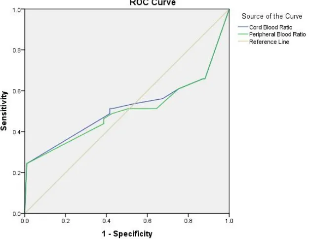
Comparison of predictive validity of Cord blood and Peripheral Blood I/T Ratio.
Test Result Variable(s)
Area Under
the Curve (AUC)
Std. Error
95% Confidence Interval of AUC
P-value
Lower Bound
Upper Bound
Cord Blood Ratio 0.515 0.064 0.389 0.641 0.784
Peripheral Blood
Ratio
Any Blood predictive value more than 0.7 good predictive validity in
predicting clinical sepsis.
The Cord blood I/T Ratio had poor predictive validity in predicting
clinical sepsis,as indicated by area under the curve of 0.515 (95% CI 0.389 to
0.641, p value 0.784)
The Peripheral Blood I/T Ratio had poor predictive validity in predicting
clinical sepsis,as indicated by area under the curve of 0.501 (95% CI 0.375 to
Figure 5: Comparison of predictive validity of Cord blood and Peripheral
Blood Platelet count
Test Result Variable(s)
Area Under
the Curve (AUC)
Std. Error 95% Confidence Interval of AUC
P-value
Lower
Bound
Upper Bound
Cord blood Platelet
Count in lakhs
0.304 0.048 0.210 0.398 <0.001
Peripheral Blood
Platelet Count in lakhs
Any Blood predictive value more than 0.7 good predictive validity in
predicting clinical sepsis.
The Cord blood Platelet count had poor predictive validity in predicting
clinical sepsis, as indicated by area under the curve of 0.304 (95% CI 0.210 to
0.398, p value <0.001)
The Peripheral Blood Platelet count had poor predictive validity in
predicting clinical sepsis, as indicated by area under the curve of 0.306 (95% CI
Figure 6: Comparison of predictive validity of Cord blood and Peripheral
Blood Micro ESR
Test Result Variable(s) Area Under the Curve(AUC)
Std. Error 95% Confidence Interval of AUC
P-value
Lower BoundUpper Bound
Any Blood predictive value more than 0.7 good predictive validity in
predicting clinical sepsis.
The Cord blood Micro ESR had good predictive validity in predicting
clinical sepsis,as indicated by area under the curve of 0.739 (95% CI 0.641 to
0.837, p value<0.001).
The Peripheral Blood Micro ESR had good predictive validity in
predicting clinical sepsis,as indicated by area under the curve of 0.740 (95% CI
Table 4
Descriptive analysis of Clinical Sepsis in study population (N=142)
ClinicalSepsis Frequency Percentage
Present 41 28.87%
Absent 101 71.13%
Among the study group, 41 babies ( 28.87%) had clinical sepsis and 101
Figure 7
Pie chart of clinical sepsis in study population
28.87%
71.13%
Table 5 :Descriptive analysis of Blood CS in study population (N=142)
Blood CS Frequency Percent
NG 120 84.51%
staph aureus 10 7.04%
pseudomonas 5 3.52%
klebsiella 4 2.82%
enterococci 2 1.41%
CONS 1 0.70%
Table 6: Association of clinical sepsis with blood culture of study population
Blood CS
ClinicalSepsis
Present Absent
CONS 0 (0%) 1 (0.99%)
enterococci 2 (4.88%) 0 (0%)
klebsiella 4 (9.76%) 0 (0%)
NG 20 (48.78%) 100 (99.01%)
pseudomonas 5 (12.2%) 0 (0%)
staph aureus 10 (24.39%) 0 (0%)
Among the clinical sepsis,21 babies (51.23%) had positive blood culture
Table 7:
Comparison of mean of various parameters with clinical sepsis
across study groups (N=142)
Parameter
Mean ±SD
P value Present (N=41) Absent (N=101)
C Blood TC 11736.59±17077.08 10222.99±3673.13 .398
P Blood TC 9273.73 ± 5609.9 10406.72 ± 3580.3 .153
C Blood ANC 7697.9 ± 14396.04 6023.48 ± 3051.41 .267
P Blood ANC 5596.29 ± 3954.39 6243.51 ± 2964.91 .288
C Blood Ratio 0.08 ± 0.08 0.05 ± 0.04 .024
P Blood Ratio 0.08 ± 0.09 0.05 ± 0.04 .024
C blood Platelet Count in
lakhs
1.76 ± 0.56 2.17 ± 0.61 <0.001
P Blood Platelet Count in
lakhs
1.78 ± 0.62 2.22 ± 0.58 <0.001
C Blood Micro ESR 13 ± 3.83 9.88 ± 3.14 <0.001
Table 8
Comparison of mean Lab parameter across study groups (N=142)
Lab Parameter Cord Blood Peripheral Blood P-value
Total count 10660.01 ± 9631.9 10079.59 ± 4276.05 0.414
ANC 5909.89 ± 3339.22 6056.64 ± 3279.83 0.021
I/T Ratio 0.06 ± 0.06 0.06 ± 0.06 0.396
Micro ESR 10.79 ± 3.63 10.93 ± 4.14 0.471
Platelet Count in
lakhs
The mean Cord Blood total count was 10660.01 ± 9631.9 and in peripheral
blood total count, it was10079.59 ± 4276.05, the difference in total count between
the two groups was statistically not significant (p value 0.414).
The mean Cord Blood ANC was 5909.89 ± 3339.22 and in peripheral blood
ANC it was 6056.64 ± 3279.83 , the difference in ANC between the two groups
was statistically significant (p value 0.031).
The mean Cord Blood I/T Ratio was 0.06 ± 0.06 and in peripheral blood
I/T Ratio it was 0.06 ± 0.06 ,the difference in I/T Ratio between the two groups
was statistically not significant (p value 0.396).
The mean Cord Blood MicroESR was 10.79 ± 3.63 and in peripheral blood
MicroESR it was 10.93 ± 4.14 , the difference in MicroESR between the two
groups was statistically not significant (p value 0.471).
The mean Cord Blood platelet count was 2.05 ± 0.62 and in peripheral
blood platelet count it was 2.09 ± 0.63 , the difference in platelet count between the
DISCUSSION
This study was conducted in Government Kilpauk Medical College
Chennai.This is a tertiary care teaching centre.
Our study population comprises 142 newborns born in Govt Kilpauk
Medical College Hospital. Out of 142 babies 91 were female babies and 89 were
male babies .In the present study no statistically significant difference (p value
0.426) was found between gender of the baby and clinical sepsis.
Out of 142 babies ,41 babies developed features of clinical sepsis and among
them 21 babies had positive blood culture.
Among the 142 neonates, the predictive validity of TC in both cord and
peripheral blood were poor in diagnosing clinical sepsis ( Area under the curve
0.41 and0.397, for cord blood and peripheral blood respectively). Both the values
Madhava R. Beeram, et al (31) in their study 2011 found a similar poor
correlation of total count in both cord and peripheral blood in sepsis screening.
(6% in UCB vs 1.2% in peripheral blood, P = .02).
Richard A. Polin et al (32) in their study found that Total white blood cell
counts have little value in the diagnosis of early-onset sepsis and have a poor
positive predictive accuracy.
Christensen RD et al and Engle WD et al also found poor predictivity of
total count in diagnosis of early onset sepsis. .(33,34).
Jahnke et al and Weitzman M et al in their study, Blood leukocytes count in
the newborn babies have been considered to be so variable and unpredictable as to
be of little value for clinical diagnosis.(35,36).
Out of the 142 babies, The Cord blood ANC had poor predictive validity in
predicting clinical sepsis, as indicated by area under the curve of 0.438 (95% CI
0.314 to 0.562, p value 0.247) The Peripheral Blood ANC had poor predictive
validity in predicting clinical sepsis, as indicated by area under the curve of 0.404
Christoph P. Hornik et al in their study found a poor predictive validity of
ANC in diagnosing early onset sepsis.(AUC=0.586 in ROC CURVE)(33).
Among the 142 babies. Both the cord and peripheral blood I/T Ratio in
predicting clinical sepsis were poor ( area under the curve of 0.515 (95% CI 0.389
to 0.641, p value 0.784 and area under the curve of 0.501 (95% CI 0.375 to 0.626,
p value 0.989).
Christoph P. Hornik, et al in 2012 in their study concluded that I/T
cut-offs were associated with relatively high specificities (73.7%, 81.7%, 95.7%,
respectively) and negative predictive values (99.2%, 99.2%, 99.0%, respectively),
but positive predictive values were low (2.5%, 3.2%, 6.0%, respectively) in the
setting of a low overall proportion of positive culture(33).
Bhandari v et al also found poor predictive validity of cord blood I/T
Ratio in diagnosing sepsis.(37).
The Cord blood Platelet count had poor predictive validity in predicting
clinical sepsis,as indicated by area under the curve of 0.304 (95% CI 0.210 to
0.398, p value <0.001).
The Peripheral Blood Platelet count had poor predictive validity in
predicting clinical sepsis, as indicated by area under the curve of 0.306 (95% CI
Khair KB1,et al in 2012 in their study , platelet count were found to have
optimal sensitivities and negative predictive value in diagnosing sepsis in neonates
.(38).
Keren Rotshenker-OlshinkaD et al in 2014 found a significant correlation
between umbilical cord and peripheral venous samples in platelets count (PLT)
(r 0.54) in diagnosing sepsis in newborn.(39).
The Cord blood Micro ESR had good predictive validity in predicting
clinical sepsis,as indicated by area under the curve of 0.739 (95% CI 0.641 to
0.837, p value<0.001) .The Peripheral Blood Micro ESR had good predictive
validity in predicting clinical sepsis, as indicated by area under the curve of 0.740
(95% CI 0.643 to 0.836, p value<0.001).
Vamseedar annam et al in 2015, had similar results of good predictivity of
both cord and peripheral blood micro ESR in clinical sepsis with a p value <
0.001.(1)
Similarly Walliullah SM et al in his study found good sensitivity of
LIMITATION OF THE STUDY
This study was done in a smaller population, so the predictivity
validity of haematological indices in diagnosing early onset
neonatal sepsis is less.
In this study, cumulative value of haematological indices in
CONCLUSION
In the present study it was concluded that cord blood micro ESR can be used
in sepsis screening to predict the neonates at risk for developing early onset sepsis
instead of peripheral blood with a good positive predictive value(p value < 0.001).
Cord blood platelet count has good correlation with peripheral blood
platelet count (p value < 0.001) but has poor predictivity in diagnosing early onset
sepsis.
No statistically significant correlation was found between cord blood and
peripheral blood Total count, ANC and I/T Ratio.
Both cord and peripheral blood Total count, ANC and I/T Ratio has poor
BIBILOGRAPHY
1. Vamseedhar Annam,: Early onset neonatal sepsis. J Clin Diagn Res. 2015 Sep; 9(9): SC04–SC06.
2. Report of the National Neonatal Perinatal Database. Report 2002-2003. NNPD Network. 2005 Jan
3. Haque KN. Definitions of bloodstream infection in the newborn. Pediatric
Crit Care Med. 2005; 6:S45–49.
4. Eric s shinwell, hematologic indices and markers of infection in umbilical cord .journal of maternal and foetal medicine vol 27 2014
5. Hansen A Potential substitution of cord blood for infant blood in the neonatal sepsis evaluation.Biol Neonate. 2005;88(1):12-8. Epub 2005 Feb 10
6. Knox Baumgart, S.,Campman,E.,Mennuti, M., & Polin, R., (1981).
Biology of the Neonate, 88, 12-18.,
7. Report of the National Neonatal Perinatal Database (National Neonatology Forum) 2000.
8. Mim SLC,Medewar MS, Perkins IR et al: predicting neonatal infections by gastric aspirate; American Journal of obstetrics and gynecology.1972,114:232.
9. Avery’s Text book of newborn diseases 8th edition. p.505.
11. Belady PH, Farkouh LJ, Gibbs RS. Intra-amniotic infection and premature rupture of membranes. Clin Perinatol 1997;24:43-57.
12. Christensen RD, Rothstein G. Pitfalls in the interpretation of leukocyte counts of newborn infants. Am J Clin Pathol 1979;72: 608-611.
13. Adler SM, Denton R1:The erythrocyte sedimentation rate in newborn period J paediatrics 1975 86:942.
14. Sharm Anita, Krishna Kutty CV, Sabharwal U, Rathi S, Mohan H.Diagnostic and prognostic role of CRP and m-ESR in neonatal septicemia. Indian Pediatrics 1993 ; 30 : 347-350.
15. Manroe BL, Weinberg AG, Rosenfeld CR et al. The neonatal blood count in healthand disease. I.Reference values for neutrophilic cells.J Pediatr 1979; 95 : 89-93.
16. Mouzinho A, Rosenfeld CR, Sanchez PJ, Risser R Effect of maternal hypertension on neonatal neutropenia and risk of nosocomial infection. Pediatrics. 1992; 90:430-435.
17. Tillett WS, Francis Jr T (1930). "Serological reactions in pneumonia with a nonprotein somatic fraction of pneumococcus" . J Exp Med 52:561–585.
19. Christenden RD, et al.thrombocytopenia among extremely low birth weight
neonates.J Perinatol 2006;26(6):348-353.
20. Mathur NB, Khalil A, Sarkar R, Puri KK. Mortality in neonatal septicemia with involvement of mother in management. Indian pediatrics 1991 ; 28 : 1259-1263.
21. Gupta Piyush, Murali MV, Faridi MMA, Caul PB, Ramchandran VG,V Talwar. Clinical profile of Klebsiella septicemia in neonates.Indian Journal of Pediatrics 1993 ; 60 : 565 – 572.
22. Moreno MT, Vargas S, Poveda R, Saez Liorens X. Neonatal sepsis and meningitis in a developing Latin American Country. Pediatric Infection. Dis J 1994 ; 13 (6) : 516 – 520.
23. Kaftan ,early onset neonatal infection; Seminars in Perinatology Volume 22, Issue 1, February 1998, Pages 15-24.
24. Francisco J. Garcia, Alan L. Nager Jaundice as an Early Diagnostic Sign of Urinary Tract Infection in Infancy ;AAP may 2002 vol 109/issue 5.
25. Weitzman M:diagnostic utility of white blood cell and differential counts AMJ of diseases of children,1975,129:1183.