0022-538X/05/$08.00⫹0 doi:10.1128/JVI.79.6.3728–3736.2005
Copyright © 2005, American Society for Microbiology. All Rights Reserved.
Inhibition of Viral Replication by Ribozyme: Mutational Analysis
of the Site and Mechanism of Antiviral Activity
Zhenxi Zhang and John M. Burke*
Department of Microbiology and Molecular Genetics, The University of Vermont, Burlington, Vermont
Received 15 April 2004/Accepted 29 October 2004
A controlled mutational study was used to determine the site and mechanism of the antiviral action of ribozymes that inhibit Sindbis virus replication. A hairpin ribozyme targeting G575 of the Sindbis virus genomic RNA was designed and cloned into a minimized alphavirus amplicon vector. Cells that were stably transfected with this construct expressed low levels of a constitutive transcript containing the ribozyme plus recognition sequences for Sindbis RNA replicase. Upon infection, the ribozyme transcript was amplified to high levels by the viral replicase, resulting in decreased viral production from infected ribozyme-expressing cells. Mutations were then introduced into the viral RNA target sequence to interfere with ribozyme binding, and compensatory changes were generated in the ribozyme recognition sequence. Single mutations in the virus or ribozyme decreased the efficacy of the ribozyme’s inhibition of viral replication, and compensatory mutations restored it. To confirm that ribozyme-catalyzed RNA cleavage was actually needed for inhibition, we performed tests with a cell line expressing an inactivated ribozyme and with a virus containing a single nucleotide target mutation that allowed the ribozyme to bind but blocked cleavage at the recognition site. The results show that most of the antiviral activity of ribozymes is due to ribozyme-catalyzed cleavage at the targeted RNA sequence, but some additional inhibition seems to occur through an antisense mechanism.
The use of nucleic acids to achieve the selective inhibition of gene expression has been broadly investigated by the use of antisense, triplex, ribozyme, and RNA interference strategies (for reviews, see references 1, 16, and 22). While each ap-proach has met with success, they all face common challenges, including intracellular activity and selectivity, subcellular local-ization, target site accessibility, delivery or expression, metab-olism, and toxicity. The development of effective nucleic acid inhibitors of gene expression has not proven to be as facile as some had anticipated. Even in cases in which significant reduc-tions of the targeted gene product have been realized, most studies have not been designed to determine if the inhibition resulted from the intended mechanism and site of action.
Hammerhead and hairpin ribozymes are small, sequence-specific RNA endonucleases whose sequence-specificity can be readily manipulated while retaining catalytic efficiency. These ribo-zymes have been used in several viral inhibition studies (6–9, 11, 20, 32). In our laboratory, we have focused on expressing hairpin ribozymes that are engineered to inhibit the replication of Sindbis virus in cultured cells. We have shown that combi-natorial target site selection methods are an effective means of identifying target sites within viral RNA transcripts (31) and that hairpin ribozymes can be effectively engineered by using rules derived from the results of in vitro genetic and biochem-ical studies (30). Hairpin ribozymes are efficient and highly selective RNA endonucleases in the test tube and within eu-karyotic cells (3, 19, 24).
Sindbis virusis the type virus of theAlphavirusgenus in the
Togaviridae family (18, 23), which is a group of viruses with
single-stranded, message-sense RNA genomes that includes
human and veterinary pathogens, including the Eastern, West-ern, and Venezuelan equine encephalitis viruses. Sindbis virus has a wide host range spanning insect, avian, and many types of mammalian cells. Sindbis virus strain S.A.AR86 (21), which was utilized in our study, contains an 11,663-nucleotide (nt) single-stranded 42S RNA genome that also serves as the mRNA encoding the nonstructural proteins (nsPs; first two-thirds of the genome) and as the template for minus-strand RNA synthesis. A 26S single-stranded subgenomic RNA is also produced, driven by the subgenomic promoter on the minus-strand RNA and encoding the viral structural proteins.
The structure-rich RNA genome is infectious, and the rep-lication cycle of the virus takes place in the cytoplasm following a receptor-mediated infection. In a rapid course of events after infection, several hundred thousand genomic and subgenomic RNAs are generated, and viral particles are secreted from the infected cells at 4 to 6 h postinfection at a rate of ca. 2,000 PFU/cell every hour (23). Cell death usually occurs 24 to 36 h after infection due to virus-induced apoptosis (5).
Considering its structure-rich RNA, high level of viral RNA replication, and large amount of virus output, the Sindbis virus system presents a rigorous challenge for the development of antiviral ribozymes. Previously, we showed that engineered hairpin ribozymes expressed from a U6 (RNA polymerase III [Pol III]) promoter in clonal mammalian cell lines can effec-tively inhibit Sindbis virus replication (20). However, these experiments were not designed to determine the mechanism or site of antiviral action.
In this article, we report the results of a series of experiments in which we used a molecular genetic approach to determine the site and mechanism of antiviral activity. An analysis of the effects of compensatory mutations in ribozymes expressed from clonal cell lines and in the Sindbis virus RNA genome showed that the antiviral activity results from the action of the * Corresponding author. Mailing address: The University of
Ver-mont, 95 Carrigan Dr., 220 Stafford Hall, Burlington, VT 05405. Phone: (802) 656-8503. Fax: (802) 656-5172. E-mail: John.Burke@uvm .edu.
3728
on November 8, 2019 by guest
http://jvi.asm.org/
ribozyme at the targeted site in the Sindbis virus genome. An examination of the effects of ribozyme and viral mutations that block catalysis without affecting the binding of the ribozyme to the viral target showed that the observed antiviral activity re-sults from a combination of ribozyme-catalyzed RNA cleavage and antisense effects.
MATERIALS AND METHODS
Preparation of RNA.Oligonucleotide RNA substrates were synthesized on an Applied Biosystems 392 DNA-RNA synthesizer and deprotected by standard procedures (27). Ribozymes and long substrates were transcribed from PCR products generated for ribozyme expression vector construction (described be-low) and from PCR products corresponding to nucleotide positions 422 to 681 of the viral genome by the use of T7 RNA polymerase and were purified by electrophoresis through denaturing polyacrylamide gels.
Screening of accessible ribozyme target sites in Sindbis virus genomic RNA.A DNA fragment containing nucleotide positions 88 to 875 was amplified from a full-length cDNA plasmid of the Sindbis virus (SIN) S.A.AR86 strain (ps73, a gift from R. E. Johnston, University of North Carolina, Chapel Hill, N.C.) (GenBank accession no. U38305). It was then transcribed by T7 RNA polymerase, and the RNA was purified by electrophoresis through a denaturing polyacrylamide gel. Protocols for randomized ribozyme pool preparation, cleavage reactions with the randomized ribozyme pool, and the mapping of cleavage sites by primer sion were described previously (31). The primers employed in the primer exten-sion mapping experiments annealed to the following regions of the SIN genome: nt 286 to 305, 469 to 490, 661 to 681, and 854 to 875.
Ribozyme expression vectors.A ribozyme targeting the G575 site of the SIN genome was designed according to standard parameters for hairpin ribozymes (30). A double-stranded DNA fragment corresponding to the wild-type active four-way junction ribozyme 575 was first created by annealing two overlapping primers (top, 5⬘-GCTCGGTTCGCCGAGCGTCGACGAGGCGAAGCACAT CAGAGAAACAGATCTCTTCGGAGATCGTACATTACCTG-3⬘; bottom, 5⬘-CGAGCCCGAAGGCTCGCTTATGAAAGGCACGATCTTTCGATCGT ATCAGGTAATGTACGATCTCCGAAGAG-3⬘), and the single-stranded ar-eas were filled in by two cycles of PCR. The products were then amplified by a set of amplification primers (top, 5⬘-TAATACGACTCACTATAGGGtctagaG CTCGGTTCGCCGAGCGTCGAC-3⬘; bottom, 5⬘-CGCTCGgggcccCGAGCCC GAAGGCTCGCTTATG-3⬘) with XbaI and ApaI cleavage sites (lowercase) and a T7 promoter (underlined). The final PCR products were digested with XbaI and ApaI. The additional ribozyme variants utilized for this study included (i) the G8A ribozyme 575, (ii) the G6C ribozyme 575, (iii) the G6C G8A ribozyme 575, and (iv) ribozyme 8242. All were produced in the same way. The digested ribozyme constructs were then cloned into the XbaI- and ApaI-digested plasmid tRNA987AGLacZ (a gift from S. Schlesinger, Washington University, St. Louis, Mo.), and the sequences were confirmed.
In vitro ribozyme cleavage assays.All ribozyme cleavage assays were per-formed under single-turnover conditions as described previously (31) (also see the figure legends). Total RNAs extracted from the ribozyme-expressing cells or from SIN-infected naı¨ve BHK-21 cells were employed as a source of expressed ribozymes or as viral RNA substrates, respectively, for some experiments. The cleavage products were resolved in denaturing polyacrylamide gels and quanti-fied by radioanalytic imaging. Cleavage rates were calculated as described else-where (13).
Cell culture and transfection.BHK-21 cells were obtained from the American Type Culture Collection and cultured in Dulbecco’s modification of Eagle’s medium (Mediatech Cellgro) containing 10% fetal bovine serum, 10% tryptose phosphate broth, 2 mML-glutamine, 100 U of penicillin/ml, and 100g of
streptomycin/ml. The cells were maintained in tissue culture incubators at 37°C with 5% CO2. Cotransfections were performed with 6g of total plasmid DNA
with a 10:1 molar ratio of the plasmid encoding the ribozyme to a plasmid harboring a neomycin resistance gene marker (pSV2neo; Clontech). The DNA was mixed with 106
BHK-21 cells during exponential growth phase and with 0.5 ml of Optimem medium (Invitrogen) in a 0.4-cm-gap cuvette. Two pulses of 250 V at a 950F capacitance were applied by use of an electroporator (Genepulser II; Bio-Rad). The transfected cells were transferred to prewarmed growth me-dium, and Geneticin was added to the medium at 48 h postinfection. Individual drug-resistant colonies were isolated after 2 weeks of selection.
Site-directed mutagenesis of SIN cDNA clone. The wild-type (WT) SIN S.A.AR86 cDNA plasmid pS73 was first cleaved with XbaI and BglII to produce a 3.1-kb fragment which was cloned into the pSP72 (C578G mutation; Promega) or pSP73 (G575A mutation; Promega) plasmid. A single top primer (5⬘-GAAA
GGCGTGCGGACGCTGTAATGGATTGG-3⬘) was utilized to mutate C578 to G578 according to the instructions of the GeneEditor in vitro site-directed mutagenesis system (Promega). The G575A mutation was generated with two fully complementary primers (top, 5⬘-GCTATGAAAGGCGTGCGAACCCTG TACTGGATTGG-3⬘; bottom, 5⬘-CCAATCCAGTACAGGGTTCGCACGCC TTTCATAGC-3⬘) by use of a QuickChange site-directed mutagenesis kit (Strat-agene). The sequences of the mutated region were confirmed by DNA se-quencing, and the 3.1-kb mutated fragment was ligated back into the 11.3-kb XbaI- and BglII-digested pS73 fragment. The cloning sites (XbaI and BglII) and the mutated region in the final cDNA clones were confirmed by DNA sequencing.
In vitro transcription of infectious viral RNAs, transfection, and virus stock titration.Ten micrograms of an XbaI-linearized SIN cDNA plasmid was tran-scribed by SP6 RNA polymerase in a 100-l reaction by use of a high-yield capped RNA transcription kit (Ambion). The infectious viral RNAs were mixed with 3⫻106exponentially growing BHK-21 cells (prewashed with 1⫻
phos-phate-buffered saline) in 0.5 ml of 1⫻phosphate-buffered saline. The cells were electroporated three times at 850 V with a capacitance of 25F in a cuvette with a 0.4-cm gap and then transferred to a 100-mm-diameter dish with 10 ml of warm growth medium. Supernatants were harvested at 30 to 35 h posttransfection, when most cells were dead. The supernatants were clarified by centrifugation at 12,000⫻g for 10 min at 4°C, and aliquots were stored at⫺80°C. The concentrations of the viral stocks were determined by plaque assays using BHK-21 cells.
Viral yield assays.Cell lines and BHK-21 cells were infected with the wild-type or mutant Sindbis virus at a multiplicity of infection of 0.01 for 1 h at 37°C. The inoculum was replaced with fresh warm growth medium, and aliquots of super-natants containing released viruses were collected at 5, 12, 22, 29, 36, and 48 h postinfection and replaced with fresh growth medium. The aliquots were stored at⫺80°C, and the viral titers of the supernatants were measured later by a plaque assay.
Naı¨ve BHK-21 cells at⬃90% confluence in 60-mm-diameter dishes were infected with the diluted aliquots of the supernatants at 37°C. The dishes were rotated three times during the 1-h incubation to facilitate the absorption of viruses by the cells. The inoculum was then removed, and the cells were overlaid with 5 ml of 0.5% immunodiffusion-grade agarose (ICN) dissolved in growth medium. The cells were further incubated for 48 to 50 h before the agarose overlay was removed. One percent crystal violet (in 20% methanol–79% water) was employed to stain the cells, and the plaques were visualized after the excess dye was removed with water.
Total RNA isolation, primer extension, and direct RNA sequencing.Total cellular RNAs were prepared by the use of TRIZOL reagent (Invitrogen) ac-cording to the manufacturer’s instructions. Two primers were utilized for primer extension assays to quantitate the expression levels of the ribozyme transcripts in the cell: a ribozyme-specific primer complementary to the right side of the domain B region of the hairpin ribozyme (5⬘ -CCAGGTAATGTACGATCTCC-3⬘) was used, and a U6 snRNA-specific primer annealing to the endogenous cellular U6 snRNA molecules (5⬘-GCGCAGGGGCCATGCTAATC-3⬘) served as an internal quantification control (⬃500,000 molecules per cell). Another Sindbis virus-specific primer (5⬘-GAAGCCAATCCAGTACAGGG-3⬘) was used to detect viral RNAs. The purified total RNA (2 to 5g) was mixed with a 5⬘32P-labeled ribozyme- or U6 snRNA-specific DNA primer in hybridization
buffer (60 mM NaCl, 50 mM Tris-HCl [pH 8.0], 10 mM dithiothreitol [DTT]) (5
l total volume) and denatured in 200 ml of 90 to 95°C water for 2 min in a beaker. The beaker was then left at room temperature for⬃3 h until the water temperature dropped to⬃27°C, allowing annealing between the RNAs and primers to occur. Another 1l of 36 mM MgCl2was added to bring the Mg
2⫹
concentration to 6 mM. Two microliters of annealed sample was then mixed with 3l of deoxynucleoside triphosphate (dNTP) mix (a 375M concentration of each dNTP [or a 750M concentration of each ddNTP when limited primer extension was performed], 5 mM DTT, 60 mM NaCl, and 6 mM MgCl2) and 5
to 7 U of avian myeloblastosis virus reverse transcriptase. The elongation step was completed in a 42 to 45°C water bath for 45 min. The reactions were terminated by the addition of an equal volume of formamide loading buffer containing 10 mM EDTA, denatured at 85 to 90°C for 1 min, and resolved in a sequencing gel. Gels were quantified by radioanalytic imaging. A special dNTP-ddNTP mix was employed when direct RNA sequencing was performed. A 100
M concentration of each ddNTP was included in addition to the regular dNTP mix, which allowed a partial termination of primer elongation and the formation of a sequencing ladder on a denaturing gel.
VOL. 79, 2005 RIBOZYME INHIBITION OF SINDBIS VIRUS 3729
on November 8, 2019 by guest
http://jvi.asm.org/
RESULTS
Combinatorial target site selection.To build upon previous
experiments in which engineered hairpin ribozymes were used to inhibit Sindbis virus replication (20, 31), we made several changes which were designed to improve (i) the target site, (ii) the catalytic activity of the ribozyme under physiological con-ditions, and (iii) the expression levels and subcellular localiza-tion of the ribozyme. We chose to target the 5⬘two-thirds of the Sindbis virus genome, which encodes nonstructural proteins that are essential for viral replication and contains sequences that are not contained within the highly expressed 26S subgenomic RNA. A combinatorial library of hairpin ribozymes containing all possible sequence specificities (31) was transcribed and used to probe target site accessibility within the 800 nt at the 5⬘end of the Sindbis virus genomic RNA. We identified 16 target sites within this sequence, each of which could be cleaved by one or more ribozyme variants within the combinatorial library with a
moderate to high efficiency. Data for two of these sites, G575 and G586, are shown in Fig. 1B.
For the mutational analysis described here, we chose to de-velop ribozymes that cleave G575 of the genomic RNA. This target site permitted us to create mutations within the viral RNA in such a manner as to change the activity of the ribozyme without modifying the amino acid sequence of the encoded viral pro-tein, nsP1. Although several other potential target sites iden-tified by this screening showed stronger cleavage patterns (data not shown), their local sequences did not permit us to intro-duce proper silent mutations into the viral genome to address the specificity and mechanism of the hairpin ribozyme. The rel-atively modest activity of ribozyme 575 in vitro led us to con-struct a four-way junction (4WJ) version to reach a higher cleav-age activity under intracellular conditions, as described below.
Ribozyme design and catalytic activity. Hairpin ribozymes
[image:3.585.94.491.65.382.2]targeted to G575 of the Sindbis virus genome were designed FIG. 1. Ribozymes and viral targets. (A) Secondary structure of ribozyme-substrate complex. Green, sequences contained in standard two-way helical junction ribozyme construct; yellow, additional sequences contained in four-way helical junction ribozyme construct; pink, sequences of targeted region of Sindbis virus genomic RNA. H1 to H6, helical elements of ribozyme-substrate complex. The ribozyme shown was designed by use of the results shown in panel B. Note that the identities of ribozyme nucleotides 16 and 48 have been changed from the standard sequence in order to prevent a predicted misfolding event. (B) Combinatorial screening for optimal ribozyme cleavage sites within viral genomic RNA. A library of hairpin ribozymes containing all possible substrate specificities (31) was incubated with Sindbis virus genomic RNA sequences, and cleavage sites were mapped by the extension of 5⬘32P-labeled oligonucleotide primers with reverse transcriptase. Control lanes (lacking ribozyme and lacking magnesium) were used to identify reverse transcriptase stop sites that did not result from ribozyme-catalyzed RNA cleavage. Sequencing lanes used appropriate ddNTPs for sequence-specific chain termination. An autoradiogram of a 6% denaturing polyacrylamide gel is shown. (C) Time course of RNA cleavage with ribozymes targeted to the Sindbis genomic G575 target. Ribozymes were generated by in vitro transcription with T7 RNA polymerase and applied to cleave a 5⬘32P-labeled oligoribonucleotide substrate in a buffer containing 2 mM MgCl
2, 150 mM NaCl, 0.1 mM EDTA, and 50 mM HEPES (pH 7.5). (D) Cleavage of structured RNA substrates. The experiment was the same as that described for panel C, except that the substrate was a uniformly labeled 250-nt RNA generated by in vitro transcription under single-turnover conditions in a buffer containing 12 mM MgCl2and 40 mM Tris-HCl (pH 7.6).
on November 8, 2019 by guest
http://jvi.asm.org/
and transcribed for examinations of their catalytic activities in vitro. We generated both a minimal ribozyme, organized around a two-way helical junction (2WJ), and a 4WJ ribozyme in which two additional helices were introduced, one of which (helix 6) provided additional base pairing between the ribo-zyme and its substrate (Fig. 1A). To facilitate the dissociation of the 3⬘cleavage product and thereby increase the extent of cleavage by reducing the ligation of the cleavage products, we destabilized helix 1 in the four-way junction ribozyme by reduc-ing it from 6 to 3 bp (28). In addition, the C16-G48 base pair in helix 3 of the ribozyme was replaced with U16-A48 in order to avoid the predicted misfolding of the four-way junction construct. Results of in vitro cleavage assays are shown in Fig. 1C and D. In a standard single-turnover reaction using long (ca. 250 nt) Sindbis virus-derived sequences conducted in cleavage buffer containing a higher concentration of magnesium ions (12 mM) than that available in the mammalian cytoplasm, the four-way junction ribozyme showed a higher rate and extent of cleavage than did the two-way junction ribozyme over the time interval examined. When an analogous experiment was con-ducted with an oligoribonucleotide substrate in a buffer with a more physiological magnesium ion concentration (2 mM), the catalytic advantage of the four-way junction ribozyme was even more pronounced. The enhanced activity of the four-way junc-tion ribozyme 575 is likely due to more efficient folding into the active tertiary structure, which is stabilized by the stacking of helices 2 and 3 upon helices 6 and 5, respectively (10, 25, 26).
Because of its significantly higher activity under near-physio-logical ionic conditions, we selected the four-way junction form of ribozyme 575 for subsequent cellular expression and viral inhibition assays.
Viral genomic mutations and ribozyme mutations.For an
analysis of the site and mechanism of antiviral activity in vivo, we designed a series of mutations within the antiviral ribozyme 575 and within the infectious viral genomic RNA, at and around the intended ribozyme target, nucleotide G575. Mutations in the viral target sequence were designed to generate synony-mous codons so that the amino acid sequence of the nsP1 gene product was unchanged. To ensure that the ribozyme and viral target sequences interacted in the expected manner, we con-ducted in vitro cleavage assays with in vitro-transcribed ri-bozymes and oligoribonucleotide substrates (Fig. 2).
[image:4.585.136.442.68.344.2]The ribozyme-substrate complex was destabilized by single base substitutions at the terminal base pair of helix 1 (H1) proximal to the cleavage site, formed by C578 of the viral genomic RNA and G6 of the antiviral ribozyme 575. A C578G substitution in the viral target was inhibitory, and a G6C sub-stitution in the ribozyme was strongly inhibitory (compare Fig. 2B and C to Fig. 2A), although the association of the two mismatched ribozyme-target combinations was apparently not affected, as shown by in vitro gel mobility shift assays (data not shown). The compensatory base pair substitution (G6C in the ribozyme and C578G in the viral RNA) significantly increased the cleavage activity with respect to each of the individual FIG. 2. Mutational analysis of specificity and mechanism of antiviral action. Single base substitutions in both ribozyme 575 (G6C) and the Sindbis virus genome (C578G) were each designed to destabilize helix 1 (H1) of the ribozyme-substrate complex. All base substitutions are shown in red. When both mutations were present, full base pairing was expected to be restored. The G8A mutation reduced the catalytic activity of the hairpin ribozyme. The substrate mutations C578G and G575A generate synonymous codons and thus do not affect the amino acid sequence of the nsP1 translation product. For each mutation shown, an autoradiogram is provided that reflects the activity of the designated combination of ribozyme and substrate in an in vitro cleavage assay with an oligonucleotide substrate, conducted as described in the legend to Fig. 1.
VOL. 79, 2005 RIBOZYME INHIBITION OF SINDBIS VIRUS 3731
on November 8, 2019 by guest
http://jvi.asm.org/
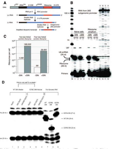
FIG. 3. Virus-induced amplification of ribozyme transcripts. (A) Amplification scheme. PRSV, promoter recognized by RNA polymerase II; tRNAAsp, partial tRNA encoded by defective interfering viral RNA; 5⬘-CSE,cis-acting element of Sindbis virus genome; PSIN26S, promoter for 26S subgenomic RNA, recognized by Sindbis virus RNA replicase; 3⬘-UTR, untranslated region required for the transcription of⫺-strand RNA. (B) High-level expression of ribozyme amplicon upon infection with Sindbis virus. The results of limited primer extension assays, in which 5⬘ -end-labeled oligonucleotide primers were extended by reverse transcriptase in a reaction containing three dNTPs and one ddNTP (ddTTP for ribozyme
on November 8, 2019 by guest
http://jvi.asm.org/
mutations, although the cleavage rate of the compensatory mutational combination was somewhat lower than that ob-served for the wild-type combination of ribozyme and substrate (Fig. 2D). These results are consistent with prior mutational studies on the hairpin ribozyme and its substrates.
Two other mutations provided important probes of ri-bozyme activity and target selectivity. First, we used mutations at ribozyme nucleotide G8, which is a component of the active site, where base substitutions are significantly inhibitory (13). The introduction of a G8A substitution into a mismatched G6C ribozyme caused a significant reduction in cleavage ac-tivity (Fig. 2E), although the incorporation of the four-way helical junction into these ribozyme constructs acted to par-tially suppress the inhibitory effects of this and other mutations (e.g., C578G). Second, a substitution of the cleavage site G (G575) nucleotide strongly inhibits cleavage because it func-tions to form a tertiary base pair with C25 of the ribozyme which is, in turn, important for positioning G8 within the active site (13–15). It is important that neither the G8A ribozyme mutation nor the G575A target site mutation inhibits the for-mation of the ribozyme-substrate complex (13, 14). Therefore, they were used as inactive controls for the present study in order to distinguish viral inhibition by ribozyme-catalyzed cleavage from antisense effects at the same site.
Cytoplasmic amplification of ribozymes upon Sindbis virus
infection.Previous expression work in our lab focused on the
use of the U6 snRNA promoter to direct the transcription of ribozymes by RNA Pol III. For this study, we explored the use of Sindbis virus infection to trigger the cytoplasmic amplifica-tion of antiviral ribozymes by the viral RNA replicase when the ribozyme was expressed constitutively at low levels as a Pol II transcript (Fig. 3A). To accomplish this, we cloned ribozyme 575 downstream of the 26S subgenomic promoter of a Sindbis virus amplicon derived from a naturally occurring defective interfering RNA, DI25 (2, 12). Note that this vector does not interfere with viral replication. Constitutive, low-level expres-sion is driven by the Rous sarcoma virus Pol II promoter, and the resulting Pol II transcript contains thecis-acting elements required for both plus- and minus-strand replication by the viral replicase (4). Stably transfected clonal cell lines express-ing ribozymes of interest were established, and ribozyme-con-taining transcripts were analyzed by dideoxy-limited primer extension assays before and after viral infection (Fig. 3B). The results showed that the transcripts were amplified by 2 orders of magnitude from their basal levels upon infection (Fig. 3C).
To ensure that the ribozyme expression levels were equivalent among the cell lines, we always infected cells under the same conditions (exponential growth phase) in subsequent experi-ments. One typical example is shown in Fig. 3D. The number of ribozyme transcripts per cell was maintained at a level ap-proximately equal to the number of viral genomic RNAs even at the latest postinfection time points used for this study (48 h), regardless of which virus strain was used for infection.
Biochemical analysis of biological transcripts. To ensure
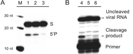
that the cellular RNA transcripts containing ribozyme se-quences possessed the expected cleavage activities, we used RNAs extracted from cell lines expressing ribozymes to cleave oligoribonucleotide substrates in vitro. The results confirmed that the ribozyme-expressing cell lines contained the expected endonuclease activities (Fig. 4A, lanes 1 and 2) and that RNAs extracted from a cell line expressing the G8A mutant ribozyme exhibited a marked reduction in activity (Fig. 4A, lane 3). In a complementary experiment, we used total RNAs extracted from virus-infected cells as substrates in cleavage experiments performed with ribozymes that were generated in vitro. These experiments showed the cleavage of viral RNA at the expected site when the antiviral ribozyme 575 was incubated with total RNAs extracted from cells infected with wild-type Sindbis virus (Fig. 4B, lane 4). As expected, no cleavage was observed with RNAs from cells infected with the uncleavable G575A mutant virus or when the ribozyme was omitted (Fig. 4B, lanes 5 and 6).
Mutational analysis of antiviral mechanism and target site.
After the characterization of stable cell lines expressing each of the ribozyme variants (the antiviral ribozyme 575, the mis-matched G6C ribozyme, and the reduced-activity G8A ri-bozyme), we used plaque assays to examine the time course of viral replication following infection by wild-type Sindbis virus or one of two engineered mutants (mismatched C578G and uncleavable G575A mutant viruses).
The abilities of ribozymes to inhibit the replication of wild-type Sindbis virus are shown in Fig. 5A to D. The expression of the antiviral ribozyme 575 resulted in an 8- to 10-fold inhibi-tion of the formainhibi-tion of infectious viral particles from 12 to 36 h after infection (Fig. 5B and D), with the time course of virus production in cells that did not express the ribozyme being used as a reference. Smaller extents of inhibition were observed at the 5-h time point, when newly synthesized viruses were only beginning to be released, and at the 48-h time point, when ribozyme-expressing cells were thriving and releasing virus at a reduced rate, while infected naı¨ve cells had
under-and ddATP for U6), are shown. A ribozyme-specific primer was used that generated a 28-nt extension product. The results were normalized by use of a primer specific for U6 snRNA, which generates a 29-nt extension product. A cell line expressing the ribozyme amplicon (A) encoding a four-way helical junction (Fig. 1A) was used, along with a control (naı¨ve) cell line that did not contain ribozyme sequences. U, G, C, and A, results from dideoxy sequencing of ribozyme amplicon within total cellular RNA extracted from virus-infected cells expressing the construct shown in panel A. The ribozyme-specific primer was used, and a strong stop was observed at a sequence corresponding to the expected 5⬘end of the ribozyme amplicon template, which is the start site of the 26S subgenomic promoter. “U6*” indicates the chain termination product at the second A of the U6 snRNA template, while “ns” indicates a nonspecific extension product of the ribozyme primer, found in the total cellular RNA, that is independent of ribozyme sequences. M, marker lane. (C) Extent of amplification of ribozyme sequences. Quantitative results of primer extension experiments conducted as described for panel B by the use of clonal cell lines expressing two-way and four-way helical junctions are shown. The number of transcripts per cell is given for cell lines that have and have not been infected with Sindbis virus. (D) Sindbis virus infection-induced amplification of ribozyme expression to a similar level among various cell lines. Cell lines expressing the 4WJ version of the ribozyme (lanes 1 to 6) and naı¨ve BHK-21 cells (lanes 7 to 9) in exponential growth phase were infected with wild-type Sindbis virus (lanes 1 to 3 and 7), the C578G strain (lanes 4 to 6 and 8), or the G575A strain (lane 9) at a multiplicity of infection of 0.01. Total RNAs were prepared at 48 h postinfection and then probed with a ribozyme-specific primer (lanes 1 to 6) or a Sindbis genomic RNA-specific primer (lanes 7 to 9) in a limited primer extension assay.
VOL. 79, 2005 RIBOZYME INHIBITION OF SINDBIS VIRUS 3733
on November 8, 2019 by guest
http://jvi.asm.org/
gone apoptosis and thus were no longer releasing virus. The expression of the mismatched G6C ribozyme resulted in a reduction in viral inhibition by a factor of 2 relative to the inhibition shown by the antiviral ribozyme 575 (Fig. 5D). A similar decrease in inhibition was observed when the 6-578 (Fig. 1A) base pair was destabilized by a C-to-G base substi-tution in the viral RNA (Fig. 5F). We concluded that a single base substitution at the viral target or within the substrate-binding domain of the ribozyme can result in a significant reduction in antiviral activity.
The expression of the reduced-activity G8A ribozyme re-sulted in a two- to threefold reduction in antiviral activity in infected cells; the extent of inhibition was slightly, but consis-tently, less than that of the mismatched G6C ribozyme at all time points (Fig. 5D). In addition, the expression of the re-duced-activity G8A ribozyme was more permissive for viral growth than that of ribozyme 575 in the cell (Fig. 5B). We concluded that the reduction in catalytic activity of the G8A ribozyme results in a two- to threefold loss of antiviral activity within infected cells; in other words, most viral inhibition re-sults from the catalytic activity of the ribozyme.
Infection with the mismatched C578G mutant virus showed that the mutant virus replicates in cells that do not express
ribozymes with kinetics that are indistinguishable from those of the wild-type virus (Fig. 5E). However, there was a dramatic change in the pattern of viral inhibition observed when the mutant virus was used to infect cells expressing the antiviral ribozyme 575 and the mismatched G6C ribozyme. The latter ribozyme preferentially inhibited the replication of the mis-matched C578G mutant virus and exhibited a reduction in its ability to inhibit the wild-type virus (compare Fig. 5F with Fig. 5D). This switch in specificity indicates that viral inhibition results from the action of the ribozyme on the intended target site within the viral genome and confirms our observation (described above) that a mutation resulting in a single base pair mismatch in the complex between the ribozyme and the viral genomic RNA reduces the antiviral activity of the ri-bozyme.
Mutant viruses whose genomes were altered to prevent ri-bozyme-catalyzed cleavage (G575A substitution) were able to replicate at rates equivalent to those of the wild-type virus in naı¨ve cells and in a cell line expressing an active ribozyme (ri-bozyme 8242) targeted to nucleotide 8242 of the genomic and 26S subgenomic RNAs. Although numerous analyses showed that ribozyme 8242 was amplified to the same level as the other ribozymes in the study (data not shown), it did not inhibit viral replication when expressed from a DI25-derived amplicon (Fig. 5B and H). The lack of inhibition by the 8242 ribozyme demonstrates that amplification of the virus-derived amplicon does not, in and of itself, interfere with viral replication.
Strikingly, equal levels of inhibition of the G575A virus were observed in cells expressing both the active antiviral ribozyme 575 and the catalytically impaired mutant ribozyme (Fig. 5H); both the active and inactive ribozymes were able to inhibit viral replication three- to sixfold. For this experiment, the observed antiviral activity can be attributed to an antisense mechanism, while the difference between the levels of inhibition observed in the experiments shown in Fig. 5D and H resulted from the ribozyme-catalyzed cleavage of Sindbis virus genomic RNA at G575. Our ribozymes may exhibit relatively high levels of an-tisense inhibition because of the additional 10 bp of ribozyme-substrate duplex that result from the inclusion of helix 6 in the four-way junction construct used for the present study (Fig. 1A) and because of the high levels of expression obtained from the viral amplicon.
DISCUSSION
[image:7.585.53.274.69.160.2]Studies in RNA biology have led to several novel strategies for the development of novel RNA-based agents that may have potential in therapeutics, including ribozymes, small interfer-ing RNAs, and aptamers. Like other strategies for gene ther-apy, there are many potential difficulties for the development of a safe and practical therapy. We have been working system-atically to identify and overcome the challenges necessary to develop ribozymes as antiviral agents and have shown that engineered hairpin ribozymes can be expressed within cells and successfully used to inhibit the replication of members of three diverse viral families, namely, human immunodeficiency virus type 1 (HIV-1), hepatitis B virus, and Sindbis virus (20, 29, 32). Prominent among the challenges in gene therapy are the issues of the biological mechanism and the site of action of the therapies being developed. For example, our previous work FIG. 4. Biochemical analysis of biological transcripts. (A) Activity
of ribozymes expressed within cells. Total cellular RNAs were ex-tracted from clonal cell lines expressing ribozymes and used for in vitro cleavage of a 21-nt oligoribonucleotide substrate. In all cases, total RNAs were harvested from cells 16 h after infection with wild-type Sindbis virus at a multiplicity of infection of 0.5. M, markers. Lane 1, 5g of total RNA from cells expressing ribozyme 575; lane 2, 10g of total RNA from cells expressing ribozyme 575; lane 3, 10g of total RNA from cells expressing the G8A reduced-activity ribozyme. Total RNAs were incubated with 5⬘-end-labeled WT synthetic RNA (21 nt) in annealing buffer (150 mM NaCl, 0.1 mM EDTA, and 50 mM HEPES, pH 7.5) for two cycles of 95°C for 2 min and 37°C for 10 min in a PCR instrument. MgCl2was added to 2 mM, and reactions were incubated for 4 h at 37°C. Reaction products were separated by elec-trophoresis through a denaturing 20% polyacrylamide gel. (B) Ri-bozyme cleavage of viral RNA synthesized within cells. BHK-21 cells were infected with either wild-type or noncleavable G575A mutant Sindbis virus at a multiplicity of infection of 0.5, and total cellular RNAs (including viral RNA) were extracted 24 h after infection. Ri-bozyme 575 (2M), generated by in vitro transcription and purified by denaturing gel electrophoresis, was incubated with the cellular RNA at 37°C for 2 h in a buffer containing 2 mM MgCl2, 150 mM NaCl, 0.1 mM EDTA, and 50 mM HEPES (pH 7.5). Cleavage products were identified by a primer extension assay, using a radiolabeled oli-gonucleotide primer complementary to the Sindbis virus RNA down-stream of the cleavage site. Lane 4, wild-type Sindbis viral RNA incubated with ribozyme 575; lane 5, mutant Sindbis virus carrying the G575A mutation, which blocks ribozyme-catalyzed RNA cleav-age at a step following the formation of the ribozyme-substrate com-plex; lane 6, wild-type Sindbis viral RNA incubated in the absence of ribozyme.
on November 8, 2019 by guest
http://jvi.asm.org/
with hairpin ribozymes to inhibit the replication of Sindbis virus is consistent with the intended mechanism, i.e., ribozyme-catalyzed cleavage of the targeted site within the viral RNA. However, these results do not rule out other plausible antiviral mechanisms, e.g., antisense inhibition, unintended cleavage of a cellular RNA whose product is essential for viral replication, or insertion of a ribozyme-encoding DNA within a host cell gene that is important for viral replication.
For this study, we used a genetic strategy to determine if the ribozyme was acting on the intended viral target site and to distinguish between viral inhibition due to ribozyme-catalyzed cleavage and that due to a general antisense mechanism in-volving binding of the ribozyme to the target site. Our ap-proach has been to generate mutations based on our under-standing of the behavior of the ribozyme-substrate complex in vitro and then to determine how these mutations affect antivi-ral activity in cell culture. To this end, we generated infectious virions with specifically designed mutations at and around the ribozyme’s intended target site within the viral genome and also constructed clonal cell lines expressing a series of ri-bozyme variants that would affect the site and mechanism through which the ribozyme would act.
In all cases, we chose single base mutations within the Sind-bis virus genome. Because the cleavage site lies within an essential coding sequence, it was necessary to find a target site and to choose mutations that would have no effect on the amino acid sequence of the translation product and to dem-onstrate that viruses containing the mutations replicated with the same growth characteristics as the wild-type strain.
The present study establishes the site of antiviral action through two complementary experiments. First, we used a compensatory mutational strategy to test the effects of single base substitutions within helix 1 of both the ribozyme and the viral RNA target. Separately, these mutations each destabi-lized the ribozyme-substrate complex and diminished the an-tiviral activity of the ribozyme. Together, they restored full base pairing within the complex and were observed to restore the full antiviral effect. Second, we mutated the cleavage site guanosine (G575) at the intended viral target and observed a reduction in antiviral activity that resulted from the loss of ribozyme-catalyzed cleavage of viral RNA at position 575.
Our work also established that the mechanism of antiviral activity includes two components, (i) ribozyme-catalyzed cleav-age at the target site and (ii) an antisense component involving the formation of an inactive ribozyme-substrate complex. The residual inhibition that was seen with two reduced-activity com-plexes, the G8A ribozyme mutation and the G575A target site mutation, was attributed to antisense inhibition, although the G8A mutation alone did not completely inactivate the catalytic activities of the four-way junction ribozymes used in our study. We believe that antisense inhibition of viral replication does not involve RNA-induced silencing complex-mediated RNA interfer-ence because all of the helices in the ribozyme-substrate complex are significantly shorter than those required for cleavage by the dicer endonuclease.
[image:8.585.44.541.70.290.2]To ensure that the population of cells was homogeneous with respect to ribozyme activity, we chose to generate clonal cell lines that constitutively expressed hairpin ribozymes within FIG. 5. Replication of wild-type and mutant Sindbis viruses in cells expressing active, inactive, and mismatched hairpin ribozymes. (A, C, E, and G) Viral growth curves. (B, D, F, and H) Extent of viral inhibition as a function of time. (A and B) Replication of wild-type Sindbis virus in cells expressing ribozyme 575, the G8A reduced-activity ribozyme, and ribozyme 8242. (C and D) Replication of wild-type Sindbis virus in cells expressing ribozyme 575, mismatched G6C ribozyme, and the G8A reduced-activity G6C ribozyme. (E and F) Replication of mismatched C578G mutant Sindbis virus in cells expressing ribozyme 575, the mismatched G6C mutant ribozyme, and the G8A reduced-activity G6C ribozyme. (G and H) Replication of noncleavable G575A mutant virus in cells expressing ribozyme 575, the G8A reduced-activity ribozyme, and ribozyme 8242. Cell lines were infected with viral strains at a multiplicity of infection of 0.01, and aliquots of supernatants were removed at the indicated time points up to 48 h after infection. Viral yields were quantitated by plaque assays with BHK-21 cells that did not express ribozymes. Each data point in the growth curves represents the mean of three independent experiments, and the error bars in the viral inhibition plots indicate standard deviations of the same three experiments.
VOL. 79, 2005 RIBOZYME INHIBITION OF SINDBIS VIRUS 3735
on November 8, 2019 by guest
http://jvi.asm.org/
a Pol II transcript that was amplified following Sindbis virus infection of the ribozyme-containing cell. The vector used, DI25, is different from common alphavirus replicon vectors, which en-code viral nonstructural proteins that lead to cytopathology and cell death (17). In our system, the amplification of ribozyme-containing transcripts is triggered by Sindbis virus infection and is supported by the proteins encoded in the incoming viral genome. Previous studies (2, 12) and our controls clearly show that the amplicon per se does not inhibit viral replication.
This amplification system has two apparent advantages. First, ribozyme-containing transcripts are amplified to very high levels that are similar to the levels attained by the Sindbis virus genomic RNAs themselves. Second, the ribozymes and viral RNA targets are colocalized within the cytoplasm. However, there is also an apparent disadvantage in that early in the infection there may not be enough ribozymes present (prior to translation of the viral nonstructural proteins) to prevent infection of the cell. Our pre-vious work has shown that the most successful ribozymes may be those that prevent the initiation of viral replication within a cell (20). Consequently, our present strategy may be improved through the generation of higher levels of ribozyme transcripts prior to viral infection and by an enhancement of ribozyme activity in the cytoplasm. In addition, Sindbis virus packaging cell lines (2) may be utilized to generate pseudoviruses that contain the ribozyme amplicon used for this study. With the wide host range of Sindbis virus, pseudoviruses may be a useful means to deliver ribozymes for therapeutic applications.
ACKNOWLEDGMENTS
We thank Joyce Heckman, Michele Shields, and other members of the Burke laboratory for valuable suggestions and critiques, Mike Fay for chemical synthesis of RNAs and for technical assistance, and Anne MacLeod for manuscript preparation. We are grateful to Sondra Schlesinger (Washington University, St. Louis, Mo.) for the DI25-based vector plasmid and for valuable discussions and to Robert Johnston and Mark Heise (University of North Carolina at Chapel Hill, Chapel Hill, N.C.) for the SIN strain S.A.AR86 cDNA plasmid and technical advice. We also appreciate the services provided by the DNA sequencing and radioanalytic imaging facilities of the University of Vermont Cancer Center.
This work was supported by NIH grant AI30534 to J.M.B.
REFERENCES
1.Akkina, R., A. Banerjea, J. Bai, J. Anderson, M. J. Li, and J. Rossi.2003. siRNAs, ribozymes and RNA decoys in modeling stem cell-based gene therapy for HIV/AIDS. Anticancer Res.23:1997–2005.
2.Bredenbeek, P. J., I. Frolov, C. M. Rice, and S. Schlesinger.1993. Sindbis virus expression vectors: packaging of RNA replicons by using defective helper RNAs. J. Virol.67:6439–6446.
3.Donahue, C. P., and M. J. Fedor.1997. Kinetics of hairpin ribozyme cleavage in yeast. RNA3:961–973.
4.Frolov, I., R. Hardy, and C. M. Rice.2001.Cis-acting RNA elements at the 5⬘end of Sindbis virus genome RNA regulate minus- and plus-strand RNA synthesis. RNA7:1638–1651.
5.Griffin, D. E., and J. M. Hardwick.1997. Regulators of apoptosis on the road to persistent alphavirus infection. Annu. Rev. Microbiol.51:565–592. 6.Inokuchi, Y., N. Yuyama, A. Hirashima, S. Nishikawa, J. Ohkawa, and K.
Taira.1994. A hammerhead ribozyme inhibits the proliferation of an RNA coliphage SP in Escherichia coli. J. Biol. Chem.269:11361–11366. 7.Lazarev, V. N., M. M. Shmarov, A. N. Zakhartchouk, G. K. Yurov, O. U.
Misurina, T. A. Akopian, N. F. Grinenko, N. G. Grodnitskaya, N. V. Kaverin,
and B. S. Naroditsky.1999. Inhibition of influenza A virus reproduction by a ribozyme targeted against PB1 mRNA. Antivir. Res.42:47–57. 8.Macejak, D. G., K. L. Jensen, S. F. Jamison, K. Domenico, E. C. Roberts, N.
Chaudhary, I. von Carlowitz, L. Bellon, M. J. Tong, A. Conrad, P. A. Pavco, and L. M. Blatt.2000. Inhibition of hepatitis C virus (HCV)-RNA-depen-dent translation and replication of a chimeric HCV poliovirus using synthetic stabilized ribozymes. Hepatology31:769–776.
9.Maeda, A., T. Mizutani, M. Hayashi, K. Ishida, T. Watanabe, and S. Namioka.1995. Inhibition of viral multiplication in acute and chronic stages of infection by ribozymes targeted against the polymerase gene of mouse hepatitis virus. Adv. Exp. Med. Biol.380:399–404.
10.Murchie, A. I., J. B. Thomson, F. Walter, and D. M. Lilley.1998. Folding of the hairpin ribozyme in its natural conformation achieves close physical proximity of the loops. Mol. Cell1:873–881.
11.Ojwang, J. O., A. Hampel, D. J. Looney, F. Wong-Staal, and J. Rappaport.
1992. Inhibition of human immunodeficiency virus type 1 expression by a hairpin ribozyme. Proc. Natl. Acad. Sci. USA89:10802–10806.
12.Olivo, P. D., I. Frolov, and S. Schlesinger.1994. A cell line that expresses a reporter gene in response to infection by Sindbis virus: a prototype for detection of positive strand RNA viruses. Virology198:381–384. 13.Pinard, R., K. J. Hampel, J. E. Heckman, D. Lambert, P. A. Chan, F. Major,
and J. M. Burke.2001. Functional involvement of G8 in the hairpin ribozyme cleavage mechanism. EMBO J.20:6434–6442.
14.Pinard, R., D. Lambert, N. G. Walter, J. E. Heckman, F. Major, and J. M. Burke.1999. Structural basis for the guanosine requirement of the hairpin ribozyme. Biochemistry38:16035–16039.
15.Rupert, P. B., and A. R. Ferre-D’Amare.2001. Crystal structure of a hairpin ribozyme-inhibitor complex with implications for catalysis. Nature410:780–786. 16.Santiago, F. S., and L. M. Khachigian.2001. Nucleic acid based strategies as potential therapeutic tools: mechanistic considerations and implications to restenosis. J. Mol. Med.79:695–706.
17.Schlesinger, S.2001. Alphavirus vectors: development and potential thera-peutic applications. Expert Opin. Biol. Ther.1:177–191.
18.Schlesinger, S., and M. J. Schlesinger.2001. Togaviridae: the viruses and their replication, p. 895–916.InD. M. Knipe, P. M. Howley, D. E. Griffin, R. A. Lamb, M. A. Martin, B. Roizman, and S. E. Straus (ed.), Fields virology, 4th ed. Lippincott Williams & Wilkins, Philadelphia, Pa. 19.Seyhan, A. A., J. Amaral, and J. M. Burke.1998. Intracellular RNA cleavage
by the hairpin ribozyme. Nucleic Acids Res.26:3494–3504.
20.Seyhan, A. A., D. Vitiello, M. T. Shields, and J. M. Burke.2002. Ribozyme inhibition of alphavirus replication. J. Biol. Chem.277:25957–25962. 21.Simpson, D. A., N. L. Davis, S. C. Lin, D. Russell, and R. E. Johnston.1996.
Complete nucleotide sequence and full-length cDNA clone of S.A.AR86, a South African alphavirus related to Sindbis. Virology222:464–469. 22.Steele, D., A. Kertsburg, and G. A. Soukup.2003. Engineered catalytic RNA
and DNA: new biochemical tools for drug discovery and design. Am. J. Pharmacogenomics3:131–144.
23.Strauss, J. H., and E. G. Strauss.1994. The alphaviruses: gene expression, replication, and evolution. Microbiol. Rev.58:491–562.
24.Vitiello, D., D. B. Pecchia, and J. M. Burke.2000. Intracellular ribozyme-catalyzedtrans-cleavage of RNA monitored by fluorescence resonance en-ergy transfer. RNA6:628–637.
25.Walter, F., A. I. H. Murchie, and D. M. J. Lilley.1998. Folding of the four-way RNA junction of the hairpin ribozyme. Biochemistry37:17629–17636. 26.Walter, N. G., J. M. Burke, and D. P. Millar.1999. Stability of hairpin
ribozyme tertiary structure is governed by the interdomain junction. Nat. Struct. Biol.6:544–549.
27.Walter, N. G., N. Yang, and J. M. Burke.2000. Probing non-selective cation binding in the hairpin ribozyme with Tb(III). J. Mol. Biol.298:539–555. 28.Yadava, R. S., A. J. Choi, L. L. Lebruska, and M. J. Fedor.2001. Hairpin
ribozymes with four-way helical junctions mediate intracellular RNA liga-tion. J. Mol. Biol.309:893–902.
29.Yamada, O., G. Kraus, B. Sargueil, Q. Yu, J. M. Burke, and F. Wong-Staal.
1996. Conservation of a hairpin ribozyme sequence in HIV-1 is required for efficient viral replication. Virology220:361–366.
30.Yu, Q., and J. M. Burke.1997. Design of hairpin ribozymes for in vitro and cellular applications. Methods Mol. Biol.74:161–169.
31.Yu, Q., D. B. Pecchia, S. L. Kingsley, J. E. Heckman, and J. M. Burke.1998. Cleavage of highly structured viral RNA molecules by combinatorial librar-ies of hairpin ribozymes. J. Biol. Chem.273:23524–23533.
32.zu Putlitz, J., Q. Yu, J. M. Burke, and J. R. Wands.1999. Combinatorial screening and intracellular antiviral activity of hairpin ribozymes directed against hepatitis B virus. J. Virol.73:5381–5387.