0022-538X/04/$08.00⫹0 DOI: 10.1128/JVI.78.23.13082–13089.2004 Copyright © 2004, American Society for Microbiology. All Rights Reserved.
CD4
⫹
CD25
⫹
T Cells Regulate Vaccine-Generated Primary and
Memory CD8
⫹
T-Cell Responses against Herpes Simplex
Virus Type 1
Felix N. Toka,
1,2Susmit Suvas,
1and Barry T. Rouse
1*
Department of Microbiology, University of Tennessee, Knoxville, Tennessee,1and Immunology Laboratory,
Department of Preclinical Sciences, Warsaw Agricultural University, Warsaw, Poland2
Received 27 May 2004/Accepted 28 July 2004
It has become evident that naturally occurring CD25ⴙregulatory T cells (Treg cells) not only influence
self-antigen specific immune response but also dampen foreign antigen specific immunity. This report extends our previous findings by demonstrating that immunity to certain herpes simplex virus (HSV) vaccines is significantly elevated and more effective if Treg cell response is curtailed during either primary or recall
immunization. The data presented here show that removal of CD25ⴙTreg cells prior to SSIEFARL-CpG or
gB-DNA immunization significantly enhanced the resultant CD8ⴙT-cell response to the immunodominant SSIEFARL peptide. The enhanced CD8ⴙT-cell reactivity in Tregcell-depleted animals was between two- and
threefold and evident in both acute and memory stages. Interestingly, removal of CD25ⴙTregcells during the
memory recall response to plasmid immunization resulted in a twofold increase in CD8ⴙT-cell memory pool. Moreover, in the challenge experiments, memory CD8ⴙT cells generated with plasmid DNA in the absence of Tregcells cleared the virus more effectively compared with control groups. We conclude that CD25ⴙTregcells
quantitatively as well as qualitatively affect the memory CD8ⴙT-cell response generated by gB-DNA vaccina-tion against HSV. However, it remains to be seen if all types of vaccines against HSV are similarly affected by CD25ⴙTregcells and if it is possible to devise means of limiting Tregcell activity to enhance vaccine efficacy.
It has been clear that the outcome of several in vivo immu-nological events is influenced by T cells that suppress the function of other cells involved in immunity. Recent focus has been on regulatory T cells (Treg cells) initially recognized to
prevent genetically susceptible mice from developing certain autoimmune diseases (2, 19, 20). Subsequently, Tregcells were
shown to influence transplantation immunity as well as im-mune and inflammatory reactions to infectious agents (3, 4, 12, 17, 22). Of particular interest, Tregcells had a pivotal influence
on the outcome of chronic parasitic infections (3). Addition-ally, it was shown that animals depleted of Treg cells showed
markedly superior acute and memory CD8⫹T-cell responses to infection with herpes simplex virus (HSV) (22). Animals lacking Treg cells in fact generated more effective protective
immunity. Subsequently, Tregcell depletion was also shown to
result in more severe immunopathological responses to virus infection.
The fact that immunity to HSV was superior and more sustained when infection occurred in Treg cell-depleted
ani-mals was considered to impact upon vaccine design. Thus, HSV is one of those clinically important agents for which there is currently no effective vaccine. Conceivably, the inhibiting activity of Tregcells could help explain the difficulty in
achiev-ing effective immunity.
In the present report, we measured the influence of Tregcells
on the immune response of mice to DNA and peptide vaccine preparations against HSV. Our results show that immunity to
DNA and peptide vaccines measured systemically were com-promised by the presence of Tregcells. Most strikingly, if Treg
cells were inhibited prior to recall in the memory phase, re-sponses to DNA vaccination were elevated twofold and ani-mals showed notably increased resistance to challenge. Our results are discussed in terms of selecting vaccine approaches that are least affected by Tregcell response as well as the need
to identify procedures that minimize the Treg cell response
during vaccination. We also comment about mechanistic events that could account for Tregcell interference.
MATERIALS AND METHODS
Animals, virus, and DNA vaccine preparation.Female C57BL/6 mice, 5 to 6 weeks of age, were purchased from Harlan Sprague Dawley (Indianapolis, Ind.). Animals were used in compliance with institutional animal health and care regulations, and all procedures used in the experiments with animals were ap-proved by the local Institutional Animal Care and Use Committee. HSV-1 KOS (American Type Culture Collection, Manasas, Va.) and vaccinia virus encoding glycoprotein B (gB) of HSV-1 were grown and plaque titrated on Vero cells and kept at⫺80°C until use. Plasmid DNA was prepared as described previously (8, 24).
Depletion of CD4ⴙCD25ⴙT cells.Before immunization, mice were depleted of CD4⫹CD25⫹regulatory T cells by intraperitoneal administration of anti-CD25 monoclonal antibody clone PC61 (American Type Culture Collection, Manassas, Va.). The antibody was used as the ammonium sulfate precipitate of hybridoma culture supernatant or as ascites produced from PC61 hybridoma in
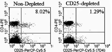
nu/numice and purified by Prosep G immunoglobulin purification kit (Millipore, Bedford, Mass.). The depletion capability of these monoclonal antibody prepa-rations did not differ significantly. Depletion efficiency was checked by staining with anti-CD25 antibody clone 7D4 (BD Bioscience, Pharmingen, San Diego, Calif.) and flow cytometry. Results (Fig. 1) show that a high level (ⱖ80%) of depletion was reached by day 4 after intraperitoneal injection of 1.2 mg of depleting anti-CD25 clone PC61.
Immunization.C57BL/6 mice 5 to 6 weeks old were injected with anti-CD25 antibody or normal rat immunoglobulin 4 days earlier to deplete CD4⫹CD25⫹ T cells and then injected with 50g of plasmid DNA encoding glycoprotein B of
* Corresponding author. Mailing address: Department of Microbi-ology, University of Tennessee, Walter’s Life Science Bldg. M409, 1414 Cumberland Ave., Knoxville, TN 37996. Phone: (865) 974-4026. Fax: (865) 974-4026. E-mail: [email protected].
13082
on November 8, 2019 by guest
http://jvi.asm.org/
HSV-1 (gB-DNA) intramuscularly or SSIEFARL peptide (HSV gB498-505) com-bined with bioactive CpG1826 (SS-CpG) (Coley Pharmaceutical Group) in the footpad. Vaccination was repeated after 3 weeks. Another group of mice were also depleted or not and infected with 106PFU of HSV-1 KOS in the footpad. HSV infection was used here as a positive control. Primary assessment of im-mune response was performed after 7 days for mice infected with HSV-1 or immunized with SS-CpG and 12 days for mice vaccinated with gB-DNA. Memory responses were assessed at 60 days post-secondary immunization. Control mice for the gB-DNA-immunized group were injected with 50g of plasmid DNA encoding-galactosidase (-galactosidase DNA), and control groups for SS-CpG were given nonbioactive SS-CpG1982 or SS-CpG2138.
ELISPOT for IFN-␥.ELISPOT plates (MultiScreen HA sterile plates, Milli-pore, Bedford, Mass.) were coated with capture anti-gamma interferon (IFN-␥) antibody in carbonate buffer, pH 9.6, overnight (BD Biosciences Pharmingen, San Diego, Calif.). Plates were then blocked with RPMI 1640 (Sigma, St. Louis, Mo.) supplemented with 10% fetal bovine serum. Responder cells from spleens or lymph nodes of immunized and control mice and stimulator cells prepared from naïve mouse spleens pulsed with HSV-gB498-505peptide and x-irradiated were added to coated plates and incubated at 37°C for 48 to 72 h and thereafter developed, and spots were counted as fully described elsewhere (24).
Intracellular cytokine staining for IFN-␥.We stimulated 106
spleen or lymph node cells per well with SSIEFARL in the presence of GolgiPlug (BD Bio-sciences Pharmingen, San Diego, Calif.) and 50 U of interleukin-2 (Hemagen) per ml for 5 h at 37°C. The cells were processed further as described by Kuma-raguru and Rouse (11). The fluorescently labeled antibodies used were pur-chased from BD Biosciences Pharmingen, San Diego, Calif..
CTL assay.A standard 4-h51
Cr release assay was performed to assess cytolytic activity of the CD8⫹ T cells isolated from immunized and control mice as described elsewhere (9, 24). Data were corrected by the formula ([experimental release⫺spontaneous release)/(total release⫺spontaneous release)]⫻100.
Challenge and virus titration.Mice were challenged at 60 days following initial immunization. A vaccinia virus challenge model, where intraperitoneal injection of vaccinia virus causes initial replication in the ovaries, was used to test efficacy of virus clearance in vaccinated animals. Female C57BL/6 mice were injected intraperitoneally with a recombinant vaccinia virus encoding gB of HSV-1 at a low dose (105PFU) and a high dose (107PFU). Ovaries were collected on days 3, 5, and 7, homogenized, clarified in phosphate-buffered saline, and used for virus titration. A conventional viral plaque assay was used.
Statistics.Where appropriate, significant differences were calculated with Student’sttest.Pⱕ0.05 was considered statistically significant.
RESULTS
Effect of CD25ⴙdepletion prior to DNA or SS-CpG vacci-nation on CD8ⴙ T-cell response.C57BL/6 mice were either depleted 4 days previously with anti-CD25 antibody clone PC61 or given normal rat immunoglobulin and exposed to infectious HSV, gB-DNA, or SS-CpG vaccine formulations. At
7 or 12 days postimmunization, both spleens and lymph nodes were collected to quantify CD8⫹T-cell responses to the im-munodominant peptide SSIEFARL by ELISPOT or intracel-lular cytokine staining for IFN-␥. The results of representative experiments are shown in Fig. 2.
As is evident, these acute-phase responses were elevated in mice that were depleted of CD25⫹Tregcells prior to antigen
exposure. This supports previous observations that depleted mice exposed to infectious virus showed increase of immune response over nondepleted animals. DNA vaccination of Treg
cell-depleted mice showed two- and threefold increase over nondepleted mice in lymph nodes and spleen, respectively. Similarly, vaccination with SS-CpG showed 2- and 2.8-fold increases in depleted versus nondepleted mice in lymph nodes and spleen, respectively.
Thus, the pattern of response observed with the two vaccine preparations in Tregcell-depleted mice, although less in extent,
was similar in profile to that of Tregcell-depleted virus-infected
mice (Fig. 2A). Taken together, these results indicated that eliminating the influence of the Treg cell population before
vaccination with gB-DNA or SS-CpG peptide enhanced the primary CD8⫹T-cell response.
Depletion of CD4ⴙCD25ⴙT cells prior to primary vacci-nation improves the memory pool of CD8ⴙT cells.We exam-ined the influence of CD4⫹ CD25⫹T-cell depletion on sys-temic T-cell memory generated with gB-DNA or SS-CpG vaccination. In the first instance, mice were depleted of Treg
cells prior to primary vaccination. Restimulation was per-formed on day 21, and memory responses were measured 60 days later. Figure 3 shows IFN-␥-producing memory CD8⫹T cells in a representative experiment. Memory CD8⫹ T-cell response in gB-DNA- or SS-CpG-immunized mice decreased two- and fourfold when measured at 60 days post-secondary immunization, respectively. Tregcell-depleted animals had
ap-proximately twofold higher responses than nondepleted mice with both types of vaccines. Higher responses dominated in the spleens than the lymph nodes. Compared to the two vaccine preparations, HSV infection (Fig. 3A) had more responding CD8⫹T cells than gB-DNA or SS-CpG immunization. These results show that removal of Tregcells prior to primary
immu-nization positively influenced the magnitude of the memory CD8⫹T cells of animals vaccinated with gB-DNA or SS-CpG. We were curious to know if depletion of Treg cells in the
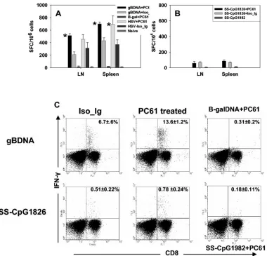
memory phase equally enhanced the T-cell response. There-fore, in the second instance mice were infected with HSV or vaccinated with gB-DNA or SS-CpG. Sixty days later the ani-mals were depleted of CD4⫹CD25⫹T cells and boosted after 5 days with gB-DNA or SS-CpG. Responses were measured 5 days later. Figures 4A and C show memory recall responses of CD8⫹T cells that produced IFN-␥upon restimulation in vitro. An approximately twofold increase in the number of IFN-␥ -producing spleen CD8⫹T cells was observed between depleted DNA-vaccinated mice and nondepleted mice. HSV-infected mice had a similar pattern of response (Fig. 4A). Finally, de-pleting Tregcells at both the primary and memory phases did
[image:2.585.46.285.72.184.2]not produce further enhancement of the immune response to gB-DNA immunization or HSV infection (Fig. 5A). There was no statistically significant difference (Pⱖ0.05) between doubly depleted or singly depleted mice. This indicated that reactiva-tion of the residual memory CD8⫹T-cell pool was also subject
FIG. 1. Depletion of CD4⫹CD25⫹T cells in C57BL/6 mice. Mice were injected intraperitoneally with 1.2 mg of anti-CD25 monoclonal antibody clone PC61. Four or 5 days later, peripheral blood mononu-clear cells were stained with CD25⫹-peridinin chlorophyll protein Cy5.5 clone 7D4 and CD4⫹-phycoerythrin. Flow cytometry analysis was done in FACScan (Becton Dickinson), and data were analyzed with CellQuest software.
VOL. 78, 2004 REGULATORY T CELLS IN VACCINATION 13083
on November 8, 2019 by guest
http://jvi.asm.org/
to regulation by Tregcells and was affected by the removal of
these cells. These results also indicated that Tregcells control
the reactivity of memory T cells and that inhibiting the function of Tregcells even when memory is established allows the
mem-ory T cells to rapidly reactivate to a higher frequency. Surprisingly, depletion of Tregcells in the memory phase of
SS-CpG-immunized mice did not lead to significant expansion of the responding CD8⫹T cells (Fig. 4B and C). Both depleted and nondepleted mice from the SS-CpG-immunized group responded similarly, and the magnitude of response was sev-eralfold lower than that observed in gB-DNA-immunized or HSV-infected mice (Fig. 4A and C). This observation indicates that even though there is enhancement of primary immune response in the absence of Tregcells, the effect had less impact
on the memory response if the primary CD8⫹T cells were generated in the absence of CD4⫹ T-cell help. Therefore,
depletion of Treg cells before SS-CpG immunization did not
contribute to the magnitude of the memory response.
Cytotoxic T lymphocytes generated by vaccination following Treg cell depletion efficiently lyse their targets. We assessed whether the CD8⫹T cells generated in CD4⫹CD25⫹depleted mice after vaccination were functional CTLs. A standard chro-mium release assay was performed on splenocytes after expan-sion for 5 days in vitro. Figures 6A and B show that in the acute phase, incubation of effector T cells from gB-DNA- and SS-CpG-vaccinated Treg cell-depleted mice with target cells
showed increased lysis of targets pulsed with gB498-505peptide.
CTL activity of cells isolated from depleted mice was higher than that of nondepleted animals. Similarly, CTL activity of cells from Treg cell-depleted SS-CpG-vaccinated mice was
[image:3.585.86.495.66.433.2]higher than that of nondepleted mice. Memory (Fig. 6C and D) CTL activity of the gB-DNA-vaccinated group showed even
FIG. 2. CD8⫹T-cell primary response to vaccination after depletion of CD4⫹CD25⫹T cells. C57BL/6 mice were either depleted of CD25⫹ T cells or injected with isotype immunoglobulin. Five days later, the animals were vaccinated with gB-DNA intramuscularly or SS-CpG in the footpad. Control mice were injected with either-galactosidase DNA or nonbioactive CpG1982 or -2138. HSV infection of depleted and nondepleted mice was used as a positive control. Spleen and draining lymph nodes were collected for analysis on either day 7 postimmunization for SS-CpG or day 12 for gB-DNA vaccination. IFN-␥ELISPOT and intracellular staining were performed as described in Materials and Methods. (A) IFN-␥ELISPOT for gB-DNA vaccination and HSV infection, (B) ELISPOT for SS-CpG vaccination, (C) intracellular staining for IFN-␥ gB-DNA- and SS-CpG-vaccinated mice. The percentage shown in each cytogram represents the mean of IFN-␥-producing CD8⫹T cells obtained from each of four mice per group in three separate experiments.ⴱ, statistically significant (Pⱕ0.05) compared to isotype immunoglobulin- and CpG1982-treated groups.
on November 8, 2019 by guest
http://jvi.asm.org/
larger differences between depleted and nondepleted mice. SS-CpG immunization resulted in a poor memory CTL re-sponse, as effector cells from such mice did not lyse the targets efficiently. When these two forms of immunization were com-pared to Tregcells from depleted virus-infected mice, the latter
had more potent CTLs (data not shown) than gB-DNA- and SS-CpG-vaccinated mice at both primary and memory phases. The difference between the depleted and nondepleted groups at the memory phase suggested that removal of Tregcell
control allowed generation of a high frequency of CTLs that efficiently lysed their target, which accounted for a better mem-ory response in terms of efficacy as described subsequently. Control lysis assays with major histocompatibility complex class I mismatched target cells or MC38 cells pulsed with an irrelevant peptide from ovalbumin showed that lysis was spe-cific to the HSV antigen-sensitized targets (data not shown).
Tregcell-depleted and vaccinated mice clear challenge virus
efficiently.To show that the enhanced immune response fol-lowing removal of regulatory T cells affected the outcome of a
challenge by virus, we measured the clearance of a recombi-nant vaccinia virus encoding gB of HSV. This challenge model utilized the fact that vaccinia virus initially replicates in the ovaries of mice, which could provide a good measure of sys-temic responses against challenge by virus. Separate groups of gB-DNA- or SS-CpG-vaccinated mice were intraperitoneally infected with two different doses of vaccinia virus gB, a low dose, 105 PFU, and high dose, 107PFU per mouse, and
fol-lowed for 7 days. Table 1 shows titers of vaccinia virus gB titrated on Vero cells from homogenized ovaries of mice chal-lenged with a low dose of virus. Challenge virus was detected in all groups of mice vaccinated with peptide, either Treg
[image:4.585.100.483.71.410.2]de-pleted or nondede-pleted, although titers were only modest. Virus replication could be detected in these mice through day 7 of observation. In contrast, the depleted and nondepleted gB-DNA-vaccinated groups showed that on day 3 replication en-sued and an approximately 2 log difference in viral titer was observed between depleted and nondepleted animals. For the
FIG. 3. CD8⫹T-cell memory response to vaccination following depletion of CD4⫹CD25⫹T cells. C57BL/6 mice were either depleted of CD25⫹T cells or injected with isotype immunoglobulin. Five days later the animals were immunized with gB-DNA intramuscularly or SS-CpG in the footpad. Control mice were injected with either-galactosidase DNA or nonbioactive CpG 1982 or 2138. HSV infection of depleted and nondepleted mice was used as a positive control. Spleen and draining lymph nodes were collected for analysis on day 60 postimmunization. IFN-␥ ELISPOT and intracellular staining were performed as described in Materials and Methods. (A) ELISPOT for gB-DNA vaccination and HSV infection, (B) ELISPOT for SS-CpG vaccination, (C) intracellular staining for gB-DNA- and SS-CpG-vaccinated mice. The percentage shown in each cytogram represents the mean of IFN-␥-producing CD8⫹T cells obtained from each of four mice per group in two experiments performed. ⴱ, statistically significant (Pⱕ0.05) compared to isotype immunoglobulin- and CpG1982-treated groups.
VOL. 78, 2004 REGULATORY T CELLS IN VACCINATION 13085
on November 8, 2019 by guest
http://jvi.asm.org/
depleted group, the virus could only be titrated on day 3 and was not detected on days 5 and 7.
Challenge with high-dose vaccinia virus gB showed that virus replicated in the ovaries of all mice irrespective of depletion
[image:5.585.104.484.66.431.2]status and vaccination (Table 2). All SS-CpG-vaccinated groups were not protected from the high virus dose challenge, and titers reached 4 logs of magnitude and could be detected throughout the observation period. Threefold difference in
FIG. 4. Effect of CD4⫹CD25⫹T-cell depletion on memory recall responses. Mice were immunized without prior depletion of Tregcells. At 60 days
after initial immunization, the mice were depleted of Tregcells with anti-CD25 monoclonal antibody and 5 days later boosted with gB-DNA or SS-CpG.
Control mice were injected with either-galactosidase DNA or nonbioactive CpG1982 or -2138. HSV infection of depleted and nondepleted mice was used as a positive control. Responses were analyzed 5 days later with IFN-␥ELISPOT and intracellular assays as described in Materials and Methods. (A) IFN-␥ELISPOT for gB-DNA vaccination and HSV infection, (B) ELISPOT for SS-CpG vaccination, (C) intracellular staining for gB-DNA- and SS-CpG -vaccinated mice. The percentage shown in each cytogram represents the mean of IFN-␥-producing CD8⫹T cells obtained from each of four mice per group in two separate experiments.ⴱ, statistically significant (Pⱕ0.05) compared to isotype immunoglobulin- and CpG1982-treated groups.
FIG. 5. Influence of double depletion of CD4⫹CD25⫹T cells on memory recall responses. Mice were depleted of Tregcells prior to vaccination
and depleted again at 60 days post-initial immunization. Control mice were injected with either-galactosidase DNA or nonbioactive CpG1982 or -2138. ELISPOT and intracellular staining for IFN-␥were performed 5 days boosting with gB-DNA or SS-CpG. (A) ELISPOT for gB-DNA vaccination and HSV infection, (B) ELISPOT for SS-CpG vaccination.
on November 8, 2019 by guest
http://jvi.asm.org/
[image:5.585.135.452.587.683.2]viral titers was shown between nondepleted and depleted gB-DNA-vaccinated mice on both days 3 and 5, and the virus could still be detected on day 7 in nondepleted gB-DNA-vaccinated group. Control groups, SS-CpG1982- and -galac-tosidase-treated mice, had the highest viral titers after both a low dose and a high dose challenge, which shows evidence of virus replication in this challenge model. Moreover, in these control animals the ovaries were hyperemic and largely edem-atous by day 7.
This challenge model indicated that depletion of CD25⫹ Tregcells led to induction of CTLs or other mechanisms that
contributed to efficient virus clearance. Although virus repli-cation still occurred, the time of clearance was reduced to at least 5 days in DNA-vaccinated Tregcell-depleted mice and 3
days in HSV-infected Tregcell-depleted mice at the high virus
challenge dose. Overall, depletion of Tregcells influenced the
efficacy of DNA vaccination.
DISCUSSION
It is evident now that naturally occurring CD25⫹Tregcells
not only influence self-antigen specific immune response (15, 18, 20) but also dampen foreign antigen specific immunity (3, 4, 22). Our initial observation with herpes simplex virus infec-tion showed that the magnitude of CD8⫹T-cell response was tightly regulated by CD25⫹Tregcells. This report extends the
previous findings by demonstrating that immunity to certain HSV vaccines is significantly enhanced and more effective if the Treg cell response is curtailed during primary or recall
immunization. The data presented here show that removal of CD25⫹Tregcells prior to SSIEFARL-CpG or gB-DNA
immu-nization significantly enhanced the resultant CD8⫹T-cell re-sponse to the immunodominant SSIEFARL peptide. This was shown by different in vitro assays, ELISPOT, CTL assay and intracellular IFN-␥ staining that measured the CD8⫹ T-cell reactivity to SSIEFARL epitope.
The enhanced CD8⫹T-cell reactivity in Tregcell-depleted
[image:6.585.137.450.69.271.2]animals was between two- and threefold and was evident in
FIG. 6. Cytotoxic T lymphocytes generated in the absence of regulatory T cells efficiently lyse peptide-sensitized targets. Splenocytes were isolated and expanded in vitro for 5 days, and CTL activity was determined as described in Materials and Methods. (A and B) CTL activity measured during the acute phase of immunization for gB-DNA and SS-CpG1826, respectively. (C and D) Memory-phase CTL activity for gB-DNA and SS-CpG1826, respectively.
TABLE 1. Mean titers of virus in the ovaries collected at days 3, 5, and 7 following challenge with 105PFU/mouse of vaccinia virus gB
Group
Mean log10titer⫾SDa
Day 3 Day 5 Day 7
HSV⫹PC61 0 0 0
HSV⫹isotype Ig 1.28⫾0.31 0 0
Phosphate-buffered saline 3.08⫾1.1 4.4⫾0.19 3.73⫾0.19 SS-CpG1826⫹PC61 1.21⫾0.40 1.11⫾0.93 1.0⫾0.67 SS-CpG1826⫹isotype Ig 1.92⫾0.14 1.01⫾0.5 1.1⫾0.41 SS-CpG2138⫹PC61 4.81⫾1.3 4.01⫾0.29 3.37⫾0.39
gB-DNA⫹PC61 1.28⫾0.62 0 0
gB-DNA⫹isotype Ig 3.00⫾1.78 2.51⫾0.2 0 -Galactosidase DNA 4.5⫾0.1 4.54⫾0.68 3.98⫾0.67
[image:6.585.43.284.600.714.2]aValues represent means for four mice per group in two separate experiments.
TABLE 2. Mean titers of virus in the ovaries collected at days 3, 5, and 7 following challenge with 107PFU/mouse of vaccinia virus gB
Group
Mean log10titer⫾SDa
Day 3 Day 5 Day 7
HSV⫹PC61 1.12⫾0.16 0 0
HSV⫹isotype Ig 2.79⫾0.21 1.10⫾0.12 0
Phosphate buffered saline 4.11⫾1.5 5.56⫾0.9 3.23⫾0.32 SS-CpG1826⫹PC61 4.12⫾0.16 4.0⫾1.01 2.15⫾0.78 SS-CpG1826⫹isotype Ig 4.790⫾0.29 4.10⫾0.24 2.29⫾0.65 SS-CpG2138⫹PC61 4.11⫾1.5 5.56⫾0.9 3.23⫾0.32
gB-DNA PC61 1.21⫾0.65 1.1⫾0.43 0
gB-DNA⫹isotype Ig 3.99⫾1.18 3.21⫾0.23 2.31⫾0.21 -Galactosidase DNA⫹PC61 4.9⫾0.13 5.3⫾1.89 4.18⫾0.77
aValues are means for four mice per group in two separate experiments..
VOL. 78, 2004 REGULATORY T CELLS IN VACCINATION 13087
on November 8, 2019 by guest
http://jvi.asm.org/
[image:6.585.298.540.609.715.2]both the acute and memory stages. Interestingly, removal of CD25⫹Tregcells during the memory phase prior to plasmid
recall immunization resulted in a twofold increase in effector cells, and virus-challenged animals cleared infection more ef-fectively. A boost of such immunity by Tregcell depletion was
not noted in CpG peptide-immunized mice. We conclude that CD25⫹Tregcells quantitatively as well as qualitatively affect
the CD8⫹T-cell immune response generated by gB-DNA vac-cination against HSV. However, it remains to be seen if all types of vaccines against HSV are similarly affected by CD25⫹ Tregcells and if it is possible to devise means of limiting Treg
cell activity to enhance vaccine efficacy.
The renaissance of Tregcells emphasized their role in
limit-ing the expression of AIDS. More recently, however, it became evident that Tregcells influence the immune response to
exog-enous antigens, especially those expressed by pathogens. Our observation that the CD8⫹ and later the CD4⫹response to HSV was limited if Treg cells were present during primary
infection raised several questions. Among these was whether the response was unique to a replicating virus and if the phe-nomenon might serve to limit the efficacy of certain vaccines. Our observation that the magnitude of the CD8⫹ T-cell re-sponse to a DNA vaccine as well as an adjuvanted peptide vaccine was elevated approximately the same as the virus when the response of Tregcell-depleted or nondepleted animals were
compared was surprising. Accordingly, we had expected that the activation of Tregcells was a combination of recognition by
viral antigen-specific Tregcells and activation, perhaps
nonspe-cific, by components of the virus or stress molecules generated by dying infected cells. However, the responses to both the DNA vaccine encoding gB and the CpG peptide vaccine were equally subject to Tregcell control, as was the response to HSV.
CD4⫹ CD25⫹ T cells were reported to influence mostly CD4⫹cells (1, 14). Here additional evidence shows that mu-rine CD4⫹CD25⫹T cells can also regulate the responses of CD8⫹cells. The fact that there was a marked difference be-tween depleted and nondepleted groups of mice indicated that clonal expansion of CD8⫹T cells was inhibited in mice not depleted of Tregcells. The mechanisms involved in the
regu-lation of antigen-specific CD8⫹T cells were not directly stud-ied, but a recent report (6) showed substantial inhibition of interleukin-2 transcription and interleukin-2 production which coincided with equally marked inhibition of interleukin-2 re-ceptor␣expression. Additionally, the same report suggested that poor performance of the CD8⫹T cells under the influence of Tregcells was due to limited transcription and production of
IFN-␥and other molecules such as perforin and granzyme B, responsible for the cytolytic activity of CD8⫹T cells.
The gB-DNA vaccine could be recognized by CD8⫹ and CD4⫹T cells, including Tregcells, but at present we do not
have positive evidence for the latter. The peptide vaccine should only be recognized by CD8⫹T cells but was regulated by Tregcells, and hence it needs to be explained how the Treg
cell function is expressed in this instance. The observation that there was a regulatory mechanism imposed on CpG peptide vaccination which inhibited the immune responses indicated that the mechanism may involve nonspecific activation of reg-ulatory T cells. Although there is no evidence of nonspecific activation of Tregcells, the use of CpG, a ligand for Toll-like
receptor 9 expressed by dendritic cells in the vaccine
prepara-tion may have induced a cytokine-chemokine milieu conducive for activation of Tregcells, since there are reports
demonstrat-ing that Treg cells are particularly sensitive to inflammatory
cytokines/chemokines (5, 10).
Direct interaction of CpG and Treg cells can be ruled out
because murine Tregcells do not express Toll-like receptor 9.
However, the finding by Caramalho et al. (7) that seven out of nine murine Toll-like receptors are expressed by Treg cells
suggests that a rather wide spectrum of inflammation-associ-ated endogenous and pathogen-specific molecules might di-rectly influence their activation. This possible line of evidence for nonspecific Tregcell activation is also seen in the study by
Moser et al. (16) in which CpG-treated dendritic cells were first pulsed with OT-I peptide and injected into mice previ-ously adoptively transferred with OT-I cells. Reponses in mice depleted of Tregcells were greatly enhanced compared to
non-depleted mice. However, examination of this effect in Toll-like receptor 9-deficient cells and animals might give insight into the mechanism of Treg cell activation in the case of
immuni-zation with a major histocompatibility complex class I-re-stricted peptide and CpG.
When Tregcells in gB-DNA-immunized mice were depleted
in the memory phase and boosted with antigen, more CD8⫹T cells were recalled and increased twofold in comparison to nondepleted animals. The control of memory T cells by Treg
cells has also been reported by Kursar et al. (12). In their studies on Listeria monocytogenes, when DNA-immunized mice were depleted of Tregcells in the memory phase and then
later restimulated, a 10-fold increase in the responding CD8⫹ T cells was observed. In a recent study the same authors showed a similar effect of memory depletion of Treg cells on
CD8⫹T cells during vaccination with nonviableListeria mono-cytogenes (13). Although we used a different antigen-vector combination, we obtained a somewhat inferior response at recall compared to that of Kursar et al. This difference in increase could result from a less restricted activation of Liste-ria-specific memory T cells (12) compared to HSV-specific cells, especially those generated by DNA encoding gB, which generally gives a weak immune response. What is not known in both cases is whether the Tregcells exert direct control on the
memory CD8⫹T cells or through other means. It is also clear that Tregcells control the generation of effector CD8⫹T cells
as well as the expansion of the T-cell memory pool of CD8⫹ upon reexposure to antigen, but what is not known is whether Treg cells play a role in the contraction and maintenance of
CD8⫹T-cell memory.
Importantly, depletion of regulatory T cells notably affected the level of memory response generated after gB-DNA vacci-nation. In contrast, depletion of Treg cells did not improve
memory to peptide or CpG immunization. The poor perfor-mance of CD8⫹T cells generated by peptide vaccination could result from the events occurring at the priming stage. It has been reported earlier that priming CD8⫹T-cell response in the absence of helper T cells impairs the memory response of those CD8⫹ T cells (25). Evidently, removal of Treg did not
alter the programming of the CD8⫹T cells to mimic that which occurs during priming in the presence of CD4⫹T-cell help.
From the immunization standpoint, it is important to under-stand what other consequences may apply to the Treg cell
manipulation approach to vaccination against microbes.
on November 8, 2019 by guest
http://jvi.asm.org/
chi and Takahashi (23) reported that injection of anti-CD25 antibodies into normal animals induced localized autoimmune disease. However, no such side effect was observed in the present study following administration of the depleting anti-body. In Sutmuller’s (21) studies on tumor vaccination involv-ing removal of Tregcells, autoimmunity developed only when
CTLA-4 was used in combination to exclude the suppressive function of CD25⫹ Treg cells. Thus, this point is critical in
understanding how to carefully manipulate such vital cells so as to benefit vaccination against viral infections. Likely, a vacci-nation protocol to include manipulation of Treg cells would
mean applying a reagent in a single dose followed shortly by the vaccine, since the immune-enhancing effect of depleting CD25⫹Tregcells was observed only after a few days of
deple-tion. Such a procedure would eliminate repeated depletion of Tregcells.
In summary, the data reported here suggest that the level of immune response observed in intact animals to DNA vaccina-tion may be a result of a higher threshold of T-cell activavaccina-tion imposed by CD4⫹CD25⫹Treg cells. Consequently,
vaccina-tion against infectious agents may be enhanced by altering the regulatory pathway involving Treg cells, which may improve
vaccine efficacy. Indeed, depletion of CD4⫹ CD25⫹ T cells improves DNA vaccine efficacy, which implies that the rational design of vaccines against viruses should consider means of circumventing the suppressive function of the regulatory T cells in inducing primary immune response or secondary sponses during boosting of existing immunity. However, it re-mains to precisely define the strategy that could allow achieve-ment of careful and successful manipulation of regulatory T cells, either a low-dose immunologic approach, which is a less likely approach, a chemical approach, or other means yet to be described.
ACKNOWLEDGMENTS
This work was supported by grants AI 14981 and AI 46462 from the National Institutes of Health.
REFERENCES
1.Alyanakian, M.-A., S. You, D. Damotte, C. Gouarin, A. Esling, C. Garcia, S. Havouis, L. Chatenoud, and J.-F. Bach. 2003. Diversity of regulatory CD4⫹T cells controlling distinct organ-specific autoimmune diseases. Proc. Natl. Acad. Sci. USA100:15806–15811.
2.Asano, M., M. Toda, N. Sakaguchi, and S. Sakaguchi.1996. Autoimmune disease as a consequence of developmental abnormality of a T-cell subpopu-lation. J. Exp Med.184:387–396.
3.Belkaid, Y.2003. The role of CD4⫹CD25⫹regulatory T cells in Leishmania infection. Expert Opin. Biol. Ther.3:875–885.
4.Belkaid, Y., C. A. Piccirillo, S. Mendez, E. M. Shevach, and D. L. Sacks.
2002. CD4⫹CD25⫹regulatory T cells control Leishmania major persistence and immunity. Nature420:502–507.
5.Bystry, R. S., V. Aluvihare, K. A. Welch, M. Kallikourdis, and A. G. Betz.
2001. B cells and professional APCs recruit regulatory T cells via CCL4. Nat. Immunol.2:1126–1132.
6.Camara, N. O., F. Sebille, and R. I. Lechler.2003. Human CD4⫹CD25⫹ regulatory cells have marked and sustained effects on CD8⫹T-cell activa-tion. Eur. J. Immunol.33:3473–3483.
7.Caramalho, I., T. Lopes-Carvalho, D. Ostler, S. Zelenay, M. Haury, and J. Demengeot.2003. Regulatory T cells selectively express toll-like receptors and are activated by lipopolysaccharide. J. Exp. Med.197:403–411. 8.Eo, S. K., M. Gierynska, A. A. Kamar, and B. T. Rouse.2001. Prime-boost
immunization with DNA vaccine: mucosal route of administration changes the rules. J. Immunol.166:5473–5479.
9.Eo, S. K., U. Kumaraguru, and B. T. Rouse.2001. Plasmid DNA encoding CCR7 ligands compensate for dysfunctional CD8⫹T-cell responses by ef-fects on dendritic cells. J. Immunol.167:3592–3599.
10.Iellem, A., M. Mariani, R. Lang, H. Recalde, P. Panina-Bordignon, F. Sini-gaglia, and D. D’Ambrosio.2001. Unique chemotactic response profile and specific expression of chemokine receptors CCR4 and CCR8 by CD4⫹CD25⫹regulatory T cells. J. Exp. Med.194:847–854.
11.Kumaraguru, U., and B. T. Rouse.2000. Application of the intracellular gamma interferon assay to recalculate the potency of CD8⫹T-cell responses to herpes simplex virus. J. Virol.74:5709–5711.
12.Kursar, M., K. Bonhagen, J. Fensterle, A. Kohler, R. Hurwitz, T. Kamradt, S. H. E. Kaufmann, and H.-W. Mittrucker.2002. Regulatory CD4⫹CD25⫹
T cells restrict memory CD8⫹T cell responses. J. Exp. Med.196:1585–1592. 13.Kursar, M., A. Kohler, S. H. E. Kaufmann, and H.-W. Mittrucker.2004. Depletion of CD4⫹T cells during immunization with nonviable listeria monocytogenes causes enhanced CD8⫹T cell-mediated protection against listeriosis. J. Immunol.172:3167–3172.
14.LeGuern, C.2003. Regulation of T-cell functions by MHC class II self-presentation. Trends Immunol.24:633–638.
15.McHugh, R. S., E. M. Shevach, and A. M. Thornton. 2001. Control of organ-specific autoimmunity by immunoregulatory CD4(⫹)CD25(⫹) T cells. Microbes Infect.3:919–927.
16.Oldenhove, G., M. de Heusch, G. Urbain-Vansanten, J. Urbain, C. Maliszew-ski, O. Leo, and M. Moser.2003. CD4⫹CD25⫹regulatory T cells control T helper cell type 1 responses to foreign antigens induced by mature den-dritic cells in vivo. J. Exp. Med.198:259–266.
17.Roncarolo, M. G., and M. K. Levings.2000. The role of different subsets of T regulatory cells in controlling autoimmunity. Curr. Opin. Immunol.12:
676–683.
18.Sakaguchi, S., N. Sakaguchi, M. Asano, M. Itoh, and M. Toda.1995. Im-munologic self-tolerance maintained by activated T cells expressing inter-leukin-2 receptor alpha-chains (CD25). Breakdown of a single mechanism of self-tolerance causes various autoimmune diseases. J. Immunol.155:1151– 1164.
19.Shevach, E. M.2000. Regulatory T cells in autoimmmunity. Annu. Rev. Immunol.18:423–449.
20.Suri-Payer, E., A. Z. Amar, A. M. Thornton, and E. M. Shevach.1998. CD4⫹CD25⫹T cells inhibit both the induction and effector function of autoreactive T cells and represent a unique lineage of immunoregulatory cells. J. Immunol.160:1212–1218.
21.Sutmuller, R. P. M., L. M. van Duivenvoorde, A. van Elsas, T. N. M. Schumacher, M. E. Wildenberg, J. P. Allison, R. E. M. Toes, R. Offringa, and C. J. M. Melief.2001. Synergism of cytotoxic T lymphocyte-associated anti-gen 4 blockade and depletion of CD25⫹regulatory T cells in antitumor therapy reveals alternative pathways for suppression of autoreactive cyto-toxic t lymphocyte responses. J. Exp. Med.194:823–832.
22.Suvas, S., U. Kumaraguru, C. D. Pack, S. Lee, and B. T. Rouse.2003. CD4⫹CD25⫹T cells regulate virus-specific primary and memory CD8⫹T cell responses. J. Exp. Med.198:889–901.
23.Taguchi, O., and T. Takahashi.1996. Administration of anti-interleukin-2 receptor alpha antibody in vivo induces localized autoimmune disease. Eur. J. Immunol.26:1608–1612.
24.Toka, F. N., M. Gierynska, and B. T. Rouse.2003. Codelivery of CCR7 ligands as molecular adjuvants enhances the protective immune response against herpes simplex virus type 1. J. Virol.77:12742–12752.
25.van Stipdonk, M. J., G. Hardenberg, M. S. Bijker, E. E. Lemmens, N. M. Droin, D. R. Green, and S. P. Schoenberger.2003. Dynamic programming of CD8⫹T lymphocyte responses. Nat. Immunol.4:361–365.
VOL. 78, 2004 REGULATORY T CELLS IN VACCINATION 13089