ENDODONTICALLY TREATED MAXILLARY
CENTRAL INCISORS: AN IN VITRO STUDY
Dissertation submitted to
THE TAMILNADU Dr. M.G.R. MEDICAL UNIVERSITY
In partial fulfillment for the Degree of MASTER OF DENTAL SURGERY
BRANCH IV
Dr.C.S.Karumaran, M.D.S., Professor, Department of Conservative Dentistry and Endodontics, Ragas Dental College and Hospital, who has helped me with his guidance, support and constant encouragement throughout my study period wherever and whenever needed.
My sincere thanks toDr. R. Indira, M.D.S., former Professor and HOD, Department of Conservative Dentistry and Endodontics, Ragas Dental College and Hospital and to Dr. S.Ramachandran, M.D.S., former Professor& Principal, Department of Conservative Dentistry and Endodontics, Ragas Dental College and Hospital,who have helped me withtheir advice and support during my post graduate curriculum.
My sincere thanks toDr. R. Anil Kumar, M.D.S., Professor and Head, Department of Conservative Dentistry and Endodontics, Ragas Dental College and Hospital, who has helped me with his advice and immense support throughout my post graduate curriculum.
I would like to extend my sincere thanks to Dr. N.S. Azhagarasan,M.D.S.,Principal, Ragas Dental College & Hospital, Chennai
andthe management of Ragas Dental College and Hospital, Chennai for their help and support.
I would like to solemnly thank Dr. Veni Ashok, M.D.S., Associate Professor, for all the help during my study period.
I would also like to thank Dr. S.M. Venkatesan, M.D.S., Dr. Shankar Narayan, M.D.S., Readers for guidance during my studyperiod.
I would also like to thank Dr. M. Sabari, M.D.S., Dr. ArrvindVikram, M.D.S., Dr. B. Venkatesh, M.D.S., Senior Lecturers for theirfriendly guidance and support.
My sincere thanks to Dr. Ragila,M.D.S., and Dr. Kushboo MDS for their guidance in biostatistics.
guidance and support in DTP and Binding works.
1 ANOVA Analysis Of Variance
2 EDTA Ethylene Diamine Tetra Acetic acid
3 HSD Honestly Significant Difference
4 ISO International Organization for
Standardization
5 N Newton
6 NaOCl Sodium Hypochlorite
7 Ni-Cr Nickel–Chromium
8 RCT Root Canal Treatment
1. INTRODUCTION 1
2. AIM AND OBJECTIVE 6
3. REVIEW OF LITERATURE 7
4. MATERIALS AND METHODS 27
5. RESULTS 33
6. DISCUSSION 39
7. SUMMARY 50
8. CONCLUSION 53
9. BIBLIOGRAPHY 54
-Table 1 Fracture Resistance for each of the samples in all groups
Table 2 Mean values and Standard Deviation in all groups
Table 3 ANOVA Comparison Table for all groups
Table 4 Multiple Comparison –Post Hoc Tukey Test
[image:9.595.100.542.154.418.2]NUMBER
TITLE
1 MAXILLARY CENTRAL INCISORS USED IN THIS STUDY
2 ACCESS OPENING OF TOOTH
3 TEETH SECTIONED 2mm CORONAL TO CEJ
4 TEETH SAMPLES EMBEDDED IN ACRYLIC BLOCK
5 CROWN FERRULE (2mm) PREPARATION
6 POST SPACE PREPARATION
7 CUSTOMIZED POST DRILL FOR STABILIZING END PREPARATION
8 GROOVES MADE IN WIRE FOR WAX PATTERN
9 WAX PATTERN FABRICATION
10 FABRICATED WAX PATTERNS
14 RESIN SEALER FOR OBTURATION
15 SCHEMATIC REPRESENTATION OF ALL EXPERIMENTAL GROUPS
16 PARALLEL DOWEL (GROUP B)
17 UNIFORMLY SERRATED PARALLEL DOWEL (GROUP C )
18 NON UNIFORMLY SERRATED PARALLEL DOWEL (GROUP D)
19 UNIFORMLY SERRATED PARALLEL DOWEL WITH STABILIZING END (GROUP E)
20 NON UNIFORM SERATES PARALLEL DOWEL WITH STABILIZING END (GROUP F)
21 ADDITION SILICONE IMPRESSION FOR METAL CROWNS
22 CROWNS LUTED WITH ZINC PHOSPHATE CEMENT
23 TOOTH ANGULATED AT ANGLE OF 45˚
INTRODUCTION
Success in endodontic therapy necessitates proper cleaning and
shaping to eliminate microorganisms and its toxins, followed by complete
obturation of the root canal system. The over all success rate of endodontic
therapy is found to be about 91 % (sjogren et al, 1990).The factors influencing
the endodontic success rate are Persistence of bacteria (intracanal and
extracanal), Inadequate filling of the canal (canals that are poorly cleaned and
obturated),Overextensions of root filling materials, Complications of
instrumentation (ledges, perforations, or separated instruments Untreated
canals (both major and accessory),Iatrogenic procedural errors such as poor
access cavity design and Improper coronal seal (leakage).
In recent times, it has been suggested that apical leakage may not be
the most important factor leading to failure of endodontic treatment, the
coronal leakage is far more likely to be the major determinant of clinical
success or failure. Hence, the time elapsed between the completion of
obturation and the placement of post endodontic resoration directly impacts
the prognosis of the endodontically treated teeth.
The type of post endodontic restoration is dictated by the amount of
remaining coronal tooth structure. If the clinical crown has all of its four walls
intact, the use of a core material will suffice. Whereas, in severely damaged
teeth with remaining one or two walls, an endodontic post with core becomes
The primary function of the core is to reinforce the lost coronal portion
of the tooth. The three basic direct core materials are amalgam, composite and
glass ionomer. Ideal physical properties of a core include: (1) high
compressive strength, (2) dimensional stability, (3) ease of manipulation, (4)
short setting time, and (5) an ability to bond to both tooth and post. Silver
amalgam demonstrated high compressive strength and rigidity; while glass
ionomer cements performed poorly as a load-bearing core material. Composite
has strength intermediate between amalgam and glass ionomer. Hence it is an
acceptable core material when substantial coronal portion of the tooth is
present.
The concept of using the root of a tooth for retention of crown is not
new. It began in 1728 when Fauchard inserted wooden dowels in canals of
teeth to aid in crown retention. The use of metal post was favoured by Black in
1869. Richmond crown was introduced in 1878 which was later modified and
redesigned as a one piece dowel and crown by Hampson EL in 1958.The main
purpose of the post is to retain the core in a tooth that has lost extensive
coronal tooth structure. Currently there are two categories of post systems
available, namely, prefabricated post and custom cast post.
Prefabricated posts have been widely used for the past 20 years. Its
time saving and can be placed at ease with simultaneous core build up. Based
on the composition, these prefabricated posts can be broadly classified into
metallic (stainless steel, titanium alloys, gold plated brass) and non-metallic
posts into tapered, parallel, smooth sided, threaded and serrated posts.
Prefabricated posts are also available in active or passive forms. Passive posts
are recommended in most cases, but there are a few indications for active
posts, primarily in short teeth where retention is minimal. Because active posts
have greater potential to cause root fractures and are more difficult to remove,
passive posts are therefore preferred for most clinical situations.
Cast post and core have a long history of clinical success and is found
to be appropriate for almost all cases. They are especially indicated for
elliptical or flared canals, teeth with little or no remaining coronal structure
and to compensate for disparity between the angulations of the root and the
planned crown. Advantages of custom fabricated cast post include better
control of the dimensions of the post compared to prefabricated posts.
Factors that influence post selection include root length, root width,
tooth anatomy, canal configuration and post adaptability, coronal structure,
position of the tooth in the arch, post design, post material, bonding ability,
retrievability, core retention and esthetics.
The length and shape of the remaining root determines the length of
the post (Holmes DC et al, 1996). It has been demonstrated that the greater the
post length, the better the retention and stress distribution (Standlee JP et al;
1978 & 1972)39. However, it may not always be possible to use a long post,
especially when the remaining root is short or curved. Mattison CD et al
(1984)39and Kvist T et al (1989)39 suggested that it is important to preserve 3
The width of the post is determined by the remaining dentin thickness
of the root. There have been different approaches regarding the selection of
post diameter and are categorized into: conservationist, preservationist, and
proportionist.
The “ferrule effect” is considered to an integral part of any cast post and core for long-term clinical success. A ferrule is defined as a vertical band
of tooth structure at the gingival aspect of a crown preparation. It adds some
retention, but primarily provides resistance form and enhances longevity.
From the studies conducted on various ferrule length (1mm,2mm,3mm), it
can be concluded ferrule with 2 mm of vertical height has been shown to
double the resistance to fracture.
Parallel sided posts were preferred because tapered threaded posts
produced the greatest dentinal stress surrounding the post. But parallel sided
posts required unnecessary removal of root dentin at the apical part. Hence,
parallel sided posts have been modified whereby parallelism is maintained but
their diameters are reduced in their apical portions where the remaining dentin
thickness is minimum.
Even with modifications made on posts in various studies, no
particular post has shown higher success rate. These studies have tested the
fracture resistance of posts under different parameters such as, post length,
post diameter, ferrule length and luting agents. Luting agents, including zinc
investigated extensively. Both zinc phosphate and glass ionomer cements are
frequently used because of their ease of manipulation along with their history
of success in luting procedures. Yet, the literature does not consistently
suggest that one luting agent is superior to another.
Pubmed search did not yield resource where in customized post and
core with dowel design modifications like Serration, stabilizing end.
This study is based on the hypothesis that serrated (uniformly and non
uniformly) passive parallel posts with stabilizing end provides better
resistance to fracture, when compared to parallel sided non serrated posts on
AIM AND OBJECTIVE
AIM
The purpose of this in-vitro study was to evaluate the influence of new
customized cast dowel designs on the fracture resistance of endodontically
treated maxillary central incisors
OBJECTIVE
This in vitro study was undertaken to gain more information on the
effect of different designs (serrations and stabilizing ends) made on
custom post on fracture resistance of endodontically treated teeth under
loading of universal testing machine.
To compare the fracture resistance of uniformly and non uniformly
serrated parallel dowel with non serrated dowels
To compare the fracture resistance of parallel dowel with stabilizing
REVIEW OF LITERATURE
P. L. Millstein, et al(1991)24 in compared the retention of serrated dowel posts using different core materials. Glass ionomer cements, composite
resin, and amalgam core materials were combined with serrated dowel posts to
form post and core assemblies. Individual post-core assemblies were mounted
with a special jig in an Instron Universal testing machine. A tensile force was
applied at a cross-head speed of 0.02 cm/min and the separation force was
recorded. Glass ionomer cement core materials were weak in tension and
metalized ionomers remained brittle and were not fracture resistant.
Composite resin and amalgam were strong in tension and relatively fracture
resistant. Segments of post-supported cores can fracture during crown
insertion or function when a restored tooth is subjected to shearing and tensile
forces. This study indicated that glass ionomer cement core materials tested in
vitro are weak in tension and prone to fracture whereas amalgam and
composite resin core materials are stronger in tension and relatively fracture
resistant.
H. A. RAY, & M. TROPE (1995)13evaluated the relationship of the
quality of the coronal restoration and of the root canal obturation on the
radiographic periapical status of endodontically treated teeth. According to a
root filling of each tooth was scored as either good (GE) or poor (PE), and the
quality of the coronal restoration similarly good (GR) or poor (PR). The apical
one-third of the root and surrounding structures were then evaluated
radiographically and the periradicular status categorized as (a) absence of
periradicular inflammation (API) or (b) presence of periradicular inflammation
(PPI). The rate of API for all endodontically treated teeth was 61.07%. GR
resulted in significantly more API cases than GE, 80% versus 75.7%. PR
resulted in significantly more PPI cases than PE, 30.2% versus 48.6%. The
combination of GR and GE had the highest API rate of 91.4%, significantly
higher than PR and PE with a API rate of 18.1%.
Brett I. Cohen, et al (1997)10determined the fractural load of four core materials supported by five post designs. A 5 x 4 factorial design was used to
assess and compare the fractural strength, and a two-way analysis of variance
was used to determine whether the buccolingual, mesiodistal, and height
dimensions differed across groupings. Mean fracture load values were
obtained. Flex&Post dowel/W-Core material at 277.1 pounds and ParaPost
dowel/ Tytin silver amalgam at 277.3 pounds recorded the greatest mean
values. These values were not statistically different. ParaPost
dowel/Ketac-Silver material had the lowest mean value (49.6 pounds). For all posts Tytin
silver amalgam and Ti-Core material were significantly stronger than
materials did not differ from each other. Results indicated that Ti-Core
composite material is at least as strong as Tytin silver amalgam.
Sidoli et al (1996)32compared parallel stainless steel or carbon-fiber posts with composite cores to cast posts and cores. Failure loads were not
reported, but no significant difference was found in the stress at fracture of
cast cores and direct cores supported by metal posts. Carbon-fiberpost
retained cores withstood significantly lower stress levels. These restorations
fractured above tooth level; in the other 2 test groups, the majority of failures
were deep, horizontal fractures.
Akkayanand Caniklioglu (1998)4compared tapered, custom-made cast posts and cores to direct cores that were built up from prefabricated
tapered or parallel posts and silver-reinforced glass-ionomer cement.
Freshly extracted maxillary canines were used in this study. A screw- type
anchor was tested but is not considered in this review because no other in
vitro studies used screw-type posts. After single crowns were fabricated, the
specimens were loaded to fracture. Significant differences were found
among the fracture loads of the 3 test groups. Higher loads were observed for
direct core restorations. The predominant mode of failure was vertical root
fracture in the cast post and-core group. Direct cores fractured horizontally;
Martinez-Insuaetal (1998)2 2compared single-rooted premolars restored with a custom-made parallel cast post and core (control) or with a
carbon-fiber post and composite core. The cast post-and-core group
withstood significantly higher fracture loads, but most fractures involved the
tooth. Fractures in the carbon-fibre group involved the cores only and
were repairable
Darel A. Orkin, (2001)11studied the retentive strengths of dowels of three different diameters in combination with one or two different-sized
threaded pins. Vented, parallel-sided Parapost (Whaledent International, New
York, N.Y.) dowels with serrated sides have a high rate of success and
predictability in the restoration of endodontically treated teeth. Prefabricated
Parapost dowels, in combination with a composite resin core, exhibit retentive
properties comparable or superior to cast dowels and cores. With the exception
of threaded posts, the retentive properties of Parapost dowels, used in
conjunction with a composite resin core, are highly rated relative to other
prefabricated systems. Threaded posts are more retentive but pose a threat of
root fracture. The use of anti-rotational elements is advised when prefabricated
Parapost dowels are placed in teeth. Threaded pins placed adjacent to and
joined to the post with a composite resin core are the anti-rotational elements
often suggested. When fitting a tooth with a cylindrical Parapost dowel that is
parallel to and in combination with the post so that the tooth is able to resist
rotational forces.
Guido heydecke(2001)14 compared the fracture strength and survival rate of endodontically treated crowned maxillary incisors with a proximal
class III cavities and different core build-ups and found that the reconstruction
of endodontically treated single rooted teeth with a proximal cavities can be
successfully performed by closure of the endodontic and additional cavities
with composite. Cementation of endodontic posts offers comparable but no
advantageous fracture resistance. Enlargement of the root canal space after
completion of endodontic treatment should be avoided and cannot be
compensated for by injection of composite resin. Less catastrophic failures
were observed without post reconstruction
Begüm Akkayan (2002)6 compared the effect of 1 titanium and 3 esthetic post systems on the fracture resistance and fracture patterns of
crowned, endodontically treated teeth restored with titanium, quartz fiber,
glass fiber, and zirconia posts and numbered as groups 1, 2, 3, and 4,
respectively. All posts were cemented with Single Bond dental adhesive
system and dual-polymerizing RelyX ARC adhesive resin cement. All teeth
were restored with composite cores, and metal crowns were fabricated and
cemented with glass ionomer cement. The mean failure loads (kg) were 66.95,
91.20, 75.90, and 78.91 for groups 1 to 4, respectively. Teeth restored with
(P<.001) than the other 3 groups. Teeth restored with glass fiber and zirconia
posts (groups 3 and 4) were statistically similar (P>.05). Fractures that would
allow repair of the tooth were observed in groups 2 and 3, whereas
unrestorable, catastropic fractures were observed in groups 1 and 4
(P<.001).They concluded that the significantly higher failure loads were
recorded for root canal treated teeth restored with quartz fiber posts. Fractures
that would allow repeated repair were observed in teeth restored with quartz
fiber and glass fiber posts.
Paulo C et al (2003) evaluated the role of composition of prefabricated esthetic posts in fracture resistance of endodontically treated teeth in vitro. The
fracture strength of CosmoPost was significantly lower than that of the other
posts [Aestheti-Post & FibreKor Post].Their study showed that the Teeth
restored with CosmoPost had post fractures, and in three specimens, those
were associated with root fractures. Teeth restored with the other two posts
presented fractures on the composite crowns. They concluded that , compared
with ceramic posts, carbon-fiber and glass-fiber prefabricated esthetic posts
provide endodontically treated teeth higher fracture resistance.
Oliver Pontiac et al(2004)26 evaluated the survival rate and fracture resistance of maxillary central incisors restored with different post and core
systems. The post and core systems investigated were a prefabricated high
precious metal post with cast core (group A), zirconia post with a
phase composite post (experimental) with a prefabricated bonded ceramic core
(group C) and the group without coronoradicular reinforcement, the access
cavity closed with a light-cured composite in combination with a
dentine-bonding agent (group D). Each specimen was intermittently loaded and
thermocycled before final stress tests in a Zwick machine. Samples restored
with a cast post and core demonstrated more vertical root fractures. It was
concluded that the preservation of both internal and external tooth structure is
of utmost importance when restoring endodontically treated teeth.
Butzet al( 2005)7compared the failure loads of different post systems subjected to thermo mechanical fatigue. Cast posts and cores were
fabricated from tapered cast-on prefabricated posts. The direct cores were
made from tapered titanium posts with dimensions identical to those of the
cast posts. The resin cores were completed with an auto polymerizing
hybrid composite. After complete cast incisor crowns were seated, all
specimens were subjected to cyclicloading. One spec- imen in each group
failed. The remaining seven specimens in each group were loaded to failure.
The differences in failure loads were not significant; a deep oblique root
fracture was observed in both test groups.
Isidoret at (2005)15used cyclic loading to compare tapered, custom cast posts and cores to direct cores with a parallel metal or carbon-fiber post
in bovine teeth. Teeth restored with cast metal posts fractured after
before failure was higher for teeth restored with carbon-fiber posts and
composite cores. Deep oblique fractures dominated the metal
post-and-core group, where as horizontal fractures dominated the direct post-and-core and
metal post group. The authors did not classify the vertical cracks in the
carbon-fiber post group as failures.
Elisabeth J. Stricker, et al (2005)35 evaluated marginal adaptation, fracture modes, and loads to failure of composite crowns with different
substructures on root-canal-treated premolars in groups as follows: group I,
untreated; group II, root-canal-treated (RCT), access cavity restored with
composite resin; group III, RCT, ferrule (2 mm), no post, standardized
composite resin crown (SRCC); group IV, RCT, ferrule, glass fiber post,
SRCC; group V, RCT, ferrule, zirconium post, SRCC; group VI, RCT, ferrule,
cast gold post, SRCC. Marginal adaptation was evaluated before and after
thermocycling and mechanical loading with scanning electron microscopy at
the tooth-to-luting composite(IF1) and the luting-composite-to-crown (IF2)
interfaces. This study Results showed a significant decrease in marginal
adaptation was found in groups III and IV after TCML at IF1. A significant
decrease was observed at IF2 in group V. Mean loads to failure did not differ
significantly between the groups with SRCCs. Those of groups II,III, and IV
did not differ from that of unrestored teeth. Half the specimens exhibited
partial root fractures, independent of the substructures used. No deep or
positive effect on marginal adaptation at IF1, but not on failure modes or loads
to failure of composite resin crowns.
RapeephanNagasiri (2005)28 evaluated the survival rate for
endodontically treated molars without crown coverage and to identify possible
related factors. They concluded that Overall survival rates of endodontically
treated molars without crowns at 1, 2, and 5 years were 96%, 88%, and 36%,
respectively. With greater amounts of coronal tooth structure remaining, the
survival probability increased. Molar teeth with maximum tooth structure
remaining after endodontic treatment had a survival rate of 78% at 5 years.
Restorations with direct composite had a better survival rate than conventional
amalgam and reinforced zinc oxide and eugenol with polymethacrylate
restorations.
Tugrul Sari et al( 2006)31evaluated the fracture resistance and fracture modes of teeth restored with nine different dowel systems. They found that
there is no significant differences among the fracture resistances of the groups.
All specimens of the pre-fabricated stainless steel dowel group fractured
catastrophically. However, even in the worst-case, five specimens of the
fiber-reinforced composite groups had favorable fracture modes. The teeth restored
with fiber reinforced composite dowels were as resistant to fracture as teeth
restored with stainless steel dowels. Fracture modes of teeth restored withfiber reinforced composite dowels were more advantageous than teeth restored with
Jefferson Ricardo Pereira, et al (2006)37 compared the fracture strengths of endodontically treated teeth using posts and cores and variable
quantities of coronal dentin located apical to core foundations with
corresponding ferrule designs incorporated into cast restorations. When the
mode of failure was evaluated, all failures in the control group occurred due to
root fracture, and all failures in the 0-mm group occurred due to core fracture.
The majority of failures in the other groups occurred due to crown
cementation failure. The results of this study showed that an increased amount
of coronal dentin significantly increases the fracture resistance of
endodontically treated teeth.
B. Ozcopuret Al (2006)25 tested the effect of different post systems on fracture strength of roots with re-attached fragments. Among the systems used
ParaPost showed the highest fracture strength among the roots with
re-attached fragments (P < 0.05). UniCore and ParaPost systems showed similar
fracture strength in the sound roots (P > 0.05). Re-attached fragments
significantly reduced the fracture strength of roots in UniCore group (P =
0.000). Ribbond post showed mostly repairable fractures. Metal post
(ParaPost) showed the highest fracture strength in the roots with re-attached
fragments; however, fracture pattern was 41% non-repairable. Re-attached
fragments significantly reduced the fracture strength of the roots in UniCore
Customized post systems EverStick and Ribbond showed mostly repairable
failure after loading in sound roots or roots with re-attached fragments.
Paulo César Maccari (2007)21 evaluated the fracture strength of teeth with flared canals and restored with two fiber-reinforced resin systems (glass
fiber: FRC Postec [IvoclarVivadent, Schaan, Liechtenstein]; quartz fiber: D.T.
Light-Post [Bisco Dental Products, Schaumburg, IL, USA]), and one custom
cast base metal (Ni-Cr) post and core system. Teeth restored with cast posts
had fracture strength twice that of teeth restored with resin posts.
Fiber-reinforced resin posts failed at a compressive force comparable to clinical
conditions, but all failures were repairable. They concluded that the fracture
strength and mode of failure in anterior teeth with flared canals varied
according to the type of post used to support a crown.
Ahed M. AL-Wahadni et al (2008)5 investigated fracture resistance and mode of failure of teeth restored with different prefabricated post systems
as follows: glass fiber posts (group 1), carbon fiber posts (group 2), and
Radix-Anchor titanium posts (group 3). Teeth were then restored with a
composite core and tested using a universal testing machine at 10 mm/min
cross-head speed. Mode of failure was identified as either reparable or
irreparable (catastrophic).The results showed that the Mean values of fracture
forces (N) for teeth restored with Radix posts (571.6) were statistically
significantly higher than teeth restored with either carbon fiber (420.6) or glass
nature. They concluded that Teeth restored with Radix-titanium posts were
more resistant to fracture than those restored with either carbon or glass fiber
posts. Most of the fracture modes were catastrophic in nature.
D. Cecchin et al ( 2009)8assessed the fracture resistance of roots that were prosthetically restored with intra-radicular posts of different lengths on
Forty-five bovine incisors were sectioned 17 mm from their apices,
endodontically treated and randomly divided into three experimental groups:
GI, fibreglass posts luted at a depth of 12 mm; GII, 8 mm and GIII, 4 mm. Endodontically treated teeth frequently require indirect restorations because of
extensive loss of healthy tooth structure as a result of carious lesion and ⁄ or trauma. In such cases, the use of intra radicular posts is recommended to
promote retention of the final restoration. However, when the coronal
restoration is associated with an intra-radicular post, the likelihood of
restoration failure increases, with root fracture being one of the most
unfavourable outcomes. No statistically significant difference was found
between GI and GII which presented the highest fracture resistance values.
Group GIII showed lower fracture resistance when compared with GI and GII.
It was concluded that the post lengths influenced the fracture resistance of
prosthetically restored roots. These results suggest that it is not necessary to
perform excessive intra-radicular postspace preparation to improve the
Aasifetal (2009)1 used single-rooted premolars with similar dimensions to compare custom tapered and prefabricated parallel posts. All
posts were cast in a base metal alloy, and complete cast crowns were
fabricated and cemented. All specimens were loaded to failure. The mode
off ailure was deep horizontal fracture, with no significant differences
between the test groups
Kıvanc¸ BH et al ( 2009)18 compared the fracture resistance of thin-walled roots after restoration with different types of post systems. Their study
groups were restored with one of the following post systems: polyethylene
wovenfibre (R), composite resin cured by light-transmitting post + glassfibre post (L), electrical glass fibre post (E), composite corono-radicular restoration (C) and cast metal post (M). Standard cores were constructed using composite
resin in the first four groups. The cast metal post group had the highest fracture strength There was no significant difference in fracture resistance
between the other four groups. Fracture resistance was affected largely by the
remaining dentine thickness in fibre post groups; however, the difference was not significant. On the contrary in the cast metal post group load failure was
inversely influenced by axio-proximal dimension of dentine walls. The cast
post group had a higher fracture strength than resin groups. The force required
John D. McLaren, et al (2009)23 in compared the fracture resistance and mode of failure of endodontically treated teeth restored with 3 different
post systems, including 2 fiber-reinforced posts (Light-Post and Snowlight)
and a stainless steel post (ParaPost XP). Groups with ParaPost XP posts
demonstrated significantly higher initial and ultimate mean failure loads when
compared with the fiber-reinforced post groups. The highest mean (SD) initial
failure load was with the Para-Post XP group with a 10-mm post length , and
the lowest was with the Snowlight group with the 5-mm post length. They
concluded that the stiffness and the load to initial fracture of the teeth restored
with ParaPost XP posts were higher compared with the fiber-reinforced post
groups.
Garbin CA and Spazzin AO et al( 2010 )12 compared stress distribution between a fractured maxillary central incisor restored with direct
composite resin only (CR) or associated with different post materials, using
finite element analysis. Stress distribution was analysed under a general condition and in the structures of the models separately. The maximum
stresses were concentrated as follows: at the cemento-enamel junction in the
model with a sound maxillary central incisor, restored with CR and with a
composite resin restoration associated with fibre posts in the enamel at the
post–enamel interface on the palatal surface of the model with a titanium post;
and in the post of the model with zirconia ceramic post. They finally
distribution of the sound tooth. The models restored with composite resin
associated with a glass or carbon fibre post had similar stress distributions to
that of the model restored with CR. The different post materials were shown to
have a substantial influence on stress distribution, with less stress
concentration whenfibre posts were used.
Robbinsetal( 2010)2 9compared tapered and parallel posts for the retention of direct amalgam core buildups. There was no difference in the
fracture loads of mandibular and maxillary canines restored with single
crowns. Unfortunately, the authors did not report the mode of failure for any
specimens.
Stewardson DA et al ( 2011)34 compared posts of different flexibility using static load testing. Hypotheses tested were (1) the flexural modulus of endodontic posts does not show a linear relationship with failure load and (2)
the flexural modulus of endodontic posts does not show an association with failure mode. Results showed that there was no significant difference in the
mean failure load of roots containing the FRC posts , but it was significantly
greater for steel post samples (P < 0.01). The mean level of fracture among the
groups was not significantly different. No root fractures were
‘favourable’.They concluded that the elastic modulus of an endodontic post does not appear to be a principal factor influencing load at failure or mode of
Vijay Kumar et al ( 2011)19 conducted an in vitro study, in which 30 extracted human maxillary central incisors, cut from cemento-enamel junction,
were divided into 3 groups of 10 teeth each and restored with fiber-reinforced
composite post–core, Ti-alloy post with fiber-reinforced composite core, and
Ni–Cr cast post–core following endodontic treatment. Ni–Cr cast post–core
group showed highest maximum force tolerated and fracture strength followed
by fiber reinforced composite post–core group and Ti-alloy post with
fiber-reinforced composite group. Maximum incident of root fracture was found in
Ni–Cr post–core group, followed by Ti-alloy with fiber-reinforced composite
core group, thus rendering the teeth non-treatable.
VivekAggarwal, et al (2011)3 evaluated and compared the fracture resistance and fracture mode of endodontically treated teeth with wide root
canals restored with various dowels such as: conventional custom-made cast
metal dowel; single glass fiber-reinforced resin dowel; glass fiber-reinforced
resin dowel with accessory fiber dowels; relined glassfiber-reinforced resin
dowel; and dowels formed with the help of polyethylene fiber
ribbon-reinforced resin composite. Results showed that the cast metal dowel groups
had the highest fracture resistance but showed non-repairable fracture in 90%
of specimens.Cast metal dowels had the highest fracture resistance but led to
non-repairable fracture while restoring the wide root canals under cyclic
dowels, and ribbon-reinforced resin provided adequate fracture resistance with
increased incidence of repairable fractures.
BurakSagsen et al ( 2013)30 compared the fracture resistance of roots filled with a bonded material[Epiphany or AH Plus], fiber posts, or titanium
post systems. They Concluded Titanium posts, fiber posts, and Epiphany root
canal filling systems were found to have no reinforcing effect on
endodontically treated roots.
RajnishAggarwal et al (2013)2 evaluated the fracture resistance ofvarious post system using different luting agents under tangential loading
after wet thermocycling. Composite resin was used as core material in all the
groups. Three type of post system: prefabricated post system (ParaPost XP),
fibre post (ParaPost fiberLux), cast nickel chromium alloy post and two type
of luting cements dual cure resin cement (ParaCem, Whaledent) and glass
ionomer cement (Fuji I, GC) were used. Results showed that the greatest
number of repairable fracture mode was recorded in fibre posts and resin
cement system. They conclude that fibre posts can be recommended as a
better alternative to the cast post and cores and prefabricated metallic posts in
the anterior region and resin cement might give additional fracture resistance
Lixian Zhang et al (2014)36 evaluated the fracture resistance of endodontically treated teeth reinforced with cast titanium posts and
prefabricated glass-fiber posts with different diameters. Of the 5 groups
{1.35mm diameter of cast titanium post; 1.5mm diameter of cast titanium
post; 1.375mm diameter of prefabricated fiber post; 1.5mm diameter of
prefabricated fiber post; resin restoration} most of cast post samples fractured
at the root middle or apical portion, while most of the fiber post samples
fractured at the root cervical or post fracture, which could be retreated. They
concluded that Human maxillary central incisors restored with cast posts could
bear higher fracture load and fiber post could protect the root from fracture
preferably.
S. I. Joephin Soundar et al ( 2014)33 evaluated fracture resistance of endodontically treated teeth restored with prefabricated zirconia post (CP),
milled zirconia post (MZ), pressable ceramic post (PC) and cast metal post
(Ni– Cr) of 1.4 and 1.7 mm diameter. Milled zirconia and prefabricated
zirconia post showed same value of fracture load with 1.4 mm diameter post.
Press able ceramic post and core showed satisfactory result with 1.7 mm post,
but showed lesser values with 1.4 mm diameter post. Prefabricated zirconia
post with pressable ceramic core (Cosmo post) exhibited higher fracture
resistance. This post and core system can be considered as ideal material of
Aysun Kara Tuncer et al (2014)16compared the fracture strength and mode of teeth restored with fiber/titanium post, polyethylene fiber, and
adhesive composite. The mesial, distal, and palatal walls of human maxillary
molar teeth were removed, so that only the buccal wall remained. Group 1,
with caries-free maxillary molars, was used as a positive control group and the
remaining groups were restored as follows: group 2, with only adhesive
composite; group 3, with polyethylene fiber and adhesive composite; group 4,
withfiber post and adhesive composite; group 5, with fiber post, polyethylene fiber, and adhesive composite; group 6, with titanium post and adhesive
composite; and group 7, with titanium post, polyethylene fiber, and adhesive composite. The study shows that, based on the fracture strength, the group of
teeth that were restored with glass fiber post, polyethylene fiber, and adhesive
composite has the most significant improvement over all the other teeth
groups. Based on the fracture mode, the teeth groups restored with only glass
fiber post, adhesive composite, polyethylene fiber, and adhesive composite have relatively more restorable fractures observed
Bekir Murat Kaya &Gulfem Ergun (2014)17 evaluated the effect of different core materials and post length on the fracture strength of different
posts (CAD/CAM zirconia post (ZR post)) and an individually formed glass
fiber reinforced composite post (FRC post).They Concluded that Longer zirconia posts with zirconia or resin-based cores can be recommended as an
in teeth restored withfiber posts were more favorable than teeth restored with zirconia posts. A higher restoring success rate can be achieved by fiber posts
rather than zirconia posts, since the failure mode for these posts would be
restorable. Additionally, post length is a more critical factor in teeth restored
with one-piece milled zirconia posts than in those restored withfiber posts.
Shu-Fen Chuang, et al(2016)9conducted a study to examine, using both experimental and finite element (FE) approaches, the influence of post
material and length on the mechanical response of endodontically treated teeth
restored with 1 of 3 prefabricated posts: stainless steel (SS), carbon fiber (CF),
and glass fiber (GF), with intra radicular lengths of either 5 or 10 mm (n=10).
After composite resin core and crown restorations, these teeth were thermal
cycled and then loaded to fracture in an oblique direction. The FE analysis
showed high stress around the apical end of the long SS post, while stress was
concentrated around the crown margins in the fiber post groups. Both long and
short fiber posts provided root fracture resistance comparable to that of SS
posts. For metal posts, extending the post length does not effectively prevent
MATERIALS AND METHODS
ARMAMENTARIUM:
1. 60 extracted central incisor
2. Distilled water
3. Endoaccess bur no. 2
4. K file 15 to 40, 40 to 80 ( Mani)
5. Gates gliddendrill(1-6 size)
6. Peeso reamer drill(1-6 size)
7. Contra angled hand piece (NSK)
8. High speed airotor hand piece (NSK)
9. Sodium hypochlorite 3 %
10. Saline(Baxter)
11. Custom made drill for stabilizing end
12. Guttapercha( Dentsply)
14. Absorbent paper points ( Dentsply)
15. Spreader 15-40 (Mani)
16. Lentulo spiral (Mani)
17. Tooth coloured acrylic resin (DPI self cure)
18. Diamond disc
19. Orthodontic wire 21 gauge (Konark)
20. Inlay wax (GC Asia)
21. RC help (Apexion dental products)
22. Endo Block (Dentsply)
23. Metal Crown (Nickel Chromium)
24. Universal testing machine(Crystal Elmec)
25. Zinc phosphate cement (GC Ellite 100)
26. Ni-Cr alloy pellets
27. Spirit lamp
28. Glass slab
30. PKT carvers
31. Addition silicone impression material (Aquasil)
MATERIAL AND METHODS
Sixty recently extracted maxillary central incisor with uniform root
lengths were selected from a total of 70 maxillary central incisors extracted for
periodontal reasons. The teeth were stored in distilled water at a temperature
of 37º C. Teeth included in the study were free from root-surface carious
lesions, fissures, resorption and had not been previously subjected to
endodontic treatment.
Access opening done in all the 60 teeth with endo access bur no 2.
Each canal was prepared to working length within 1 mm of the radiographic
apex with an initial K file #25. Canal preparation was enlarged upto3 sizes
larger (#30, #35, and #40) than the initial instrument. The root canal for each
tooth was instrumented with a conventional step-back technique to an
International Standardization Organization (ISO) file #40 at the apex. The
canals were irrigated with 2.5% sodium hypochlorite solution throughout the
preparation and dried with paper points. Each canal was obturated by cold
lateral condensation using gutta-percha points and an ISO 40 primary
gutta-percha master cone. Resin sealer (AH plus) was used as the sealer.
Obturation was completed by lateral condensation in all the teeth using
Now the 60 teeth were divided into 6 groups of 10 teeth each (group A,
B, C, D, E, F). The group A was the control group which did not receive any
post endodontic restoration. Except the control group [A], the coronal
aspects of the teeth of the other groups [B,C,D,E,F] were removed at 2mm
above the CEJ to make 2mm uniform ferrule. All specimens were prepared
with a diamond rotary cutting instrument in a high-speed hand piece with
water Spray. The finish lines for all specimens were placed at the level of the
CEJ.
Root surfaces were marked 2 mm below the CEJ and covered with
aluminium foil. All specimens were embedded in acrylic resin poured into
molds made of the addition silicone material. The teeth were embedded along
their long axes placed in a cool water bath during the polymerization of the
resin. After polymerization, the aluminium foil spacer was removed from each
acrylic blocks and the space was replaced by the addition silicone material to
simulate resilience shown by periodontal ligament in oral cavity
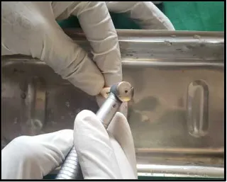
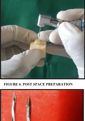
The groups B,C,D,E,F underwent post space preparation using a #6
reamer to remove gutta-percha apical to the CEJ from each filled canal,
leaving 5mm remaining in apical region. The length of the post space prepared
portion of the canal was kept uniform at 10mm. This was common for all the
groups other than group E,F where the apical 3mm of the post space
preparation was further prepared using a customized post drill to obtain a
A direct wax pattern was taken using inlay wax and 21 gauge wire
Wax pattern for all custom post made and the margins were finished. The wax
patterns were sprued, invested, and cast in a Ni-Cr alloy. The canal of each
tooth was restored with a custom made cast post and core with respective
dowel designs other than the control group A.
Group B- parallel dowel without any modifications
Group C- parallel dowel with uniform serrations at every 2 mm
Group D-parallel dowel with non uniform serration
Group E- parallel dowel with uniform serrations at every 2 mm with
stabilizing end
Group F- parallel dowel with non uniform serrations with stabilizing
end
The zinc phosphate cement mix was introduced into each root canal
with a lentulo spiral. All posts were cemented under finger pressure, held in
place until final setting. Excess cement was removed.
Final impressions for metal crowns were taken with addition silicone.
Master cast was obtained from the impression. Metal crown was fabricated
and luted with zinc phosphate cement.
The samples were positioned at 45 degrees to the long axis of teeth in
orientation in a universal testing machine. A crosshead speed of 0.5 mm/min
was applied until each sample fractured.
The load at which failure occurred was measured in Newton (N).
Failure was defined as fracture of the core material with displacement from the
post head, or when fracture affected the core or the tooth.
Access opening done , followed by cleaning and shaping ( apical enlargement upto size 40 k file). Obturation done by lateral condensation technique
Teeth were randomly divided into 6 groups A,B,C,D,E,F ( 10 teeth/group )
Teeth were sectioned 2mm coronal to CEJ for 5 groups (B,C,D,E and F)
Teeth samples were embedded in acrylic block at the level of 2 mm below CEJ
Post space preparation done with # 2 to 4 peaso reamer upto apical 5 mm gutta percha for groups B,C,D,E and F. but in groups E,F a Custom made drill was used in the apical 3mm of the preparation to obtain a stabilizing end of the
cast post (7mm+3mm)
GROUP D-Customised post and core with non-uniformly serrated parallel dowel
GROUP E-Customised post and core with uniformly serrated parallel dowel with stabilizing end
GROUP F-Customised post and core with non- uniformly serrated parallel dowel with stabilizing end
*2mm uniform ferrule is incorporated in all the groups
Posts luted with zinc phosphate cement
Addition silicone impresssion for metal crown
Metal crowns luted using zinc phosphate cement
Specimens mounted at an angle of 45˚ in universal testing machine and the load at which failure occurs is recorded
FIGURE 2. ACCESS OPENING OF TOOTH
FIGURE 4. TEETH SAMPLES EMBEDDED IN ACRYLIC BLOCK
FIGURE 6. POST SPACE PREPARATION
[image:56.595.172.450.369.622.2]FIGURE 8: GROOVESMADE IN WIRE FOR WAX PATTERN
[image:57.595.141.455.390.642.2]FIGURE 10: FABRICATED WAX PATTERNS
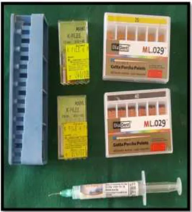
[image:58.595.155.443.411.649.2]FIGURE 12A: INSTRUMENTS USED FOR RCT
[image:59.595.172.425.386.617.2]FIGURE 13: IRRIGANTS USED
[image:60.595.156.437.401.643.2]GROUP C:UNIFORMLY SERRATED PARALLEL DOWEL
GROUP D: NON
UNIFORMLY SERRATED PARALLEL DOWEL
GROUP E: UNIFORMLY
SERRATED PARALLEL DOWEL WITH STABILIZING END
[image:61.595.113.541.99.682.2]GROUP F: NON UNIFORMLY SERRATED PARALLEL DOWEL WITH STABILIZING END
FIGURE 15: SCHEMATIC REPRESENTATION OF ALL EXPERIMENTAL GROUPS
FIGURE 16: PARALLEL DOWEL (GROUP B)
FIGURE 17: UNIFORMLY SERRATED PARALLEL DOWEL (GROUP C )
[image:62.595.107.550.110.244.2]FIGURE 19: UNIFORMLY SERRATED PARALLEL DOWEL WITH STABILIZING END (GROUP E )
FIGURE 21: ADDITION SILICONE IMPRESSION FOR METAL CROWNS
FIGURE 23: TOOTH ANGULATED AT ANGLE OF 45˚
[image:65.595.157.476.357.636.2]FIGURE 25:
INTRAORAL PERIAPICAL RADIOGRAPH OF POST SPACE PREPERATION
[image:66.595.109.334.107.665.2]RESULTS
Data obtained from universal testing machine in newtons were
recorded and tabulated for each specimen. It was then entered in an excel
spread sheet and analyzed using SPSS (Statistical Package for Social
Sciences) V.23 software. Kolmogorov Smirnov test was used to check
normality and the data was found to follow normal distribution. p value < 0.05
was considered to be statistically significant and one-way ANOVA and Post
hoc Tukey HSD tests were done.
Table 1-Denotes the fracture resistance for each of the samples in all
the groups measured from universal testing machine in Newton (N).
Table 2 -Denotes mean values and standard deviation among the
groups A, B, C, D, E and F .The mean values of all the groups showed that the
highest fracture resistance was seen in the group E (uniformly serrated
parallel dowel with stabilizing end) and the lowest fracture resistance was seen
in the group restored with parallel dowel (group b) other than the control
group A. When all the groups (B, C, D, E, F) were compared with the control
group A, the p value obtained was highly significant(p value < 0.001**).
Further, multiple comparison among groups were performed using
posthocTukey HSD test
Table 3-Denotes the multiple comparison among the groups which
did not receive any post showed inferior strength than other groups which
received a post. But when intra group comparison was done with the other
groups the p value was insignificant showing that the custom made post
fabricated had no difference in whichever technique and preparations done.
Graph 1 Denotes the mean fracture resistance among the 6 groups .
The Group E (uniformly serrated parallel dowel with stabilizing end) had
Table 1- Denotes the fracture resistance for each of the samples in all the
groups measured from universal testing machine in Newton (N).
GROUP A GROUP B GROUP C GROUP D GROUP E GROUP F
588 1470 1274 1765 1569 1863
686 1372 1372 1667 1667 1569
882 1274 1863 980 1372 1274
588 1176 1765 1078 1470 1372
784 1618 980 1323 1961 1470
490 1667 1078 1421 1863 1765
539 931 1372 1372 1078 980
882 1274 1520 1470 1176 1078
735 980 1470 1225 1470 1323
Table 2 -Denotes mean values and standard deviation among the GROUPS A,B,C,D,E & F
Experimental groups
sample
size(n) Minimum Maximum Mean
Std. Deviation
GROUP-A 10 441 882 661.5 157.089
GROUP-B
10 931 1667 1318.3 244.149
GROUP-C 10 980 1863 1426.3 276.097
GROUP-D
10 980 1765 1362.4 240.155
GROUP-E
10 1078 1961 1524.4 276.115
GROUP-F
10 980 1863 1421.4 277.599
Table 3: Comparison of all the six groups (ANOVA)
Sum of
Squares Df Mean Square F Sig.
Between Groups 4917318.283 5 983463.657
15.884 0.001 Within Groups 3343407.900 54 61914.961
[image:73.595.94.502.224.518.2]Table 4: Multiple Comparisons (Post hoc Tukey test) (I) groups (J) groups Mean Difference (I-J) Std. Error Sig. 1 2
-656.800* 111.27
9 .000
3
-764.800* 111.27
9 .000
4
-700.900* 111.27
9 .000
5
-862.900* 111.27
9 .000
6
-759.900* 111.27
[image:74.595.110.470.596.730.2]9 .000
Table 5: Comparison Of Groups With Different Dowel Designs (ANOVA)
Sum of
Squares df
Mean
Square F Sig.
Between
Groups 241560.920 4 60390.230
.871 .489 Within Groups 3121315.40
0 45 69362.564
Total 3362876.32
Graph 1 Denotes the mean fracture resistance among the 6 groups 0 200 400 600 800 1000 1200 1400 1600 1800
GROUP A GROUP B GROUP C GROUP D GROUP E GROUP F
DISCUSSION
Prognosis is the forecast of the course of a disease. In the context of
endodontics, therefore, this term was said to be positively correlated with the
technical quality of the disinfection and obturation of the root canal space.
However recently, attention has been given to procedures carried out after
completion of the endodontic treatment and their impact on the prognosis of
the treated teeth. Delay in these procedures allows the passage of
microorganisms and their by-products to the apical region of the root, a
potential cause of failures. The consequences of these events may be important
in determining the long-term success of endodontic treatment. The two factors
that govern the placement of a post endodontic restoration are biologic and
mechanical.
Ray and Trope (1995)13 studied the biologic aspect by evaluating the
relationship between the quality of the coronal restoration and the quality of
the root canal filling by examining the radiographs of endodontically treated
teeth. They observed that a combination of good restorations and good
endodontic treatments resulted in absence of periapical inflammation in 91.4%
of the teeth, whereas poor restorations and poor endodontic treatments resulted
in the absence of periradicular inflammation in only 18.1% of the teeth
examined. Furthermore, where poor endodontic treatments were followed by
concluded that apical periodontal health depended significantly more on the
coronal restoration than on the technical quality of the endodontic treatment.
Mechanical aspect deals with cusp fracture which is a common occurrence in
the heavily restored dentition. But endodontically treated teeth with
intra-coronal restorations are at higher risk and the occurrence of unrestorable
sub-gingival cusp fractures is more common. Fennis et al. found that, there was a
positive correlation between endodontically treated teeth and subgingival
fracture location. An endodontically treated posterior tooth may have a cavity
depth three to four times greater than a vital tooth - hence significantly greater
risk of fracture. A twenty year retrospective study showed that in
endodontically treated posterior teeth restored with amalgam without cusp
coverage, fracture was a significant problem. Maxillary bicuspids with MOD
restorations showed the lowest survival rate overall (28% fractured within
three years, 57% after ten years and 73% after twenty years). The most serious
fractures were found in the maxillary second molar and accounted for the
majority of the extractions due to vertical fracture. It was concluded that silver
amalgam without cusp coverage was unsuitable for restoration of multiple
surfaces of the endodontically treated tooth (Ray and Trope 1995)13.
It is therefore evident that endodontically treated posterior teeth with
intra-coronal restorations show a high risk of unrestorable cusp fracture. Hence, the
use of crowns can significantly improve the success of endodontic treatment in
Analysis of the reason for all extractions of endodonticallytreated teeth over a
period of one year revealed that almost 60% of these were unrestorable tooth
fractures, 32% involved periodontal problems and only 7% were endodontic
failures. The major reason for failure is unsuccessful restorative treatment and
to a lesser extent, inadequate endodontic therapy. Close to half of all failures
were due to fracture of the natural coronal tooth structure and appeared to
involve either uncrowned teeth or crowned teeth without definitive anchorage.
They were deemed unrestorable due to the extent of lost tooth structure. Teeth
with crowns showed longer clinical life than non-crowned teeth (Rapeephan
Nagasiri,2004)28.
A high incidence of failure for posterior endodontically treated teeth without
cusp coverage has been reported.In a retrospective study of uncrowned
endodontically treated teeth, the overall survival rates of molars without
crowns at 1, 2, and 5 years were 96%, 88% and 36% respectively.The amount
of remaining tooth structure was a significant factor in tooth survival.
Aqualina et al found that endodontically treated teeth without crowns failed at
6 times greater rate than uncrowned teeth. The presence of a crown was more
important than the type of foundation restoration, although it was noted that
teeth with posts demonstrated better survival.
The clinical longevity of endodontically treated maxillary and mandibular
anterior teeth does not appear to be dependent on coronal coverage. Separation
tooth fracture is less common, provided adequate coronal dentin remains.
Placement of a crown is not necessary just because an anterior tooth is
endodontically treated. It becomes a matter of clinical judgment depending on
whether the tooth can be adequately restored to form and function with direct
restorative materials. The presence of moderate class III, or class IV, bonded
composite restorations can frequently provide excellent longevity, provided
that the endodontic access is not excessive.
However, in the case of badly mutilated tooth with one or two remaining
walls, post endodontic restoration with a post and core system followed by the
placement of a crown becomes a necessity. This post and core system is
considered to reinforce the tooth structure and aid in the retention of the core.
Post and core systems broadly falls under 2 categories- prefabricated
post and cores, custom made post and cores. Prefabricated posts are popular
because of their ease of placement, less chair time, lower cost and the ability
to restore a tooth for immediate crown preparation; they also rely principally
on cement for retention (Goerig AC et al; 1983). As prefabricated posts are
cylindrical, they are best suited for circular canals (i.e. maxillary incisors)
rather than teeth with wide buccolingual root canals. But prefabricated post
rely principally on the cement for retention. They also have decreased core
retention and potential for rotation.
In the cast post/core system the core is an integral extension of the post, and
This construction prevents dislodgment of the core from the postand root
when minimal tooth structure remains. For many years, the cast metal post and
core has been the traditional method for fabricating the foundation
restorationof a prosthetic crown.
For any post system used, there are two factors that dictate its success:
retention and resistance form. Retention form prevents the removal of the post
along its path of insertion or the long axis of the tooth. Whereas, resistance
form prevents dislodgement of the post when forces are directed in an apical
or oblique direction and prevents any movement of the post under occlusal
forces. Retention and resistance are inter related and often inseparable
qualities.
Factors that affect the retention and resistance form include taper, freedom of
displacement, length of the post, substitution of internal features in post,
presence of ferrule and cementing material. Taper of the post should be kept at
a minimum to increase retention; hence, in this study parallel posts were used.
Freedom of displacement should be minimized, which is explained by limiting
the number of paths along which the post can be removed. So in this study,
custom cast post system was selected since it gives a more intimate fit to the
canal and provides better resistance against torque and twisting forces. The
length of the post is directly proportional to the resistance and retention of the
post. However, excessive removal of the root canal dentin results in
compromised root that is prone to fracture. Hence, an optimum post length