JOURNAL OFVIROLOGY, July 2006, p. 6225–6234 Vol. 80, No. 13 0022-538X/06/$08.00⫹0 doi:10.1128/JVI.02506-05
Copyright © 2006, American Society for Microbiology. All Rights Reserved.
Demonstration of Nicking/Joining Activity at the Origin of DNA
Replication Associated with the Rep and Rep
⬘
Proteins
of Porcine Circovirus Type 1
Tobias Steinfeldt, Tim Finsterbusch, and Annette Mankertz*
Division of Viral Infections (FG12), Robert Koch-Institute, 13353 Berlin, GermanyReceived 30 November 2005/Accepted 10 April 2006
The replication of porcine circovirus type 1 (PCV1) is thought to occur by rolling-circle replication (RCR), whereby the introduction of a single-strand break generates a free 3ⴕ-hydroxyl group serving as a primer for subsequent DNA synthesis. The covalently closed, single-stranded genome of PCV1 replicates via a double-stranded replicative intermediate, and the two virus-encoded replication-associated proteins Rep and Repⴕ have been demonstrated to be necessary for virus replication. However, although postulated to be involved in RCR-based virus replication, the mechanism of action of Rep and Repⴕis as yet unknown. In this study, the ability of PCV1 Rep and Repⴕto “nick” and “join” strand discontinuities within synthetic oligonucleotides corresponding to the origin of replication of PCV1 was investigated in vitro. Both proteins were demonstrated to be able to cleave the viral strand between nucleotides 7 and 8 within the conserved nonanucleotide motif (5ⴕ-TAGTATTAC-3ⴕ) located at the apex of a putative stem-loop structure. In addition, the Rep and Repⴕ proteins of PCV1 were demonstrated to be capable of joining viral single-stranded DNA fragments, suggesting that these proteins also play roles in the termination of virus DNA replication. This joining activity was demonstrated to be strictly dependent on preceding substrate cleavage and the close proximity of origin fragments accomplished by base pairing in the stem-loop structure. The dual “nicking/joining” activities associated with PCV1 Rep and Repⴕ are pivotal events underlying the RCR-based replication of porcine circoviruses in mammalian cells.
Porcine circovirus type 1 (PCV1) and PCV2 are members of the familyCircoviridae(40, 49). Circoviruses infect birds and vertebrates (30). They are characterized by covalently closed circular single-stranded DNA (ssDNA) genomes. PCV1 (46, 48) is widespread in swine, but a cytopathogenic effect has not been observed in tissue culture cells and the virus is not associated with any animal disease (3, 47). In contrast, PCV2 (4, 13, 36, 37) is the etiological agent of a new emerg-ing multifactorial disease in swine, named postweanemerg-ing mul-tisystemic wasting syndrome (1, 12). The genomes of PCV1 and PCV2 are similarly organized and display a homology of 68 to 76% identity (30).
Upon infection, the viral ssDNA genome is converted into a double-stranded intermediate and serves as a template for viral replication. The origin of replication is located within the in-tergenic region between the two major open reading framesrep
andcap(34) and overlaps with the promoter of therepgene. The origin is the initial point of plus-strand synthesis and comprises characteristic sequence motifs (Fig. 1). An inverted repeat forms a putative stem-loop structure with a nonamer (5⬘-T/AAGTATTAC-3⬘) in its apex, conserved in all circovi-ruses. Adjacent to this structure, hexamer and pentamer re-peats are found. The minimal binding site (MBS) for the PCV replication proteins Rep and Rep⬘comprises the 3⬘part of the inverted repeat plus the two inner hexamers (44). It is sug-gested that recruitment of the replication proteins for
initia-tion of replicainitia-tion at the double-stranded origin is mediated by binding to the MBS. The inverted repeat comprising the con-served nonamer is also found in the virus families Geminiviri-dae(20, 27, 42, 43) andNanoviridae(7, 17, 41). These plant-infecting viruses possess a circular covalently closed ssDNA genome, which is replicated by rolling-circle replication (RCR). For geminiviruses, the initial point of viral-strand DNA synthesis has been mapped to the putative stem-loop within the nonamer in the intergenic region. Rep is the sole viral protein essential for replication, binding, and cleaving the viral origin of replication in a strand-specific manner within the nonamer between nucleotides 7 and 8 (21, 29, 45). While the Rep protein is covalently attached to the 5⬘ end after cleavage (29, 45), unidirectional leading-strand synthesis is ini-tiated at the generated 3⬘-hydroxyl group, probably by a host DNA polymerase. After one or more rounds of replication, the regenerated origin is recognized and cleaved again by Rep, thereby terminating the reaction and releasing the newly syn-thesized single-stranded viral genome via a nucleotidyltransfer reaction (29).
In contrast to geminiviruses, not only one but two products of the rep gene are indispensable for PCV replication (32). Besides the full-length protein Rep, a spliced isoform of the replication protein called Rep⬘ has been identified. Rep⬘ is truncated, and the C terminus is expressed in a different read-ing frame. Both proteins have been shown to bind to double-stranded oligonucleotides carrying the conserved sequence motifs of the plus-strand origin in vitro (44). Besides its role in replication, the Rep protein is a transcriptional repressor ofrep
gene expression. This function is also exerted by binding to the MBS (31). The Rep protein shows homology to the replication * Corresponding author. Mailing address: Division of Viral
Infec-tions (FG12), Robert Koch-Institute, Nordufer 20, 13353 Berlin, Germany. Phone: 49 30 4547 2516. Fax: 49 30 4547 2598. E-mail: [email protected].
6225
on November 8, 2019 by guest
http://jvi.asm.org/
proteins of nanoviruses and geminiviruses. Based upon se-quence comparisons, three conserved regions (I, II, and III), typical of replication enzymes mediating replication by RCR (25), as well as a P loop for deoxynucleoside triphosphate binding, have been identified (Fig. 1) (34). RCR (16) has been described for plasmids of bacteria (18, 24), bacteriophages (5), and ssDNA viruses of plants (22, 27), birds (6, 38), and mam-mals (2). The striking conservation of the involved replication initiator proteins and their cognate origins of replication led to the assumption that PCV replication is achieved by RCR as well (34, 35).
In this study, initiation and termination of viral replication
by purified PCV1 Rep and Rep⬘ fusion proteins have been
investigated in vitro. The influence of sequence specificity and secondary structures with respect to the enzymes’ cleavage/ joining activities has been determined. Evidence for cleavage as a condition for joining is presented.
MATERIALS AND METHODS
Construction of plasmids.All plasmids were constructed by cloning of PCR-generated fragments.
For the generation of pTriEx-6HN-rep and pTriEx-6HN-rep⬘, PCR fragments were amplified with high-fidelity polymerase (Roche Diagnostics, Mannheim, Germany) from plasmids pORF4A (encoding Rep) (33) and pAM4 (encoding Rep⬘) (32), using primers F245 (5⬘-CGGGATCCAAGCAAGAAAAGCGGC-3⬘)
and B226 (5⬘-GGAATTCGATGTGATAACAAAAAAGACTCAGT-3⬘). PCR
fragments were restricted with EcoRI and BamHI and cloned into EcoRI- and BamHI-restricted vector pTriEx-6HN.
For the generation of vector pTriEx-6HN, oligonucleotides F524 (5⬘-TCATC
ATCATCACCATCACAGCAGCGGCGAGCT-3⬘) and B525 (5⬘-CGCCGCTG
CTGTGATGGTGATGATGATGA-3⬘) were hybridized and cloned into
EcoRV- and SacI-restricted vector pTriEx1.1 (Merck Biosciences, Schwalbach am Taunus, Germany), enabling the expression of N-terminally His-tagged fu-sion proteins.
All plasmids were sequenced to exclude PCR-acquired misincorporations.
Expression and purification of PCV1 replication proteins Rep and Repⴕ.For the expression of N-terminally His-tagged fusion proteins PCV1 Rep and Rep⬘,
Escherichia coli RosettaBlue(DE3)pLacI competent cells (Merck Bio-sciences) were transformed with pTriEx-6HN-rep or pTriEx-6HN-rep⬘. Ad-ditionally, as a negative control, an empty vector (pTriEx-6HN) was trans-formed. Bacteria were grown at 33°C in LB medium supplemented with ampicillin (50g/ml), chloramphenicol (30g/ml), and glucose (1%) to an optical density at 588 nm of 0.75. Expression was induced by the addition of an equal volume of LB medium supplemented with ampicillin (100g/ml) and IPTG (isopropyl--D-thiogalactopyranoside) (0.5 mM) for 2 h at 33°C.
[image:2.585.65.502.65.289.2]FIG. 1. Map of PCV1 and localization of the plus-strand origin. A linear map of the circular genome of PCV1 is depicted. Open reading frames are indicated by open bars at the top, transcripts for Rep and Rep⬘with splice sites by horizontal arrows. The viral origin of replication, located between the divergently transcribedrepandcapgenes, is enlarged. The expanded view elucidates the putative stem-loop structure with adjacent hexamer and pentamer sequences framed by boxes and ovals. The nonamer sequence in the apex of the stem-loop is shown in boldface, with the cleavage site indicated by an arrow.
FIG. 2. Purified His-Rep and His-Rep⬘fusion proteins. His-Rep (lane 2) and His-Rep⬘(lane 3) fusion proteins were expressed inE. coli
cells and subsequently purified by affinity chromatography using Ni-nitrilotriacetic acid agarose beads. Rebuffered and concentrated puri-fied proteins were fractionated by sodium dodecyl sulfate-polyacryl-amide gel electrophoresis and visualized by staining with Coomassie brilliant blue dye. The apparent molecular masses were determined by comparison with a protein standard (lane 1) with the indicated molec-ular masses to the left of the gel.
6226 STEINFELDT ET AL. J. VIROL.
on November 8, 2019 by guest
http://jvi.asm.org/
Purifications of His-tagged fusion proteins were performed with Ni-nitrilo-triacetic acid HisBind resin after cell lysis using BugBuster Protein Extraction Reagent in combination with Benzonase Nuclease and rLysozyme Solution according to the recommendations of the manufacturer (Merck Biosciences). Purified recombinant proteins Rep and Rep⬘ were concentrated and re-buffered in Tris-EDTA buffer, pH 7.5, using Microcon centrifugal filter de-vices (Millipore Corporation, Bedford, MA). Aliquots of the purified proteins were fractionated by sodium dodecyl sulfate-polyacrylamide gel electro-phoresis and visualized by staining with Coomassie brilliant blue dye.
In vitro cleavage and cleavage/joining reactions.DNA cleavage and cleavage/ joining reactions were performed with oligonucleotides corresponding to se-quences of the PCV1 plus-strand origin (see Fig. 3).
In the cleavage reaction, 0.5 pmol of Cy5 5⬘-end-labeled DNA substrate was incubated with 500 ng of purified recombinant Rep or Rep⬘fusion protein or with the negative control in cleavage buffer (50 mM Tris-HCl, pH 7.5, 150 mM NaCl, 10 mM MgCl2, 5 mM dithiothreitol, 1 mM EDTA) in a total volume of 30
l for 90 min at 37°C. Following incubation, the cleavage products were analyzed by electrophoresis in 5.5% polyacrylamide gels under nondenatur-ating conditions at room temperature or containing 6.7 M urea at 65°C after proteinase K digestion and visualized using the Fluorescent Image Analyzer
FLA-2000 (Fuji, Stamford, CT). The same procedures were followed for the cleavage/joining reaction with the exception that nondenaturating polyacryl-amide gels were run at 65°C.
RESULTS
Cloning and bacterial expression of PCV1 replication pro-teins.For analysis of the catalytic activities of the PCV1 rep-lication proteins, Rep and Rep⬘ were expressed as recombi-nant N-terminally His-tagged fusion proteins from plasmids pTriEx-6HN-rep and pTriEx-6HN-rep⬘ inE. coliafter induc-tion with IPTG. On the basis of a Coomassie-stained poly-acrylamide gel, the apparent molecular masses of His-Rep and His-Rep⬘corresponded to 36 kDa and 20 kDa (Fig. 2, lanes 2 and 3).
In vitro cleavage of the viral strand by PCV1 Rep and Repⴕ.
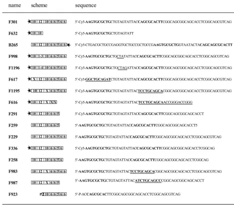
[image:3.585.53.520.76.486.2]Viral replication proteins mediating replication by RCR cleave FIG. 3. Names, schemes, and sequences of oligonucleotides. For schematic presentation, single sequence elements are indicated by numbers of nucleotides, Cy5-end labeling is represented by a black star, and phosphorylation is indicated with a P. Inverted repeats framing the nonamer are shown in boldface, and base alterations are underlined in the oligonucleotide sequences.
VOL. 80, 2006 CLEAVAGE/JOINING AT THE ORIGIN OF PCV1 6227
on November 8, 2019 by guest
http://jvi.asm.org/
the viral origin, thereby introducing a ssDNA break, or “nick.” A subsequent ligation reaction is needed for the sealing and maturation of the newly synthesized viral ssDNA. To investi-gate the abilities of Rep and Rep⬘of PCV1 to cleave the viral DNA and reseal the resultant nick, recombinant proteins were incubated with synthetic oligonucleotides encompassing se-quence motifs of the viral origin of replication (all oligonucle-otides are shown in Fig. 3).
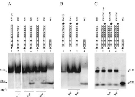
F301 was used as a substrate in the cleavage reaction. The 61-mer was labeled with Cy5 at its 5⬘end and comprised the inverted repeat with the conserved nonamer (10-12-10) (Fig. 4) and four adjacent hexamers (6-6-5-6-6) corresponding to the viral plus strand (Fig. 4A, lane 1). Incubation with the fusion protein His-Rep, as well as with His-Rep⬘, resulted in the appearance of an additional band representing the 5⬘-terminal cleavage product (Fig. 4A, lanes 4 and 6). Specificity of cleav-age could be confirmed by the absence of catalytic activity in the negative control (Fig. 4A, lane 2). The size of the cleavage product was determined by comparison with an oligonucleo-tide marker 20 nucleooligonucleo-tides (nt) in size (F632) (Fig. 4A, lane 7),
indicating that cleavage occurs between nucleotides 7 and 8
within the conserved nonamer. In contrast, Rep and Rep⬘
cleavage of the viral minus strand (B265) (Fig. 4B, lanes 2 and 3) and the double strand (F301/B265) (Fig. 4C, lanes 3 and 4) was not observed. To rule out possible retention of the cleav-age product at the top of the gel due to binding of the replicase to the undisintegrated double strand, products were resolved after protease digestion on urea gels run at 65°C. The cleavage activities of Rep and Rep⬘ were strictly dependent on the presence of divalent cations in the cleavage buffer (Fig. 4A, lanes 3 and 5). Mg2⫹could be replaced by 10 mM Ba2⫹, Mn2⫹,
or Ca2⫹(data not shown). Catalytic activity of Rep occurred in
the absence of ATP but was stimulated after addition of ATP, whereas influence of ATP on the cleavage activity of Rep⬘was not detected (data not shown).
[image:4.585.64.521.63.392.2]Sequence and structure requirements for cleavage.The se-quence specificity of the cleavage reaction mediated by Rep and Rep⬘ was investigated with truncated and mutated plus-strand origin fragments (Fig. 3). After mutation of the first 3 nucleotides of the nonamer from 5⬘-TAGTATTAC-3⬘to 5⬘-C FIG. 4. PCV1 His-Rep and His-Rep⬘ cleave origin fragments of PCV1 within the conserved nonamer of a putative stem-loop structure. Bacterial overexpressed and subsequently purified fusion protein (500 ng) was incubated with 0.5 pmol oligonucleotide in the cleavage reaction in vitro. Unless otherwise noted, reactions were supplemented with Mg2⫹. Oligonucleotides of viral- or complementary-strand polarity
represent-ing the conserved sequences of the PCV1 origin of replication were labeled with Cy5 at the 5⬘end. Catalytic activities of His-Rep and His-Rep⬘ were determined with single-stranded (A, lanes 3 and 4, 5 and 6; B, lanes 2 and 3) and double-stranded (C, lanes 3 and 4) substrates. As a negative control (n. c.) plasmid pTriEx-6HN was used (A, lane 2). Samples in panels A and B were resolved on a native polyacrylamide gel at room temperature, samples in panel C on a denaturating polyacrylamide gel containing 6.7 M urea at 65°C in comparison to oligonucleotides of defined sizes (A, lanes 1 and 7; B, lanes 1 and 4; C, lanes 1, 2, and 5). The positions and sizes of oligonucleotides are marked, and cleavage products are highlighted by black arrowheads. See the legend to Fig. 3 for the remaining symbols.
6228 STEINFELDT ET AL. J. VIROL.
on November 8, 2019 by guest
http://jvi.asm.org/
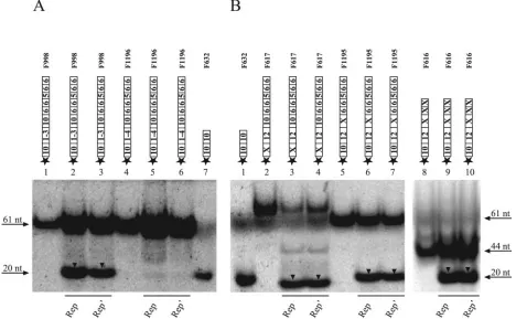
TATATTAC-3⬘ (F998), cleavage activity was still detectable (Fig. 5A, lanes 2 and 3), while sequence alterations of the first
4 nucleotides from 5⬘-TAGTATTAC-3⬘ to 5⬘-CTAGATTA
C-3⬘ (F1196), as well as of the entire nonamer (data not shown), resulted in a loss of cleavage activity of His-Rep and His-Rep⬘(Fig. 5A, lanes 5 and 6). These results indicate that only part of the conserved sequence 5⬘-TAGTATTAC-3⬘ spec-ifies the sequence requirements for the cleavage of origin
frag-ments of PCV1 by Rep and Rep⬘ and thereby corroborate
recently published data (8, 10).
The influence of secondary structures on cleavage was investigated with oligonucleotides disabled in formation of a hairpin structure. Oligonucleotides comprising a mutated 5⬘ part (F617) and 3⬘part (F1195) of the inverted repeat were
efficiently cut by Rep and Rep⬘, demonstrating that the
formation of a hairpin structure is not essential for cleavage in vitro (Fig. 5B, lanes 3 and 4, 6 and 7). In the case of F617, the forward reaction had gone almost to completion, i.e., only cleavage, but no religation, was observed, indicating that in contrast to the dynamic cleavage/joining reaction of F1195, the self-rejoining reaction for F617 may be seriously impaired.
Previously, the MBS comprising the 3⬘part of the inverted repeat and the two adjacent hexamers was found to be neces-sary for binding of Rep and Rep⬘ to double-stranded origin fragments in vitro (44). For determination of the influence of these elements upon cleavage of ssDNA origin fragments by
Rep and Rep⬘, F616 carrying altered sequences was used.
Cleavage of F616 was detected after the addition of Rep or Rep⬘(Fig. 5B, lanes 9 and 10). Since the cleavage activities of Rep and Rep⬘were not altered by preventing the formation of a stem-loop structure, these results signify different sequence requirements for binding to double-stranded and single-stranded origin fragments by Rep and Rep⬘in vitro. While the MBS is necessary for recognition and binding of double-stranded DNA (dsDNA) molecules, the nonamer was suffi-cient for cleavage of single-stranded origin fragments by Rep and Rep⬘.
[image:5.585.59.527.71.360.2]PCV1 Rep and Repⴕ exhibit joining activity. The joining activities of PCV1 Rep and Rep⬘were determined by incuba-tion with two substrates differing in size, with only one of them labeled at its 5⬘end. Joining was indicated by the appearance of a newly labeled product of different size, i.e., the transfer of the label from one molecule to the other (Fig. 6A). For deter-mination of a PCV1 Rep/Rep⬘ intrinsic joining activity, an equimolar mixture of the unlabeled 49-mer F259 and the Cy5 5⬘-end-labeled 61-mer F301 was incubated with purified His-Rep or His-His-Rep⬘in vitro. After ligation of the cleavage prod-ucts, an additional Cy5 5⬘-end-labeled product corresponding in size to the 49-mer F291 was observed (Fig. 6B, lanes 3 and 4). Combination of the Cy5 5⬘-end-labeled 49-mer F291 and the unlabeled 61-mer F229 resulted in the formation of a product with a mobility corresponding to that of the 61-mer F301 (Fig. 6B, lanes 8 and 9). We concluded that PCV1 Rep FIG. 5. Sequence requirements for His-Rep- and His-Rep⬘-mediated cleavage reactions. Cy5 5⬘-end-labeled oligonucleotides corresponding to mutated and truncated plus-strand origin fragments were incubated with purified His-Rep (A, lanes 2 and 5; B, lanes 3, 6, and 9) or His-Rep⬘(A, lanes 3 and 6; B, lanes 4, 7, and 10) fusion protein in vitro. All samples were resolved on a native polyacrylamide gel in comparison to oligonucleotides of defined sizes (A, lanes 1, 4, and 7; B, lanes 1, 2, and 5). The positions and sizes of oligonucleotides are marked, and cleavage products are highlighted by black arrowheads. See the legend to Fig. 3 for the remaining symbols.
VOL. 80, 2006 CLEAVAGE/JOINING AT THE ORIGIN OF PCV1 6229
on November 8, 2019 by guest
http://jvi.asm.org/
and Rep⬘possess a nucleotidyltransferase activity, ligating or-igin fragments in vitro.
Sequence requirements for joining. The inverted repeat within the origin may serve as a fastener bringing the two restricted strands into close proximity for subsequent ligation. To test this hypothesis, mutagenized plus-strand origin frag-ments were used in the cleavage/joining assay. Base alterations were located either upstream (F617) or downstream (F983) of the conserved nonamer sequence, thereby inhibiting formation of a hairpin structure. These modifications resulted in loss or
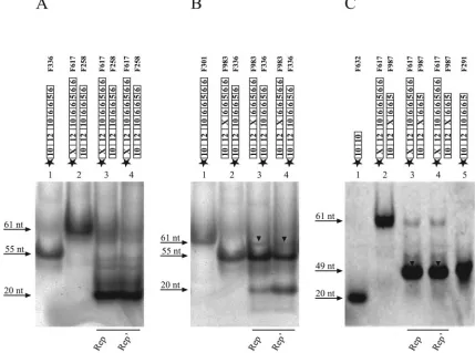
significant reduction of the joining activities of Rep and Rep⬘ in vitro (Fig. 7A and B, lanes 3 and 4). This result is in correlation with the observed restriction pattern of oligonucle-otides F617 and F1195 (Fig. 5B, lanes 3 and 4, 6 and 7), suggesting a limited self-rejoining reaction for F617, but not for F1195, in vitro. Additionally, we used two oligonucleotides carrying corresponding mutations upstream (F617) or down-stream (F987) of the conserved nonamer sequence, which should restore hairpin formation after the ligation of cleavage
[image:6.585.68.521.72.537.2]products. Incubation of Rep and Rep⬘ with F617 and F987
FIG. 6. PCV1 His-Rep and His-Rep⬘exhibit joining activities. (A) Schematic representation of the Rep- and Rep⬘-mediated joining reaction. (B) An equimolar mixture of two oligonucleotides of viral-strand polarity, with only one labeled with Cy5 at its 5⬘end and varying in sizes and numbers of hexamer motifs, was incubated with purified His-Rep (lanes 3 and 8) or His-Rep⬘(lanes 4 and 9) fusion protein in the cleavage/joining reaction in vitro. All samples were resolved on a native polyacrylamide gel next to oligonucleotides of defined sizes (lanes 1, 2, 5, 6, and 7). The positions and sizes of oligonucleotides are marked. Recombinant products, 49 nt and 61 nt in size, are highlighted by black arrowheads. See the legend to Fig. 3 for the remaining symbols.
6230 STEINFELDT ET AL. J. VIROL.
on November 8, 2019 by guest
http://jvi.asm.org/
demonstrated that the joining activities of Rep/Rep⬘were re-stored completely (Fig. 7C, lanes 3 and 4). According to these results, ligation of restricted origin fragments in vitro is inde-pendent of specific sequences flanking the nonamer, as long as base pairing is possible. Interestingly, a cleavage product was not observed (Fig. 7C, lanes 3 and 4), indicating that joining of substrates F617 and F987 is efficient, but subsequent cleavage of the newly formed ligation product (49 nt) is impaired by the sequence alterations upstream and downstream of the nonamer.
Cleavage is a prerequisite for joining. The dependence of the joining reaction on preceding substrate cleavage was inves-tigated. After incubation of purified His-Rep or His-Rep⬘ fu-sion protein with an equimolar mixture of oligonucleotides mimicking a putative Cy5 5⬘-end-labeled 5⬘ cleavage product (F632) and a 5⬘-phosphorylated 3⬘cleavage product (F923), no joining activity was detectable (Fig. 8A, lanes 5 and 6), dem-onstrating that substrate cleavage has to precede joining. This assumption is supported by the restoration of joining activity after the addition of the unlabeled 61-mer (F229) instead of the 3⬘ cleavage product F923 (Fig. 8C, lanes 2 and 3). The 5⬘-terminal cleavage product, but no ligation, was observed after incubation of the labeled 55-mer (F336) and F923 with purified His-Rep (Fig. 8B, lane 3) or His-Rep⬘fusion protein
(Fig. 8B, lane 4). These results demonstrate that substrate cleavage is a condition for joining of origin fragments. More-over, it supports the hypothesis that the replication proteins are associated with the 5⬘end of the 3⬘cleavage product, as has been shown for other RCR systems (29, 45).
DISCUSSION
In replicons replicating by RCR, DNA cleavage and ligation are pivotal steps during ssDNA synthesis. After recruitment of the replicon-encoded initiator protein by binding to the dou-ble-stranded origin, replication is initiated by introduction of a strand- and site-specific nick within the origin sequence, gen-erating a free 3⬘-hydroxyl group. While this serves as a primer for DNA synthesis, the 5⬘-phosphate group is displaced during DNA replication. After one or more rounds of replication, the nascent strand is cleaved again within the regenerated origin, and the 5⬘-phosphate end is ligated to the newly created 3⬘ -hydroxyl group, resulting in the release of unit length mono-mers (23).
Due to their limited coding capacities, small DNA viruses often encode multifunctional proteins that accomplish differ-ent tasks during infection. A case in point is the simian virus 40 large T antigen involved in replication, transcription, and ac-FIG. 7. Sequence requirements for His-Rep- and His-Rep⬘-mediated joining reaction. Cy5 5⬘-end-labeled oligonucleotides corresponding to mutated plus-strand origin fragments were incubated with purified His-Rep (A, B, and C, lanes 3) or His-Rep⬘(A, B, and C, lanes 4) fusion protein in vitro. All samples were resolved on a native polyacrylamide gel in comparison to oligonucleotides of defined sizes (A and B, lane 1; C, lanes 1 and 5). The positions and sizes of oligonucleotides are marked, and recombinant products, 61 nt and 49 nt in size, are highlighted by black arrowheads. See the legend to Fig. 3 for the remaining symbols.
VOL. 80, 2006 CLEAVAGE/JOINING AT THE ORIGIN OF PCV1 6231
on November 8, 2019 by guest
http://jvi.asm.org/
[image:7.585.76.505.73.392.2]tivation of the cell cycle of the host (14), or the AL1 protein of tomato golden mosaic virus, which binds dsDNA and catalyzes ssDNA cleavage and ligation (15, 29). In contrast to the gemini-viruses employing only one essential replicase, two proteins of PCV1, Rep and Rep⬘, have been proven to be indispensable for viral replication. The multifunctional character of the rep-lication proteins Rep and Rep⬘of PCV1 has been established in previous studies (30). Here, we demonstrate for the first time the cleavage/joining activities of Rep and Rep⬘ at the origin of replication, i.e., both are able to function as RCR initiator proteins in vitro. Our results showed that Mg2⫹, but
not ATP, had to be present and that Rep/Rep⬘ discriminate between strand polarities at the viral origin of replication, corroborating a prior study characterizing the Rep protein of banana bunchy top virus (19).
In this study, investigation of sequential and structural de-pendencies was performed with mutated and truncated oligo-nucleotides. Mutation of the entire nonamer sequence of PCV1, as well as its first 4 nucleotides, resulted in total loss of cleavage activity, while alteration of the first 3 nucleotides within the nonamer did not impede the activities of Rep and Rep⬘. Thus, the canonical nonamer sequence T1AGTATTAC9
can be condensed to the hexamer T4ATTAC9, sufficient for
cleavage and ligation of origin fragments in vitro, while 6 nucleotides within the octamer A2XTAXTAC9 are essential
for PCV DNA replication in vivo (8, 10).
Since oligonucleotides disabled in formation of a hairpin structure by sequence alteration of only one part of the in-verted repeat were cut efficiently, formation of this secondary-structure element is not a prerequisite for cleavage of origin fragments in vitro. In contrast to this finding, joining of origin fragments in vitro was reduced by mutations affecting the right arm of the hairpin and even more strongly impaired by muta-tions in the left arm. Removing the specific sequence without disturbing the base pairing of the restricted products restored the joining activity. These results indicate that base pairing is essential for joining of the origin fragments in vitro and that downstream sequence alterations can be better compensated for than upstream mutations.
The MBS of PCV1 has been characterized previously. It comprises the right part of the putative stem-loop and the two adjacent hexamers and is essential for recruiting Rep and Rep⬘ to double-stranded origin fragments (44). In contrast to bind-ing to dsDNA, the specific sequence of these elements is not FIG. 8. Cleavage is a prerequisite for joining. (A) an equimolar mixture of two oligonucleotides of viral-strand polarity, mimicking the labeled 5⬘cleavage product and the phosphorylated 3⬘cleavage product, were incubated with His-tagged fusion protein Rep (lane 5) or Rep⬘(lane 6) in the cleavage/joining reaction in vitro. (B) An equimolar mixture of two oligonucleotides of viral-strand polarity, mimicking the labeled 61-mer and the phosphorylated 3⬘cleavage product, were incubated with His-tagged fusion protein Rep (lane 3) or Rep⬘(lane 4) in the cleavage/joining reaction in vitro. (C) An equimolar mixture of two oligonucleotides of viral-strand polarity, mimicking the labeled 5⬘cleavage product and the unlabeled 61-mer, were incubated with His-tagged fusion protein Rep (lane 2) or Rep⬘(lane 3) in the cleavage/joining reaction in vitro. All samples were resolved on a native polyacrylamide gel next to oligonucleotides of defined sizes (A, lanes 1 and 2; B, lane 1; C, lane 4). The positions and sizes of oligonucleotides are marked. Recombinant products, 61 nt in size, are highlighted by black arrowheads. See the legend to Fig. 3 for the remaining symbols.
6232 STEINFELDT ET AL. J. VIROL.
on November 8, 2019 by guest
http://jvi.asm.org/
required for recognition and cleavage of single-stranded origin fragments in vitro. These results are in agreement with results observed for PCV1 replicating in cultured cells, suggesting specific interaction of the replication proteins with the nonamer (9, 11). Nevertheless, in vitro cleavage activity relies upon se-quences flanking the nonamer upstream and downstream, be-cause sequence alterations enclosing both parts led to complete inhibition of cleavage despite the presence of the hexamer se-quences.
Since the ssDNA genome of PCV is converted into a double-stranded replicative form, cleavages of single-double-stranded and double-stranded oligonucleotides were compared in this study.
Although Rep and Rep⬘ bind to double-stranded templates,
cleavage activity was observed only for single-stranded tem-plates. This apparent contradiction may be resolved by the hypothesis that cruciform extrusion provides the ssDNA con-formation necessary for cleavage in vivo, as in the cases of the plasmids of the pT181 family (24, 39). In our assay, this struc-ture may be formed but not maintained, because stabilizing factors are missing. Alternatively, local melting at the viral origin of replication by an ATP-dependent helicase activity of the Rep protein may have to precede cruciform extrusion. Sequence alignments revealed that the C termini of PCV Rep proteins encompass a putative helicase domain (30) related to the helicases of other ssDNA and dsDNA viruses (26). Al-though the impact of the C-terminally located GKS box of PCV1 Rep on replication in vivo was demonstrated (32), ATP binding or hydrolysis is not necessary for the in vitro cleavage activity of PCV1 Rep.
Termination of viral replication is accomplished after RCR by ligation of the 5⬘-phosphate to the newly created 3⬘-hydroxyl group and release of a circular ssDNA molecule. Rep and Rep⬘both catalyze the joining of oligonucleotides resembling fragments of the viral origin in vitro without ATP. Cleavage is a condition for joining, since oligonucleotides mimicking the cleavage products were not ligated by Rep and Rep⬘and the activity was restored by addition of the uncleaved origin frag-ment. Since covalent association of protein and DNA has been demonstrated for the Rep proteins of geminiviruses (29) and conservation of energy provides an explanation for the inde-pendence of the cleavage/joining reaction from ATP, these findings suggest that Rep and Rep⬘are linked to the 5⬘ -phos-phate of the 3⬘ part of the cleaved origin fragment in vitro. Current experiments using 3⬘-biotinylated oligonucleotides support this interpretation (T. Finsterbusch, unpublished data).
A model for PCV1 Rep/Repⴕ-catalyzed RCR.After conver-sion of the viral ssDNA into the double-stranded replicative
intermediate, Rep and Rep⬘ of PCV1 bind to the hexamer
sequences within the origin of replication. The initial step for the subsequent RCR of the viral DNA is the site-specific cleav-age of the viral strand between nucleotides 7 and 8 within the conserved nonamer. Cruciform extrusion provides the single-stranded conformation of the nonamer that is indispensable for cleavage and is possibly introduced by the helicase activity of Rep. Cleavage results in generation of a free 3⬘-hydroxyl group for priming of plus-strand DNA synthesis. After at least one round of replication, a second cleavage terminates DNA synthesis and a Rep/Rep⬘-mediated nucleotidyltransfer
reac-tion results in the release of a newly synthesized circular ssDNA molecule.
In bacteriophage⌽x174, the replication initiator protein RepA contains two closely spaced tyrosyl groups at its active site, which participate alternately during initiation and ter-mination of replication (50). In geminiviruses, Tyr-103 has been described as an active amino acid involved in the cleavage/joining reaction of tomato yellow leaf curl virus (28); the second catalytic center is provided by another Rep protein in a Rep dimer. A complex of Rep and Rep⬘may be responsible for the execution of this biochemical reaction in the case of PCV1. Although experimental evidence is still missing, the idea of Rep/Rep⬘acting as a dimer or multimer in initiating and terminating RCR is supported by the for-mation of homo- and heterocomplexes of the replication proteins in vitro and in vivo (Finsterbusch, unpublished). On the basis of complete loss of endonuclease activity by mu-tation of Tyr-93 of the Rep protein, we assume that Tyr-93 is the predominant amino acid for cleaving the phosphodi-ester bond (T. Steinfeldt, T. Finsterbusch, and A. Mankertz, unpublished data).
ACKNOWLEDGMENTS
This work was supported by the Deutsche Forschungsgemeinschaft (Ma 2126/2-1) and the European Union (5th FP, QLK2-CT-1999-00307, and 6th FP, 513928, PCVD).
REFERENCES
1.Allan, G., B. Meehan, D. Todd, S. Kennedy, F. McNeilly, J. Ellis, E. G. Clark, J. Harding, E. Espuna, A. Botner, and C. Charreyre.1998. Novel porcine circoviruses from pigs with wasting disease syndromes. Vet. Rec.142:467– 468.
2.Allan, G. M., and J. A. Ellis.2000. Porcine circoviruses: a review. J. Vet. Diagn. Investig.12:3–14.
3.Allan, G. M., F. McNeilly, J. P. Cassidy, G. A. Reilly, B. Adair, W. A. Ellis, and M. S. McNulty.1995. Pathogenesis of porcine circovirus; experimental infections of colostrum deprived piglets and examination of pig foetal material. Vet. Microbiol.44:49–64.
4.Allan, G. M., F. McNeilly, S. Kennedy, B. Daft, E. G. Clarke, J. A. Ellis, D. M. Haines, B. M. Meehan, and B. M. Adair.1998. Isolation of porcine circovirus-like viruses from pigs with a wasting disease in the USA and Europe. J. Vet. Diagn. Investig.10:3–10.
5.Baas, P. D., and H. S. Jansz.1988. Single-stranded DNA phage origins. Curr. Top. Microbiol. Immunol.136:31–70.
6.Bassami, M. R., D. Berryman, G. E. Wilcox, and S. R. Raidal. 1998. Psittacine beak and feather disease virus nucleotide sequence analysis and its relationship to porcine circovirus, plant circoviruses, and chicken anaemia virus. Virology249:453–459.
7.Boevink, P., P. W. Chu, and P. Keese.1995. Sequence of subterranean clover stunt virus DNA: affinities with the geminiviruses. Virology207:354–361. 8.Cheung, A. K.2005. Detection of rampant nucleotide reversion at the origin
of DNA replication of porcine circovirus type 1. Virology333:22–30. 9.Cheung, A. K.2004. Detection of template strand switching during initiation
and termination of DNA replication of porcine circovirus. J. Virol.78:4268– 4277.
10.Cheung, A. K. 2004. Identification of an octanucleotide motif sequence essential for viral protein, DNA, and progeny virus biosynthesis at the origin of DNA replication of porcine circovirus type 2. Virology324:28–36. 11.Cheung, A. K.2004. Palindrome regeneration by template strand-switching
mechanism at the origin of DNA replication of porcine circovirus via the rolling-circle melting-pot replication model. J. Virol.78:9016–9029. 12.Clark, E. G.1997. Presented at the Proceedings of the American Association
of Swine Practitioners, 28th Annual Meeting, Quebec City, Canada. 13.Ellis, J., L. Hassard, E. Clark, J. Harding, G. Allan, P. Willson, J. Strokappe,
K. Martin, F. McNeilly, B. Meehan, D. Todd, and D. Haines.1998. Isolation of circovirus from lesions of pigs with postweaning multisystemic wasting syndrome. Can. Vet. J.39:44–51.
14.Fanning, E., and R. Knippers.1992. Structure and function of simian virus 40 large tumor antigen. Annu. Rev. Biochem.61:55–85.
15.Fontes, E. P., P. A. Eagle, P. S. Sipe, V. A. Luckow, and L. Hanley-Bowdoin. 1994. Interaction between a geminivirus replication protein and origin DNA is essential for viral replication. J. Biol. Chem.269:8459–8465.
VOL. 80, 2006 CLEAVAGE/JOINING AT THE ORIGIN OF PCV1 6233
on November 8, 2019 by guest
http://jvi.asm.org/
16.Gilbert, W., and D. Dressler.1968. DNA replication: the rolling circle model. Cold Spring Harbor Symp. Quant. Biol.33:473–484.
17.Gronenborn, B.2004. Nanoviruses: genome organisation and protein func-tion. Vet. Microbiol.98:103–109.
18.Gruss, A., and S. D. Ehrlich.1989. The family of highly interrelated single-stranded deoxyribonucleic acid plasmids. Microbiol. Rev.53:231–241. 19.Hafner, G. J., M. R. Stafford, L. C. Wolter, R. M. Harding, and J. L. Dale.
1997. Nicking and joining activity of banana bunchy top virus replication protein in vitro. J. Gen. Virol.78:1795–1799.
20.Heyraud, F., V. Matzeit, M. Kammann, S. Schaefer, J. Schell, and B. Gronenborn.1993. Identification of the initiation sequence for viral-strand DNA synthesis of wheat dwarf virus. EMBO J.12:4445–4452.
21.Heyraud-Nitschke, F., S. Schumacher, J. Laufs, S. Schaefer, J. Schell, and B. Gronenborn.1995. Determination of the origin cleavage and joining domain of geminivirus Rep proteins. Nucleic Acids Res.23:910–916.
22.Ilyina, T. V., and E. V. Koonin.1992. Conserved sequence motifs in the initiator proteins for rolling circle DNA replication encoded by diverse replicons from eubacteria, eucaryotes and archaebacteria. Nucleic Acids Res.20:3279–3285.
23.Khan, S. A.2005. Plasmid rolling-circle replication: highlights of two decades of research. Plasmid53:126–136.
24.Koepsel, R. R., R. W. Murray, W. D. Rosenblum, and S. A. Khan.1985. The replication initiator protein of plasmid pT181 has sequence-specific endo-nuclease and topoisomerase-like activities. Proc. Natl. Acad. Sci. USA82: 6845–6849.
25.Koonin, E. V., and T. V. Ilyina.1993. Computer-assisted dissection of rolling circle DNA replication. Biosystems30:241–268.
26.Koonin, E. V., and T. V. Ilyina.1992. Geminivirus replication proteins are related to prokaryotic plasmid rolling circle DNA replication initiator pro-teins. J. Gen. Virol.73:2763–2766.
27.Laufs, J., I. Jupin, C. David, S. Schumacher, F. Heyraud-Nitschke, and B. Gronenborn.1995. Geminivirus replication: genetic and biochemical char-acterization of Rep protein function, a review. Biochimie77:765–773. 28.Laufs, J., S. Schumacher, N. Geisler, I. Jupin, and B. Gronenborn.1995.
Identification of the nicking tyrosine of geminivirus Rep protein. FEBS Lett. 377:258–262.
29.Laufs, J., W. Traut, F. Heyraud, V. Matzeit, S. G. Rogers, J. Schell, and B. Gronenborn.1995. In vitro cleavage and joining at the viral origin of repli-cation by the replirepli-cation initiator protein of tomato yellow leaf curl virus. Proc. Natl. Acad. Sci. USA92:3879–3883.
30.Mankertz, A., R. Caliskan, K. Hattermann, B. Hillenbrand, P. Kurzendoerfer, B. Mueller, C. Schmitt, T. Steinfeldt, and T. Finsterbusch.2004. Molecular biology of Porcine circovirus: analyses of gene expression and viral replica-tion. Vet. Microbiol.98:81–88.
31.Mankertz, A., and B. Hillenbrand.2002. Analysis of transcription of Porcine circovirus type 1. J. Gen. Virol.83:2743–2751.
32.Mankertz, A., and B. Hillenbrand.2001. Replication of porcine circovirus type 1 requires two proteins encoded by the viralrepgene. Virology279: 429–438.
33.Mankertz, A., J. Mankertz, K. Wolf, and H. J. Buhk.1998. Identification of a protein essential for replication of porcine circovirus. J. Gen. Virol.79: 381–384.
34.Mankertz, A., F. Persson, J. Mankertz, G. Blaess, and H. J. Buhk.1997.
Mapping and characterization of the origin of DNA replication of porcine circovirus. J. Virol.71:2562–2566.
35.Meehan, B. M., J. L. Creelan, M. S. McNulty, and D. Todd.1997. Sequence of porcine circovirus DNA: affinities with plant circoviruses. J. Gen. Virol. 78:221–227.
36.Meehan, B. M., F. McNeilly, D. Todd, S. Kennedy, V. A. Jewhurst, J. A. Ellis, L. E. Hassard, E. G. Clark, D. M. Haines, and G. M. Allan.1998. Charac-terization of novel circovirus DNAs associated with wasting syndromes in pigs. J. Gen. Virol.79:2171–2179.
37.Nayar, G. P., A. Hamel, and L. Lin.1997. Detection and characterization of porcine circovirus associated with postweaning multisystemic wasting syn-drome in pigs. Can. Vet. J.38:385–386.
38.Niagro, F. D., A. N. Forsthoefel, R. P. Lawther, L. Kamalanathan, B. W. Ritchie, K. S. Latimer, and P. D. Lukert.1998. Beak and feather disease virus and porcine circovirus genomes: intermediates between the geminivi-ruses and plant circovigeminivi-ruses. Arch. Virol.143:1723–1744.
39.Noirot, P., J. Bargonetti, and R. P. Novick.1990. Initiation of rolling-circle replication in pT181 plasmid: initiator protein enhances cruciform extrusion at the origin. Proc. Natl. Acad. Sci. USA87:8560–8564.
40.Pringle, C. R.1999. Virus taxonomy at the XIth International Congress of Virology, Sydney, Australia, 1999. Arch. Virol.144:2065–2070.
41.Rohde, W., J. W. Randles, P. Langridge, and D. Hanold.1990. Nucleotide sequence of a circular single-stranded DNA associated with coconut foliar decay virus. Virology176:648–651.
42.Saunders, K., A. Lucy, and J. Stanley.1991. DNA forms of the geminivirus African cassava mosaic virus consistent with a rolling circle mechanism of replication. Nucleic Acids Res.19:2325–2330.
43.Stanley, J.1993. Geminiviruses: plant viral vectors. Curr. Opin. Genet. Dev. 3:91–96.
44.Steinfeldt, T., T. Finsterbusch, and A. Mankertz.2001. Rep and Rep⬘ pro-tein of porcine circovirus type 1 bind to the origin of replication in vitro. Virology291:152–160.
45.Timchenko, T., F. de Kouchkovsky, L. Katul, C. David, H. J. Vetten, and B. Gronenborn.1999. A single Rep protein initiates replication of multiple genome components of faba bean necrotic yellows virus, a single-stranded DNA virus of plants. J. Virol.73:10173–10182.
46.Tischer, I., H. Gelderblom, W. Vettermann, and M. A. Koch.1982. A very small porcine virus with circular single-stranded DNA. Nature295:64–66. 47.Tischer, I., W. Mields, D. Wolff, M. Vagt, and W. Griem.1986. Studies on
epidemiology and pathogenicity of porcine circovirus. Arch. Virol.91:271– 276.
48.Tischer, I., R. Rasch, and G. Tochtermann.1974. Characterization of pa-povavirus-and picornavirus-like particles in permanent pig kidney cell lines. Zentbl. Bakteriol. Orig. A226:153–167.
49.Todd, D., M. McNulty, A. Mankertz, P. Lukert, J. Randles, and J. Dale. 2000.Circoviridae, p. 299–303.InM. H. V. van Regenmortel, C. M. Fauquet, D. H. L. Bishop, E. B. Carstens, M. K. Estes, S. M. Lemon, J. Maniloff, M. A. Mayo, D. J. McGeoch, C. R. Pringle, and R. B. Wickner (ed.), Seventh report of the International Committee on Taxonomy of Viruses. Academic Press, San Diego, Calif.
50.van Mansfeld, A. D., H. A. van Teeffelen, P. D. Baas, and H. S. Jansz.1986. Two juxtaposed tyrosyl-OH groups participate in phi X174 gene A protein catalysed cleavage and ligation of DNA. Nucleic Acids Res.14:4229–4238.
6234 STEINFELDT ET AL. J. VIROL.