0022-538X/90/115542-11$02.00/0
Copyright © 1990, AmericanSociety for Microbiology
Cysteine
Mutants
of
Herpes
Simplex Virus
Type 1
Glycoprotein D
Exhibit
Temperature-Sensitive
Properties
in Structure
and
Function
DEBORAH LONG,l 2* GARY H. COHEN,"2 MARTIN I. MUGGERIDGE,12 AND ROSELYN J. EISENBERG2,3
DepartmentofMicrobiology1 and Center for Oral Health Research,2 School of Dental Medicine, and Departmentof Pathobiology,3 School of Veterinary Medicine, University of
Pennsylvania, Philadelphia, Pennsylvania 19104-6003
Received11June 1990/Accepted 28July1990
Wepreviously constructedseven mutations in thegene forglycoproteinD(gD) of herpes simplexvirustype
1 inwhich thecodonfor one of the cysteine residueswasreplaced byaserinecodon.Eachof the mutantgenes
wasclonedinto aeucaryotic expression vector, andtheproteinsweretransientlyexpressedinmammalian cells.
Wefoundthatalteration of anyof thefirstsixcysteine residueshadprofound effectsonprotein conformation
andoligosaccharide processing.Inthis report, weshow that fiveofthe mutant proteinsexhibit temperature-sensitivedifferencesinsuchpropertiesasaggregation, antigenic conformation, oligosaccharide processing, and transport to the cell surface. Using acomplementation assay, we have now assessed theability ofthe mutant
proteins to function in virus infection. This assay tests the ability ofthe mutant proteins expressed from
transfected plasmids torescueproductionof infectiousvirionsofagD-minus virus, F-gDl),in Vero cells.Two
mutantproteins, Cys-2 (Cys-106 to Ser) and Cys-4 (Cys-127toSer),wereable tocomplement F-gDlI at31.5°C
but not at 37°C. The rescued viruses, designated
F-gDIO(Cys-2)
andF-gDlP(Cys-4),
were neutralized asefficiently aswild-typevirus byanti-gD monoclonal antibodies, indicating thatgDwas present in the virion envelope in a functional form. Both
F-gDO(Cys-2)
and F-gD,I(Cys-4) functioned normally in a penetration assay.However,theinfectivity oftheseviruses wasmarkedly reducedcomparedwith that of the wildtypewhenthey werepreincubatedattemperaturesabove37°C.Theresults suggestthat mutationsinvolving Cys-106or
Cys-127ingD-1 conferatemperature-sensitivephenotypeonherpessimplexvirus. These and otherproperties ofthecysteine-to-serinemutants allowed us topredictadisulfidebonding pattern forgD.
Glycoprotein D (gD) ofherpes simplex virus (HSV) is a
structural component of the virion envelope which stimu-lateshigh titers of neutralizing antibody (8, 10, 15-18, 20, 30,
36,40, 47) and isessential for virus entry (24, 29) in tissue
culture. In addition, studies using anti-gD monoclonal
anti-bodies (MAbs)have implicated gD in adsorption (18), pen-etration (19, 20), and fusion of infected cells (5, 20, 40). Although the actual function(s)ofthe gDmolecule remains
unknown,recentstudies suggest that gD bindstoareceptor
afterthe virion has attachedtothecells (4, 23).In addition, the basic fibroblast growth factor receptor may bind HSV and serve as a portal of entry for the virus (25). The gD-1
polypeptide containsthreeutilized N-linkedoligosaccharide sites (12, 50, 51), twoorthree0-linkedoligosaccharides (6), andsixcysteineresidues (50, 51) in its extracellular domain. A seventh cysteine residue is located in the hydrophobic
membrane-anchoring domain near the carboxy terminus of theprotein. The first six cysteines are strictly conserved in both number andspacing in gD-2, but the seventh cysteine is absent (28, 50). A similar pattern of cysteine spacing is also found intwogDhomologs,pseudorabies virusgpSO(42) and bovineherpesvirus gIV (G. Kiel, personal communication).
Conservation ofcysteines implies that these residues prob-ably play an important structural role, possibly by formation ofdisulfide bonds. Disulfide bonds can stabilize the native
structures of proteins (26, 33, 41, 44, 46, 52) and may be
important for glycosylation and cellular transport of
mem-*Corresponding author.
braneproteins (43, 56). Conformational epitopes relyingon
disulfide bond formation have also been describedfor
gly-coproteins ofa variety of viruses (9, 35, 49, 55, 56). Wepreviously showed thatreplacement ofthecysteine of gD-1atresidue333byserine(Cys-7)hadaminimal effecton
carbohydrate processing or the reactivity of MAbs that recognize discontinuous epitopes (54). In contrast, the
re-placement of any one of the other six cysteine residues
(Cys-1 through Cys-6)resulted ineitheramajorreductionor
completeloss ofantibody binding. Inaddition, mutationsof
anyof these sixcysteines hadprofoundeffectson
oligosac-charide processing of gD. We found there was little or no
accumulation of the mature form of gD, which contains
complex oligosaccharides (11, 14, 34). This result implied
that transport of the mutants to the Golgi apparatus was
impaired. We postulated that thefirst six cysteine residues
form three disulfide bonds whicharerequired fortheproper
folding of gD and that impaired processing was due to
misfolding of the protein. During these studies, we noted minor variations in the amount of antigenic activity and processing from experiment to experiment. This led us to
question whether any of the mutants were temperature sensitive (ts). Therefore, we reexamined each of the gD-1
cysteinemutants by carryingoutthe transfectionsateither 31.5or39.5°C. Wefound thatmutantsCys-1, Cys-2, Cys-4,
Cys-5, and Cys-6 were tsfor one or more of the following properties: conformation, formation of aggregates,
process-ing, and transport to the cell surface. When we analyzed
each of the mutants in a complementation assay for virus function (38), only two mutant proteins, Cys-2 and Cys-4,
5542
on November 10, 2019 by guest
http://jvi.asm.org/
could complementthegD-minusvirus,
F-gDP
(24), and the complementation was ts. Furthermore, both F-gD,B(Cys-2) andF-gDP(Cys-4)
functioned normally in a virus penetration assay (20, 21)carried out at 37°C (the temperature used for nonpermissive infection). However, they were more suscep-tible to thermal inactivation than was F-gD3(WT). On the basis of the properties of the cysteine mutants, a model for theintracellulardisulfide bond patternof gD is presented.MATERIALSANDMETHODS
Cell culture and virus. Conditions for the growth and maintenance of COS-1 cells were previously described (13). Vero cells were grown in Dulbecco modified Eagle medium
containing 5% fetal bovine serum. VD60 cells (29) were grownin the same medium supplemented with 1 mM
histi-dinol.
F-gDP
virus (29) was propagated and titered in VD60cells without histidinol.
Construction of gD cysteine mutants. The construction of plasmids containing gD-1 cysteine mutations was previously described (54). pDL100 and pDL101 were derived from pWW31and pWW38, respectively. The latter plasmids were
discovered to have a C-to-A mutation in the codon for residue 54 of gD, changing the glutamine to a proline. To
correctthese unwanted mutations, a 203-bp SacII fragment
(containingtheC-to-Amutation) was excised from plasmids
pWW31 and pWW38 and replaced with a SacII fragment
isolatedfrom the wild-type gD expression vector pRE4 (13).
Plasmid DNA was prepared and sequenced (7). Plasmid DNA used in transfections was subjected to two CsCl
gradient centrifugation steps.
Expression of gD cysteine mutants inCOS-1 cells. COS-1 cells were transfected as previously described (13), with
modifications. The DNA was added, and the cells were
incubatedfor 16 h at 37°C, washed, and overlaid with fresh medium. The cells were then incubated at either 31.5 or
39.5°C for an additional 32 h. Cytoplasmic extracts were
prepared as described previously (13, 48).
Polyclonal antibodies and MAbs.Rabbit anti-gDserum(15) was used for Western immunoblotting and for indirect
im-munofluorescence experiments. MAbs DL11 (group
Tb)
(37), DL2 (group VI), and DL6 (group II) (17, 22; M. I.Mug-geridge, S. R. Roberts, V. J. Isola, G. H. Cohen, and R. J.
Eisenberg,inM. V. H. VanRegenmorteland A. R.Neurath, ed., Immunochemistry ofVirusesII, inpress) wereisolated
in ourlaboratory. HD1(group Ia) (37)waskindly provided by Lenore Pereira; ABD, RIP, and VID (group III) (V. J.
Isola, unpublished data) were provided by Claude
Des-Granges; and LP2 (group Ia) was provided by Anthony
Minson(36).
Westernblotanalysisand endo Hdigestion. Sodium
dode-cyl sulfate-polyacrylamide gel electrophoresis of proteins
under denaturing and nonreducing native conditions was
performedin
10%
acrylamide gels(9). Proteins weretrans-ferred to nitrocellulose (2) and then probed with antibody
and with
'25I-protein
A(ICN)asdescribedpreviously(8).In somecases,theproteinwas digested with endo-,B-N-acetyl-glucosaminidase H (endo H; Boehringer Mannheim)beforeelectrophoresis (54).
Immunoblot assay. Cytoplasmic extracts were applied to
nitrocellulose by using a slot blot apparatus (Hoefer) and then incubated with agD-specific MAb. The nitrocellulose was washed with a blocking solution, incubated with 125I-protein A, washed again, and exposed to X-ray film. To
quantitate MAb binding, individual slots were cut out and counted in a gammacounter. The reaction of each extract
with MAb DL6 was used to normalize the results obtained with the other MAbs (37, 48). Background was determined by applying equal amounts of an extract from mock-trans-fected cells and reacting it with the appropriate MAb.
Cell surface immunofluorescence. COS-1 cells seededonto
glass cover slips were transfected with purified plasmid DNA,incubated at 37°Cfor16 h,and then shifted to 31.5 or 39.5°C for another 32 h before assay. The
immunofluores-cenceprotocolwascarried out aspreviously described (31, 48). Briefly, cover slips were washed in phosphate-buffered saline (PBS), fixed in 3% paraformaldehyde, and reacted with anti-gD serum,followed by a goat
anti-rabbit-tetrame-thylrhodamine isothiocyanate conjugate. The fixed cells werepermeabilized with 0.8% Nonidet P-40, incubated with anti-gD, and labeled witha goatanti-rabbit-fluorescein iso-thiocyanate conjugate. The cells were viewed with a Leitz
epifluorescence microscope.
Complementation assay. gD-1 cysteine mutants were
testedfor theirabilityto rescuetheinfectivity of
F-gDp
virus in Vero cells incubatedat31.5 or37°C. The lattertempera-ture waschosen asthe nonpermissive temperature because F-gD, and its parent virus (strain F) do not replicate at
39.5°C because ofa mutation in the gene for ICP4(1). The
complementation assay wasperformed as described
previ-ously (38) when carriedout at37°Cexceptthatthe cellswere
lysed by sonicationrather thanby Douncehomogenization.
For assays carried out at 31.5°C, intracellular virus was
harvestedat 36to48 hpostinfection.
Neutralization of rescued virus. Virus collected from the
complementationassaywasincubatedfor 1 h at31.5°C with variousdilutions of an anti-gD MAb, DL11 (group lb). The virus was then titered on VD60 cells at 31.5°C. The results
are expressed as the antibody dilution that gave a 50%
reduction in plaques compared with the value for acontrol
sample of virus that wasincubated without antibody.
Penetration assay.Theprocedure is derived from that used by Highlander et al. (20). Confluent VD60 cells in 22-mm-diametertissue culture disheswereinfectedwith 100to200 PFUof virusprepared fromthecomplementationassay.The
plateswereincubatedat4°Cfor2h, washedtwicewithPBS,
overlaid with medium, and shifted to 31.5 or 37°C. At
various times after the temperature shift, the infected cells weretreated withacid-glycine(pH 3.0) (3)for1min,washed twice with PBS, and overlaid with medium. After 24 h at
37°C,
plaques,
representing
intracellularvirus,
werecounted, and the percentage of PFU surviving acid treat-ment wascalculated; 100%is the number ofplaquesformed
on cells not treated with acid-glycine. Penetration at each time pointis thendefinedbythefollowing formula:percent
survivors = 100 x numberof PFU(acid
treated)/number
of PFU (PBS control).Measurement of heat sensitivity of rescued virus. Virus
preparations were diluted in Dulbecco modified Eagle
me-diumcontaining 5% fetal bovineserumandincubatedfor1h
at various temperatures, and the residual
infectivity
wastitered at 31.5°C on VD60 cells. To determine the rate of virus inactivation, virus samples were incubated at 37 or
45°Candsamplesweretakenatvarious
times, placed
onice,
and titeredon VD60 cellsat31.5°C.RESULTS
Thesevenmutated formsofgD,
designated
Cys-1
through
Cys-7,and the
plasmids
containing
themarelisted in Table1.Dideoxynucleotide
sequencing
confirmedthe cysteine-to-serinechange,but uponadditionalsequencing
wefound thaton November 10, 2019 by guest
http://jvi.asm.org/
TABLE 1. Properties of gD-1 cysteine mutants expressed inCOS-1 cells at 37oCa
Residue Aggre- EndoH Discon-Plasmid mutated Protein gates resis- tinuous toSer gates tance epitopes
pRE4 None Wildtype - + +
pWW53 Cys-66 Cys-1 + +
pWW47 Cys-106 Cys-2 + - +
pDL101d Cys-118 Cys-3 + -
-pWW48 Cys-127 Cys-4 + + b
pDL100d Cys-189 Cys-5 + - +
pWW54 Cys-202 Cys-6 + -
-pWW50 Cys-333 Cys-7 - + +
pRSVntEPA Vector None
a Summarized from information in Wilcox et al. (54) as well as new information regarding pDL101and pDL100.
b5to10% ofthat seenwith wild-typegD.
' +,Weakorvariablereaction.
dAlteredasdescribed in Materialsand Methods tocontain the cysteine-to-serinechangeand to remove the extraneousmutationatamino acid54.
C, COi ,l,In(
) LO)u) ) (fUn )
A 3: OOC000 O
gD
d
i
1 2 3 4 5 6 7 8
C\tCO LOCD
r-( cmn V) (1 cs (
C)
X)on
00f
souB ¢ DO O OO VOO
VFW
JII 4m6h,
12457two mutants, pWW31 (Cys-5) and pWW38 (Cys-3),
con-tainedan additional change atamino acid 54 (glutamine to proline) ofthemature protein.This mutationwas corrected as described in Materials and Methods, and all of the mutantswere reexamined for effects on antigenic structure and processingat37°C (Table 1). The properties oftheCys-3
and Cys-5 proteins were essentially the same as those
reported previously (54).
Properties of the mutant proteins expressed at 31.5 and
39.5°CinCOS-1cells. (i) AggregationasdetectedbyWestern blotanalysisundernonreducing conditions. Previously,when cellsweretransfectedand incubatedat37°C,weused native
gels to demonstrate that Cys-1 through Cys-6 proteins
ag-gregate (Table 1) (54). These aggregates were not present when the proteins were electrophoresed under reducing
conditions. We speculated that aggregation was due to aberrant intermolecular disulfide bonding of the mutant proteinswithin cysteineresidues 1 to6. Asimilar
phenom-enon has beenreportedfor otherproteins (32, 39, 45).Here
we asked whether aggregationwasaffected by the tempera-ture at which transfection and protein synthesis occurred. COS-1 cells were transfected at either 31.5 or 39.5°C, and
cytoplasmic extracts were electrophoresed on nonreducing
native gels, transferred to nitrocellulose, and reacted with
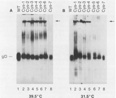
polyclonal anti-gD-1 serum(Fig. 1). TheCys-7 protein (lane 8) synthesized ateither temperature comigrated with wild-type (pRE4) gD-1 (lane 1; 59 RDa) and did not aggregate. This result is consistentwithpreviousresults obtainedwith this mutant at 37°C (Table 1) (54). When mutants Cys-1 through Cys-6 were synthesized at 39.5°C, gD migrated predominantly as a single polypeptide; in addition,
higher-molecular-weightaggregate'swerefound whichwere
distrib-uted in the separating gel, at the stacker-separating gel interface(arrowsin Fig. 1),andatthetopof the stacking gel.
When the mutants were expressed at 31.5°C, there was
generally less evidence of aggregation, although higher-molecular-weight bands were still seen at the top of the
stacking gel for mutants Cys-1, Cys-2, Cys-3, and Cys-4.
Theseresultssuggestthatsomeof themutantshaddifferent structural properties when expressed at the two
tempera-tures. It should be noted that theseaggregates could result from aberrant intermolecular disulfide bonds between gD
molecules. Alternatively, or in addition, the aggregates
couldform betweengDandother unidentified cell proteins. (ii) Processing of the gD Cys mutants. Previously we
39.5° C 31.50C
FIG. 1. Western blotanalysisofwild-typegD-1andgD-1 cyste-ine mutants expressed at 39.5 or 31.5°C, electrophoresed under nonreducing native conditions.Cytoplasmicextractspreparedfrom cells transfectedat39.50C(A)or31.5°C(B)wereelectrophoresedon
10% native gels, transferred to nitrocellulose, and reacted with polyclonal anti-gD-1 serum and 125I-protein A. WT, Wild-type gD-1(pRE4).Theseparation between thestacking andseparatinggel isindicated bythe arrow.PanelB isacomposite oftwogels.
showed that four of the mutant proteins (Cys-2, Cys-3,
Cys-5, andCys-6) synthesizedat37°C containedonly
high-mannose oligosaccharides (Table 1). In contrast, Cys-1and
Cys-4 were partially processed (5 to 10% compared with wild-type gD) beyond the precursor stage. Here we
exam-inedprocessingof eachofthemutantssynthesized ateither
39.5°C (Fig. 2A) or 31.5°C (Fig. 2B). Cytoplasmic extracts
were treated with endo H, electrophoresed under reducing conditions, transferred to nitrocellulose, and reacted with
anti-gD-1 serum. Mutants Cys-1 through Cys-6
synthesized
at
39.5°C
werecompletely
sensitive to endoH,
indicatingthat these proteins contained only high-mannose
oligosac-charides (precursor forms). Cys-7 and wild-type gD were both almost completely endo H resistant, indicating that these proteins contained
predominantly
complexoligosac-charides(productforms).
When the mutants were expressed at 31.5°C, several patternsofendoHdigestionwereobserved
(Fig.
2B).First,
the Cys-7 protein showed the same mobility shift as did wild-typegD. Inboth cases, onlya small proportion ofthe
protein was endo H sensitive. Second, Cys-3 was
entirely
endo H sensitive, showing that the carbohydrate moieties presentonthisproteinareall of thehigh-mannosetype. The third pattern was exhibited by the remainingfive mutants,
Cys-1, Cys-2, Cys-4, Cys-5, and Cys-6. In each case, a
significant portionof the N-linkedoligosaccharideswasendo H resistant when the proteins were synthesized at
31.50C,
even though there was no evidence ofprocessing ofthese
mutants tocomplexformsat39.5°C.At37°C (54),therewas
partial processing ofCys-1 and Cys-4. Taken together, the results suggest that Golgi-associated oligosaccharide proc-essingofCys-1, Cys-2, Cys-4, Cys-5, and Cys-6mutantsis ts.
(iii) Transportof the mutantgDproteinsto the cellsurface. The ts difference in processing of some of the mutant
proteins suggestedthattheir transportwithin the cell andto
on November 10, 2019 by guest
http://jvi.asm.org/
[image:3.612.61.301.101.217.2]C\ CO CT LO CO t_
I I
Uft U Ch LI) U:In C O
>1 >N >- >1 > > >1
: 0 0 0 000 0
m m m
1m1
m ,, m lrm m 1EndoH
-±+-+-+---
-+A
(39.50C)
69
-46- 0
B
(31.50
C)
69
-46-
ell5n*
a
*et
- gD
- pgD
FIG. 2. Endo HanalysisofgD-1cysteinemutants synthesized at 39.5 or31.5°C. Cytoplasmic extracts prepared from cells transfected at 39.5°C (A)and31.5°C (B)weremocktreated (-) or treated (+) with endo H,electrophoresedunderdenaturing conditions, transferred to nitrocellulose,andreacted withanti-gD-1serum and '25I-proteinA. Molecularweight markers of 46,000 and 69,000 are indicated. WT, wild type.
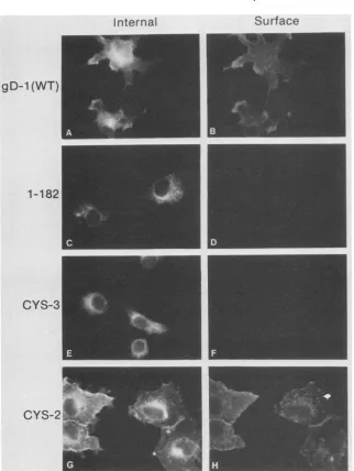
the cellsurface might also be ts. Cells were transfected with plasmids containing the wild-type or mutant forms of gD,
incubated at either 31.5 or 39.5°C, then examined by a
double-labelindirect immunofluorescence assaythat detects cell surface and internalexpression of gD in the same cell (Fig. 3 and 4). A mutant form of gD-1, truncated after residue 182 [gDt(182)] and therefore lacking the transmembrane
region, was used as a negative control for cell surface
staining (Fig. 3D and 4D)
(48).
All of the proteins were detectedinternallyatboth temperatures (Fig. 3 and 4, panels A, C, E, andG). At 31.5°C, all of the cells transfected witheither wild-type gD (Fig. 3B) or Cys-7 (not shown) that
exhibited internal fluorescence also had gD on the cell
surface. Thiswasalso the casefor Cys-2 (Fig.3H) aswell as
Cys-1, Cys-4, Cys-5, and Cys-6 (not shown). In contrast, Cys-3 exhibited no cell surface fluorescence on any cells at this temperature(Fig.3F). Theseresults suggested that allof the mutants except Cys-3 were transported to the cell
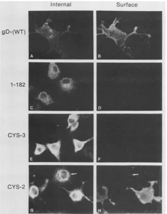
surfaceat31.5°C.At39.5°C (Fig.
4),
the samebasic pattern was observed, with one important difference. For Cys-1,Cys-2, Cys-4, Cys-5, andCys-6, only 15to35% ofthecells
exhibitinginternalfluorescencewerealsostainedonthe cell
surface (shown forCys-2 in Fig. 4G and H). These results suggest that transport ofCys-1, Cys-2, Cys-4, Cys-5, and
Cys-6was ts.TransportofCys-7tothecell surface occurred
atbothtemperatures(notshown), and transportofCys-3 did notoccur ateither temperature(Fig. 3Fand 4F).
(iv) Antigenic analysis. Thus far,wehave shown that five ofthe cysteine mutants (Cys-1, -2, -4, -5, and -6) exhibitts
differences in aggregation, processing, and transport. We
asked whether there were also ts differences in protein conformation as detected by alterations in antigenic struc-ture.Toaddress this question, we usedapanelofMAbs in
aslot blot assaytoexamine theantigenic propertiesof each
mutantprotein expressedat31.5or39.5°C. Results
for
three MAbsare showninFig. 5. DL6, agroup IIMAb thatbindsto acontinuous epitope (residues 272 to 279), was used to
normalize the total amountofgD loaded in
each
slot(Fig.5A).
MAbs HD1 (group Ia) (37) and ABD (group III) recognize distinct discontinuous epitopes (Fig. SB and C) and wereused
todetect differences in the conformation of the mutant forms ofgD. Three patterns ofreactivity wereobserved. First, the Cys-7 mutant expressed at either tem-perature showed good binding of ABD but somewhat
re-duced bindingof HD1. Thebinding ofotherMAbs in group Iresembled thatofHD1, and thebinding of other MAbs in groupIIIresembledthat of ABD. These results suggest that theabsenceofcysteine7 had someeffect, albeitlimited,on
protein structure. Second, Cys-3 and Cys-6 expressed at
either 31.5 or 39.5°C were unreactive with either HD1 or
ABD as well as with five other MAbs representing other
antigenic sites (notshown). This findingindicated that cys-teines 3 and 6 are indispensable for the correct antigenic conformation of gD. Third, mutants Cys-1, Cys-2, Cys-4,
andCys-5 exhibited majorreductions inbindingtoboth HD1 and ABD.However, in eachcasethere wasatleasttwo-to
threefoldmorereactivitywith these MAbs when theproteins
wereexpressed at 31.5°C than when they wereexpressedat
39.5°C. A similar step-up in reactivity of Cys-1, Cys-2, Cys-4,andCys-5synthesizedat31.5°Cwasfoundwitheach
ofthe five other MAbs todiscontinuous epitopesthatwere
tested(resultsnotshown).Theseresults suggest thatfolding ofCys-1, Cys-2, Cys-4,and Cys-5 is ts.
Functional analysis of gD cysteine mutants. (i) Rescue of
F-gDoi
virus by gD cysteine mutants expressed in trans. Acomplementation assay was used to examine whether the
gD-1 Cysmutants werefunctionalin vivo(3, 38).The assay
uses
F-gD3,
a mutantvirus thatwasderivedfromHSV-1(F) by replacementof thegD-codingsequenceand partof thegI gene with the ,-galactosidase gene of Escherichia coli(29).It can replicate in VD60 cells, which contain integrated copies ofthegD gene, butnotin Verocells, fromwhich the VD60 cell line was derived (29). Vero cells infected with
F-gDP
produce noninfectious virions lacking gD. In thecomplementation assay, gD transiently expressed from transfected plasmids is tested for its ability to rescue pro-duction of infectious F-gD, virions in Vero cells. In this case,Verocells firstweretransfected with
plasmids
contain-ing wild-type gD-1or oneof theCysmutantsand thenwere infected with
F-gDP
and incubated at37°C
as the nonper-missive temperature. BecauseF-gDP
was derived from strain F, it contains a ts mutation in theimmediate-early
ICP4 gene and does notgrow at 39.5° (1; D. Long,
unpub-lisheddata). Intracellular viruswascollected after24to48
h,
0 el 40
61044a
1.11,11111111W m isina41
4
0
on November 10, 2019 by guest
http://jvi.asm.org/
[image:4.612.147.459.85.266.2]Internal
gD-1 (WT)
1-182
CYS-3
CYS-2
Surface
FIG. 3. Immunofluorescenceanalysisof cellsexpressing wild-typeandmutantforms ofgD-1at31.5°C. Double-label immunofluorescence wasusedtodetectgD-1
internally
and on thecellsurface. Cellswerefixedwithparaformaldehydeand reacted withanti-gDserumand then with goatanti-rabbit-tetramethylrhodamine isothiocyanatetolabelgD-1 on thecell surface(B,D, F,andH). CellswerethenpermeabilizedwithNonidet P-40 andreacted with anti-gD-1 serumandanti-rabbit-fluorescein isothiocyanatetodetectgD-1 inside the cell (A,C, E, and G). Proteinsanalyzedwerewild-type (WT) gD-1 (Aand B),agD-1 truncationmutantcontaining residues 1to182(CandD),Cys-3 (Eand F),andCys-2 (G andH).
and virus titersweredeterminedonVD60monolayers (Fig.
6). Whencellsweretransfectedwith thewild type or Cys-7, the titers were at least 50-fold higher than those obtained when cells were transfected with vector alone. Thus, cyste-ine 7atresidue 333 may not be critical for the function of gD. Although it remains functional at 37°C, it appears that Cys-7 is actually better than wild-type gD-1 at rescuing F-gD,. Thus, its ability to rescue at 31.5°C may actually be some-what impaired. In contrast to the results for Cys-7, virus
titers obtained from cells transfected with any one of the other sixcysteinemutants weresimilar tobackground levels when theexperiment was carried out at 37°C. Thus,
altera-tion of any one of these six cysteines to serine renders gD
nonfunctional.However, when the assay was carried out at
31.5°C,twoof the mutant proteins, Cys-2 and Cys-4,
exhib-ited complementationactivity. Titers for
F-gDP(Cys-2)
were16-fold higher than background levels, and the titers for F-gD,(Cys-4)were13-foldhigher. Thus, theseproteinswere
functional when synthesized at the lower temperature,
though the level ofcomplementation was never ashigh as
withwild-type gD. The viruses produced in the
complemen-tation assay and used in the following experiments are
designated as F-gD,B(WT),
F-gDP(Cys-2),
F-gD,B(Cys-4),and
F-gDP(Cys-7).
(ii) Neutralization ofrescued virus. To confirm that these
virions actually contained gD in their envelope, we deter-mined whetheragDMAb, DL11 (groupIb), couldneutralize the infectivity of virus obtained in the complementation
assay (Table 2). DL11 neutralized both F-gD,B(Cys-2) and
F-gDP(Cys-4)
asefficientlyasit neutralizedF-gDP(WT).
Weconclude thatCys-2 andCys-4mutantproteinsare
incorpo-rated into the virus envelope of
F-gDP
at the permissiveon November 10, 2019 by guest
http://jvi.asm.org/
[image:5.612.157.479.68.496.2]Internal
gD-(WT)
1-182
CYS-3
Surface
CYS-2
FIG. 4. Immunofluorescenceanalysis of cells expressing wild-type (WT) andmutantforms of gD-1at39.5°C. Double-label immunoflu-orescence wasusedtodetectgD-1internally(A,C,E, andG)and on thecell surface (B, D, FandH)asdescribedin thelegendtoFig.3. Thearrowsin panels G andHpointto acell that exhibits internalbut not cellsurface fluorescence.
temperature. Moreover, thestructure of themutantprotein incorporated into the virus envelope at 31.5°C appears, at
leastbythecriterion oftheneutralizationassayusedhere,to
be indistinguishable fromthatofwild-type gD-1.
(iii) Penetration of
F-gDP(Cys-2)
andF-gDO(Cys-4)
virions intoVD60cells. Since gD is essential forHSV-1penetration (24, 29), wedecidedtocomparetheabilityofF-gDO(Cys-2)
and F-gD,(Cys-4) topenetrate cells at31.5 or37°C. VD60cellswereincubated with virus produced in the
complemen-tation assay for2 h at4°Ctoallow for attachmentand were
thenshifted eitherto 31.5or37°C; after variousamountsof
time,
extracellular viruswasinactivated withlow-pHglycinebuffer (20, 21), and the remaining (intracellular) virus was
allowedtodevelopinto visibleplaques. Wefoundno
differ-encein therateofpenetrationof any of the viruses testedat
either temperature (Fig. 7). In each case, penetration pro-ceeded more rapidly at 37°C (Fig. 7B) thanat 31.5°C (Fig. 7A). Previous studieshave shown thatpenetrationof HSVis temperaturedependent(21). Wefound that 50% of the total
input virus became resistant to acid inactivation within 22 minof incubationat 37°C(Fig. 7B), whereasat31.5°C (Fig.
7A), 50% of the inputvirus becameacid resistantwithin 60 min. The results suggest thatCys-2andCys-4, once
synthe-sized at31.5°C and incorporated intovirions, were ableto function normally so that penetration was unaffected at
37°C.
Alternatively, penetration
occurred before thermal inactivation ofF-gD,(Cys-2)orF-gD3(Cys-4)
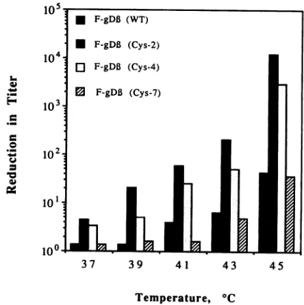
at37°C.(iv) Thermalsusceptibility studies. We examined the
ther-mal stability of each ofthe viruses obtained inthe
comple-mentation assay byincubatingthematvarious temperatures for 1 hand then
testing
theirinfectivity (Fig. 8).
We found that theinfectivityofF-gD,B(Cys-7)
andF-gD,(WT)
wasnotsignificantly affected at temperatures below
43°C.
Incon-trast, the infectivity of
F-gD3(Cys-2)
andF-gD,B(Cys-4)
decreased
approximately
50%at37°C,
andat45°C
therewasa4-logdrop in virus titer.
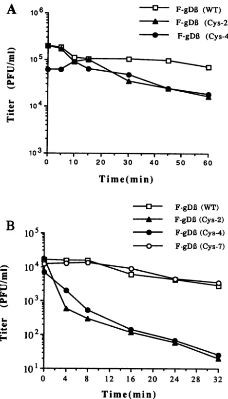
Therateof heatinactivationwasdetermined
by
incubating
virus preparations at 37 or 45°C forincreasing
amounts ofon November 10, 2019 by guest
http://jvi.asm.org/
[image:6.612.146.472.77.496.2]o
0
o 0
A LOcrz CO)
A co co
Mock
WT
Cys-1
41
_w
4
0 0
o 0
& T
B CLO C
,4'a
4I,Amok
0 0
LO L)
C 0)CY) COI
_p
4
._W
amv
Cys-2
4m
4mCys-3
Cys-4
Cys-5
Cys-6
Om 4
a~m
-_a
Cys-7 mm am
DL6
HD1
ABDFIG. 5. Slot immunoblotanalysis, using MAbs,of wild-type gD-1 and gD-1cysteine mutantsexpressed at 39.5 or31.5°C. Cytoplasmic extractsofcells transfected withwild-type(WT) or mutant gD-1 genes wereappliedtonitrocellulose byusingaslotblotapparatus(Hoefer). Membraneswereincubated with thefollowingMAbs: DL-6(continuousepitope,groupII)(A), HD-1(discontinuous epitope,groupIa) (B), and ABD(discontinuous epitope, group III) (C).
timeandthentitering the residual virus. Compared with the
titer ofF-gD,(WT), thetiters of F-gD,(Cys-2) and
F-gDP
(Cys-4) were not significantly affected by preincubation at 37°Cforless than 30 min(Fig.9A). Itshould be noted that by 30 min at this temperature approximately 60% of the virus would have penetrated the cell(Fig. 7B). Longer incubation
times decreased infectivity by approximately 10-fold. At
45°C (Fig.9B), theinfectivity ofbothF-gD,(WT) and
F-gDP
(Cys-7) slowly decreased; approximately 50% of these
vi-80
-60
40
20
M
370C* 31.50C
inE
L
J_
now gD-1 Cys-1 Cys-2 Cys-3 Cys4 Cys-5 Cys-6 Cys-7 Formof Protein in Virus
FIG. 6. Rescueof
F-gDP
by gD-1cysteinemutantsexpressed intrans at 37 or 31.5°C. Vero cells were transfected with a test
plasmid, infected 16to18 h later with F-gD,B, and incubatedat37or
31.5°C for 24to48h. Intracellular viruswascollectedandtiteredon
VD60 cells. Dataare expressedasthetiter obtained with the test
plasmid divided by the titer obtained when cells weretransfected
withthecontrol plasmid, pRSVntEPA.Titersfor pRSVntEPAwere
approximately 104 at 37°C and 6 x 10² at 31.5°C. The results representtheaveragesof threeseparate experiments.
ruses wereinactivatedafter 20 min, and therewas afourfold decrease in virus titer after 32 min. In contrast,
F-gDP(Cys-2) and
F-gDP(Cys-4)
wereboth significantly more sensitive toinactivation at45°C; approximately 50% of these viruseswere inactivated within 4 min, and there was almost a
complete lossofinfectivity after 32 min. DISCUSSION
We havepreviouslydescribed theconstructionand
char-acterization of seven cysteine gD-1 mutants (54). These
results indicatedthatsix ofthesevencysteinesareimportant
inmaintaining the native structureofgD, probably through theformationof disulfide bonds. In thisstudy,wefound that
five of the seven mutants were ts for one or more of the
following properties: aggregate formation, conformation, processingfromahigh-mannoseprecursor toaproductform containing complex oligosaccharides, and transport to the
cell surface. Furthermore, two mutant proteins, Cys-2 and
Cys-4,rescued theinfectivityofagDnull mutant,
F-gDp,
ina tsfashion.
[image:7.612.164.469.73.287.2]Thesubstitution of serine for anyofthefirst sixcysteines ofgD-1 resulted in the appearanceofhigh-molecular-weight forms, presumably due to aggregation ofthe protein as a
TABLE 2. Neutralization of virusproducedin the complementationassay
Antibody dilution
Rescuedvirus" (105) for50%
plaquereductionb
F-gDP(WT)
... 1.28F-gD,B(Cys-2)... 0.96
F-gD,(Cys-4)
... 1.28"Namesrefer to thephenotypeof thevirus obtainedin the complementa-tion assay.
bEqualamountsof rescued virus were incubated withvariousdilutionsof DL11 at31.5'Cfor 1h,and themixturewasadded toVD60cells. After3 to 4daysat31.5°C,plaqueswerecounted;the averagesoftriplicate samplesare shown.
-
--
-_ au
U1)
5-i
5-U)
(U)
O4
on November 10, 2019 by guest
http://jvi.asm.org/
[image:7.612.66.305.447.645.2]HSV gD-1 CYSTEINE MUTANTS EXHIBIT ts PROPERTIES
-0
*
,&a
c)
-0-- F-gDB (WT)
-*-- F-gDB (Cys-2)
*@-
F-gDB(Cys-4)F-gDl (Cys-7)
20 40 60 80 100 120
Time(min)
10-'
* F-gDB (WT)
* F-gDB (Cys-2)
10O4
D F-gDB (Cys-4)
Mi F-gDB (Cys-7)r
102 101
1i0
37 39 41 43 45
B
Temperature, ICFIG. 8. Effectofpreincubationtemperatureoninfectivity. Virus
100 sampleswereincubated for1hattheindicatedtemperaturesandthe
titerswere determined onVD60 cell
monolayers
at31.5°C.80- *
_oligosaccharides added to these proteins remained endo H
sensitive, suggesting that atthis high temperature much of
o
theprotein was notreaching theGolgi compartment, whereprocessing to endo H resistance occurs. In contrast, when
/the
proteins were expressed at 31.5°C, all of the mutantsce
40- L//F-gDB(WT) except Cys-3 were transported to the cell surface and a
F-gDB(Cys-2) significant amount of the accumulated protein was endo H
e
-/0
*F-gDBi
(Cys-2) resistant. Inthesestudies, weexamined cell surface expres-20-4 F-gDB (Cys-4) sion only in a qualitative way. Whitt et al. (53) have shownthat the ability of some of the mutated G proteins of
vesicular stomatitis virus to rescue ats mutant, tsO45, was
,_ ,_ ,_ ,_ .,_,_proportionalto theamountof each mutantproteinonthe
0
20 40 60.
80 00 120surface. It would be of interest to do similar studies with
Cys-2and Cys-4. However, itshould be notedthat vesicularTime(min) stomatitis virus matures at the plasma membrane, whereas
HSVmatures intracellularly.
FIG. 7. Ratesofpenetration ofviruses obtained from the
com-plementationassaycarriedoutat31.5or
37°C.
Virus samples (100to Our studies with MAbs that recognize discontinuous200 PFU) preparedfromthe complementation assay(Fig. 6) were
epitopes
suggest that there are temperature-sensitivestruc-addedto
VD60
cell monolayers and allowedtoadsorbtoVD60 cells tural differences in four of the Cys mutant proteins.How-for 2 h at
4°C.
The cultures were then incubated at31.5°C
(A) or ever, the MAb reactivities of the mutant proteinssynthe-37°C
(B). At the times indicated, cells were washed with acid- sized at31.5°C
never reached wild-type levels. Ourglycine buffer (pH 3.0). Virus plaques were counted after 24 h at conclusion is that although all of the disulfide bonds are
37°C.
The resultsarereportedaspercentsurvivors when compared critical for correct folding and maintenance of antigenic with controls in which PBS wassubstituted for acid-glycine buffer. structure, theabsence ofanyone ofthese bondsiscompen-satedto some degree by lowering the temperature.
resultof incorrect disulfide bonding. Several of the mutants To determine whether any of the cysteine-to-serine
appeared to be less aggregated when synthesized at
31.5°C
changesaffected thefunction of gD, we usedacomplemen-than at
39.5°C.
One explanation is that folding occurs more tationassayin whichgD transiently expressed bya plasmidslowly, decreasing the inappropriate hydrophobic interac- can rescue the infectivity ofF-gD,, a virusstrain of HSV-1 tions, resulting in a more accurately folded polypeptide at that lacks a gD gene. In this assay, Cys-7 was found to
the lower temperature (27). In addition, the processing function normally, which is consistent with our prediction
whichoccurs at
31.5°C
mayalso beimportant for achieving that this cysteine residue is not involved in formation oformaintaining the native conformation (32, 39). disulfide bonds. Noneof the other six mutants were able to Allsevenmutantproteins accumulated intracellularly,and complement at 37°C. However, when the assay was
per-all but Cys-3wereobserved tobepresentonthecellsurface formed at 31.5°C, two mutant proteins, Cys-2 and Cys-4, when expressed at
31.5°C.
In contrast, when the transfec- were able to complement, albeit at a lower level than tionswerecarried outat39.5°C
onlyasmallfraction of cells wild-type gD-1. Neutralization of the infectivity of virionsexpressing Cys-1,-2, -4, -5,and-6internallywerealsofound containingCys-2orCys-4withthegD-specificMAbDL11 is
to express the protein on the cell surface. In addition, the evidence that these forms of gD become incorporated into
A
U, so
*
-:C
a
VOL.64,1990 5549
on November 10, 2019 by guest
http://jvi.asm.org/
[image:8.612.317.539.76.299.2] [image:8.612.49.288.79.493.2]---- F-gDB (WT) S F-gDB (Cys-2) * F-gDB (Cys-4)
7 369
66 106 118127 18
s-S
-S-19202 333
FIG. 10. Modelofpredicted intramolecular disulfidebondingof gD.The essential features ofgD-1aredepictedas a stickfigure.The sevencysteineresidues aredesignated byaC, and their positionsin the gD-1 sequence are indicated beneath. The box surrounding Cys-7represents the transmembrane region. The protein contains three sites for addition of N-linked oligosaccharides, depictedas ballons. The disulfide bond pattern (-S-S-) predicted from the propertiesofthegD-1 cysteinemutants is shown.
10 20. 3 4 5 I0
1 0 20 30 40 50 60
Time(min) Cys-4 mutations will produce virions with aninteresting ts phenotype. Recombination experiments using these mutant
forms ofgDare nowinprogress. Itis of interesttonotethat
-0----
F-gDB
(WT)Highlander
et al.(20) reported
a slower rate of entry of -*-- F-gDB (Cys-2) HSV-1(KOS-321) into Vero cells at 37°C than we observedin our study. This discrepancy could be due to strain 0 F-gDI3 (Cys-4) differences in one or more of the envelope proteins. This
-0-- F-gDB(Cys-7) issue will best be resolved
by comparing
theentry kinetics of the recombinant viruses with that of KOS-321 in the sameexperiment.
Our
experiments
suggest thatCys-1 through Cys-6
form three disulfide bonds (54). In addition,preliminary
experi-ments using
[14C]iodoacetamide
are consistent with thisnotion(Long, unpublisheddata). Toaddress thequestion of which cysteine residuepairsmightbeinvolvedinthesethree intramolecular disulfide
linkages,
wecompared
thepheno-types of the various gD-1 Cys mutants. This strategy was usedto predict disulfide bonds of the Tac receptor protein
and wasconfirmedbyapreliminary peptide map of
cystine-linked enzymatic fragments (43). As another example, the
differing propertiesoftwocysteinemutantsof theenvelope
0 4 8 12 16 20 24 28 32 protein of human immunodeficiency virus type 1 suggested
Time(min)
that thesetwocysteines might
notbe bondedtoeachother (49). The properties of the cysteine mutants predict the ateofvirusinactivation at 37 and45°C.Virussamples patternofdisulfidebonding
forgD-1shown inFig.
10.Cys-2Led at37°C (A)or45°C (B) for thetimesindicatedand (residue 106) and Cys-4 (residue 127) are paired because they
at4°C.Residual virus titers were determined on VD60 were similar in allproperties and were the only mutants to
ers at31.5°C. function in the complementation assay. Cys-1 (residue 66)
andCys-5 (residue 189)arepaired becausetheyboth
exhib-nvelopeat31.5°C. Inaddition, it ispossible that ited similarts
properties,
including ability
tobind to MAbsandCys-4aresynthesizedat37°C,theyarenot thatrecognizediscontinuous
epitopes,
processing
toendo Hd into the envelope. Perhaps the other Cys resistance,andtransporttothe cell surface. Inaddition,and not complementbecause they are notincorpo- unlikeCys-2andCys-4, neither of these mutantswasable to
the virion at either temperature. Future experi- function in the
complementation
assay. In contrast, Cys-3 attempt toaddress these questions bypurifying (residue118)
andCys-6 (residue
202)
bothshowedalack of us virions produced in the complementation as-reactivity
to MAbs to discontinuousepitopes;
however, aminingthem forthe presence of gD. Cys-6 diddisplay some tsprocessing
thatwas not seenforresting that when incorporated into the envelope Cys-3. Thus,thesetwo
cysteines
aredisulfide bonded in ourboth Cys-2 and Cys-4 displayed normal entry model
partly by
default.Confirmation of this disulfide bondboth 31.5 and 37°C. Viruses containing either of pattern
by
biochemicalanalysis
is nowin progress. these proteins were fairly stable when preincubated at 37°Cbut more thermolabile than viruses containing wild-type gD-1 whenpreincubatedat 45°C. One possibility is that the
mutant proteins are more sensitive to thermal inactivation
duringsynthesis at 37°C, whereas once the protein is
incor-porated into the envelope it is not as easily denatured. Furtherexperiments will be necessary to clarify this issue. Thecomplementationresultspredict that both the Cys-2 and
ACKNOWLEDGMENTS
Thisinvestigationwassupportedby Public Health Servicegrants
DE-08239 from the National Institute of Dental Research and AI-18289 from the National Institute of Allergy and Infectious Diseases andby agranttoG.H.C. and R.J.E. from the American Cyanamid Co. D.L. is apredoctoral trainee supported by Public
Health Service grant NS-07180 from the National Institute of NeurologicalDiseases andStroke.
A
E
Pu ;r
no
106.
10 5I
0
B
105
1-1
E
1-1-.
._N
FIG. 9. R wereincubat then chilledz
cellmonolay
the virione
when Cys-2
incorporate
mutantsdo
rated into t mentswill
noninfectioi
sayandexr
Itisinter
at 31.5°C,
kinetics at
1 4
1
on November 10, 2019 by guest
http://jvi.asm.org/
[image:9.612.331.568.73.155.2] [image:9.612.78.312.74.483.2]WethankD.C.Johnsonforthe VD60 cells and
F-gDp
virus; W. Wilcox forthe mutants constructed by site-directed mutagenesis; D. L.Sodorafor the discovery of the mutation at amino acid 54 and forhelp inpreparation of the manuscript; L. Pereira, C. Desgranges, and A.Minsonfor antibodies; and M. Cohen for excellent technical assistance.LITERATURE CITED
1. Arsenakis, M., J. Hubenthal-Voss, G. Campadelli-Fiume, L. Pereira, and B. Roizman. 1986.Constructionand properties of a cell line constitutively expressing the herpes simplex virus glycoproteinBdependentonfunctionala4proteinsynthesis. J. Virol.60:674-682.
2. Burnette, W. N. 1981. "Western blotting". Electrophoretic transfer of proteins from SDS-polyacrylamidegels to unmodi-fied nitrocellulose and radiographic detection with antibody and radioiodinated proteinA. Anal. Biochem. 112:195-203. 3. Cai, W., S. Person, C. DebRoy, and B. Gu. 1988. Functional
regions and structural features of the gB glycoprotein of herpes simplex virustype 1. J. Mol.Biol. 201:575-588.
4. Campadelli-Fiume, G., M. Arsenakis, F. Farabegoli, and B. Roizman. 1988. Entry ofherpes simplex virus1in BJ cellsthat constitutively express viral glycoprotein D is by endocytosis and results indegradation of the virus. J. Virol. 62:159-167. 5. Campadelli-Fiume, G., E. Avitabile, S. Fini, D. Stirpe, M.
Arsenakis, and B. Roizman. 1988. Herpes simplex virus glyco-protein D is sufficient to induce spontaneous pH-independent fusion inacell linethatconstitutively expresses the glycopro-tein.Virology 166:598-602.
6. Campadeili-Fiume,G., L. Poletti, F. Dall'Olio, and F. Serafini-Cessi. 1982. Infectivity and glycoprotein processing of herpes simplex virustype 1 growninaricin-resistantcellline deficient inN-acetylglucosaminyl transferase I.J. Virol.43:1061-1071. 7. Chen,E.U.,and P. H.Seeburg. 1985. Supercoil sequencing:a
fast and simple method for sequencing plasmid DNA. DNA 4:165-170.
8. Cohen, G. H., B. Dietzschold, M. Ponce de Leon, D. Long, E. Golub, A. Varrichio, L. Pereira, and R. J. Eisenberg. 1984. Localization andsynthesis ofanantigenic determinant of herpes simplex virus glycoprotein D that stimulates production of neutralizing antibody. J. Virol.49:102-108.
9. Cohen, G. H., V. Isola, J. Kuhns, P. W. Berman, and R. J. Eisenberg. 1986. Localization of discontinuousepitopes of her-pes simplex virus glycoprotein D: use of a nondenaturing ("native" gel) system of polyacrylamide gel electrophoresis coupled withWestern blotting. J. Virol. 60:157-166.
10. Cohen,G. H., M. Katze, C.Hydrean-Stern,and R.J. Eisenberg. 1978. Type-common CP-1 antigen of herpes simplex virus is associated witha 59,000-molecular-weight envelope glycopro-tein.J. Virol. 27:172-181.
11. Cohen,G.H., D. Long, and R. J.Eisenberg.1980.Synthesis and processing of glycoproteins gD and gC of herpes simplex virus type 1.J. Virol. 36:429-438.
12. Cohen, G. H., D. Long, J. Matthews,M.May,andR.Eisenberg. 1983. Glycopeptides ofthe type-common glycoprotein gD of herpessimplexvirus types 1and2. J.Virol.46:679-689. 13. Cohen,G.H., W. C. Wilcox, D. L.Sodora,D.Long, J. Z. Levin,
and R.J. Eisenberg. 1988. Expression of herpessimplex virus type 1glycoproteinD deletionmutantsin mammaliancells. J. Virol.62:1932-1940.
14. Eisenberg, R., C. Hydrean-Stern, and G. H. Cohen. 1979. Structural analysis of precursor and product forms of
type-common envelope glycoprotein D (CP-1 antigen) of herpes simplex virustype 1. J. Virol.31:608-620.
15. Eisenberg, R. J., D. Long,L.Pereira, B. Hampar,M.Zweig,and
G.H. Cohen. 1982. Effect of monoclonalantibodiesonlimited proteolysis of native glycoprotein gD of herpes simplex virus types 1 and 2byuseof monoclonalantibody. J.Virol. 41:478-488.
16. Eisenberg, R. J., M. Ponce deLeon, and G. H. Cohen. 1980. Comparative structural analysis ofglycoprotein gD ofherpes
simplex virustypes 1 and 2. J. Virol.35:428-435.
17. Eisenberg, R. J., M. Ponce de Leon,L. Pereira,D. Long,and
G. H. Cohen. 1982. Purification ofglycoprotein gD ofherpes simplexvirustypes 1and 2byuseof monoclonalantibody.J. Virol. 41:1099-1104.
18. Fuller,A.O.,andP. G.Spear. 1985.Specificitiesofmonoclonal and polyclonal antibodies that inhibit adsorption of herpes simplexvirus tocells and lack of inhibitionbypotent
neutral-izingantibodies.J. Virol. 55:475-482.
19. Fuller, A. O., and P. G. Spear. 1987. Anti-glycoprotein D antibodies thatpermit adsorptionbut block infectionby herpes simplex virus 1 prevent virion-cell fusionat the cell surface. Proc.Natl. Acad. Sci. USA 84:5454-5458.
20. Highlander,S.L.,S. L.Sutherland,P.J.Gage,D.C.Johnson, M. Levine,andJ. C. Glorioso. 1987. Neutralizingmonoclonal antibodies specific for herpes simplex virus glycoprotein D inhibit viruspenetration.J. Virol.61:3356-3364.
21. Huang,A. S.,and R. R. Wagner. 1964. Penetration ofherpes simplex virus into human epidermoid cells. Proc. Soc. Exp.
Biol. Med. 116:863-869.
22. Isola,V.J.,R.J. Eisenberg,G. R.Siebert,C.J. Heilman,W.C. Wilcox,andG. H. Cohen.1989. FinemappingofantigenicsiteII ofherpes simplexvirusglycoproteinD. J. Virol. 63:2325-2334. 23. Johnson, D. C., R. L. Burke, and T. Gregory. 1990. Soluble herpessimplexvirusglycoproteinD bindsto alimited number of cellsurface receptorsandinhibits virus entryinto cells. J. Virol.64:2569-2576.
24. Johnson,D.C.,andM. W.Ligas. 1988.Herpessimplexviruses lackingglycoproteinDareunable toinhibit viruspenetration:
quantitativeevidence forvirus-specificcellsurfacereceptors.J. Virol. 62:4605-4612.
25. Kaner, R. J., A. Baird, A. Mansukhani, C. Basilico, B. D.
Summers,R. Z.Florkiewicz,and D. P.Haijar.1990. Fibroblast
growthfactorreceptorisa portalof cellularentryforherpes simplexvirus-1. Science 248:1410-1413.
26. Karnik,S.S., T. P.Sakmar,H.-B.Chen,and H. G. Khorana. 1988. Cysteine residues 110 and 187 are essential for the formation ofcorrect structureinbovinerhodopsin.Proc.Natl. Acad.Sci. USA 85:8459-8463.
27. Kim,P. S.,and R. L. Baldwin. 1982. Specificintermediatesin the foldingreactions of small proteins and the mechanism of
protein folding. Annu.Rev. Biochem. 51:459-489.
28. Lasky,L.A.,D.Dowbenko,C. C.Simonsen,and P. W. Berman. 1984. Protection of mice from lethal herpes simplex virus infectionbyvaccination withasecretedform ofcloned glyco-proteinD. Bio/Technology2:527-532.
29. Ligas,M.W.,andD. C.Johnson. 1988.Aherpessimplexvirus
mutant in which glycoprotein D sequences are replaced by
0-galactosidase
sequences binds tobut is unable to penetrate into cells. J. Virol. 62:1486-1494.30. Long, D.,T.J. Madara,M. Ponce deLeon,G. H.Cohen,P. C. Montgomery, and R. J. Eisenberg. 1984. Glycoprotein D
pro-tects mice against lethal challenge with herpes simplex virus types 1and2. Infect. Immun.37:761-764.
31. Machamer, C. E.,R. Z. Florkiewicz,and J. K. Rose. 1985. A singleN-linkedoligosaccharideateither ofthetwonormalsites issufficient fortransportof vesicular stomatitisvirus Gprotein tothecell surface. Mol. Cell. Biol. 5:3074-3083.
32. Machamer, C. E., and J. K. Rose. 1988. Vesicularstomatitis virus Gproteinswith alteredglycosylationsitesdisplay temper-ature-sensitive intracellulartransport and are subject to
aber-rantintermolecular disulfidebonding.J. Biol.Chem. 263:5955-5960.
33. Matsumura,M.,G.Signor,and B. W. Matthews.1989. Substan-tial increase ofprotein stability by multiple disulphidebonds. Nature(London)342:291-293.
34. Matthews, J. T., G. H. Cohen, and R. J. Eisenberg. 1983. Synthesis andprocessingofglycoproteinDofherpessimplex
virus types 1 and 2 inanin vitro system. J.Virol.48:521-533. 35. McDougal,S.J.,J.K. A.Nicholson,D.Cross,S. P.Cort,M.S.
Kennedy,andA.C. Mawle. 1986. Bindingof the human
retro-virusHTLV/LAV/HIVtotheCD4(T4)molecule:conformation
dependence, epitopemapping, antibody inhibition,and poten-tial foridiotypicmimicry.J. Immunol. 137:2937-2944. 36. Minson,A.C.,T.C.Hodgeman,P.Digard,D.C.Hancock,S. E.
on November 10, 2019 by guest
http://jvi.asm.org/
Bell, and E. A. Buckmaster.1986. An analysis of the biological properties of monoclonal antibodies against gD of herpes sim-plex virus and identification of amino acid substitutions that confer resistance to neutralization. J. Gen. Virol. 67:1001-1013. 37. Muggeridge, M. I., V. J. Isola, R. A. Byrn, T. J. Tucker, A. C. Minson, J. C.Glorioso,G. H. Cohen, and R. J. Eisenberg. 1988. Antigenic analysis of a major neutralization site of herpes simplexvirus glycoprotein D, using deletion mutants and mono-clonal antibody-resistant mutants. J. Virol. 62:3274-3280. 38. Muggeridge, M. I., T.-T. Wu, D. C. Johnson, J. C. Glorioso,
R.J. Eisenberg, and G. H. Cohen. 1990. Antigenic and func-tional analysis of a neutralizationsiteof HSV-1 glycoprotein D. Virology 174:375-387.
39. Ng,D.T.W.,S. W. Hiebert, and R. A. Lamb. 1990. Different roles of individual N-linked oligosaccharide chains in folding, assembly, and transport of the simian virus 5 hemagglutinin-neuraminidase. Mol. Cell. Biol. 10:1989-2001.
40. Noble, A.G.,G. T.-Y. Lee, R. Sprague, M. L. Parish, and P. G. Spear. 1983. Anti-gD monoclonal antibodies inhibit cell fusion inducedby herpes simplex virus type 1. Virology 129:218-224. 41. Pace, C. N., G. R. Grimsley, J. A. Thomson, and B. J. Barnett. 1988. Conformational stability and activity of ribonuclease Ti withzero, one, and two intact disulfide bonds. J. Biol. Chem. 263:11820-11825.
42. Petrovskis, E. A., J. G. Timmins, M. A. Armentrout, C. C. Marchioli,R.J. J. Yancey, and L. E. Post. 1986. DNA sequence ofthe genefor pseudorabiesvirus gp5O, aglycoproteinwithout N-linkedglycosylation.J. Virol. 59:216-223.
43. Rusk, C.M.,M. P. Neeper,L.-M. Kuo, R. M. Kutny, and R. J. Robb. 1988. Structure-function relationships for the IL-2 sys-tem. V. Structure-activity analysis of modified and truncated forms ofthe Tac receptorprotein: site-specific mutagenesis of cysteine residues.J. Immunol. 140:2249-2259.
44. Sauer, M. K., and D. J. Donoghue. 1988. Identification of nonessential disulfide bonds and altered conformations in the v-sis protein, a homolog of the B chain of platelet-derived growth factor. Mol. Cell. Biol.8:1011-1018.
45. Scheele, G., and R. Jacoby. 1982. Conformational changes associatedwithproteolytic processingof presecretory proteins allow glutathione-catalyzed formationof native disulfide bonds. J. Biol. Chem. 257:12277-12282.
46. Schultz, S. C., G.Dalbadie-McFarland,J. J. Neitzel, and J. H.
Richards. 1987. Stability of wild-type and mutant RTEM-1 P-lactamases: effect of the disulfide bond. Proteins Struct. Funct. Genet. 2:290-297.
47. Showalter, S. D., M. Zweig, and B. Hampar. 1981. Monoclonal antibodiesto herpessimplexvirus type 1proteins, includingthe immediate-early protein ICP4. Infect. Immun. 34:684-692. 48. Sodora, D. L., G. H. Cohen, and R. J. Eisenberg.1989. Influence
of asparagine-linked oligosaccharides on antigenicity, process-ing, andcell surface expression of herpes simplex virus type 1 glycoprotein D. J. Virol. 63:5184-5193.
49. Tschachler, E., H. Buchow, R. C. Gallo, and M. S. J. Reitz. 1990.Functionalcontribution ofcysteineresidues to the human immunodeficiency virus type 1 envelope. J. Virol. 64:2250-2259.
50. Watson, R. J. 1983. DNA sequenceoftheherpessimplexvirus type 2glycoproteinDgene. Gene26:307-312.
51. Watson, R. J., J. H. Weis, J. S. Salstrom, and L. W. Enquist. 1982. Herpessimplex virus type-1 glycoprotein D gene: nucle-otide sequence and expression in Escherichia coli. Science 218:381-384.
52. Wetzel, R. 1987. Harnessing disulfidebonds using protein engi-neering. Trends Biochem. Sci. 12:478-482.
53. Whitt, M. A., L. Chong, andJ. K. Rose. 1989. Glycoprotein cytoplasmicdomain sequencesrequired forrescueofa vesicu-larstomatitisvirusglycoproteinmutant.J. Virol. 63:3569-3578. 54. Wilcox, W. C., D. Long, D. L. Sodora, R. J. Eisenberg, and G. H. Cohen. 1988. The contribution of cysteine residues to antigenicity and extentofprocessing ofherpes simplex virus type 1glycoproteinD.J.Virol.62:1941-1947.
55. Winkler, G., F. X. Heinz, and C. Kunz. 1987. Characterization ofadisulfide bridge-stabilized antigenic domain of tick-borne encephalitis virus structural glycoprotein. J. Gen. Virol. 68: 2239-2244.
56. Wright, K. E., M. S. Salvato, and M. J. Buchmeir. 1989. Neutralizing epitopesoflymphocytic choriomeningitisare
con-formationalandrequirebothglycosylationanddisulfidebonds forexpression. Virology 171:417-426.
57. Yamamoto, T., R. W. Bishop, M. S. Brown, J. L. Goldstein, and D. W. Russel. 1986. Deletion in cysteine-rich region ofLDL receptorimpedes transport to cell surfacein WHHL rabbits. Science232:1230-1237.