0022-538X/86/080292-09$02.00/0
Copyright ©1986, American Society for Microbiology
Deletions in the N-Terminal Coding Region of the
v-sis
Gene:
Determination of the Minimal Transforming Region
MONICA K. SAUER,1 MARK HANNINK,2AND DANIELJ. DONOGHUE2*Departments of Chemistry2 and
Biology,'
University of California, SanDiego, La Jolla, California 92093 Received20 December1985/Accepted 21 April 1986Thegeneproductof the v-sisgeneisclosely relatedtothe B chain ofplatelet-derived growth factor (PDGF). However, v-sisalso encodes additional amino acids atits N and C termini, whichare notrepresented inthe
sequencedata of PDGF. We have constructedaseries ofN-terminaldeletionmutantsin the v-sisgenetodefine theminimum region required for transformation. Thesemutantswereassayedforbiologicalactivity by using retroviral expression vectors which donate a signal sequence, required for translocation across the rough
endoplasmic reticulum,tothemutantgeneproduct. Theminimaltransforming regionof the v-sisgeneproduct
defined bythis analysis has 15 residues missingat the N terminuswhencomparedwith the PDGF-B chain. Thereareonlytwo residuesseparating theclosesttransformingandnontransforminggeneproducts. Mutant geneproducts lacking boththe basicdipeptide processing site andthe N-linkedglycosylationsitewerefound
to be biologically active, indicating the dispensability of those processing steps. These results delimit the minimal transformingregionofthev-sisgeneproducttoresidues127through 214,atotal of 21residues smaller
than thePDGF-Bchain.
The v-sis oncogene is the transforming gene of simian sarcomavirus,anacutelytransforming retrovirus whichwas
originallyisolatedfromawoolly monkey sarcoma (4, 30). It aroseby recombinationbetween the envelope (env) geneof
simian sarcoma-associated virus and the cellular gene c-sis, resulting in the formation of a fused env-sis open reading
frame (4). The open reading frame consists of 813
nucleo-tides, potentially encoding a protein of 271 amino acids.
There are six in-frame ATG codons in the open reading frame,thefirstthreebeing within the envregion, and ithas
previously been shown that the first ATG is used in the
synthesis ofthe nascent gene product (11). Between the second andthird methioninesthere isasignalsequence.The
signal peptide directsthecotranslationaltranslocationofthe nascentprotein across the membrane of the rough
endoplas-mic reticulum and is then cleaved off within the rough
endoplasmic reticulum, removing approximately 4
kilo-daltons (kDa) from the N terminus of the protein (11, 24). The v-sis gene also encodes an N-linked glycosylation site consensussequence,Asn-(Met)-Thrresidues 93to95. (Note thatamino acid residues are numbered from the N-terminal
methionine ofthe nascent v-sis geneproduct.) Addition ofa
complex polysaccharide to Asn 93 occurs cotranslationally and, inconjunctionwith signalsequencecleavage, resultsin theformationofaglycoprotein of28(25)or32kDa(11). The glycoprotein, commonly termed p28sis, dimerizes in the
rough endoplasmic reticulum. During transport through the
Golgicomplex to the plasma membrane it is proteolytically processed at a basic dipeptide, Lys-Arg residues 110 and 111, to yield a 42-kDa dimer composed of two 20-kDa subunits (25).
The cellular
homolog,
c-sis,
is the gene for theB chainofplatelet-derivedgrowthfactor(PDGF) (5, 18, 32). PDGF isa potent mitogen for cells of mesenchymal origin, including fibroblasts, glial cells, and smooth-muscle cells (27). It is normally synthesized in megakaryocytes and stored in the
*Corresponding author.
alpha granules ofplatelets (19). Binding of PDGF to the PDGF receptor results in the activation oftyrosine protein kinaseactivity (22) andautophosphorylation of the receptor. PDGF isadimericprotein of 27to 35 kDa and is composed of 12- to 18-kDa subunits (17, 32). Peptide sequencing suggests that PDGF consists of two distinct polypeptide chains, A and B. The amino acid sequence of the PDGF-B chain ishomologous toresidues 112 to220of the predicted v-sis protein (17, 18, 32).
Both the intracellularand the secretedforms ofthe v-sis
proteinhave beenshowntohavemitogenicactivity forNIH 3T3 cells (3, 6, 15, 23), and the secreted form stimulates
autophosphorylation of the PDGF receptor (6, 15, 23).
Dimerization is required for the mitogenic activity of both PDGF and the v-sis protein (23). The v-sis gene encodes 10
cysteine residues, 9 of which are also present in the pre-dicted c-sis gene product. Of these, eight cysteines are within theregion which is homologous to the PDGF-Bchain.
There are51 residues atthe C terminus and 111 residues at the N terminus of the v-sis gene product which are not present in the PDGF B chain. Previousworkindicates that deletionofupto57amino acidsatthe Cterminus ofthe v-sis geneproducthas noeffectonbiological activity (12). Other
reports have demonstrated a requirement for a signal se-quence attheNterminus ofthe v-sis geneproduct (11, 20).
The role of the N-terminal region beyond the signal se-quence, though, has not been studied extensively. In one report, a deletion mutant with the signal sequence region
fused directly to thePDGF-B homologousregionwas fully
transforming (20), but no mutants with deletions extending
into the PDGF-B homologous region were studied.
In the present study we have delimited the minimal
transforming regionofv-sis. We haveconstructed deletions inthe N-terminalcoding regionof thev-sisgeneand assayed
themutantgenesfortheirbiologicalactivities. Theseresults,
in conjunction with our earlier analysis of the C-terminal coding region of v-sis, delimit the minimal transforming regionof thev-sisgeneproducttoresidues 127through214. 292
on November 10, 2019 by guest
http://jvi.asm.org/
TABLE 1. v-sis mutantsandcorresponding expression clones M-MuLV-derived SV40-derivedexpression expressionclones clones" Mutant"
Vector Plasmid Vector Plasmid
used used
v_siswt pDD145 pJC119 pMH29
v-sis92 pDD184 pDD188
v-sis'ol pDD185 pDD195
v-sis'02 pDD184 pDD191 pRB34 pDD205
v-sist09 pDD185 pDD199
v-sismll pDD184 pDD187
v_sis1t2 pDD184 pDD189
v-sis1t4 pDD184 pDD193
v_sis120 pDD185 pDD196 pRB34 pDD206
v-Sist21 pDD186 pDD197 pRB34 pDD208
v-sisl27 pDD185 pDD194 pRB34 pDD210
v-sis129 pDD186 pDD198 pRB34 pDD209
v-sis132 pDD186 pDD192 pRB34 pDD211
v_sis127/214
pDD185 pMS059 pRB34 pMS060aThe number indicates the first remaining v-sis-encoded residue in the deletedgeneproduct.
bNote that not all mutant genes were inserted intoSV40-derivedclones.
This represents 15 residues fewer at the N terminus and 6 residues fewer at the C terminus than the homologous PDGF-B chain.
MATERIALS AND METHODS
Construction of deletion mutants. The v-sis gene which served as the starting clone for BAL 31 mutagenesiswas a deleted v-sisfragment contained in the plasmid pDD140, in which nucleotides 1 through 271 numbered from the first ATG codon are deleted. This plasmid has the v-sis gene
flanked by XhoI sites and a unique Sall site immediately
upstreamof thev-sis gene. ThepDD140plasmidwas
linear-ized by digestion at the Sall site and then treated with Klenow fragment to fill in the 5' overhang. The DNA was treated with BAL 31 exonuclease at 0.4 U/,ug of DNA for
timesbetween 20 and 100s. Followingasecondtreatmentof the molecules with Klenow fragment,
XhloI
linkers(CCTCGAGG) were ligated to the DNA, yielding sisgenes
deleted tovarious distances and flanked byXhoI sites. The exact extent of each of the deletions was determined by Maxam-Gilbert sequencing (21). A total of 12 deletion mu-tantswere constructed in this way, with deletion endpoints varying between nucleotides 273 and 392 of the v-sis open
reading frame (the numbers represent the first remaining
v-sis nucleotide) (see Fig. 1).
Construction of M-MuLV-derived expression vectors. To allow the expression of the deleted v-sisgenes in NIH 3T3
cells, the genes were inserted into retroviral expression
vectors. However, since the genes do not encode their own
signalsequence, which is knowntobe requiredfor
transfor-mation, special vectors were constructed. For detailsof the construction see Fig. 2. The proviral clone of Moloney murine leukemia virus (M-MuLV), p836
(14).
served asthe parentalplasmid.Theplasmid pMS00wasderivedfromthe M-MuLV derivative pDD102 (the construction of which is described by Bold and Donoghue [2]) by the insertion ofa unique Clal site immediately downstream of the uniqueXhoIsite which serves as the site of insertion for genes to be expressed in the pDD102 retroviral vector. It should be noted that there is a second Clal site in M-MuLV at nucleotide 4980,butthis site is methylated inDNA grownin
dam+ strains of Escherichia coli and is resistant to cleavage
by CIal. The retroviral vector pMS020 has deletions in the gagand polgenesand has most of the ens' gene deleted. It does, however, retain the 5' splice donor site and the 3'
splice acceptor site for the ens' gene. Inserted genes must
provide theirown ATGcodon for initiation of translation. A XhoI-to-ClaI fragment encompassing the v-sis signal sequence coding region was inserted into pMS020 to yield pDD180. To do this we used a subclone of the v-sis gene,
pDD179, inwhich the 5' SallI site is convertedto aXhoI site and the SstI siteatnucleotide 172 is convertedto aC/al site. The Xhol-to-ClaI fragment of pDD179 was inserted into pMS020 between the unique XhoI and Clal sites. The Xlzol site was destroyed by digestion with X/zol, followed by treatment with Klenow fragment and religation. Next, the Clal site at the 3' end of the signal sequence region was convertedto auniqueXhiol site. To be able toinsertdeleted genesbeginning in all three reading frames, the site conver-sion was done with Xliol linkers of 3 sizes: an 8-mer (CCTCGAGG), a 10-mer (GCCTCGAGGC), and a 12-mer (GCACTCGAGTGC). These reconstructions yielded pDD184, pDD185, and pDD186, respectively (see Fig. 2). The nucleotide sequence across the junctions of all three retroviral vectors was confirmed by Maxam-Gilbert se-quencing (21) (data not shown).
Thedeleted v-sis genes wereinserted into the appropriate expression vectors asXhoI-to-Xhol fragments. Colony hy-bridizations (9) were used todetect the presence of inserts, and restriction mapping was used to confirm the correct orientation of the insert for expression. Some of the plas-mids, including pDD188, pDD189, pDD191, and pDD193, were sequenced from a unique restriction site within the deleted v-sis gene across the junction to verify that the correct reading frame was maintained from the signal se-quence coding region into the v-sis coding region. These expression vectors, with thesissignal sequence region fused to various deleted v-sis genes, were used to transfect NIH 3T3cells. Theexpression vectors used for each mutant gene and the resultant plasmids are given in Table 1.
Biological assays. The protocol used for the focus assay was essentially asdescribed previously (1, 8). All cells were grown in Dulbecco modified Eagle medium with 10% calf serum. A1-,ugportion of each DNA, along with1,ugofDNA ofareplication-competent clone of M-MuLV (15) and 20 pg of sheared calfthymus DNA, was transfected as a DNA-calcium phosphate coprecipitate onto a semiconfluent monolayer of NIH 3T3 cells grown in a 60-mm dish. The calcium phosphate coprecipitate wasformed by adding the DNA in400 .I1of 0.25 MCaCl. to 400pl1 of2x HBS (50mM HEPES [N-2-hydroxyethylpiperazine-N'-2-ethanesulfonic acid], 280mMNaCl, 10mM sodiumphosphate [pH 7.0]) and allowing it to precipitate for 45 min. The coprecipitate was added directly to the cells in medium and left for approxi-mately 12 to 18 h. The cells were then trypsinized and split onto four 10-cm dishes. They were refed at 3-day intervals and scored for the appearance of fociat7days posttransfec-tion.
The conditioned medium was collected when the assays were scored and titrated onto fresh monolayers of NIH 3T3 cells grown in 60-mm dishes. For the virus titers, 200 p.l of theappropriate dilution of conditioned medium,containing4 p.g of polybrene per ml,wasallowed toadsorbtothe cells at 37°C for 1 h.Thecells werethenwashed and refedand were scoredfor the presence of foci after 6days to calculate the titerof rescuable focus-forming virus.
Construction of a clone with the minimal transforming region. A subclone of the v-sis genewas constructed which
on November 10, 2019 by guest
http://jvi.asm.org/
M-MuLV-sis
LTR AtV\ltD
SalIAn .wt
*env -v-sis qaaA
---XhoI
-:ATG signal NMT KR TAA
I,. I , .1
LTR
Xhol "
' 1 200
MUTANT
V-SIS92
172101
v-sis I
*102 .109
v-SIS
.111
V-SIS I - I
v-i
112
v-sis
l.-114
v-sis F
120
v-sis I
121
v-sIS l - I
127
v-sIS i
-v_siS129
.132
v-sis h - I
2
7/t214
v-sls F
400 600 800 1000
273
-I
3Q1 31
i
-1
325 330
333
I i
339
i
358 359 379 383 392
379 643 813
FIG. 1. N-terminal deletion analysis ofthe v-sisgene. ThestructureofaM-MuLV-derivedexpressionvectorwith thecompletev-siswl
geneinsertedisdisplayedatthetopof thefigure.TheSalIandXhoI sites bracketing the sissignalsequenceand the v-sisgene areindicated.
Anexpandedmapofthenucleotidesequenceis shownbelow,with thepositionsof thefollowingimportant landmarks noted: the firstATG; the signal sequence coding region; the region encoding the N-linked glycosylation site, Asn-Met-Thr (NMT); the region encoding the proteolytic cleavagesite Lys-Arg(KR);and the translationterminatorcodon TAA. Nucleotides arenumbered fromthe firstATGcodonof
the env-sisopenreadingframe andcanbeconvertedtothe nucleotide numbersofthesimian sarcomavirussequence (4)by adding3,657.
The seriesof13 v-sisdeletion mutantswasconstructed asdescribed in thetext.The nucleotide positionateachendpointisindicated, as
determinedby Maxam and Gilbertsequencing (21),withthe numberreferringtothe firstremaining nucleotide of v-sis after the XhoI linker. Inthecaseofv-sisI27/214,thenucleotide positionof the C-terminaldeletionendpoint wasdeterminedpreviously (12) andrefers tothe last remaining nucleotideof v-sis. Thebiological activityofeach of the deletionmutantswasassayedby its abilitytoinducefoci in NIH 3T3 cells andbythe titerofrescuablefocus-formingvirus. Symbols: +,minimum value of 1.4 x 10⁴ FFU/pmoloftransfectedDNA; -,less than1.0
x 101FFU/pmol. Inallcasesin whichapositive valuewasobtained in the focusassaythetiterof rescuable virus in theconditioned media
wasat least2.0 x 105 FFU/ml, andwheneveranegative valuewasobtainedthe titerwasbelow5.0FFU/ml.
combined the most extensive transforming deletions of the N-terminal and C-terminal coding regions. Afragmentfrom pLH11, encoding the N-terminal region of v-sis127, was
recombined in vitro withafragment encodingtheC-terminal
region of v-sis214 from plasmid pMS008. This yielded a
subclone of thev-sis gene, pMS058, encoding residues 127 through 214, i.e., with nucleotides 379 through 642. The translationtermination codonTAAis present in frameatthe end of the mutant gene. The v-sis gene is flanked byXhoI sites andwasinserted as anXhoI-to-XhoIfragment intothe
retroviral expressionvectorpDD185, yielding pMS059 (Ta-ble 1).
TransientexpressioninCos-1cells. Toallowtheexpression ofthedeleted v-sisgenesin Cos-1cells, they wereinserted intoasimian virus 40(SV40)-derived expressionvector.The parental plasmid waspJC119 (29).This vectorhasadeleted
copyof theSV40genome, withauniqueXhoIsitereplacing
mostof the VP1openreadingframe. Itretains, however,the latepromoterandthe5' and 3' splice sites whichareused for
the expression ofgenes inserted at the XhoI site. Plasmid pRB34wasderived frompJC119 byconversionof theunique XhoI siteintoauniqueXbaI site. Seven of the deleted v-sis
geneswereexcised from the M-MuLVexpressionvectorsas
XbaI-to-XbaI fragments. These fragments encompassed both thesignal sequencecodingregion,which was donated
by the retroviral vector, and the deleted v-sis gene. The XbaI-to-XbaIfragments wereinserted into theuniqueXbaI site of pRB34. Colony hybridizations and restriction
map-pingwere again usedto verify the presence of the inserted genesinthecorrectorientation. Theplasmids resultingfrom theinsertion of the deleted v-sisgenesinto theSV40-derived expressionvectoraregiveninTable 1. The v-siswtgenewas
inserted intopJC119 as anXhoI-to-XhoI fragment.
Immunoprecipitations. The SV40 expression plasmid
1165 nucleotides
:FOCUS :ASSAY
1+
1+
!+
+
+
+
+
+
<I-I+
U-to V-.polA^ w--_
-1
on November 10, 2019 by guest
http://jvi.asm.org/
[image:3.612.101.496.75.427.2]TABLE 2. Biological activities of v-sis deletion mutants
ta Deleted Deleted Junction Molwt Focusassay
Mutanta nuceio- residues' residuesc (kDa)d (FFU/pmol)' tides'
v-sisW, 26.4 6.7 x 10⁴
v-SiS92 173-272 58-91 EASPRGL 23.1 6.7 x 10⁴ v-sis0l' 173-300 58-100 EASASRG 22.1 1.4 x 10⁴ v-sis102 173-302 58-101 EASPRGE 22.1 1.4 x 10⁵
v-sis'0 173-324 58-108 EASASRG 21.3 4.9 x 10⁴ v-sis"ll 173-329 58-110 EASPRGR 21.1 1.9 x 10⁴ v-sis"2 173-332 58-111 EASPRGS 20.9 8.6 x 10⁴ v-sis114 173-338 58-113 EASPRGG 20.7 9.6 x 10⁴ v-sis120 173-357 58-119 EASASRE 20.2 2.9 x 10⁴ v-sis12' 173-358 58-120 EASALEEP 20.2 7.7 x 10⁴ v-sis127 173-378 58-126 EASASRC 19.5 2.4 x 10⁴ v-sis129 173-382 58-128 EASALEET 19.4 <1.0 x 101 v-sis132 173-391 58-131 EASALEAE 19.0 <1.0 x 101 v-sisl271214 173-378, 58-126, EASASRC 13.6 2.4 x 10⁴
643-813 215-271
aNumber refers to the first remaining v-sis encoded residue from the
deleted gene.
IThe v-sis signal sequence region inserted into the M-MuLV-derived
expression vectors terminates atnucleotide 172, thusencoding amino acid residues 1through57.Thedeletionendpoints refertothe first nucleotide of the deletedv-sisgeneortothefirst residue encodedby the deletedv-sisgene. Nucleotides are numbered from the first ATG codon of the env-sis open readingframe.The numbers can beconvertedtothe numbers of thepublished parental virus(simiansarcomavirus) sequencebyadding 3,657(4). Amino acid residuesarenumbered fromthefirst methionine residue inthefull-length v-sisgeneproduct.
' Owingtothelinkers used in theconstruction of the M-MuLV-derived expression vectors,thejunctions between thesignalsequenceregionandthe remainder ofthedeletedv-sisgeneproduct contain fiveorsixextraresidues, which differ slightly dependingonwhich ofthethreeexpressionvectors was used. Thejunction residuesaregiven in thesingle-lettercode.Theunderlined residuesarethose encodedbyv-sis.
d Predictedmolecularweight, inkilodaltons, of themutantgeneproducts after signal sequence cleavage and without N-linked glycosylation or proteolyticcleavage.
eThe biological activity was assayed as described in Materials and Methods.
DNAs were transfected into Cos-1 cells by the DEAE-dextran method. Semiconfluent monolayers ofCos-1 cells grownin 60-mm dishes were washed twice with Tris-saline
(10 mM Tris [pH 7.4], 150 mM
NaCI),
a solution ofTris-saline containing 5 ,ug ofthe DNA and 500 ,ug of DEAE-dextran (molecular weight, 2.0 x
106)
perml was added tothecells, andthemixturewas incubatedat
37°C
for10min.After the DNA solution was removed, the cells were shocked withasolution of 10%dimethyl sulfoxide (DMSO) inTris-saline at roomtemperaturefor 1min.Thecellswere then washed twice with Tris-saline and refed with fresh
medium containing 100 FM chloroquine for 2 h. The cells wererefed withfresh medium and allowedtogrowfor 48 h to begin expressing the transfected genes. They were then
metabolicallylabeledwith 100
pCi
each of[35S]cysteine
and[35S]methionine
in Dulbecco modified Eagle medium minuscysteineand methionine for2h.
For the immunoprecipitation of intracellular
proteins,
the cells were washed with versene and then the cell lysateswere prepared in 600
pl
of RIPA buffer (10 mM sodiumphosphate [pH 7.0], 150 mM NaCl, 1% Nonidet P-40, 1%
sodium deoxycholate, 0.1% sodium dodecyl sulfate [SDS],
and 1% Trasylol, Mobay Chemical Corp.). Cellular debris waspelletedbefore the
supernatant
wasaliquoted forimmu-noprecipitation. The antiserum used was raised
against
bacterially synthesized
p28v-sis.
Immunoprecipitations were done as describedpreviously
(16), with Staphylococcusaureus tocollect theimmunoglobulinG. The
immunoprecip-itatesweresuspendedinsample buffer (50 mM Tris [pH6.8], 2%SDS,20%2-mercaptoethanol, 10%glycerol), boiled for 2
min,andclarified by pelleting before electrophoresis on 15% SDS-polyacrylamide gels. For the samples which were ana-lyzed under nonreducingconditions, the immunoprecipitates were suspended in sample buffer without 2-mercaptoeth-anol. For the immunoprecipitation of secreted proteins, the conditioned medium was collected from the labeled cells. The medium was frozen and lyophilized prior to
immuno-precipitation. In experiments to assay for the effect of tunicamycin, tunicamycin was present at 4 ,ug/ml for 2 h prior to labeling and throughout the labeling period. The immunoprecipitations were then performed in the same manner asdescribed above.
RESULTS
N-terminal deletions of v-sis. It is known that the v-siswt geneproduct is homologous to the PDGF-Bchain (5, 18, 32). More precisely, the protein encoded by v-sis potentially contains 271amino acids, ofwhichresidues 112through 220 arehomologous to the PDGF-B chain. (Note that numbering of theamino acids begins with the firstmethionineresidue of the nascent v-siswt gene product.) The first 52 residues are encoded by thevirally derived env sequences and include a signal sequence which is required for translocation ofthe nascent protein across the rough endoplasmic reticulum. Earlier studies involving the use ofdeletion analysis and site-specific mutations within the signal sequence have shown that the N-terminal signal sequence is required for transformation by v-sis (10, 11, 20). However, todelimitthe region of the v-sis genebeyondthe signalsequence whichis necessary for transformation, we constructed deletions which extend into the N-terminal portion of the PDGF-homologous domain.
N-terminal deletion mutants of the v-sis gene were con-structed by BAL 31 mutagenesis. A subclone of the v-sis gene, pDD140, was linearized by digestion at aunique Sall site just upstream of the v-sis gene and then treated with BAL 31 exonuclease. After treatment with Klenow
frag-ment,XhoI linkers were ligated into the moleculesto allow for the insertion of the v-sis mutantgenes, asXhoI-to-XhoI fragments, into theexpression vectorsdescribedbelow. The endpoints of the deletions were determined by nucleotide sequencing and are presented in Fig. 1 and Table 2. In this way, 12 deletion mutants were constructed, terminating at
nucleotides between 273
(v-sis92)
and 392(v-sis132)
(Fig. 1).The mutants were named with regard to the numberofthe first amino acid residue encoded by the v-sis gene. The corresponding plasmid numbers are givenin Table 1.
Construction of retroviral expression vectors. To express the mutant genes in eucaryotic cells, we constructed retro-viral vectors in which the genes are substituted in place of
the env geneof M-MuLV. The vectorscontain 5' and 3'long
terminal repeats as well as the 5' and 3' splice sitesoftheenv gene. Since it had previously been shown that a signal
sequence is required for a biologically active v-sis gene product, we constructed a set of retroviral expression vec-tors containing the v-sis signalsequence coding region(Fig.
2).This was donebyinsertingafragment encoding thesignal
sequenceinto a M-MuLV derived vector,pMS020.Aunique
restriction enzyme site was introduced at the end of the signal sequence coding region to allow for insertion of the deleted genes. To accommodate deleted genes beginning in any of the three reading frames, three XhoI linkers of different lengths were used, resulting in the construction of
on November 10, 2019 by guest
http://jvi.asm.org/
A
M-MuLV
LTR, gag
5'SS
1.0kb
pMS020
pol
x1oI
In_ s
HindM 3SS hal
/w
/
env LTR
Clal
J_LTR
-, 0
/
/~~~~~~".
/
-... / I1/
5'SS Hinam 3'SS3h4W1,&b
LTRI
tf
# LTRB
M-MuLV Vector
5'SS
10.5kb
pDD184
pDD185
pDD186
C
Hindm 3'SSSallXI
SaTI Xhol
signal
'...ATG.o signa
aI
ATG @
TcxisequenceF
TCGTGCTGAGTGCHindm _ v v ,
FIG. 2. Construction of retroviral expressionvectors. (A)GenomeofM-MuLV, withthesplice sites andtheretroviralgenesindicated. Therestrictionenzymesitesused in theconstructionoftheexpressionvectors arealsoshown,with theircorresponding nucleotidepositions. pMS020 isderivedfrompDD102,theconstruction of which is detailedby Bold andDonoghue(2). InpMS020a uniqueClaI sitehas been inserted intothe vectoradjacenttotheunique XhoI site. This allows thedirected insertion ofXhoI-to-ClaI fragmentsinto the vector. This ClaI site isnot the same one as is present in theoriginalM-MuLVsequence. (B) M-MuLV-derivedexpressionvectors usedinthisstudy. Details of their construction aregiven in Materials andMethods. A fragment encompassingthe v-sis signal sequence-coding region was inserted into pMS020 betweentheXhoIand theClaI sites, and thentheXhoI sitewasremovedand the Clalsitewasconverted to a new, uniqueXhoI site. The indicated Sallsite iswithinthesignalsequencefragment.InpDD184an8-merXhoI linkerwasligatedinto the vector after thesignalsequenceregion,inpDD185a10-merXhoIlinker wasligated into thevector,and inpDD186a12-merXhoIlinkerwasused. This allowed the insertion of deletedv-sisgenesbeginning inanyofthethreereading frames. (C) Entire plasmidswiththesplicesites, long terminalrepeats, and important restrictionenzyme sites denoted. All three plasmids are about 16 kilobases in size.
the three retroviral expression vectors pDD184, pDD185, and pDD186. These manipulations result in the addition of codons for five or six new residues before the first codon donated by the deletedv-sisgenes. Thecodonsareshownin
Fig. 2,and the residues across the junctions of the deletion mutantproteins aregivenin Table 2.
Biological assays. The deletion mutantsof the v-sis gene were assayed for their ability to inducefocus formation in NIH 3T3 cells. The M-MuLV-derived expression vectors
providefor theexpressionof inserted genesby using theenv
transcriptionpathway. The deletedv-sisgeneswereinserted
into theretroviralvectorsandtransfected intoNIH3T3cells by the DNA-calciumphosphatecoprecipitatemethodto test the effect of each deletion on the transforming activity (1). Since theexpression vectors aredefectivein both their gag and pol genes and have had their env gene replaced, a replication-competent clone of M-MuLV (14) was cotrans-fectedtoallowfor viralspreadandvirusrescue.Theresults of the focus assaysareshown inFig. 1. Biologicalactivities of up to 6.7 x
104
FFU/pmol of transfected DNA were obtained from transfections with thev-siswtgene (Table 2).Mutantsencodinggeneproductswith thesissignalsequence
on November 10, 2019 by guest
http://jvi.asm.org/
[image:5.612.161.472.75.502.2]1 2 3 4 5 6 7
B - _ -*
st
A
.4-56
_4-39
-F23
- _ 20
'sFa"
FIG. 3. Immunoprecipitation ofmutantv-sisgene p
anti-p28sis serum. Cos-1 cells were transfectedwith p
(lane 1), orwith the v-sisI20, v-sisl2l, v-sis129, v-sis127, v-sis, genes(lanes 2, 3, 4, 5, 6, and 7,respectively), i
the SV40 expression vector as described in text. ( analyzed under nonreducing conditions; (B) identicals
lyzed under reducing conditions. In panel A thearrows
dimericform of the wild-type v-sis protein at56kDa, form of the three transforming v-sis mutant gene
approximately 39 kDa, and themonomericforms of the
products between 23 and 20 kDa. In panel B thearrows
v-sis' proteinat32 kDa and themutantv-sisgeneprod between23 and 20 kDa.
theminimal transforming region ofv-sis,whenassayedwith
asignal sequence, canbe definedasextending fromresidue
127 to residue 214. This is considerably smaller than the
PDGF-B chain, which corresponds to residues 112 through 220.
As mentioned above, the insertion of the deleted genes
into the three different expression vectors results in the
formation of different junctions encoding five or six extra
amino acids(Table2). However,theseextraamino acidshad noeffectontransformation, sincedeletion mutantsretaining the same sis-encoded amino acids but withdifferentamino acids across their junctions had the same transformation capabilities. Also, the biological activities of the v-sis
dele-_4-32 tion mutants correlated only with the extents of the
dele-_-32 ~~tiQIlS.
Transient protein expression and dimerization. To study
23 the
proteins
encodedby
themutant v-sis genes,expression
4-20 assaysin Cos-1cells
wereperformed. Transientexpression from SV40 promoters inCos-1cells allows higher levels of the desired gene product to be made than are possible in NIH 3T3cells. Althoughit ispossibletodetectexpressionof mutantv-sis geneproducts inNIH3T3cells, the low levelof expression precludes their routine use for the experiments described below. In previous studies, including our earlier work with v-sis, Cos-1 cells were shown to proteolyticallyiroducts with process, glycosylate, and secrete certain proteins properly
oJC119 alone (7, 11, 26, 29, 31). In experiments done with these deletion
v_SiS132, or mutantgenes, a
SV40-derived
expression vector constructedinserted
into frompJC119
wasused.
pJC119 has adeleted
copy of the(A)
Samplessamples
ana-indicatethe
the dimeric 1 2 3 4 5 8 7 8
products at
mutantgene sindicatethe luctsranging
fused to residues up to and including residue 127 had biological activities comparabletothat of thewild-typegene
product. In contrast, mutants encodinggene products with
the signalsequencefused to residue 129or beyond had no
detectablebiological activity. This sharp decreasein biolog-icalactivity between v-sis127 and
v-sisI29
allowsonetoplace theN-terminal limit of the transforming region of the v-sisgeneproduct, when assayed with the v-sis signal sequence,
betweenaminoacid residues 127 and 129. It should be noted here thatthe PDGF-B chain begins with the residue
corre-spondingtoresidue 112 of thenascentv-siswtprotein. Thus ithas 15 residues presentat the N terminus which are not present in the smallest transforming v-sis gene product described here.
We previously described deletionsintheC-terminal cod-ing region of v-sis which located the C terminus of the minimaltransformingregiontobetween residues 204 and 214 (12). The
v-sis127/214
gene wasconstructedby combining the twogeneswhichdelimit the Nterminusand the C terminus oftheminimaltransforming region (Fig. 1). The 5'endofthis gene was donated by v-sis127, while the 3' end was fromv-sis214. The v-sis214 gene was previously shown to be the smallesttransforming C-terminal deletionmutantgene.The
v-sisl271214
geneextends from nucleotides 379to642,therebyencoding residues 127 to 214. The v-sis1271214 gene was
assayed inone ofthe M-MuLV-derived expressionvectors describedabove(Table 1)andwasfoundtohaveabiological
activity comparable tothat ofv-sisWt (Table 2). Therefore,
- 32
_ _4-29
A a.a _-20
FIG. 4. Immunoprecipitations ofv-sis proteins from cells with
and without tunicamycin treatment. Cos-1 cells were transfected
with the v-sis'29gene(lanes 1 and 5), the v-sis127gene(lanes 2 and 6),the v-sis132gene(lanes3 and7)orthe v-sisw' gene(lanes4 and8), and cell lysates were immunoprecipitated with anti-p28sis serum.
Theimmunoprecipitateswereanalyzed by SDS-polyacrylamide gel
electrophoresis under reducing conditions. Samples in lanes 1
through4werefrom cells whichwerenottreated withtunicamycin,
while those in lanes 5 through 7 were from tunicamycin-treated
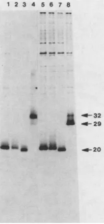
cells. The arrows indicate the glycosylated form of the v-sis' proteinat32kDa, the unglycosylatedform of thewild-typeprotein at 29kDa, andtheunglycosylatedforms of the mutantv-sisgene productsatapproximately 20 kD.
A B
1 23 4567
.@!!U
B.i1
W 6i$;
on November 10, 2019 by guest
http://jvi.asm.org/
[image:6.612.53.292.74.320.2] [image:6.612.379.485.384.612.2]SV40 genome inserted as a
BamHI
fragment in a pBR322 derivative (29). Most of the VP1 open reading frame,includ-ing the firstATG, has been deleted and replaced by a unique XhoI site. However, the late promoter and the 5' and 3' splice sites remain. Genes inserted at the unique
XhoI
siteareexpressed by using the late-transcription pathway, with
thevector providing for initiation, splicing, and polyadenyla-tion. To insert the v-sis signal sequence, donated by the
M-MuLV-derived expression vector, along with the deleted
v-sis genes, the genes were excised with
XbaI.
XbaI
cuts onceinthe M-MuLV region upstream of thesignal sequenceand again in the 3' long terminal repeat downstream ofthe
deleted genes. Therefore, a new SV40-derived expression vector, pRB34, was constructed, with the
XhoI
site of pJC119 converted to a unique XbaI site. This vector wasused forinsertion of the deleted v-sis genes as
XbaI-to-XbaI
fragments.
Five of the mutant v-sis genes, complete with the
vector-donated v-sis signal sequence region, were inserted into
pRB34 in the correct orientation for expression. These
plasmids were transfected into Cos-1 cells and metabolically
labelledwith [35S]methionine and
[35S]cysteine.
Cell lysates were immunoprecipitated with antiserum raised againstbacterially synthesized
p28sis.
Immunoprecipitates were an-alyzed on SDS-polyacrylamide gels under reducing and nonreducing conditions. Cells transfected with thev-siswt
geneexpressed a 32-kDa reduced protein (Fig. 3B, lane 7), which migrated as a dimer of the expected size when analyzed under nonreducing conditions (Fig. 3A, lane 7). Cells transfected with the five mutant v-sis genes yielded
immunoprecipitable
proteins of approximately the predicted molecular weights (Table 2) when electrophoresed underreducing conditions (Fig. 3B, lanes 2 through 6). The gene
product ofv-sis121 (Fig. 3B, lane 3) has an altered mobility
when analyzed on reducing SDS-polyacrylamide gels, mi-grating slightly more slowly than expected. When immuno-precipitates were analyzed under nonreducing conditions,
the gene
products
encodedby
thev-sisI2'
and
theV_SiS1
geneswerefoundtodimerize
(Fig.
3A,
lanes2and3).
Lesseramounts ofthe dimerized forms of the proteins encoded by theV-SiS129,
V-sis127,
and v-sis32 genes were observed (Fig. 3A, lanes 4, 5, and 6, respectively). In all cases, significantamounts of the monomer forms were also detected. Loss of N-linked glycosylation in the mutant v-sis gene
products. The v-siswt gene product undergoes N-linked
glycosylation
at Asn residue 93 (11, 24). Of the mutant v-sisgenes examined in this study, only the
v-sis92
gene retained the coding region for the consensus glycosylation siteAsn-Met-Thr. This region is deleted from all the other mutant v-sis genes, and their gene products are not expected to be
glycosylated. This was demonstrated by
immunoprecipita-tion analysis ofthe v-sis-encoded proteins from transfected
Cos-1 cells that had been treated with tunicamycin.
Immu-noprecipitates
of cells transfected with the v-siswt geneexhibited aproteinof 29 kDa when labeling was done in the
presenceoftunicamycin(Fig. 4, lane 8), as compared with a
proteinof 32kDa in the absence of tunicamycin (lane 4). The
migration ofthe gene products encoded by the mutant v-sis
genes was notaffectedby treatment with tunicamycin (lanes (1 and 5, 2 and 6, and 3 and 7), confirming that N-linked
glycosylation did not occur.
DISCUSSION
Wehaveconstructeda series of deletions in the v-sis gene which define the N-terminus of the minimal transforming
v-sis
NMTKR C CCCCC CC C
M
.I.I
.))I(( IL1 20 271amino acids
112 127 214 220
SLGSLSVAEPAMIAECKTRTEVFF .o oo o oV TLEDHLACKCEIVAAAARAVT Minimal Transforming Region
PDGF-B Homologous Region
FIG. 5. Minimal transforming region of the v-sis geneproduct. A map of the full-length v-sis gene product is shown, with the first methionine (M) residue and the 10 cysteine (C) residues indicated. Also indicated are the sequence Asn-Met-Thr (NMT), residues 93 through 95, which is the site of N-linked glycosylation; and the Lys-Arg basic dipeptide (KR), residues 110 and 111, which is the site of proteolytic cleavage. The signal sequence is indicated by a curlicue. The
v-siswt
initial translation product contains 271 resi-dues. The minimal transforming region of the v-sis gene, as delim-ited by this analysis of the N terminus in conjunction with the previous data on the C terminus (12), extends from residue 127 to residue 214. This region is indicated by the heavy line. Below the map of the full-length v-sis gene product is an expansion of the residues between 112 and 220. These correspond to the residues which are homologous to the PDGF-B chain. The extent of the minimal transforming region, as defined by this study, is indicated below the sequence. The PDGF-B chain homology spans the v-sis minimal transforming region and extends beyond it at both the C-terminal and N-terminal ends. The cysteine residues are distin-guished by asterisks. However, there are five other cysteines within this region which are not represented in this partial sequence. region of v-sis (Fig. 5). The deleted genes were assayed fortheir ability to induce the formation of foci in NIH 3T3 cells by using a M-MuLV-derived expression vector encoding the v-sis signal sequence. Mutants encoding a protein in which residues 58 through 126 are deleted are fully transforming,
while mutants encoding proteins with deletions extending
beyond residue 128 are biologically inactive. Furthermore, a mutant v-sis gene encoding deletions at both the N terminus and the C terminus of the PDGF-B homologous region also retains full biological activity. The minimal transforming region of the v-sis gene product therefore corresponds to
residues 127 through 214. This is 21 residues smaller than the
PDGF-B homologous region, which corresponds to residues 112 through 220.
Only two residues,
Cys127
andLys128,
separate theendpoints of the closest transforming and nontransforming deletion mutant gene products, which suggests that those residues are important for transformation by v-sis. The full-length v-sis gene encodes 10cysteine residues. Cysteine 28 is encoded by the portion of the viral env gene retained in the fused env-sis open reading frame and is removed along
with
the signal sequence. The next eight cysteine residues are within the PDGF-B homologous region, while the last cysteine is outside that region at residue 256. This final cysteine is not required for transformation or dimerization, since C-terminal truncated gene products with endpoints between residues 214 and 246 are still capable of dimeriza-tion and transformadimeriza-tion (12). However, one or both cysteine residues 208 and 210 are required for dimerization andtransformation, since a mutant gene product with an endpoint at residue 214 was able to dimerize and to trans-form cells, while a mutant
gene
product with an endpoint at residue 204 was incapable of both processes. The present study indicates that cysteine residue 127 is also requiredforon November 10, 2019 by guest
http://jvi.asm.org/
[image:7.612.326.562.79.182.2]transformation. All mutant v-sis genes which encode this residue areabletotransformNIH3T3cells andarecapable of dimerization. However, its role in dimerization is not
clear, for the v-sis127 gene product dimerizes to a lesser
degree than the v-siswt or the other transforming gene products examined in this study. This difference may reflect
the contribution of local secondary structure to either the efficiency of formationorthestability of the disulfide bonds.
Itshould be notedthat mutantproteins wereanalyzed with
Cos-1 cells rather than NIH 3T3 cells, on the basis of evidence that the processing of secreted proteins occurs
indistinguishably in these two cell lines (7, 11, 26, 29, 31).
However, there remains the formal possibility that subtle
differences between these cell lines may account for the
observations reported here concerning dimerization and
stability of the mutant gene products in Cos-1 cells, which
were correlated with transformation assays in NIH 3T3 cells.
Thev-sisWtgeneproduct is proteolytically processedafter
the basicdipeptide, Lys-Arg residues 110and111,toyielda
20-kDa PDGF-related protein as detected under reducing conditions (25). This protein exists as a dimer under
nonreducing conditions. Ofthe mutants studied here, only the four encoding the largest gene products retain the Lys-Argcodons. Otherdeletionmutants, however, lackthe Lys-Argdipeptide,yetstill encodegeneproducts whichare
capable of dimerization and transformation.The dispensabil-ity of N-terminal proteolytic processing is supported by previous work in which the Lys-Arg codonswere
mutagen-izedtoAsn-Thrwithnoreduction in thebiological activity of
the mutant (11).
There isasingleconsensussitefor N-linkedglycosylation in the v-sis gene product. This is the Asn-(Met)-Thr se-quence at residues 93 through 95. Only the
v-sis92
generetainsthe codonsfor this glycosylation site. All the other
mutant v-sis geneproducts studied here, both
transforming
and nontransforming, lack the N-linked
glycosylation
site. Thiswasdirectly demonstrated by the factthattunicamycin
treatmentof cellsexpressingthesemutantsfailedtoalter the
mobility of the v-sis proteins. These results indicate that
N-linked glycosylation isdispensablein termsofthe
biolog-ical activity ofthe v-sis gene product.
From the results of this study, in conjunction with our
previous work (12), we have delimited the minimal
trans-forming region
of thev-sisgeneproduct
ascorresponding
to88 amino
acids,
residues 127through
214. This issignifi-cantly smaller than the PDGF-B
chain,
whichcorresponds
toresidues 112 through 220 of the v-sis gene product. The
minimal transforming gene product encoded
by
thev-sisl271214
gene has 6fewer residuesatthe C terminusand 15fewer residues at the N terminus than does the PDGF-B
chain.Proteolytic processingandN-linkedglycosylationare notrequired for transformation by v-sis, even thoughthese processes do occur in the formation ofthe mature v-sisWt proteinand intheformation ofthePDGF-B chain from the nascent c-sis gene product.
Dimerization,
however, is nec-essaryfor thebiological activity ofv-sis. Additional studieson the roles of individual cysteine residues are currently being undertaken by means of site-directed mutagenesis.
Internal deletions and linker scanning will also be used to
further characterize the minimaltransforming region of the v-sisgene product.
ACKNOWLEDGMENTS
We thank Jay Sweet for his gift of antisera raised against the bacterially synthesized v-sis gene product. We thank Linda
Haberstroh for herhelp in the construction of the deletionmutants and RickBold and KarenMay for excellent technical assistance.
This workwassupportedbyPublic Health Service grant CA40573 from the National Cancer Institute. M.K.S. was supported by a postgraduatescholarshipfrom the Natural Sciences and Engineer-ing Research Council of Canada. D.J.D. gratefully acknowledges receiptof support from theChicago CommunityTrust/Searle Schol-arsProgram.
LITERATURE CITED
1. Andersson, P., M. P. Goldfarb,and R. A.Weinberg. 1979. A defined subgenomic fragment of invitro synthesized Moloney sarcomavirusDNA caninduce cell transformation upon trans-fection. Cell16:63-75.
2. Bold, R. J., and D. J. Donoghue. 1985. Biologically active deletion mutants of the v-mos oncogene assayed with novel retroviralvectors.Mol. Cell. Biol. 5:3131-3138.
3. Deuel,T.F., J.S.Huang, S. S. Huang,P.Stroobant,and M. D. Waterfield. 1983.Expressionofaplatelet-derivedgrowth factor-likeproteininsimiansarcomavirus transformed cells. Science 221:1348-1350.
4. Devare,S. G., E. P. Reddy, J. D.Law, K. C. Robbins, S.R. Tronick,andS. A. Aaronson.1983. Nucleotide sequence of the simiansarcomavirus genome: demonstrationthat itsacquired cellular sequences encode thetransforming protein p28sis.Proc. Natl. Acad.Sci. USA 80:731-735.
5. Doolittle,R. F.,M. W.Hunkapiller, L. E.Hood, S. G. Devare, K.C. Robbins, S. A. Aaronson, and H. N. Antoniades. 1983. Simiansarcomavirusoncgene, v-sis,is derived fromthegene (orgenes) encoding aplatelet-derived growth factor. Science 221:275-277.
6. Garrett, J. S.,S. R. Coughlin,H. L. Niman, P. M. Tremble, G. M. Giels, and L. T. Williams. 1984. Blockade of autocrine stimulation in simiansarcomavirus-transformed cells reverses
down-regulation of platelet-derived growth factor receptors. Proc. Natl.Acad. Sci. USA 81:7466-7470.
7. Gluzman, Y. 1981. SV40-transformedsimian cells support the replicationofearly SV40mutants. Cell23:175-182.
8. Graham,F.L.,and A.J.Vander Eb.1973.Anewtechniquefor the assayofinfectivityof human adenovirus5 DNA. Virology 52:456-467.
9. Grunstein, M.,and D.S.Hogness.1975.Colonyhybridization:a methodfor the isolation of cloned DNA's thatcontainaspecific
gene. Proc. Natl. Acad. Sci. USA 72:3961-3965.
10. Hannink, M., and D. J. Donoghue. 1984. Requirement for a
signal sequence in the biological expression of the v-sis oncogene. Science226:1197-1199.
11. Hannink, M., and D.J. Donoghue. 1986. Biosynthesis of the v-sisgeneproduct:signal sequence cleavage,glycosylationand proteolytic processing. Mol. Cell. Biol. 6:1343-1348.
12. Hannink,M.,M. K.Sauer,and D.J. Donoghue.1986.Deletions in theC-terminalcoding regionof thev-sisgene:dimerization is requiredfor transformation. Mol. Cell. Biol. 6:1304-1314. 13. Heldin, C.-H.,and B. Westermark.1984.Growth factors:
mech-anism ofactionandrelationtooncogenes. Cell 37:9-20. 14. Hoffman, J. W., D. Steffen, J. Gusella, C.Tabin, S.Bird, D.
Cowing,and R. A.Weinberg.1982. DNAmethylation affecting
the expression of murine leukemia proviruses. J. Virol. 44:144-157.
15. Huang, J.S.,S. S.Huang,and T. F. Deuel. 1984.Transforming proteinof simiansarcomavirus stimulatesautocrine growthof SSV-transformed cells through PDGF cell-surface receptors. Cell 39:79-87.
16. Huang, J. S., S. S. Huang, and T. F. Deuel. 1983. Human
platelet-derived growthfactor: radioimmunoassay and discov-ery ofaspecific plasma binding protein.J.Cell Biol. 97:383-388. 17. Johnsson, A.,C.-H.Heldin,A.Wasteson,B.Westermark,T. F. Deuel, J. S. Huang, P. H. Seeburg, A. Gray, A. Ulirich, G. Scrace,P.Stroobant,andM. D. Waterfield.1984. Thec-sisgene encodes a precursor of the B-chain ofplatelet-derived growth factor. EMBOJ. 3:921-928.
18. Josephs, S. F., C. Guo, L. Ratner, and F. Wong-Staal. 1984. Humanproto-oncogene nucleotide sequencescorrespondingto
on November 10, 2019 by guest
http://jvi.asm.org/
the transforming region of simian sarcoma virus. Science 223:487-491.
19. Kaplan, D. R., F. C. Chao,C. D. Stiles, H. N. Antoniades, and C. D. Scher. 1979. Platelet alpha granules contain a growth
factorforfibroblasts. Blood53:1043-1052.
20. King, C. R., N. A. Giese, K. C.Robbins, andS. A. Aaronson. 1985.Invitro mutagenesis of thev-sistransforminggenedefines
functional domains of its growth factor-related product. Proc. Natl. Acad. Sci. USA82:5295-5299.
21. MIxam, A. M., and W. Gilbert. 1980. Sequencing end-labeled DNAwithbase-specific chemical cleavages. Methods Enzymol. 65:499-560.
22. Nishimura, J., J. S. Huang, and T. F. Deuel. 1982. Platelet-derivedgrowthfactorstimulates tyrosine specific protein kinase in swiss mouse 3T3 cell membranes. Proc. Natl. Acad. Sci. USA79:4303-4307.
23. Owen, A. J., P. Pantazis, and H. N. Antoniades. 1984. Simian
sarcomavirus-transformed cells'secrete amitogen identicalto platelet-derivedgrowthfactor. Science 225:54-56.
24. Robbins, K. C., F. Leal, J. H. Pierce, and S. A. Aaronson.1985. The v-sis/PDGF-2 transforming gene product localizes to cell membranes but is not a secretory protein. EMBO J. 4:1783-1792.
25. Robbins, K. C., H. N. Antoniades, S. G. Devare, M. W. Hunkapiller, andS. A. Aaronson. 1983. Structural similarities between sarcoma virus gene product(s) and human platelet-derivedgrowth factor. Nature(London) 305:605-608.
26. Rose, J. K., andJ. E. Bergmann. 1982. Expression from cloned cDNA of cell-surface secreted forms of the glycoprotein of vesicular stomatitis virus in eucaryotic cells. Cell 30:753-762.
27. Ross, R., J. Glomset, B. Kariya, and L. Harker. 1974. A platelet-d,erived serum factor that stimulatestheproliferationof arterial smooth muscle cells in vitro. Proc. Natl. Acad. Sci. USA 71:1207-1210.
28. Sporn,M.B.,and A. B. Roberts.1985.Autocrinegrowthfactors and cancer. Nature (London) 313:745-747.
29. Sprague,J., J. H. Condra, H.Arnheiter, and R. A. Lazzarini. 1983. Expression ofarecombinant DNA clone codingfor the vesicular stomatitis virus nucleocapsid protein. J. Virol. 45:773-781.
30. Theilen, G. H., D. Gould, M. Fowler, and D. L. Dungworth. 1971. C-type virus in tumor tissue of a woolly monkey (Lagothrix spp.) with fibrosarcoma. J. Natl. Cancer Inst. 47:881-889.
31. Warren, T. G., and D. Shields. 1984. Expression of preprosomatostatin in heterologous cells: biosynthesis, post-translational processingand secretion of mature somatostatin. Cell39:547-555.
32. Waterfield, M. D., G.T. Scrace, N. Whittle, P. Stroobant, A. Johnsson, A. Wasteson, B. Westermark, C.-H. Heldin, J. S. Huang,and T. F. Deuel. 1983. Platelet-derivedgrowth factor is structurally relatedtotheputative transforming proteinp28s1s of simiansarcoma virus. Nature (London) 304:35-39.