Open Access
Research article
Roles of TNF-
α
gene polymorphisms in the occurrence and progress
of SARS-Cov infection: A case-control study
Shixin Wang
†1, Maoti Wei*
†2, Yi Han
1, Keju Zhang
1, Li He
1, Zhen Yang
1,
Bing Su
3, Zhilun Zhang
4, Yilan Hu
2and Wuli Hui
2Address: 1Proteomics Laboratory, Medical College of Chinese People's Armed Police Force, Tianjin, China, 2Department of Epidemiology, Medical
College of Chinese People's Armed Police Force, Tianjin, China, 3Department of Respiratory, Pingjin Hospital, Medical College of Chinese People's
Armed Police Force, Tianjin, China and 4Department for Emergency Diseases, Center for Disease Control and Prevention, Tianjin, China
Email: Shixin Wang - [email protected]; Maoti Wei* - [email protected]; Yi Han - [email protected];
Keju Zhang - [email protected]; Li He - [email protected]; Zhen Yang - [email protected]; Bing Su - [email protected]; Zhilun Zhang - [email protected]; Yilan Hu - [email protected]; Wuli Hui - [email protected]
* Corresponding author †Equal contributors
Abstract
Background: Host genetic factors may play a role in the occurrence and progress of SARS-Cov
infection. This study was to investigate the relationship between tumor necrosis factor (TNF)-α
gene polymorphisms with the occurrence of SARS-CoV infection and its role in prognosis of patients with lung interstitial fibrosis and femoral head osteonecrosis.
Methods: The association between genetic polymorphisms of TNF-α gene and susceptibility to severe acute respiratory syndromes (SARS) was conducted in a hospital-based case-control study including 75 SARS patients, 41 health care workers and 92 healthy controls. Relationships of
TNF-α gene polymorphisms with interstitial lung fibrosis and femoral head osteonecrosis were carried
out in two case-case studies in discharged SARS patients. PCR sequencing based typing (PCR-SBT)
method was used to determine the polymorphisms of TNF-αgene in locus of the promoter region
and univariate logistic analysis was conducted in analyzing the collected data.
Results: Compared to TT genotype, the CT genotype at the -204 locus was found associated with
a protective effect on SARS with OR(95%CI) of 0.95(0.90–0.99). Also, TT genotype, CT and CC
were found associated with a risk effect on femoral head necrosis with ORs(95%CI) of
5.33(1.39–20.45) and 5.67(2.74–11.71), respectively and the glucocorticoid adjusted OR of CT was
5.25(95%CI 1.18–23.46) and the combined (CT and CC) genotype OR was 6.0 (95%CI 1.60–22.55)
at -1031 site of TNF-αgene. At the same time, the -863 AC genotype was manifested as another
risk effect associated with femoral head necrosis with OR(95%CI) of 6.42(1.53–26.88) and the
adjusted OR was 8.40(95%CI 1.76–40.02) in cured SARS patients compared to CC genotype.
Conclusion: SNPs of TNF-αgene of promoter region may not associate with SARS-CoV infection. And these SNPs may not affect interstitial lung fibrosis in cured SARS patients. However, the -1031CT/CC and -863 AC genotypes may be risk factors of femoral head necrosis in discharged SARS patients.
Published: 29 February 2008
BMC Infectious Diseases 2008, 8:27 doi:10.1186/1471-2334-8-27
Received: 30 September 2007 Accepted: 29 February 2008 This article is available from: http://www.biomedcentral.com/1471-2334/8/27
© 2008 Wang et al; licensee BioMed Central Ltd.
Background
TNF, the gene encoding tumour necrosis factor (TNF), resides in the central part (class III region) of the major histocompatibility complex (MHC) surrounded by a large number of other immunological genes [1]. Because of the special locus of this gene, it can be deduced that this gene may associate with many diseases, and this hypothesis was confirmed by many research results [2,3]. TNF-α is a key mediator of the inflammatory response and is critical for host defense against a wide variety of pathogenic microbes. However, the over-expression of this cytokine may lead to badness in disease recovery. The dual role of TNF, acting as an agent of both innate immunity and inflammatory pathology, poses a considerable challenge for gene regulation [2], and this regulation mainly located on promoter region of this gene. The capacity for cytokine production in an individual has a major genetic compo-nent, and striking differences existed among individuals in terms of their ability to produce cytokines. Several bial-lelic polymorphisms had been described within the TNF-α gene, including seven in the promoter region at posi-tions -1031T→C, -863C→A, -857C→T, -376G→A, -308G→A, -238G→A and -163G→A base pairs from the transcription start site [4,5]. Moreover, a number of stud-ies had shown that the TNF-α promoter polymorphism had a significant effect on its transcriptional activity [6,7].
Severe acute respiratory syndrome (SARS) is a newly described human infectious disease caused by a novel coronavirus-SARS-CoV. SARS-CoV infection is important because of its high infectivity and unpredictable clinical course, which is characterized by a high mortality rate [8]. Till now, many researchers had reported that susceptibili-ties to infection SARS-CoV may associated with HLA,
MXA, OAS-1 and CLEC4M gene polymorphisms [9-13], yet these results were variable in different populations. For example, Ng reported that SARS-CoV infection was associated with HLA-B*0703 and HLA-DRB1*0301 in HongKong population [9], however, Lin's results showed that HLA-B*4601 and HLA-B*5401 were closely related to SARS-CoV infection [10]. Chan reported that CLEC4M
was attributed to SARS-CoV infection [13], but Zhi's results failed to support this conclusion.7 These differ-ences may be attributed to the study population used in each report, also the complex mechanism infection to SARS-CoV should be considered as another factor of these differences. In order to explore more host factors influenc-ing the occurrence of SARS-CoV infection, we studied the polymorphisms of TNF-α gene at the promoter region, which had been ascribed to polymorphisms within the regulatory regions or signal sequences of cytokine genes [14].
After discharging from hospital, interstitial lung fibrosis was observed in SARS patients. Clinical data showed that
the prevalence rate of this change was 21%(42/200) in cured SARS patients nine months from the discharge [15]. TNF-α was one of the earliest cytokines implicated in the pathogenesis of lung fibrosis diseases and, together with IL-1, has been found to over-expressed in regenerated type II pneumocytes in human lung, thus enhanced fibroblast proliferation [16]TNF-α polymorphisms have been dis-covered significantly associated with increased risk of developing pulmonary fibrosis [17,18]. Given that genetic variation may potentially alter inflammation and fibrosis in the lung, the aim of this case-case control study was to examine the TNF-αpolymorphisms with interstitial lung fibrosis in SARS patients.
In spite of interstitial lung fibrosis in cured SARS patients, another sequela – femoral head necrosis was also observed in this population and the prevalence rate was 22.07%(49/221) and 23.1%(18/78) in Tianjin and Bei-jing patients respectively [15,19]. The cause of this disease was still unknown and there were arguments about it. For example, some author considered SARS-CoV as the cause of femoral head necrosis, yet other authors disagreed with this view [20,21]. Previous studies showed that femoral head necrosis may caused by hormone usage [20], yet our data failed to agree with this point. So, it need further study to explore the cause of this sequela and TNF-α pol-ymorphisms were considered first in this report.
In this paper, we aimed to study whether polymorphisms in TNF-αpromoter region were associated with SARS-CoV infection, development, and progression of interstitial lung fibrosis and femoral head necrosis in cure SARS patients.
Methods
SubjectsThis study was reviewed and approved by ethics commit-tees in the Medical College of CPAFP. The study popula-tion comprised 75 SARS patients in Pingjin hospital, Tianjin, China, 41 health care workers of the same hospi-tal, who had come into contact with SARS patients but had not developed into SARS, and 66 individuals having no contact history with SARS patients. Among 75 SARS patients, 55 could be classified into severe and light SARS according to their clinical condition history during the hospitalized period and this population also had the his-tory of hormone therapy by reviewing the clinical treat-ment. Anti-SARS-CoV antibodies of the serum samples were tested by SARS ELISA kits (Huada Diagnostics Ltd, Beijing, China).
associations between gene polymorphisms with disease. Three kinds of hormone were used in SARS patients including methylprednisolone, deltadehydrocortisone and dexamethasone. In order to simplify analyzing, delta-dehydrocortisone and dexamethasone dosage were calcu-lated into methylprednisolone using the following equation: 4 mg methylprednisolone = 5 mg deltadehydro-cortisone = 0.75 mg dexamethasone. Lash therapy means more than 320 mg methylprednisolone were used in a single day.
Cured SARS patients with interstitial lung fibrosis were diagnosed by respiratory experts according to CT results following the standard proposal for therapy and diagnosis of SARS patients issued by Chinese Ministry of Health in 2004 [22]. Interstitial lung fibrosis of SARS patients man-ifested as irregular patch and strip shadow or high density strip shadow and honeycomb interstitial lung fibrosis, these changes could combine with the bronchiectasis.
Femoral head necrosis was diagnosed using magnetic res-onance imaging (MRI). An MRI scan of a normal femoral head would show uniformly high signal intensity on T1-and T2-weighting throughout the femoral head. Agree with one of the following image could be diagnosed as femoral head necrosis in SARS patients: Abnormal signal with clear margin in cartilage of femoral head, or double thread image, or fracture or joint dent under cartilage, or T1WI low signal, T2WI and STIR high signal of the mar-row cavity edema with blur edge [22].
Genomic DNA extraction and polymerase chain reaction Leucocytes were isolated within 12 h of blood collection using Percoll reagent. Then genomic DNA was extracted using cell DNA extraction kit (Tiangen BioTec Co, Beijing, China, patch number: 2004-08-13) according to the man-ufacturer's instructions. Primers were designed according to Gewaltig [23]. Standard 50-μL polymerase chain reac-tions (PCRs) contained 5 μl(6.7 μM) forward primer 5'-GATGGACTCACCAGGTGAG-3', 5 μl(6.7 μM) reverse primer 5'-CTCATGGTGTCCTTTCCAGG-3', 5 μl buffer [150 mm (NH4)2SO4, 500 mM Tris-Cl(pH = 8.8), 500 μM EDTA-Na2,15 mM MgCl2,100 mM β-Mercaptoethanol], 0.5 μL DNA polymerase (Tiangen BioTec Co, Beijing, China), 3 μl DNA template. Amplification was carried out in a thermal cycler TC312 (Techne, Duxford Cambridge, UK) with cycle parameters of 5 min at 94°C (initial
dena-turation), 35 rounds of 94°C 30 s, 60°C 100 s and 72°C 150 s, and a final extension for 5 min at 72°C. The reac-tions were carried out in molecular BioProducts 200 μL capped tubes, as these gave optimal heat transfer in the thermal cycler.
Sequencing of TNF-αgene fragments
The TNF-α gene 1279 bp fragments in this paper were sequenced in double directions with forward primer GATGGACTCACCAGGTGAG-3' and reverse primer 5'-CTCATGGTGTCCTTTCCAGG-3' and Invitrogen company (Invitrogen Co, Shanghai, China) using ABI 373 thermal cycler carried out this job. The homozygote genotype of each SNP site manifested as a single peak, yet the hetero-zygote with an ambiguous nucleotide position of a dou-ble color peak in the Big Dye chemistry pictures. According to reading the sequence graphs, the genotype was determined.
Statistics
The differences in values between two groups were evalu-ated by Chi analysis for frequencies or student t test for quantitative index and binary logistic regression was done using SPSS 11.5 software (SPSS Inc, Chicago, Illinois, USA).
Results
Demographic characteristics of the populations
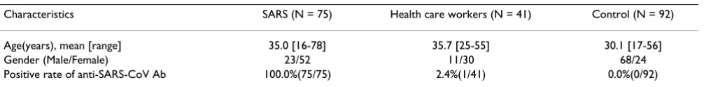
A total of 75 SARS patients, 41 health care workers and 66 individuals were included in this study. All the popula-tions were Chinese Han ethnic. The mean age was 35.0 years for SARS, 35.7 for health care workers and 30.1 for individual controls (SARS Vs HCW, P > 0.05; SARS Vs
individual control, P > 0.05). The proportion of male was 30.6% in SARS, 26.8% in health care workers and 69.5% in individual controls (SARS Vs HCW, P > 0.05; SARS Vs
individual control, P < 0.05). The sera positive rate anti-SARS-CoV antibody was 100.0% in SARS, significantly higher than that of health care workers and individual controls (SARS Vs HCW, P < 0.05; SARS Vs individual con-trol, P < 0.05) (Table 1).
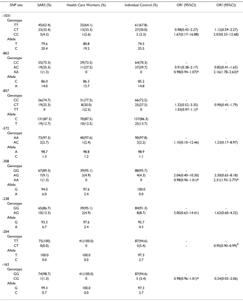
TNF-αpolymorphisms and SARS-Cov infection
TNF-αgenotype frequencies were variable in SARS, health care workers and individual controls. There were no dif-ferences of TNF-α genotype distribution at the -1031(T→C), -863(C→A), -572(A→C), -308(G→A) and
-Table 1: Demographic characteristics of the populations
Characteristics SARS (N = 75) Health care workers (N = 41) Control (N = 92)
Age(years), mean [range] 35.0 [16-78] 35.7 [25-55] 30.1 [17-56]
Gender (Male/Female) 23/52 11/30 68/24
[image:3.612.56.559.670.732.2]238(G→A) among the three populations. However, the CT genotype was less frequent in SARS group when com-pared with individual controls at the -204 locus (X2 = 4.20, P = 0.04). Compared to TT genotype, the CT geno-type at the -204 locus were found associated with a protec-tive effect on SARS with OR(95%CI) of 0.95(0.90–0.99) (Table 2).
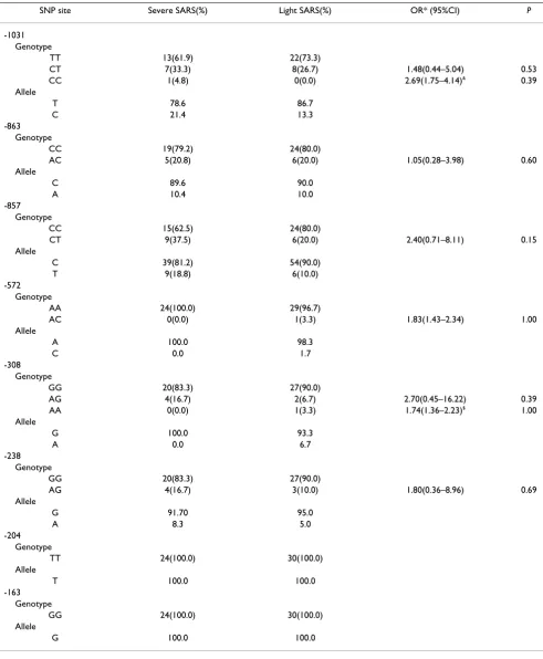
According to the clinical history, symptoms of SARS patients were classified into light and severe. Because of the complicated clinical condition during SARS outbreak, some patients' histories were incomplete and could not be classified following the severity standard [22]. The severe SARS referred to those with one or more of the following: (1) dyspnea, more than 30 times per min respiratory fre-quencies in still condition;(2) oxygenation index less than 300 mmHg; (3) shock or multiple organ dysfunction syn-drome. Among all 75 patients, fifty-four were classified into light and severe. And there were no association of
TNF-αpolymorphisms and SARS severity (Table 3).
Glucocorticoid using in SARS with interstitial lung fibrosis or femoral head necrosis
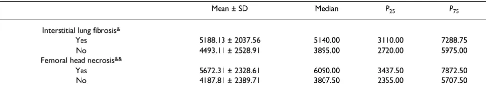
Glucocorticoid using dosage, method and lasting period were not associated with interstitial lung fibrosis or femo-ral head necrosis in binary logistic analysis in SARS patients (Table 4). And there was no difference of hor-mone using dosage between the interstitial lung fibrosis and non-interstitial lung fibrosis group(t = 0.72, P = 0.47) and this trend was also observed in the femoral head necrosis and non-femoral head necrosis group(t = 1.90, P
= 0.064) (Table 5).
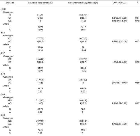
TNF-αpolymorphisms and interstitial lung fibrosis Allele frequencies of TNF-αpolymorphisms were listed in Table 6 and there were no significant differences between interstitial lung fibrosis and non-interstitial lung fibrosis in SARS patients at promoter region of TNF-αgene.
TNF-α polymorphism and femoral head necrosis
Allele frequencies of TNF-α gene were compared in SARS patients between femoral head necrosis and non-femoral head necrosis (Table 7). The -1031 CT and CC genotypes were more frequent in SARS patients with femoral head necrosis(53.7% and 6.7%, respectively) than in non-fem-oral head necrosis(20.0% and 0.0%, respectively). Com-pared to TT genotype, CT and CC were found associated with a risk effect on femoral head necrosis with ORs(95%CI) of 5.33(1.39–20.45) and 5.67(2.74–11.71), respectively. The adjusted OR of CT was 5.25(95%CI 1.18–23.46) and the combined (CT and CC) genotype OR was 6.0 (95%CI 1.60–22.55). Also, the -863 AC gen-otype accounted for 43.7% of femoral head necrosis group but 10.8% of non-femoral head necrosis. Com-pared to CC genotype, the AC genotype was manifested as
another risk effect associated with femoral head necrosis with OR(95%CI) of 6.42(1.53–26.88) and the adjusted OR was 8.40(95%CI 1.76–40.02) in cured SARS patients.
Discussion
Four years after SARS occurrence, many problems still remained unknown to us. Till now, many researchers have reported that susceptibility to infection SARS-CoV may associate with HLA, MXA, OAS-1 and CLEC4M gene pol-ymorphisms, yet the results are variable in different pop-ulations [9-13]. These differences may be attributed to the study population used in each report, also the complex mechanism infection to SARS-CoV should be considered as another factor of these differences. In order to explore more host factor influence the occurrence of SARS-CoV infection, we studied the polymorphisms of TNF-α gene at the promoter region, which have been ascribed to pol-ymorphisms within the regulatory regions or signal sequences of cytokine genes [14]. Allele distributions at -1031, -863, -857, -572, -238 and -163 were almost the same among the SARS, the health care workers and indi-vidual controls, but a higher A allele frequency in SARS population when compared with the control at the -308 locus(X2 = 8.96, P = 0.003). Though previous study showed that TNF-α-308 AG genotype was associated with the clearance of Hepatitis B virus and the infection of Heli-cobacter pylori cagA subtype infection [24,25], our results failed to show the role of this locus in SARS-CoV infection and this conclusion agreed with that of Chong WP et al
[26]. We found that there was a weak protective effect of CT genotype at -204 locus of TNF-α gene against SARS-CoV infection. The -204 locus was a new discovered SNP site of TNF-α promoter region and its role in infectious diseases might need further study. At the same time, no obvious association between the polymorphisms of TNF-αpromoter region with the severity of SARS was observed. However, Lu reported that the -238G/A polymorphism of
TNF-α associated with the outcomes of hepatitis B virus infection [27]. Thus, the roles of TNF-αgene in infectious diseases should be further studied.
Table 2: TNF-α gene polymorphism in promoter region in cured SARS patients, health care workers and individual control
SNP site SARS (%) Health Care Workers (%) Individual Control (%) OR1 (95%CI) OR2 (95%CI)
-1031 Genotype
TT 45(63.4) 25(64.1) 61(67.8) - -CT 23(32.4) 13(33.3) 27(30.0) 0.98(0.43–2.27) 1.12(0.59–2.27) CC 3(4.2) 1(2.6) 2 (2.2) 1.67(0.17–16.88) 2.03(0.33–12.68) Allele
T 79.6 80.8 74.5 C 20.4 19.2 25.5 -863
Genotype
CC 55(73.3) 29(72.5) 64(70.3) - -AC 19(25.3) 11(27.5) 27(29.7) 0.91(0.38–2.17) 0.82(0.41–1.63) AA 1(1.3) 0 0 0.98(0.94–1.07)& 2.16(1.78–2.63)&
Allele
C 86.0 86.3 85.2 A 14.0 13.7 14.8 -857
Genotype
CC 56(74.7) 31(77.5) 66(72.5) - -CT 19(25.3) 8(20.0) 25(27.5) 1.32(0.52–3.35) 0.90(0.45–1.79) TT 0 1(2.5) 0 1.03(0.97–1.1)$
-Allele
C 131(87.3) 70(87.5) 157(86.3) T 19(12.7) 10(12.5) 25(13.7) -572
Genotype
AA 73(97.3) 40(97.6) 90(97.8) - -AC 2(2.7) 1(2.4) 2(2.2) 1.10(0.10–12.46) 1.23(0.17–8.97) Allele
A 98.7 98.8 98.9 C 1.3 1.2 1.1 -308
Genotype
GG 67(89.3) 39(95.1) 88(95.7) - -AG 7(9.1) 2(4.9) 4(4.3) 2.04(0.40–10.30) 2.30(0.65–8.18) AA 1(1.3) 0 0 0.98(0.96–1.01)& 2.31(1.93–2.77)&
Allele
G 94.0 97.6 100.0 A 6.0 2.4 0.0 -238
Genotype
GG 65(86.7) 39(95.1) 84(91.3) - -AG 10(13.3) 2(4.9) 8(8.7) 3.00(0.63–14.41) 1.62(0.60–4.32) Allele
G 93.3 97.6 95.7 A 6.7 2.4 4.3 -204
Genotype
TT 75(100) 41(100.0) 87(94.6) - -CT 0(0.0) 0 5(5.4) - 0.95(0.90–0.99)$ Allele
T 100.0 100.0 97.3 C 0.0 0.0 2.7 -163
Genotype
GG 74(98.7) 41(100.0) 87(94.6) - -CG 1(1.3) 0 5 (5.4) 0.98(0.96–1.01)& 0.24(0.03–2.06)
Allele
G 99.3 100.0 97.3 C 0.7 0.0 2.7 Note: OR1 was calculated by comparing SARS with HCW;
OR2 was calculated by comparing SARS with individual control;
Table 3: TNF-α gene polymorphism and the progress of SARS
SNP site Severe SARS(%) Light SARS(%) OR* (95%CI) P
-1031 Genotype
TT 13(61.9) 22(73.3)
CT 7(33.3) 8(26.7) 1.48(0.44–5.04) 0.53
CC 1(4.8) 0(0.0) 2.69(1.75–4.14)& 0.39
Allele
T 78.6 86.7
C 21.4 13.3
-863 Genotype
CC 19(79.2) 24(80.0)
AC 5(20.8) 6(20.0) 1.05(0.28–3.98) 0.60
Allele
C 89.6 90.0
A 10.4 10.0
-857 Genotype
CC 15(62.5) 24(80.0)
CT 9(37.5) 6(20.0) 2.40(0.71–8.11) 0.15
Allele
C 39(81.2) 54(90.0)
T 9(18.8) 6(10.0)
-572 Genotype
AA 24(100.0) 29(96.7)
AC 0(0.0) 1(3.3) 1.83(1.43–2.34) 1.00
Allele
A 100.0 98.3
C 0.0 1.7
-308 Genotype
GG 20(83.3) 27(90.0)
AG 4(16.7) 2(6.7) 2.70(0.45–16.22) 0.39
AA 0(0.0) 1(3.3) 1.74(1.36–2.23)$ 1.00
Allele
G 100.0 93.3
A 0.0 6.7
-238 Genotype
GG 20(83.3) 27(90.0)
AG 4(16.7) 3(10.0) 1.80(0.36–8.96) 0.69
Allele
G 91.70 95.0
A 8.3 5.0
-204 Genotype
TT 24(100.0) 30(100.0)
Allele
T 100.0 100.0
-163 Genotype
GG 24(100.0) 30(100.0)
Allele
G 100.0 100.0
organs, resulting in acute lung injury and, subsequently, multi-organ dysfunction [29]. So, TNF-αgenetic variation may potentially alter inflammation and fibrosis in the lung. After discharging from hospital, interstitial lung fibrosis was observed in cured SARS patients and the prev-alence rate was 21%(42/200). TNF-α was one of the earli-est cytokines implicated in the pathogenesis of lung fibrosis disease and, together with IL-1, has been found to over-express in regenerating type II pneumocytes in human lung, thus enhancing fibroblast proliferation [16].TNF-αgenetic polymorphisms have been found sig-nificantly associated with increased risk of developing pulmonary fibrosis [17,18]. During the progress of idio-pathic pulmonary fibrosis, activated epithelial cells are thought to release potent fibrogenic molecules and cytokines, such as TNF-α and TGF-β1, which in turn foster the transformation of fibroblasts into myofibroblasts and promote their production of extracellular matrix mole-cules and a vicious cycle of injury and abnormal epithelial healing sets the stage for progressive fibrosis and architec-tural distortion of the lung parenchyma [7]. However, our data failed to show that alleles at -1031(T→C), -863(C→A), -857(C→T), -572(A→C), -308(G→A) and -238(G→A) were related to interstitial lung fibrosis when compared with non-interstitial lung fibrosis in SARS patients. At the same time, -204 and -163 were homozy-gote genotype of TT and GG respectively in SARS patients and no relationship of genotype with interstitial lung fibrosis could be calculated. This result implicated that there maybe a different mechanism of interstitial lung
fibrosis of SARS compared with idiopathic pulmonary fibrosis.
[image:7.612.55.556.100.194.2]Femoral head necrosis, another sequela of discharged SARS patients, prevailed with a rate of 22.07%(49/221) in Tianjin [15]. However, the cause of this sequela was still unknown and there were arguments about it. For exam-ple, some author considered SARS-CoV as the cause of femoral head necrosis, yet other authors disagree with this view [20,21]. Previous studies showed that femoral head necrosis may caused by hormone usage, our data was far to agree with this conclusion. There was no obvious asso-ciation between hormone using including hormone dos-age, method and lasting period with femoral head necrosis in binary logistic analysis in SARS patients. We found that the -1031 CT and CC genotypes were more fre-quent in SARS patients with femoral head necrosis(53.7% and 6.7%, respectively) than in non-femoral head necro-sis(20.0% and 0.0%, respectively). And CT and CC were related with a risk effect on femoral head necrosis with ORs (95%CI) of 5.33(1.39–20.45) and 5.67(2.74–11.71), respectively when compared to TT gen-otype. The hormone using adjusted OR of CT was 5.25(95%CI 1.18–23.46) and the combined (CT and CC) genotype OR was 6.0 (95%CI 1.60–22.55). Also, the -863 AC genotype accounted for 43.7% of femoral head necro-sis group but 10.8% of non-femoral head necronecro-sis. Com-pared to CC genotype, the AC genotype was manifested as another risk effect associated with femoral head necrosis with OR(95%CI) of 6.42(1.53–26.88) and the adjusted OR was 8.40(95%CI 1.76–40.02) in cured SARS patients. Table 4: Binary logistic analysis of hormone using in SARS patients
Factors Interstitial lung fibrosis Femoral head necrosis
P OR 95.0% C.I. P OR 95.0% C.I.
Glucocorticoid 0.97 1.00 0.99 1.00 0.11 1.00 0.99 1.00
Time 0.78 0.97 0.81 1.18 0.96 1.00 0.84 1.18
Lash therapy 0.51 2.13 0.23 20.13 0.27 0.32 0.04 2.43
Constant 0.68 3.48 0.13 88.28
Table 5: Cumulative hormone usage (mg) in cured SARS patients classified according to interstitial lung fibrosis or femoral head necrosis
Mean ± SD Median P25 P75
Interstitial lung fibrosis&
Yes 5188.13 ± 2037.56 5140.00 3110.00 7288.75
No 4493.11 ± 2528.91 3895.00 2720.00 5975.00
Femoral head necrosis&&
Yes 5672.31 ± 2328.61 6090.00 3437.50 7872.50
No 4187.81 ± 2389.71 3807.50 2355.00 5707.50
&: hormone dosage between interstitial lung fibrosis and non-interstitial lung fibrosis, t = 0.72, P = 0.47;
[image:7.612.57.556.618.711.2]TNF-α can activate activity of osteoclasts and accelerate absorption of the bone and cartilage and induce occur-rence of oxygen free radicals an d lipid peroxidation, which can induce ischemic necrosis of the femoral head [30]. So, the ploymophism TNF-αgene may be directly or indirectly attributed to the occurrence of femoral head necrosis in SARS patients.
Conclusion
[image:8.612.57.548.99.574.2]In conclusion, there may be no association of TNF-α pol-ymorphisms in promoter region with SARS-Cov infection. Also, TNF-α gene polymorphisms may no affect the occurrence of interstitial lung fibrosis in cured SARS patients. However, the polymorphisms may relate with femoral head necrosis.
Table 6: TNF-α gene polymorphisms and the occurrence of interstitial lung fibrosis in SARS patients
SNP site Interstitial lung fibrosis(%) Non-interstitial lung fibrosis(%) OR* (95%C.I.) P
-1031 Genotype
TT 14(70) 12(57.1)
CT 6(30) 8(38.1) 0.64(0.17–2.38) 0.51
CC 0 1(4.8) 1.08(0.93–1.27)‡ 0.48
Allele
T 85.00 76.19
C 15.00 23.81
-863 Genotype
CC 17(77.3) 16(72.7)
AC 5(22.7) 6(27.3) 0.78(0.20–3.08) 0.73
Allele
C 88.64 38
A 11.36 13.64
-857 Genotype
CC 15(68.8) 17(77.3)
CT 7(31.8) 5(22.7) 1.59(0.42–6.07) 0.50
Allele
C 84.09 88.64
T 15.91 11.36
-572 Genotype
AA 21(95.5) 22(100)
AC 1(4.5) 0 0.96(0.87–1.05)& 0.50
Allele
A 97.73 100.00
C 2.27 0.00
-308 Genotype
GG 21(95.5) 18(81.8)
AG 1(4.5) 4(18.2) 0.21(0.02–2.10) 0.17
Allele
G 97.73 90.9
A 2.27 9.1
-238 Genotype
GG 20(90.9) 18(81.8)
AG 2(9.1) 4(18.2) 0.45(0.07–2.76) 0.33
Allele
G 95.45 90.9
A 4.55 9.1
Note:*:OR was calculated using non-interstitial lung fibrosis as control. &: Odds ratio replaced with interstitial lung fibrosis group;
Competing interests
The author(s) declare that they have no competing inter-ests.
Authors' contributions
SXW conceived of the study, and participated in its design and coordination.
MTW, co-first author, carried out the molecular genetic studies and drafted the manuscript.
YH, KJZ and LH carried out the molecular genetic studies
[image:9.612.57.548.100.583.2]ZY, BS and ZLZ participated in field investigation and samples collection of the study
Table 7: TNF-α gene polymorphism and femoral head necrosis in cured SARS patients.
SNP site Femoral head necrosis (%) Non-femoral head necrosis (%) OR* (95%C.I.) P
-1031 Genotype
TT 6(40) 28(80)
CT 8(53.7) 7(20) 5.33(1.39–20.45) 0.01
CC 1(6.7) 0 5.67(2.74–11.71)$ 0.04
Allele
T 66.67 90.00
C 33.33 10.00
-863 Genotype
CC 9(56.3) 33(89.2)
AC 7(43.7) 4(10.8) 6.42(1.53–26.88) 0.01
Allele
C 78.12 94.59
A 21.88 5.41
-857 Genotype
CC 12(75) 26(70.3)
CT 4(25) 11(29.7) 0.78(0.21–2.99) 0.73
Allele
C 87.50 85.14
T 12.50 14.86
-572 Genotype
AA 16(100) 36(97.3)
AC 0 1(2.7) 1.44(1.21–1.73)‡ 0.70
Allele
A 100 98.7
C 0 1.3
-308 Genotype
GG 14(87.5) 32(86.5)
AG 2(12.5) 4(10.8) 1.14(0.19–6.98) 0.89
AA 0 1(2.7) 1.44(1.19–1.74)‡ 0.70
Allele
G 93.37 91.89
A 6.25 8.11
-238 Genotype
GG 13(81.3) 33(89.2)
AG 3(18.7) 4(10.8) 1.90(0.37–9.71) 0.35
Allele
G 90.62 94.59
A 9.38 5.41
Note: *:OR was calculated using non-femoral head necrosis as control. $:OR replaced by femoral head necrosis;
YLH and WLH participated in the design of the study and performed the statistical analysis
All authors contributed to writing of the final manuscript
All authors read and approved the final manuscript
Acknowledgements
This paper was supported by the Tianjin Science Fund (No.
05YFSZSF02900) and the Science Fund of Medical College of CPAFP(No. WY-2005-14).
References
1. The MHC sequencing consortium: Complete sequence and gene map of a human major histocompatibility complex. The MHC sequencing consortium. Nature 1999, 401:921-923. 2. Sariban E, Imamura K, Luebbers R, Kufe D: Transcriptional and
posttranscriptional regulation of tumor necrosis factor gene expression in human monocytes. J Clin Invest 1988, 81:1506-10. 3. Richardson A, Sisay-Joof F, Ackerman H, Usen S, Katundu P, Taylor T, Molyneux M, Pinder M, Kwiatkowski D: Nucleotide diversity of the TNF gene region in an African village. Genes and Immunity
2001, 2:343-348.
4. Higuchi T, Seki N, Kamizono S, Yamada A, Kimura A, Kato H, Itoh K:
Polymorphism of the 5'-flanking region of the human tumor necrosis factor (TNF)-alpha gene in Japanese. Tissue Antigens
1998, 51:605-612.
5. Juszczynski P, Kalinka E, Bienvenu J, Woszczek G, Borowiec M, Robak T, Kowalski M, Lech ME, Baseggio L, Coiffier B, et al.: Human leuko-cyte antigens class II and tumor necrosis factor genetic pol-ymorphisms are independent predictors of non-Hodgkin lymphoma outcome. Blood 2002, 100:3037-3040.
6. Kroeger KM, Carville KS, Abraham LJ: The -308 tumor necrosis factor-alpha promoter polymorphism effects transcription.
Mol Immunol 1997, 34:391-399.
7. Verma S, Slutsky AS: Idiopathic Pulmonary Fibrosis-New Insights. N Eng J Med 2007, 356(13):1370-1371.
8. Lee N, Hui D, Wu A, Chan P, Cameron P, Joynt GM, Ahuja A, Yung MY, Leung CB, To KF, Lui SF, Szeto CC, Chung S, Sung JJ: A major outbreak of severe acute respiratory syndrome in Hong Kong. N Engl J Med 2003, 348:1986-94.
9. Ng MH, Lau KM, Li L, Cheng SH, Chan WY, Hui PK, Zee B, Leung CB, Sung JJ: Association of Human-Leukocyte-Antigen Class I (B*0703) and Class II (DRB1*0301) Genotypes with Suscep-tibility and Resistance to the Development of Severe Acute Respiratory Syndrome. J Infect Dis 2004, 190(3):515-8. 10. Lin M, Tseng HK, Trejaut JA, Lee HL, Loo JH, Chu CC, Chen PJ, Su
YW, Lim KH, Tsai ZU, Lin RY, Lin RS, Huang CH: Association of HLA class I with severe acute respiratory syndrome corona-virus infection. BMC Med Genet 2003, 4(1):9.
11. He J, Feng D, de Vlas SJ, Wang H, Fontanet A, Zhang P, Plancoulaine S, Tang F, Zhan L, Yang H, Wang T, Richardus JH, Habbema JD, Cao W: Association of SARS susceptibility with single nucleic acid polymorphism of OAS-1 and MxA gene: a case-control study. BMC Infectious Disease 2006, 6:106-112.
12. Hamano E, Hijikata M, Itoyama S, Quy T, Phi NC, Long HT, Ha le D, Ban VV, Matsushita I, Yanai H, Kirikae F, Kirikae T, Kuratsuji T, Sasa-zuki T, Keicho N: Polymorphisms of interferon-inducible genes OAS-1 and MxA associated with SARS in the Vietnamese population. Biochemical and Biophysical Research Communications
2005, 329(4):1234-1239.
13. Chan VS, Chan KY, Chen Y, Poon LL, Cheung AN, Zheng B, Chan KH, Mak W, Ngan HY, Xu X, Screaton G, Tam PK, Austyn JM, Chan LC, Yip SP, Peiris M, Khoo US, Lin CL: Homozygous L-SIGN (CLEC4M) plays a protective role in SARS coronavirus infec-tion. Nat Genet 2006, 38(1):38-46.
14. Danis VA, Millington M, Hyland VJ, Grennan D: Cytokine produc-tion by normal human monocytes: inter-subject variaproduc-tion and relationship to an IL-1 receptor antagonist (IL-1Ra) gene polymorphism. Clin Exp Immunol 1995, 99(2):303-310.
15. Jiao ZS, Yuan SL, Cao Q, Wang SX, Wu HW, Yang Z, Su B, Yu QC, Lu WY, Li YC, Tian HG, Zhang Y: The Follow-up Study on the
Health Status of Convalescent Patienrs Recovered from SARS in Tianjin. Chinese Journal of Prevention and control of Chronic non-communicable Diseases 2006, 14(6):175-178.
16. Pan LH, Ohtani H, Yamauchi K, Nagura H: Co-expression of TNF alpha and IL-1 beta in human acute pulmonary fibrotic dis-eases: an immunohistochemical analysis. Pathol Int 1996,
46(2):91-9.
17. Whyte M, Hubbard R, Meliconi R, Whidborne M, Eaton V, Bingle C, Timms J, Duff G, Facchini A, Pacilli A, Fabbri M, Hall I, Britton J, John-ston I, Di Giovine F: Increased risk of fibrosing alveolitis associ-ated with interleukin-1 receptor antagonist and tumor necrosis factor-a gene polymorphisms. Am J Respir Crit Care
2000, 162(2 Pt 1):755-8.
18. Pantelidis P, Fanning GC, Wells AU, Welsh KI, Du Bois RM: Analysis of tumor necrosis factor-a, lymphotoxin-a, tumor necrosis factor receptor II and interleukin-6 polymorphisms in patients with idiopathic pulmonary fibrosis. Am J Resp Crit Care Med 2001, 163(6):1432-6.
19. Xie L, Liu Y, Fan B, Xiao Y, Tian Q, Chen L, Zhao H, Chen W:
Dynamic changes of serum SARS-Coronavirus IgG, pulmo-nary function and radiography in patients recovering from SARS after hospital discharge. Respiratory Research 2005, 6:5-11. 20. Gao HS, Wang SX, Cao X, Hui WL, Li YM, Yang Z, SU B: Nested case-control study of avascular necrosis of femoral head dur-ing SARS patients' convalescence. Acta Academiae Medicinae CPAPF 2005, 14(1):11-13.
21. Li YM, Wang SX, Gao HS, Wang JG, Wei CS, Chen LM, Hui WL, Yuan SL, Jiao ZS, Yang Z, Su B: Factors of avascular necrosis of femo-ral head and osteoporosis in SARS patients' convalescence.
Zhonghua Yi Xue Za Zhi 2004, 84(16):1348-53.
22. Chinese Ministry of Health: Proposal for therapy and diagnosis of SARS,2004 edition issued by Chinese Ministry of Health in 2004. [http://www.jdzx.net.cn/editor/UploadFile// 20060703105949294.doc]. Accessed 3rd April 2008
23. Gewaltig J, Mangasser-Stephan K, Gartung C, Biesterfeld S, Gressner AM: Association of polymorphisms of the transforming growth factor-beta1 gene with the rate of progression of HCV-induced liver fibrosis. Clin Chim Acta 2002, 316(1–2):83-94. 24. Kim YJ, Lee HS, Yoon JH, Kim CY, Park MH, Kim LH, Park BL, Shin HD: Association of TNF-alpha promoter polymorphisms with the clearance of hepatitis B virus infection. Human Molec-ular Genetics 2003, 12(19):2541-2546.
25. Yea SS, Yang YI, Jang WH, Lee YJ, Bae HS, Paik KH: Association between TNF-αpromoter polymorphism and Helicobacter pylori cagA subtype infection. J Clin Pathol 2001, 54(9):703-706. 26. Chong WP, Ip WK, Tso GH, Ng MW, Wong WH, Law HK, Yung RW, Chow EY, Au KL, Chan EY, Lim W, Peiris JS, Lau YL: The inter-feron gamma gene polymorphism +874 A/T is associated with severe acute respiratory syndrome. BMC Infect Dis6:82. 2006 May 4
27. Lu LP, Li XW, Liu Y, Sun GC, Wang XP, Zhu XL, Hu QY, Li H: Asso-ciation of -238G/A polymorphism of tumor necrosis factor-alpha gene promoter region with outcomes of hepatitis B virus infection in Chinese Han population. World J Gastroenterol
2004, 10(12):1810-1814.
28. Zhang Y, Li J, Zhan Y, Wu L, Yu X, Zhang W, Ye L, Xu S, Sun R, Wang Y, Lou J: Analysis of serum cytokines in patients with severe acute respiratory syndrome. Infect Immun 2004, 72(8):4410-5. 29. He L, Ding Y, Zhang Q, Che X, He Y, Shen H, Wang H, Li Z, Zhao L,
Geng J, Deng Y, Yang L, Li J, Cai J, Qiu L, Wen K, Xu X, Jiang S:
Expression of elevated levels of pro-inflammatory cytokines in SARS-CoV-infected ACE2+ cells in SARS patients: rela-tion to the acute lung injury and pathogenesis of SARS. J Pathol 2006, 210(3):288-97.
30. Yuan P, He X, Zhou H, Wang G, Wang D, Li H: Experimental study on gufusheng in treatment of steroid-induced ischemic necrosis of femoral head in rabbits. J Tradit Chin Med 2005,
25(4):300-3.
Pre-publication history
The pre-publication history for this paper can be accessed here: