THE BIOSYNTHESIS OF PORPHYRINS
AND HAEMS
A THESIS
submitted for the degree
of
DOCTOR OF PHILOSOPHY
in the
Australian National University
by
ROBERT JOHN PORRA
V
i
T h i s t h e s i s e m b o d i e s work c a r r i e d o u t i n t h e L a b o r a t o r i e s o f t h e B i o c h e m i s t r y S e c t i o n o f t h e D i v i s i o n o f P l a n t I n d u s t r y , C . S . I . R . O . , C a n b e r r a , d u r i n g a p e r i o d a s a n e x t r a m u r a l s t u d e n t o f t h e A u s t r a l i a n N a t i o n a l U n i v e r s i t y , w h i l e s u p p o r t e d b y
ii
A C K N O W L E D G E M E N T S
It is a pleasure to express my sincere thanks to my Supervisor, Dr. J.E. Ealk, for his constant interest,
and for constructive criticism and valuable advice
throughout the course of this work. I am also indebted
to Professor A.H. Ennor for his advice, continued interest in the work and for his help in many other matters.
I also wish to thank various members of the staff of the Division of Plant Industry, C.S.I.R.O., and of the Department of Biochemistry, Australian National University for many helpful discussions.
I also wish to thank Dr. O.T.G. Jones for his
assistance in a part of the work described in Chapter IV. Thanks are also due to Mr. C. Totterdell and his staff, who prepared the photographs, and especially to
I wish to thank my wife, Katrine, for her
encouragement at all times and more particularly for
her patience and understanding in the latter stages
iv
In compliance with the regulation of the
Australian National University, my contribution to
this work may best be described by quoting from a
joint letter of May 11 to the Registrar from
Professor A.H, Ennor and Dr. J.E. Falk.
"The experimental work was carried out by
Mr. Porra, with the exception that one of us
(Jo E. Falk) made minor contributions to Chapter II,
and Dr. O.T.G. Jones to Chapter IV".
CHAPTER I.
TABLE OF CONTENTS
Page* GENERAL INTRODUCTION.
1* Porphyrins and tetrapyrrole
pigments* 1
2. The biosynthesis of porphyrins
and haems*
3
3* Glycine in haem biosynthesis* 8
4* Acetate in haem biosynthesis* 10
5* The citric acid cycle and haem
biosynthesis* 10
6* 6-aminolaevulic acid in haem
biosynthesis* 10
7* The synthesis of
6-amino-laevulic acid* 19
8* The common monopyrrole precursor
of tetrapyrrole pigments* 24
9* The conversion of
6-amino-laevulic acid to porpho
bilinogen* 25
10* Tetrapyrrole intermediates and
the porphyrinogens in haem
V I
Page*
11*
The enzymic conversion of
porphobilinogen to uroporphyrin
ogens*
33
12* Consideration o f the h ypothetical
mechanisms fo r porphobilinogen
T Q
condensations*
13*
The conversion of uroporphyrin
ogens to coproporphyrinogens*
43
14*
The enzymic conversion of copro
porphyrinogen to protoporphyrin*
46
15«
The enzymic incorporation of metals
in to protoporphyrin*
51
16*
The p ro sth etic groups of n atu rally
occurring haemoproteins*
5417*
The regulation of tetrap yrrole
sy n th esis in liv in g c e lls *
58
18
*
The purpose of the present
in v estig a tio n s*
81
CHAPTER I I .
THE EFFECT OF OXYGEN TENSION ON
PORPHYRIN BIOSYNTHESIS IN AVIAN
ERYTHROCYTE PREPARATIONS
63
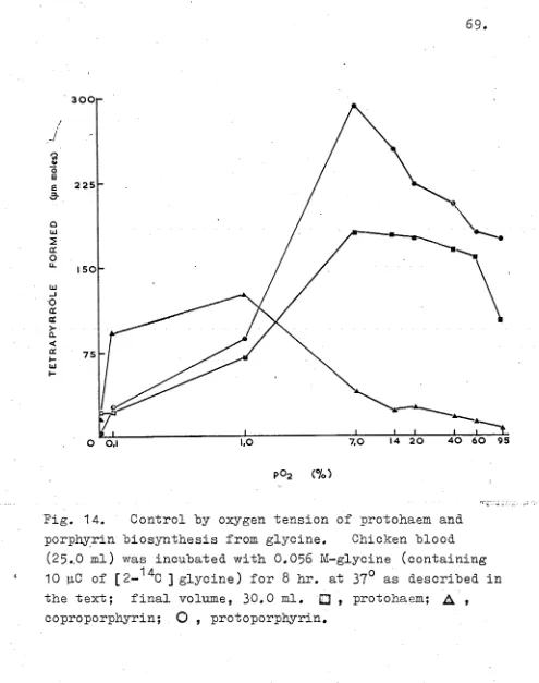
Introduction
Page.
Results 68
Discussion 85
Summary 88
CHAPTER III. A STUDY OP COPROPORPHYRINOGEN
OXIDATIVE DECARBOXYLASE PROM BEEP LIVER
Introduction 90
Experimental 91
Results 100
Discussion 137
Summary 142
CHAPTER IV. STUDIES ON PERROCHELATASE
AND ITS POSSIBLE SIGNIPICANCE IN THE BIOSYNTHESIS OP HAEMO- PROTEIN PROSTHETIC GROUPS
Introduction 144
Exp e r ime n t al 1 47
Results 160
Discussion 184
viii
Abbreviations
P o r p h y r i n isomers. After first mention in the text,
the Roman numerals III and IX are omitted f r o m the names
of the series III porphyrins a n d haems un l e s s reopiired
for clarity.
P o r p h y r i n side-chain abbreviations: E, EOH,
- C H ( 0 H ) C H 3 ; F, -CHO; H, -H; M, - C H 3 ; MOH, - C H 20H;
P, - C H 2 ,CH2 .C00H; V, - C H : C H 2 ; X, -CH(OH).CHgR»
(R l = alkyl side-chain); Y, - C H : C H R ” (Rn = alkyl
s i d e - c h a i n ) ; Z, - C H ( C H 3 ).S.GH2 ,CH(NH2 ) C 0 0 H .
A L A
A.R.
AT P
B V L P
CoA
Coprogen-ase
EDTA
ESF
N A P
PBG-p 0 2
6 -aminolaevulic acid
analytical reagent
adenosine triphosphate
mono vinyl-(j?-)hy dr oxyethyldeut er
o-porphyrin
coenzyme A
coproporphyrinogen oxidative
decarboxylase
ethylene diamine tetraacetic
acid
erythropoietic s timulating factor
nicotine adenine dinucleotide
por p hob i 1 ino ge n
Protogen-oxidase
Sedormid
TEAE-cellulose
Urogen-ase
protoporphyrinogen oxidase
2-(isopropyl-4-pentenoyl) urea
triethylaminoethyl-cellulose
uroporphyrinogen oxidative decarboxylase
THE BIOSYNTHESIS OP PORPHYRINS AND HAEMS
C H A P T E R I GENERAL INTRODUCTION 1• Porphyrins and tetrapyrrole pigments«
The occurrence of haems and chlorophylls in nature has created considerable interest over a long period.
These pigments and their porphyrin precursors have long been the subject of chemical investigation. Perhaps the most significant of the early studies occurred between 1867 and 1871, when Thudichum (1867) and Hoppe-Seyler
(1871) prepared metal free porphyrins from haem compounds.
Since that time, however, a vast knowledge of the chemistry of many tetrapyrroles has accumulated, due mainly to the studies of Willstätter and Hans Pischer. Between the years 1912 and 1944, Willstätter studied chlorophyll and its porphyrin derivatives, while Pischer examined the haems and many related porphyrins. Much of the work of this period on the chemistry of pyrroles, porphyrins and metallo-porphyrins has been recorded by Pischer & Orth
(1937).
The correct tetrapyrrole ring structure for the porphyrins was first proposed by Küster (1912),
2
.
>jc
1929, when the complete synthesis of protoporphyrin IX
(Fig.
1
t (4)) and protohaem IX (Fig. 1, (5
)) by Fischer& Zeile (I929)t firmly established the cyclic structure
of the porphyrin nucleus. Fischer's investigations,
including the novel proof of the cyclic nature of the porphyrin structure by the method of "progressive pairing of quadrants", has been reviewed by Corwin (1943)*
The recognition of abnormalities in porphyrin
metabolism in various pathological conditions stimulated much of the early interest in porphyrin and haem metabolism,
H
but the impact of Fischer*s and Willstatter*s studies aroused further interest in the biological aspects of
bile pigments, porphyrins and metallo-porphyrins. Methods
were developed to study these compounds in biological
materials and these biochemical investigations resulted in an increased awareness of the multitude of biological
processes with which the tetrapyrrole pigments are intimately
associated. The results of these earlier biochemical
investigations and the techniques which arose from them have been reviewed by Lemberg & Legge (1949).
The biological importance of the chlorophyll pigments of both plant and bacterial origin in the process of photo synthesis is well known, though the way in which the
3
.
chlorophylls participate is still not understood in detail* The haemoproteins, which include cytochromes, catalases, peroxidases, haemoglobins and myoglobins are known to be intimately concerned with photosynthesis, cellularrespiration, hydrogen peroxide metabolism, oxygen and carbon dioxide transport, oxygen storage and possibly symbiotic nitrogen fixation*
Haemoproteins occur in nearly all living cells but they are absent from some anaerobic micro-organisms (Keilin, 1925; Fujita & Kodama, 1934; Frei, Heedmuller & Almasy,
1934)* The anaerobic mode of life, however, is not
characterized by the absence of haemoproteins as was previously suggested by Keilin (1925), since it is now known that many anaerobes possess cytochromes (cf. Kamen,
I960).
It is interesting that the only well-established function of the porphyrins and their reduced hexahydro- derivatives, the porphyrinogens, in the normal cellular metabolism of animals, plants and micro-organisms is that
of precursor in the biosynthesis of the biologically active chlorophyll- and haemo-proteins (Fig* 9)*
2* The biosynthesis of porphyrins and haems*
.
techniques to trace a biosynthetic pathway* This
biosynthetic sequence has been studied extensively in
animal tissues, especially avian erythrocytes, and studies have been extended to porphyrin and chlorophyll biosynthesis in green plants and micro-organisms*
The course of the investigations into haem biosynthesis
has followed a well established biochemical pattern* The
first experiments were carried out in vivo* Later
experiments were conducted in vitro upon intact cells and
later upon cell-free preparations* Amongst the more recent
studies are reports of the isolation and part purification of individual enzymes concerned in haem synthesis*
The biosynthesis of porphyrins, haems and chlorophylls has been the subject of many reviews in recent years
(Shemin, 1955, 1956 a, b, c; Rimington, 1954, 1955, 1957,
1958; Bogorad, I960; Margoliash, 1961; Granick &
Mauzerall, 1961)*
Prior to 1945, nothing was known about the biosynthesis
of protoporphyrin or protohaem* Fischer’s theory that
5
The first insight into the long chain of biosynthetic reactions which results in the formation of protohaem
followed the isotopic labelling experiments of Shemin &
Rittenberg (1945, 1946 a, b). These workers showed
1 5
that [ ^N) glycine was incorporated into the circulating haem of man and animals and contributed equally to the labelling of all four nitrogen atoms of the tetrapyrrole
nucleus. Bloch & Rittenberg (1945) found that the
2
administration of H labelled acetate led to the incorp
oration of deuterium into circulating haem. In this way,
glycine and acetate were found to be precursors of haem. As early as 1946 and following the discovery that glycine and acetate were precursors of protohaem, Lemberg
(1946, 1955 a, b) and Lemberg & Legge (1949) suggested that uroporphyrin, coproporphyrin and protoporphyrin were precursors of protohaem rather than degradation products
as suggested earlier by Fischer. This theory was based
upon the assumption that the decarboxylation and oxidative decarboxylation reactions involved in the conversion of uroporphyrin to protoporphyrin, by way of coproporphyrin,
were more likely than the reverse carboxylations. Lemberg,
of the citric acid cycle in haem synthesis. Lemberg reasoned that if uroporphyrin was the first tetrapyrrole unit in the synthesis of haem, then the monopyrrole
precursor (Fig. 1, (1)) would likewise bear acetic- and
propionic- acid side-chains. It was proposed that such
a precursor might be formed by the condensation of two molecules of «(-oxoglutarate, formed from acetate by citric
acid cycle activity, with two molecules of glycine. A
reaction sequence similar to that proposed by Lemberg is shown in Figure 1•
Many of the individual reactions postulated have subsequently been proved incorrect but in broad outline the sequence predicted by Lemberg in 1946 was found to be correct.
The discovery that haem could be formed enzymically in vitro greatly assisted the investigation of the sequence
of reactions involved in haem biosynthesis. Shemin,
London & Rittenberg (1948) first demonstrated haem
synthesis in vitro from glycine by a suspension of duck
erythrocytes. Haem synthesis by cell free systems was
later demonstrated using bone marrow homogenates (Altman, Salomon & Noonan, 1949), rabbit spleen homogenates
haemolysed duck erythrocytes (Shemin & Kumin, 1952 a; London & Yamasaki, 1952).
Another study which greatly influenced investigations of haem biosynthesis was made by Wittenberg & Shemin (1950)
and Shemin & Wittenberg (1951)* These workers devised a
stepwise chemical degradation of protohaem and proto porphyrin whereby each carbon atom of the tetrapyrrole
nucleus could be unequivocally isolated. With this technique
it was possible, using labelled substrates, to determine
the activity and origin of any particular carbon atom in the porphyrin nucleus.
3. Glycine in haem biosynthesis.
Following the discovery that acetate and glycine were precursors of circulating haem in both man and animals,
Radin, Rittenberg & Shemin (1950 a) studied haem synthesis in vitro from [«(-^C ] glycine and [carboxy-^C] glycine in
duck blood. They found that the «(-carbon atom, but not the
carboxy-carbon atom, was incorporated into the haem formed.
15
They studied also the simultaneous incorporation of N and
1A.
labelled «(-carbon atoms from glycine into haem. They
showed that eight carbon atoms and four nitrogen atoms of
the haem molecule were derived from glycine. This
9
each nitrogen atom used* showed that for every two molecules of glycine involved one nitrogen atom was lost during the
synthesise This nitrogen atom is lost as ammonia during
the condensation of four molecules of porphobilinogen (PBG) (Fig<>
i, (i))
to form a tetrapyrrole (see Fig*i)<>
V/ittenberg & Shemin (1950), using their haem
degradation procedure, showed that the eight carbon atoms
•4 A
of the haem nucleus derived from[oc— ^C] glycine were
incorporated into the positions shown in Fig* 2 0 The
origin of the other twenty six carbon atoms of the haem molecule was still unknown»
Fig» 2. The incorporation of the <=<-carbon atom of
.
The i n c o r p o r a t i o n o f g ly c in e i n t o haem h as beenco n firm ed i n whole a n im a ls (M uir & N eu b e rg e r, 1 9 5 0 ), i n
c h ic k e n b lo o d (D re s e l & F a lk , 1956 a ) , i n washed c h ic k e n
e r y th r o c y te s (D re s e l & F a lk , 1956 a ) , i n c h ic k e n r e d - c e l l
h a e m o ly sa te s (D re s e l & F a lk , 1954, 1956 c ) , and i n legume
r o o t n o d u le hom ogenates (Richmond & Salom on, 1955)*
4. A c e ta te i n haem b i o s y n t h e s i s .
R a d in , R i tte n b e r g & Shemin (1950 b ) found t h a t th e
l a b e l l e d ca rb o n atom s of [ « ( - ^ C ) a c e t a t e and [ c a rb o x y -^ C ]
a c e t a t e were in c o r p o r a te d i n t o haem when in c u b a te d w ith
duck b lo o d . The in c o r p o r a t i o n of th e «(-carbon atom i n t o
haem was much more e f f i c i e n t , how ever, th a n th e in c o r p o r a t i o n
of th e c a rb o x y -c a rb o n atom .
Shemin & W itte n b e rg (1951) in c u b a te d duck blood
s e p a r a t e l y w i t h [ « - ^ C ] a c e t a t e and [ c a r b o x y -^ C ] a c e t a t e
and th e n d eg rad ed th e l a b e l l e d haem fo rm ed . They i s o l a t e d
each c a rb o n atom o f th e t e t r a p y r r o l e n u c le u s and m easured
i t s r a d i o a c t i v i t y . I n t h i s way th e y found t h a t th e o th e r
tw e n ty - s ix carb o n atom s of th e haem m o le c u le , n o t a r i s i n g
from g l y c i n e , were d e r iv e d from th e «(- and c a rb o x y -c a rb o n
atom s of a c e t a t e .
5* The c i t r i c a c id c y c le and haem b i o s y n t h e s i s .
showed that the labelling patterns in all four pyrrole
units of haem derived from each form of labelled acetate were similar (Fig* 3)* These results were the first
experimental indication of a common monopyrrolic precursor for all four pyrrole rings of haem and thus supported
Lemberg’s view that the first tetrapyrrole formed during the biogenesis of protohaem might be uroporphyrin, since uroporphyrin contains only one type of monopyrrole ring*
• 1 1.
Frca jc ar b oxy-'1 acetate From
acetate
Fig* 3* Labelling patterns in the pyrrole units of haem synthesised from [carboxyl*C] and [c<-1 ^C] acetate* The figures indicate the relative specific activity of the labelled carbon atoms*
.
asymmetrical four carbon compound (Fig# 6)# The
asymmetrical nature of the intermediate formed from
acetate is indicated by the unequal incorporation of ^ C from [carboxy-^C] acetate between the carboxyl group of the propionic acid sidechain and the carbon atom in the
position *y to the carboxyl carbon (Fig, 3)* Shemin &
Wittenberg (1951) suggested that this four carbon compound was a derivative of succinic acid, perhaps succinyl-
coenzyme A (succinyl-CoA),
Shemin & Kumin (1952 a, b) incubated both intact and haemolysed erythrocytes with [1:4~^C] succinate and showed that succinate was incorporated into the pyrrole moieties
as a unit (Fig, 4), This incorporation was stimulated by
the addition of coenzyme A (CoA) (Shemin & Kumin, 1952 b), again suggesting that the asymmetrical four carbon compound
was succinyl-CoA. That succinyl-CoA is definitely involved
in haem biosynthesis has been demonstrated (Chapter 1, section 7)•
The labelling patterns obtained when [1:4~^C] succinate and [ 2 :3~^C] succinate were incubated in the
13o
° G O O H ° C O O H
° C O O H
C H 0 ° C O O H
1
21
C K 0
2 1 2
d C H 0
C l d
^ C O O H
0 H o
>
p 1
d' d CHp
G C O -R
+ 2 G l y c i n e
--- --->
c h2 c h2
y-— . .v
H
Pig* 4« Incorporation of the carboxyl carbon atoms of
14
[1:4- C ]succinate into the pyrrole moieties of protohaen©
Assuming that succinyl-CoA does condense with glycine during the biosynthesis of haems, consideration of Figure 5 shows that the incorporation of the carboxyl- carbon atoms can only proceed by reversal of reaction A and therefore will be unaffected by the inhibition of
reaction B by malonate® The methylene carbon atoms of
succinate, however, can be incorporated both by reversal of reaction A and by conversion to succinyl—CoA by reactions
B, C 9 D, E,
P,
G- and H* Thus, the incorporation ofmethylene carbon atoms of succinate will be partly inhibited
by malonate* Experiments carried out in the presence and
absence of malonate confirmed this prediction* This result
EO-C-COOH
h2c-c o o h
HC-COOH ti o HC-COOH
3 H oC-C00H
21 o H oC-C00H
CH^CO-CoA
0=C-C00H I Q
H2C-C0GH
K 0C-CCCK
2 I
CH0
I 2
0 =C—CoA
H2G-C00H EO-C-°GOOH
h2c-°g o o h
i
H oC-C00H 2 I o HC-COOH 0=C-%00H
si
H^C-COOH 2 l CH2 0»C-C00H e CO. PORPHYRINS V HAEMPig. 5. The participation of the citric acid cycle in porphyrin and haem biosynthesis. The fate of the carboxyl carbon atoms of succinate. Only the significant steps of the citric acid cycle are shown.
1 5*
i n th e b i o s y n t h e s i s of haem i s an in te r m e d ia te i n th e
o x id a tiv e d e c a r b o x y la tio n of < * -o x o g lu ta ra te to s u c c in a te *
More c o n c lu s iv e ev id e n c e f o r th e p a r t i c i p a t i o n o f th e
c i t r i c a c id c y c le i n haem s y n th e s is was o b ta in e d by W ris to n ,
Lack & Shemin (1955)» who showed th e c o n v e rs io n o f [ 1 :5 - 3
c i t r a t e , [ 5 - ] « - o x o g l u t a r a t e and [ 1 :2 - *C] « - o x o g l u t a r a t e
t o haem by h a e m o ly sa te s of duck e r y th r o c y te s * The l a b e l
d i s t r i b u t i o n p a t t e r n found i n th e haem m o le cu le a f t e r
in c u b a ti o n w ith each s u b s t r a t e was i d e n t i c a l t o t h a t p r e d ic te d
on t h e a ssu m p tio n t h a t succinyl-C oA from th e c i t r i c a c id c y c le
was in v o lv e d i n th e s y n t h e s i s a s shown i n F ig u re 6*
T h is work by Shemin and h i s s c h o o l on th e r o l e o f th e
c i t r i c a c id c y c le i n haem s y n t h e s i s and l a t e r work on th e
s y n t h e s i s of 6 -a m in o la e v u lic a c id (ALA) (Fig* 6 , ( 7 )) and
i t s p a r t i n haem s y n t h e s i s h a s b een e x t e n s iv e ly rev iew ed
by Shemin (1955, 1956 a , b , c)*
Brown (1958 a ) , s tu d y in g ALA s y n th e s is (Fig* 6) i n
c h ic k e n r e d - c e l l s , d e m o n stra te d t h a t ALA fo rm a tio n from
g ly c in e and c e r t a i n c i t r i c a c id c y c le in te r m e d ia te s was
i n h i b i t e d by f l u o r o c i t r a t e and p a ra p y ru v a te * These l a t t e r
compounds a r e i n h i b i t o r s o f a c o n ita s e and « - o x o g l u t a r a t e
o x id a tiv e d e c a rb o x y la s e r e s p e c tiv e ly * T hese r e s u l t s
f u r t h e r co n firm ed th e p a r t i c i p a t i o n o f th e c i t r i c a c id
with «c-oxoglutarate, citrate, and isocitrate was enhanced
by the addition of «c-lipoic acid* Synthesis from succinate,
although unaffected by «fr-lipoic acid was stimulated by
adenosine triphosphate (ATP)* This result again confirms
that the unsymmetrical four-carbon intermediate in ALA synthesis is succinyl-CoA formed either by the oxidative decarboxylation of <<-oxoglutarate (Gunsalus, 1954) or from succinate by succinyl-CoA synthetase*
There have been reports of porphyrin synthesis
(Lascelles, 1956a, 1959? Cooper, 1956) and bacteriochloro phyll synthesis (Lascelles, 1959, I960) by various
Rhodopseudomonads from glycine and <<-oxoglutarate or
succinate, suggesting that tetrapyrrole synthesis in micro organisms also might be dependant upon citric acid cycle
activity* Lascelles (1957) has also demonstrated porphyrin
synthesis by the protozoon Tetrahymena vorax (T. vorax) from glycine with acetate, «c-oxoglutarate, succinate or fumarate*
6* &-aminolaevulic acid in haem biosynthesis*
It has been proposed that succinyl-CoA and glycine condense to give <ac-amino-ß-oxoadipic acid (Pig* 6, (6)) and that this compound decarboxylates either enzymically or spontaneously to give ALA (Pig* 6)*
A M , 6-succinamidolaevulic acid (Pig« 7> ( 10)) and
suecinamidoacetic acid (Pig« 7 9 (9 ))« Lascelles (1953)
incubated intact cell suspensions of Rhodopseudomonas spheroldes (Rps« spheroides) with compounds ( 9 ) and (10) but neither compound supported porphyrin synthesis«
•
17.
C O O H i C H 0
1 £
C O O H C H 2
C O O K Ah
'-z il 1 2 C H 0
C O O H 1
C O O H 1
C H 2 C O O H c n 2 ... C H 0 • ? H 2 - 2 H-jO v 1 1 C O - C o A
J> 1 <-
----C O C O
k
h
\ 2
c h~
/ 2 +
C H ^ H g C O O H
C H . N H 2 C O O H
(6) C o A
c h2
VsN H 2
(7) C 0 2
C O
X C H 2 N H 2
o
H
' ^ O E 2 K H 2
(8 )
Pig« 6 « She proposed condensation of glycine with
succinyl-CoA to form «<«-amino-ß— oxoadipic acid (6) and
ALA (7)o The conversion of ALA to PBG- (8 ) is also shown*
C O O H
G C O H 8
C O O H
\
C H 0 j 2 C H 0
! d C H 03 2
C H 0 1 2 C E 0
i 2 C H 0i -
C O
S N H ^ CO
i C H 0
C O C H ^ C O O H
*
(9) ( 1 0 )
Shemin & Russell (1953) and independently and almost simultaneously Heuberger & Scott (1953) and Dresel & Falk (1953) demonstrated the biosynthesis of haem, porphyrins
and PBG from ALA. It was found that ALA could replace both
glycine and succinate (Shemin & Russell, 1953)* When
unlabelled ALA was incubated with avian erythrocytes
14
together with C labelled glycine or succinate, the activity
of the haemin recovered was significantly reduced (Shemin
& Russell, 1953; Dresel & Falk, 1956 c)* This result
indicated that ALA was a normal intermediate in haem biosynthesis*
Similar conversion of T ^ N ] ALA and [1:4-^C] ALA to protoporphyrin and haem has been observed by Berlin, Neuberger & Scott (1956 a, b).
Rigorous evidence for the participation of ALA in haem biosynthesis has been provided by Shemin, Abramsky & Russell (1954) and Shemin, Russell & Abramsky (1955),
14
who have shown identical C labelling patterns in haem
synthesized by haemolysates incubated with [ 5 - ^ C ] ALA or
[2-^ C ]glycine. Likewise, identical patterns have been
obtained using either [1:4-1*C] A L A or [1s4-^C] succinate
as substrates. It has been found that the incorporation
of [ 1:4-^C] ALA into haem is far more efficient than the
1 9
.
a r e i n a c c o r d w i t h t h e schem e shown i n F i g u r e 6 .I t h a s b e e n shown t h a t p o r p h y r i n s c a n h e s y n t h e s i z e d fr o m ALA by t h e m i c r o - o r g a n i s m s T. v o r a x ( L a s c e l l e s , 1 9 5 7 ) a n d R p s . s p h e r o i d e s ( L a s c e l l e s , 1956a, 1 9 5 9 ) .
S h e m in , C o r c o r a n , R o sen b lu m & M i l l e r ( 1 9 5 6 ) a n d C o r c o r a n & Shem in ( 1 9 5 7 ) h a v e d e m o n s t r a t e d t h a t t h e
p o r p h y r i n - l i k e m o i e t y o f v i t a m i n i s d e r i v e d fro m ALA. 7 o The s y n t h e s i s o f & - a m i n o l a e v u l i c a c i d .
T h a t ALA was i n v o l v e d i n haem s y n t h e s i s was no l o n g e r
i n d o u b t b u t l i t t l e was known a b o u t i t s s y n t h e s i s . A l l a v a i l a b l e e v i d e n c e s u g g e s t e d t h a t i t was fo rm e d by t h e c o n d e n s a t i o n o f a s u c c i n y l d e r i v a t i v e w i t h g l y c i n e . The enzyme r e s p o n s i b l e , 6 - a m i n o l a e v u l i c a c i d s y n t h e t a s e
( A L A - s y n t h e t a s e ) i s a p a r t i c u l a t e enzyme a n d h a s b e e n s t u d i e d i n s u b c e l l u l a r p a r t i c l e s ( L a v e r , N e u b e r g e r & U n d e n f r i e n d , 1 9 5 8 ) , i n d i s r u p t e d s u b - c e l l u l a r p a r t i c l e s
( G i b s o n , L a v e r & N e u b e r g e r , 1 958 a , b ) a n d h a s a l s o b e e n s o l u b i l i z e d ( G i b s o n , 1 9 5 8 ; K i k u c h i , Shem in & Bachmann, 1 9 5 8 ; S h e m in , K i k u c h i & B achm ann, 1 9 5 8 ; K i k u c h i , Kumar, Talmage & S h em in , 1 9 5 8 ; K i k u c h i , Kumar & S h e m in , 1 9 5 9 ) .
The slo w r a t e o f s y n t h e s i s o f ALA an d i t s r a p i d u t i l i z a t i o n by n o r m a l h a e m a t o p o i e t i c t i s s u e s made t h e
.
however, from glycine and «x-oxoglutarate or succinate in
the erythrocytes of chickens, made anaemic by the adminis
tration of phenylhydrazine, was demonstrated by Laver
et al. (1958). These workers, moreover, isolated a
particulate fraction from these erythrocytes which could
synthesize ALA but lacked the enzymes concerned with its
further metabolism. The formation of ALA from glycine and
o(-oxoglutarate or succinate by these particles was found to
be an aerobic process and was stimulated by the addition of
CoA, pyridoxal phosphate and M g +* and ions. No
other dialysable cofactors appeared to be needed. Similar
cofactor requirements for ALA synthesis have been reported
in extracts of Rhodospirillum rubrum (R s p . rubrum) and
ftps* spheroides (Kikuchi, Kumar et_ al. 1958) and in
chicken red-cell preparations (Granick, 1958). Earlier
studies of the effect of vitamin B group deficiency on
porphyrin and haem synthesis in T. vorax (Lascelles, 1957)
and in duck red-cells (Schulman & Richert, 1955, 1956) had
also suggested that CoA and pyridoxal phosphate might be
involved in ALA synthesis.
When the particles from anaemic blood were incubated
with glycine, pyridoxal phosphate and synthetic
21
particles were impermeable to this succinyl derivative*
Gibson et al* (1958 a, b), however, demonstrated ALA
synthesis from glycine, pyridoxal phosphate, and succinyl- CoA after freeze-drying the particles, which presumably
+ + « * t
ruptured the encasing membranes* No Mg ions, PO^ ions
or oxygen were required under these conditions, suggesting
that they participate in the formation of ’’high energy"
intermediates in the formation of succinyl-CoA* That
succinyl-CoA is a substrate of ALA formation in Rps*
spheroides extracts has been confirmed by Shemin, Kikuchi
& Bachmann (1958) and Kikuchi, Kumar et_ al* (1958)*
Lascelles (1959» I960) has obtained results of a similar nature using the facultative photosynthetic micro
organism, Rps* spheroides* Introduction to anaerobic
and illuminated conditions causes Rps* spheroides to under go an adaptation to a photosynthetic mode of existence which is marked by a stimulation of porphyrin and bacteriochloro-
phyll synthesis* These compounds could be synthesized
from cc-oxoglutarate and glycine only when the cells were
grown anaerobically in the light* The synthesis of these
tetrapyrroles was diminished when the cells were grown aerobically and completely suppressed when grown in the
dark* When ALA was used as substrate, aerobic and dark
ATP is required for the activation of precursors
of ALA. Studies on cell-free extracts, using succinate,
glycine, pyridoxal phosphate and ATP as substrates, have shown that ALA-synthetase and ALA-dehydrase are synthesized during the adaptation to photosynthetic conditions
(Lascelles, 1959, 1960).
The specificity of the freeze-dried particles (Gibson jst al. 1958 b) was studied and amino-acetone was formed when succinyl-CoA was replaced by acetyl CoA. Other aminoketones were formed with propionyl CoA and glutaryl-CoA but the fastest reaction occurred with
succinyl-CoA. The freeze-dried particles formed very
little ALA from «<-oxoglutarate and glycine unless
nicotine-adenine-dinucleotide (NAD) was added. This
suggested that the synthesis of ALA from oc-oxoglutarate involves the formation of succinyl-CoA by the NAD-
dependant «c-oxoglutarate oxidase (cf. Gunsalus, 1954).
Gibson and his co-workers obtained evidence suggesting that pyridoxal phosphate was firmly bound to the
enzyme. In this regard, it is interesting to note that
Kikuchi, Kumar & Shemin (1959) proposed the prior formation of an enzyme-pyridoxal phosphate complex before reacting
2 3
.
complex th e n co n d en ses w ith su c cin y l-C o A .
G ibson e t a l . , (1958 b) have su g g e ste d t h a t th e
r e a c t i v e form o f g ly c in e i s a S c h i f f * s b a s e form ed by
th e r e a c t i o n of th e amino a c id w ith p y r id o x a l p h o s p h a te ,
th e «(-carb o n atom o f g ly c in e th u s b e in g a c t i v a t e d .
C o n d e n sa tio n w ith succinyl-C oA th e n r e s u l t s i n th e f o rm a tio n
o f o c-am in o -p -o x o ad ip ic a c id w hich i s r e a d i l y d e c a rb o x y la te d
a t pH7 (N e u b e rg e r, 1 9 6 1 )9 fo rm in g ALA.
To e x p l a in th e p o s s i b le r o l e of b i o t i n i n ALA
s y n t h e s i s ( L a s c e l l e s , 1956a, I 9 6 0 ) , N eu b erg er (1961) h a s
s u g g e s te d th e f o rm a tio n of an « - a m in o - p -o x o a d ip a te -
p y r id o x a l phosphate-enzym e com plex i n w hich th e «(-am ino-p-
o x o a d ip a te may be s t a b l e . N euberger h a s su g g e ste d t h a t
enzyme-bound b i o t i n may be r e q u ir e d to r e l e a s e c a rb o n
d io x id e and ALA and r e g e n e r a te A L A -synthetase and p y r id o x a l
p h o s p h a te . I t i s known t h a t b i o t i n i s n o t removed from
i t s apoenzyme by d i a l y s i s and t h a t b i o t i n enzymes a r e
a s s o c i a t e d w ith th e m etab o lism of ca rb o n d io x id e (Lynen,
1 9 5 9 ).
S tim u la tio n of ALA fo r m a tio n by Fe++ io n s u n d er a
v a r i e t y of c o n d i tio n s (Brown, 1958 a , b , c) h a s s u g g e ste d
t h a t F e++ io n s a r e r e q u ir e d f o r A L A -synthetase a c t i v i t y .
.
the activation or condensation of glycine, and that the metal ions may stimulate by the stabilization of a glycine- pyridoxal phosphate Schiff*s base compound (cf Metzler,
Ikawa, & Snell, 1954; Patwardhan, 1958)* Lascelles
(1956a), however, has shown that porphyrin synthesis in Rps. spheroides from glycine and «-oxoglutarate, but not from ALA, is inhibited by the presence of iron salts.
8. The common monopyrrole precursor of tetrapyrrole
pigment s♦
The identification of the monopyrrolic precursor of porphyrins and haems was a long and difficult task.
Sachs (1931) and Waldenstrom (1935) found that the urine of patients suffering from acute porphyria contained a compound which gave a red colour with
p-dimethylamino-it
benzaldehyde (Ehrlich*s reagent). Later, Waldenstrom
& Vahlquist (1939) obtained a concentrate of this substance, which they called porphobilinogen (PBG), and their investi
gations led them to believe that the compound was a dipyrrylmethane.
Westall (1952), however, obtained pure PBG and
Cookson & Rimington (1953, 1954) elucidated its structure (Pig. 6 (8)).
25a
s y n t h e s i s of haem and p o rp h y rin s h as b een d e m o n stra te d
i n c ru d e o r p a r t l y p u r i f i e d e x t r a c t s from a v ia n e r y th r o
c y te s ( F a lk , D re s e l & R im in g to n , 1953; D re s e l & F a lk ,
1953, 1956 b ; B o o ij & R im in g to n , 1957; Lockwood &
R im in g to n , 1957; Lockwood & B enson, I9 6 0 ; Sano, 1 9 5 8 ),
from mammalian r e d - c e l l s (R im ington & B o o ij, 1957; Sano,
1 9 5 8 ), from r a t - l i v e r (S c h w a rtz , 1954; S chw artz &
W atson, 1 9 5 7 ), from C h l o r e l l a (Bogorad & G ra n ic k , 1953 a ) ,
from s p in a c h le a v e s (B ogorad 1955 a , b , c; 1958 a ) , from
wheat germ (Bogorad 1955 c , 1958 b ) , from e x t r a c t s o f many
le a v e s (B ogorad, 1958 a ) and from R ps* s p h e ro id e s (H oare
& H e a th , 1958 a , b , 1959; H eath & H o are, 1959 a , b)*
U sing i s o t o p i c d i l u t i o n te c h n iq u e s , D re s e l & F a lk
(1956 c) showed t h a t PBG i s a norm al in te r m e d ia te i n th e
b i o s y n t h e s i s of haem by c h ic k e n r e d - c e l l h ae m o ly sa tes*
D re s e l & F a lk (1956 a ) a l s o found t h a t PBG, when in c u b a te d
w ith c h ic k e n b lo o d o r washed c h ic k e n r e d - c e l l s , was n o t
c o n v e rte d to p o rp h y rin s o r haem and su g g e s te d t h a t th e
r e d - c e l l s were im perm eable to PBG*
9* The c o n v e rs io n o f 6 -a m in o la e v u lic a c id t o p o r p h o b ilin
ogen*
The c o n v e rs io n o f ALA t o PBG by ALA-dehydrase h a s
& Falk, 1953, 1956 b), in intact cells of Rps. spheroides (Lascelles, 1956a) and in intact animals (Berlin, Neuberger
& Scott, 1956 a, b). There have been several reports of
the part purification of ALA-dehydrase from ox-liver
(Gibson, Neuberger & Scott, 1955; Iodice, Richert &
Schulman, 1958), from avian erythrocytes (Granick, 1954;
Schmid & Shemin, 1955; Granick & Mauzerall, 1958 a),
from pigeon and rat liver (Schulman, 1955) from rabbit- reticulocytes (Granick & Mauzerall, 1958 a), from
Rps. spheroides (Lascelles, 1959, I960) and from
Corynebacterium diphtheriae (Gibson al., 1955).
In an extensive study of the distribution of ALA- dehydrase in the subcellular particles of rat-liver
homogenates, Gibson et al, (1955) showed that the enzyme was present almost exclusively in the soluble fraction of
the cells. Using rabbit tissues, the enzyme was shown to
be present not only in liver cells but also in kidney, bone-marrow, blood and spleen*
The activity of ALA-dehydrase has been found to
fluctuate during various metabolic derangements. Gibson
et al. (1955) found that ALA-dehydrase activity in rabbit- spleen and blood was increased several fold during anaemia induced by phenylhydrazine and also in the liver during
Sedormid-induced porphyria. Lascelles (1959,1960).reported an
27. increase in ALA-dehydrase activity in Rps. spheroides
during adaptation to a photosynthetic existence when bacteriochlorophyll synthesis is greatly increased*
Gibson £t al. (1955) isolated ALA-dehydrase from
acetone dried powders of ox-liver and purified it 270-fold. This electrophoretically pure preparation, but not the
crude extracts, required glutathione or cysteine for
maximum activity. Activity of the crude extracts,
however, was diminished by thiol inhibitors* The enzyme
was found to be inhibited by ethylenediamine-tetracetic
acid (ELTA) and by tris buffer* This latter inhibition
was removed by addition of phosphate buffer* No require
ment for a metal could, however, be demonstrated directly.
Iodice £t al. (1958), however, reported the presence of
copper and magnesium in an electrophoretically pure preparation of ALA-dehydrase and also claimed that the activity of the enzyme isolated from copper-deficient
animals is low. Wilson, Iodice, Schulman & Richert
(1959)» however, have more recently shown that the removal of copper from the enzyme does not affect its activity* The inhibitions by EDTA and tris thus remain unexplained.
COOH
in
CCGH I CH0
I 2
—CH0 i 2
2 -P-0
< b' \ i“2
c e2 C-0— a
- K-,0
ch2n h2
COCK I CH-,
l 2
CH0
l 2
c = o I CH-, X .✓ COOK I CH0
I 2 CH-,
i 2
C \
- H20
c k2n h2
(11)
COOH
COOH
Fig« 8o A p o s s i b l e m e c h a n i s m f o r P E G f o r m a t i o n i n v o l v i n g a h y p o t h e t i c a l k e t i m i n e intermediate®
O n l y one e l e c t r o p h o r e t i c a l l y pure enzyme was requ i r e d f o r the r e a c t i o n a n d G r a n i c k & M a u z e r a l l (1958 a), who have
shown that the kine t i c s of P B G f o r m a t i o n i n d i c a t e the f o r m a t i o n o f a s imple 1s1 enzyme-substrate complex* h a v e p o s t u l a t e d the f o r m a t i o n of a keti m i n e interme d i a t e
(Pig. S 9 (11))*
10* T e t r a p y r r o l e i n t e r m e d i a t e s a n d the p o r phyrinogens in h a e m b i o s y n t h e s i s *
The s e q u e n c e of the porphyrins in the b i o s y n t h e s i s of haem* i l l u s t r a t e d in F i g u r e 1 9 ha s b e e n confirmed by various workers® A study of the kine t i c s of p o r p h y r i n f o r m a t i o n f r o m P 3 G in h a e m o l y s a t e s of c h i c k e n erythrocytes (Falk* D r esel & Ei m i n g t o n , 1953; Falk* 1955; Dre s e l & Falk, 1956 b)* i n d i c a t e d that u r o ~ and coprc- porphyrins w e r e
2 9
.
o f haem.
The seq u en ce o f p o rp h y rin s was f u r t h e r c o n firm ed by
i s o l a t i n g , by v a r io u s m e th o d s, th e enzymes c o n v e rtin g PEG
t o p r o to p o r p h y r in .
Bogorad & G ra n ick (1953 a ) d e m o n stra te d th e accum ula
t i o n o f u ro p o rp h y rin s I and I I I and th e i n h i b i t i o n o f th e
enzymes r e s p o n s i b le f o r copro— and p r o to — p o rp h y rin
f o rm a tio n by g e n t l e h e a t tr e a tm e n t o f C h l o r e l l a e x t r a c t s .
U sing c h ic k e n re d —c e l l h a e m o ly s a te s , i t was p o s s i b le t o
d e m o n stra te u r o - and c o p ro - p o r p h y rin fo rm a tio n from PBG
u n d e r a n a e r o b ic c o n d i tio n s b u t p r o to p o r p h y r in c o u ld o n ly
be form ed by s u b s e q u e n tly c o n tin u in g th e in c u b a tio n i n a i r
(F a lk , D re s e l & R im in g to n , 1953? D re s e l & F a lk , 1956 b ) .
A s tu d y o f th e s u b c e l l u l a r f r a c t i o n s o f c h ic k e n r e d - c e l l s
( D r e s e l, 1955? D re s e l & F a lk , 1956 b ) showed t h a t s o lu b le
enzymes, p r e s e n t i n t h e s u p e r n a t a n t , w ere r e s p o n s i b l e f o r
th e c o n v e rs io n o f PBG to u r o — and c o p r o - p o rp h y r in s .
P r o to p o rp h y rin fo rm a tio n c o u ld be d e m o n stra te d o n ly on
a d d i t i o n o f e r y th r o c y te p a r t i c l e s o r r a t - l i v e r m ito c h o n d ria
t o th e s u p e r n a ta n t.
A d d itio n a l c o n f ir m a tio n o f th e se q u en ce o f p o rp h y rin s
i n haem b io s y n t h e s is h as been o b ta in e d by H oare & H eath
(1958 a , b ) and H eath & Hoare (1959 a , b ) who have
-p o r -p h y r i n s by R -p s . s -p h e r o i d e s u s i n g f r o z e n a n d t h a w e d c e l l s u s p e n s i o n s , a c e t o n e - d r i e d p o w d e r s o f c e l l s a n d c e l l - f r e e e x t r a c t s . S t u d i e s o f t h e k i n e t i c s o f p o r p h y r i n f o r m a t i o n by a c e t o n e - d r i e d c e l l s (Hoa re & H e a t h , 1958 a )
i n d i c a t e d t h a t u r o p o r p h y r i n was a p r e c u r s o r o f c o p r o p o r p h y r i n . As f u r t h e r c o n f i r m a t i o n , H o a r e & H e a t h (1958 a , b ) d e m o n s t r a t e d i n h i b i t i o n o f c o p r o p o r p h y r i n f o r m a t i o n a n d a c c u m u l a t i o n o f
u r o p o r p h y r i n I I I b y r e p e a t e d f r e e z i n g a n d t h a w i n g o f w h o l e c e l l s a n d by s t o r a g e o f c e l l - f r e e e x t r a c t s a t - 1 5 ° C .
P a r t l y p u r i f i e d enzymes h a v e now b e e n o b t a i n e d f r o m b o t h p l a n t a n d a n i m a l o r i g i n w h i c h w i l l c o n v e r t PEG t o u r o p o r p h y r i n s I a n d I I I ( B o g o r a d , 1955 a , b , c , 1958 a , b ;
G r a n i c k & M a u z e r a l l , 1 9 5 8 a ) *
I t h a s b e e n f o u n d , h o w e v e r , t h a t u r o - p o r p h y r i n i s n o t c o n v e r t e d e f f i c i e n t l y t o p r o t o p o r p h y r i n o r haem by h a e m o l y s a t e s o f a v i a n e r y t h r o c y t e s ( D r e s e l , 1 9 5 5 ) a nd
c o p r o p o r p h y r i n n o t a t a l l a n d so i t was s u g g e s t e d t h a t t h e t r u e t e t r a p y r r o l e i n t e r m e d i a t e s i n haem b i o s y n t h e s i s a r e n o t t h e p o r p h y r i n s b u t r e d u c e d p o r p h y r i n s ( W i t t e n b e r g & S h e m in , 1 9 5 0 ; D r e s e l , 1 9 5 5 ; R i m i n g t o n , 1 9 5 5 ; D r e s e l & F a l k , 1956 c ) . E v i d e n c e t o s u p p o r t t h i s t h e o r y was
o b t a i n e d by B o g o r a d ( 195 5 a , b ) a n d G r a n i c k (1955 a ) who d e t e c t e d compounds a b s o r b i n g a t 500 mp, d u r i n g t h e
31.
aut oxidation, the absorption at 500 mp, diminished and
these compounds were converted to uro- and copro porphyrins.
Neve, Labbe & Aldrich (1956) demonstrated the enzymic conversion of synthetic uroporphyrinogen III (Fig. 9, (12)),the reduced (hexa-hydro) form of uroporphyrin III, to protohaem by haemolysates of avian erythrocytes.
That the porphyrinogens were the true intermediates in the biosynthesis of haem was indicated by chemical
considerations. The simple condensation of four molecules
of PBG would lead, on theoretical grounds, to the formation of a tetrapyrrole with four methane bridges and four imino nitrogens.
That the porphyrinogens can be utilized as substrates and that the naturally occurring tetrapyrrole intermediates are reduced porphyrins has been confirmed by Heath & Hoare
(1959 a, b), Hoare & Heath (1958 a, 1959), Granick &
Mauzerall (1958 a, b) Mauzerall & Granick (1958), Bogorad (1958 a, b, c) Sano & Granick (1961) and Porra & Falk (1961). A modified scheme for the conversion of PBG to haem (cf Fig.
1) is shown in Figure 9.
The compounds absorbing at 500 mji (see above) have subsequently been shown to be intermediates in the
porphyrins (Bogorad* 1958 c; Mauzerall & Granick, 1358)
FR0T0HA2U (Pig. 1, (5))
33t
11« The enzymic c o n v e r s i o n o f p o r p h o b i l i n o g e n t o
u r o p o r p h y r i n o g e n s .
l i t t l e p r o g r e s s h a s b e e n made i n t h e u n d e r s t a n d i n g o f t h e e n z y m ic m e c h a n i s m by w h i c h f o u r m o l e c u l e s o f PBG a r e c o n d e n s e d t o f o r m t h e f i r s t a s y m m e t r i c a l t e t r a p y r r o l e i n t e r m e d i a t e o f t h e i s o m e r I I I s e r i e s « The e x p e r i m e n t a l i n v e s t i g a t i o n o f PBG- c o n v e r s i o n t o u r o p o r p h y r i n o g e n h a s , h o w e v e r , y i e l d e d some i n t e r e s t i n g r e s u l t s .
B o g o r a d (1955 a , b , c , 1958 a ) h a s p r e p a r e d an enzyme, p o r p h o b i l i n o g e n d e a m i n a s e ( PBG—d e a m i n a s e ) , f r o m an a c e t o n e - d r i e d p ow d e r o f s p i n a c h l e a v e s . T h i s enzyme
c o n v e r t e d PBG t o u r o p o r p h y r i n o g e n I« B o g o r a d (1955 c , 19 58 b ) h a s a l s o p r e p a r e d a n enzyme f r a c t i o n f r o m w h e a t germ e x t r a c t s w h i c h c o n t a i n e d an enzyme, u r o p o r p h y r i n o g e n i s o m e r a s e , w h i c h s y n t h e s i z e d u r o p o r p h y r i n o g e n I I I f r o m PBG i n t h e p r e s e n c e o f a d d e d PBG—d e a m i n a s e . U r o p o r p h y r i n
og e n i s o m e r a s e consu med v e r y l i t t l e PBG i n t h e a b s e n c e o f PB G -d ea m in a se a n d was u n a b l e t o c o n v e r t u r o p o r p h y r i n I
o r u r o p o r p h y r i n o g e n I t o t h e s e r i e s I I I i s o m e r «
B o g o r a d (1955 c , 19 58 b ) p r e p a r e d a n o t h e r w h e a t
germ f r a c t i o n w h i c h c o n v e r t e d PBG t o u r o p o r p h y r i n o g e n I I I .
uroporphyrinogen isomerase is a very labile enzyme. That this isomerase is labile has also been demonstrated in enzymic preparations from avian erythrocytes (Booij & Rimington, 1957; Lockwood & Rimington, 1957; Lockwood & Benson, I960), from mammalian red-cells (Rimington & Booij, 1957), from rat-liver homogenates (Schwartz &
Watson, 1957), from Chlorella (Bogorad & Granick, 1953 a), from spinach (Bogorad, 1958 a) and from Rps. spheroides
(Heath & Hoare, 1959 b). While these inactivations of
the isomerase occur at about 60°C the deaminase, which converts PBG to uroporphyrinogen I, is more stable but is inactivated by boiling (Hoare & Heath, 1958 a).
Lockwood & Benson (i960), using electrophoretic techniques, were unable to demonstrate the presence of two enzymes in their purified Hporphobilinogenasew
preparation from avian erythrocytes, which converts PBG
to uroporphyrinogen III. They suggested that a single
enzyme possessing both deaminase and isomerase activities might be involved and that the isomerase activity but not the deaminase is destroyed by mild heat treatment, cyanide or azide.
35
.
during the conversion (Dresel & Falk, 1956 b; Granick
& Mauzerall, 1958 a; Bogorad, 1958 a, b, c; Lockwood
& Benson, I960)* Bogorad (1955 b, 1958 a) has shown
that the enzymic condensation of four molecules of PBG is accompanied by the liberation of four molecules of NH^
from the aminomethyl group of porphobilinogen. The
liberation of in the non-enzymic condensation of PBG
was demonstrated by Westall (1952).
Several mechanisms advanced to account for formation of uroporphyrinogen III have involved the formation of free formaldehyde from the aminomethyl group of PBG, while others have involved the migration or incorporation of formaldehyde. Experimental investigation has shown that free formaldehyde is produced and incorporated during the non-enzymic synthesis of porphyrins from PBG (Shemin, Russell & Abramsky, 1955;
Lockwood & Benson, I960; Bogorad & Marks, I960). Free
formaldehyde, however, was not incorporated into porphyrin ogens during the enzymic conversion of PBG (Lockwood &
Rimington, I960; Bogorad & Marks, I960). Bogorad & Marks
(i960), using partly purified enzyme preparations, have shown that only minute amounts of free formaldehyde are formed and have suggested that this could be accounted for
by simultaneous low levels of non-enzymic conversion. It
.
c o n d e n s a tio n s may in v o lv e d i f f e r e n t m echanism s.
Hoare & H eath (i9 6 0 ) have i s o l a t e d u r o p o rp h y r in and
c o p ro p o rp h y rin o f th e u n - n a t u r a l iso m e r I I s e r i e s from
in c u b a ti o n s o f a d ip y rry lm e th a n e ( P ig . 10 (1 8 )) w ith cru d e
e x t r a c t s o f R ps. s p h e r o id e s . The o th e r d i p y r r y l m ethanes
u s e d ( P ig . 10, ( 1 5 ) ( 1 6 ) ( 1 7 ) ) w ere found t o be c o m p le te ly
i n a c t i v e a s s u b s t r a t e s f o r t e t r a p y r r o l e f o rm a tio n b u t were
found to be n o n -c o m p e titiv e i n h i b i t o r s o f PBG -deam inase.
Compound (1 8 ) was n o t c o n v e rte d t o p o r p h y rin by p u r i f i e d
PBG-deaminase p r e p a r a t i o n s from B ps, s p h e ro id e s (H oare &
H e a th , I9 6 0 ) and s p in a c h le a v e s (B ogorad, I9 6 0 ; C a rp e n te r
& S c o t t , 1 9 6 1 ).
An i n t e r e s t i n g compound known a s p s e u d o u ro p o rp h y rin
h a s been d e t e c t e d d u rin g th e enzymic c o n v e rs io n o f ALA and
PBG to p o rp h y rin s ( F a lk , 1955? F a lk , D r e s e l, Benson &
K n ig h t, 1956; F a lk & D r e s e l, I 9 6 0 ) . T h is compound
re se m b le s u r o p o rp h y r in i n i t s s p e c tr o s c o p ic and s o l u b i l i t y
p r o p e r t i e s b u t d i f f e r s s l i g h t l y i n i t s c h ro m a to g ra p h ic
b e h a v io u r. P se u d o u ro p o rp h y rin on ch em ical d e c a r b o x y la tio n
y i e l d s c o p ro p o rp h y rin I I I . T h is compound i s o n ly form ed
i n s i g n i f i c a n t am ounts d u r in g th e enzymic s y n t h e s i s o f
p o rp h y rin s (F a lk & D r e s e l, I9 6 0 ) and i t h a s b een s u g g e s te d
t h a t p se u d o u ro p o rp h y rin may be an o x id a tio n p r o d u c t o f a
37
*(1-5 )
07)
Pig« IO© Various dipyrrylmethanes tried as substrates
for enzymic tetrapyrrois formation*
formation of uroporphyrinogen III fron P3G- or during the decarboxylation of uroporphyrinogen III to coproporphyrin
ogen III« Some evidence has been found for a cofactor
H o are, 1959 a , b ; Hoare & H e a th , 1 9 6 0 ).
I t h a s been shown by S c h w a rtz , I k e d a , M i l l e r &
W atson (1959) t h a t PBG i s a ls o u t i l i z e d a s a s u b s t r a t e
i n th e enzymic f o rm a tio n o f th e p o r p h y r in - lik e m o ie ty
o f v ita m in
12. C o n s id e r a tio n o f th e h y p o t h e t i c a l m echanisms f o r
p o r p h o b ilin o g e n c o n d e n s a tio n .
A g r e a t many h y p o t h e t i c a l m echanisms f o r th e enzymic
f o rm a tio n o f p o rp h y rin o g e n s from PBG, b a s e d m a in ly on
c h e m ic a l a n a l o g i e s , have been p ro p o se d . They a r e to o
numerous to be d is c u s s e d h e r e i n d e t a i l . The most
d i f f i c u l t problem h a s been t o c o n c e iv e an enzymic mechanism
t o a c c o u n t f o r th e p r e f e r e n t i a l f o rm a tio n o f th e a sy m m e tric a l
iso m e r I I I o f u ro p o rp h y rin o g e n r a t h e r th a n th e sy m m e tric a l
iso m e r I .
A c o n c is e re v ie w o f th e s e h y p o th e s e s h a s been
p ro v id e d by R im ington (1 9 5 8 ). A c r i t i c a l rev ie w o f th e s e
h y p o th e s e s i n th e l i g h t o f r e c e n t e x p e rim e n ta l e v id e n c e
h a s been made by M a rg o lia s h (1961) who c l a s s i f i e s th e s e
mechanism s i n t o f o u r main g ro u p s .
Group 1 . H y p o th eses b a se d on th e f o rm a tio n o f b ra n c h e d
p o ly p y r r y l m ethane s t r u c t u r e s .
39. Russell & Abramsky (1955), the "TM tetrapyrryImethane
theory of Bogorad & Granick (1953a) and the octapyrryl- methane theory of Wittenberg (1959).
The tripyrryImethane theory derived some support as a result of the identification of a tripyrrylmethane
intermediate in the chemical synthesis of dipyrrylmethanes (Corwin & Andrews, 1937) and porphyrins (Andrews, Corwin
& Sharp, 1950)* The formation of this tripyrrylmethane
provided an explanation for the appearance of more than one type of dipyrrylmethane (Corwin & Andrews, 1937). This theory was further supported by the synthesis of prodigiosin by cultures of Serratia marcescens (formerly B. prodigiosus). since this pigment had been assigned a radial tripyrrylmethene structure (Fig. 11,(19)) by Wrede
& Rotthaas (1934). It was also shown, as in the case of
haem, that glycine and acetate were precursors of
prodigiosin (Hubbard & Rimington, 1950). It was later
shown by Marks & Bogorad (i960), however, that ALA was not a precursor of prodigiosin, demonstrating that the pyrrole rings of prodigiosin and haem do not arise from
a common biosynthetic pathway. Later work (Treibs &
a radial tripyrrylmethene but possesses a linear structure (Fig, 11 (20)),
0 9) (2 0)
Fig, 11o Structure of prodigiosin (20), The
7 '
tripyrrylmethene structure (19) previously assigned to the molecule is now known to be incorrect.
The tripyrrylmethane theory also proposed the
formation of one molecule of opsopyrroledicarboxylic acid (3-carboxymethylpyrrole-4-ß-propionic acid) and one
molecule of formaldehyde for every molecule of uroporphyrin« ogen III formed. The experimental evidence already
discussed does not support such a theory.
Furthermore3 the formation of polypyrryl methane
ir anaerobic conditions it is unlikely, though not impossible, that such an oxidation to the pyrromethene level occurs.
Group 2. Hypotheses involving oxidized derivatives such
as "porphobilinogen aldehyde” (opsopyrrole-2-aldehyde). Such a mechanism has been proposed by Shlyk (1956)
(cf. Lockwood & Benson, I960). This mechanism necessitates
the loss of one pyrrole unit in tetrapyrrole formation and
involves two oxidation steps; the formation of the
aldehyde compound and the formation of a pyrromethene
intermediate. Such oxidations are unlikely under anaerobic
conditions. It has already been shown that there is no
loss of pyrrole units during the enzymic formation of tetrapyrroles.
Group 3. Hypotheses involving exchange or condensation
reactions of linear pyrrole polymers.
The main theories of this type have been advanced by Jackson & MacDonald (1957) and Godnev & Rotfarb (1959)« These two theories required the incorporation of
opsopyrryldicarboxylic acid. Carpenter & Scott (1959)
have shown that this compound is not incorporated into haem by avian haemolysates, but it was found to be a competitive inhibitor of FBG-deaminase (Bogorad, 1958 a; Carpenter & Scott, 1959)*
Group 4. Hypotheses involving the intramolecular migration of side-chains.
These theories include those of Cookson & Rimington (1954), Treibs & Ott (1958), Robinson (1955), Bullock,
Johnson, Markahm & Shaw (1958) and Lockwood & Benson (i960). The theory advanced by Cookson & Rimington is based on the chemical conversion of FBG to porphyrins in dilute
acid. One scheme proposed by Cookson & Rimington involves
the formation and incorporation of formaldehyde and is
supported by the fact that dimedon, a formaldehyde trapping
agent, inhibits porphyrin formation. Formaldehyde, however,
is not produced or incorporated during the enzyme reaction* This particular scheme, however, is only one of several envisaged by the overall theory.
Carpenter & Scott (1961) have shown that PBG—
deaminase does not utilize isoporphobilinogen. This
compound differs from porphobilinogen only in the relative position of the aminomethyl group with respect to the
acetic-and propionic-acid side-chains. At first sight
this result appears to make unlikely all schemes involving intra-molecular rearrangement of the side-chain from the
2 to 5 position of the pyrrole ring. The rearrangements